
part 9 Medicine - Pearson Education · Obstructive pulmonary diseases: emphysema, chronic...
44
The following items provide an overview to the purpose and content of this chapter. The Standard and Competency are from the National EMS Education Standards. STANDARD • Medicine (Content Area: Respiratory) COMPETENCY • Applies fundamental knowledge to provide basic emergency care and transporta- tion based on assessment findings for an acutely ill patient. Respiratory Emergencies 16-1. Define key terms introduced in this chapter. 16-2. Explain the importance of being able to quickly rec- ognize and treat patients with respiratory emergencies. 16-3. Describe the structure and function of the respiratory system, including: a. Upper airway b. Lower airway c. Gas exchange d. Inspiratory and expiratory centers in the medulla and pons 16-4. Demonstrate the assessment of breath sounds. 16-5. Describe the characteristics of abnormal breath sounds, including: a. Wheezing b. Rhonchi c. Crackles (rales) 16-6. Explain the relationship between dyspnea and hypoxia. 16-7. Differentiate respiratory distress, respiratory failure, and respiratory arrest. 16-8. Describe the pathophysiology by which each of the following conditions leads to inadequate oxygenation: a. Obstructive pulmonary diseases: emphysema, chronic bronchitis, and asthma b. Pneumonia c. Pulmonary embolism d. Pulmonary edema e. Spontaneous pneumothorax f. Hyperventilation syndrome g. Epiglottitis h. Pertussis i. Cystic fibrosis j. Poisonous exposures k. Viral respiratory infections 16-9. As allowed by your scope of practice, demonstrate administering or assisting a patient with self- administration of bronchodilators by metered-dose inhaler and/or small-volume nebulizer. 16-10. Differentiate between short-acting beta 2 agonists appropriate for prehospital use and respiratory medi- cations that are not intended for emergency use. 16-11. Describe special considerations in the assessment and management of pediatric and geriatric patients with respiratory emergencies, including: a. Differences in anatomy and physiology b. Causes of respiratory emergencies c. Differences in management 16-12. Employ an assessment-based approach in order to recognize indications for the following interventions in patients with respiratory complaints/emergencies: a. Establishing an airway b. Administration of oxygen c. Positive pressure ventilation d. Administration/assistance with self-administration of an inhaled beta 2 agonist e. Expedited transport f. ALS backup 16-13. Given a list of patient medications, recognize medica- tions that are associated with respiratory disease. 16-14. Use reassessment to identify responses to treatment and changes in the conditions of patients presenting with respiratory complaints and emergencies. OBJECTIVES • After reading this chapter, you should be able to: CHAPTER PART 9 Medicine 16 445
Transcript of part 9 Medicine - Pearson Education · Obstructive pulmonary diseases: emphysema, chronic...
445
# 109912 Cust: Pearson Education / NJ / CHET Au: Mistovich Pg. No. 445 Title: Prehospital Emergency Care 10e Server:
C / M / Y / K Short / Normal
DESIGN SERVICES OF
S4carliSlePublishing Services
The following items provide an overview to the purpose and content of this chapter. The Standard and Competency are from the National EMS Education Standards.
Standard • Medicine (Content Area: Respiratory)
CompetenCy • Applies fundamental knowledge to provide basic emergency care and transporta-tion based on assessment findings for an acutely ill patient.
Respiratory Emergencies
16-1. Define key terms introduced in this chapter. 16-2. Explain the importance of being able to quickly rec-
ognize and treat patients with respiratory emergencies. 16-3. Describe the structure and function of the respiratory
system, including:a. Upper airwayb. Lower airwayc. Gas exchanged. Inspiratory and expiratory centers in the medulla
and pons 16-4. Demonstrate the assessment of breath sounds. 16-5. Describe the characteristics of abnormal breath
sounds, including:a. Wheezingb. Rhonchic. Crackles (rales)
16-6. Explain the relationship between dyspnea and hypoxia.
16-7. Differentiate respiratory distress, respiratory failure, and respiratory arrest.
16-8. Describe the pathophysiology by which each of the following conditions leads to inadequate oxygenation:a. Obstructive pulmonary diseases: emphysema,
chronic bronchitis, and asthmab. Pneumoniac. Pulmonary embolismd. Pulmonary edemae. Spontaneous pneumothoraxf. Hyperventilation syndromeg. Epiglottitish. Pertussis
i. Cystic fibrosisj. Poisonous exposuresk. Viral respiratory infections
16-9. As allowed by your scope of practice, demonstrate administering or assisting a patient with self- administration of bronchodilators by metered-dose inhaler and/or small-volume nebulizer.
16-10. Differentiate between short-acting beta2 agonists appropriate for prehospital use and respiratory medi-cations that are not intended for emergency use.
16-11. Describe special considerations in the assessment and management of pediatric and geriatric patients with respiratory emergencies, including:a. Differences in anatomy and physiologyb. Causes of respiratory emergenciesc. Differences in management
16-12. Employ an assessment-based approach in order to recognize indications for the following interventions in patients with respiratory complaints/emergencies:a. Establishing an airwayb. Administration of oxygenc. Positive pressure ventilationd. Administration/assistance with self-administration of
an inhaled beta2 agoniste. Expedited transportf. ALS backup
16-13. Given a list of patient medications, recognize medica-tions that are associated with respiratory disease.
16-14. Use reassessment to identify responses to treatment and changes in the conditions of patients presenting with respiratory complaints and emergencies.
objeCtiveS • After reading this chapter, you should be able to:
CHAPTER
part 9 Medicine
16
445
M16_MIST9137_10_SE_CH16.indd 445 2/28/13 5:27 PM
www.bradybooks.com446
# 109912 Cust: Pearson Education / NJ / CHET Au: Mistovich Pg. No. 446 Title: Prehospital Emergency Care 10e Server:
C / M / Y / K Short / Normal
DESIGN SERVICES OF
S4carliSlePublishing Services
reSpiratory anatomy, phySiology, and pathophySiology
The respiratory system can be divided into three por-tions. The first two are the upper and lower airways, with the vocal cords (or glottic opening) being the transition between the two. The primary purpose of the upper and lower airways is the conduction of air into and out of the lungs. The third portion of the respiratory system consists of the lungs and acces-sory structures, which work in concert with the upper and lower airways to allow the oxygenation of body cells and the elimination of carbon dioxide from the bloodstream.
introduCtionFew things are more frightening to the patient than the inability to breathe easily, and one of the most common symptoms of a respiratory emergency is shortness of breath. A number of other signs and symptoms may ac-company difficulty in breathing, which is also known as respiratory distress. Respiratory conditions may present very similarly; this is because many of these findings are from the body’s attempt to improve breathing adequacy, not necessarily from the specific respiratory condition. As such, many of your treatment modalities are similar for these conditions. It is important for you to recognize the signs and symptoms of respiratory emergencies, complete a thorough patient interview and physical as-sessment to determine the cause, and provide immedi-ate intervention.
apnea p. 448bronchoconstriction p. 449bronchodilator p. 449dyspnea p. 448hypercarbia p. 449
hypoxemia p. 448hypoxia p. 448metered-dose inhaler (MDI) p. 464pulsus paradoxus p. 476respiratory arrest p. 450
respiratory distress p. 449respiratory failure p. 449small-volume nebulizer p. 464spacer p. 464tripod position p. 472
Key termS • Page references indicate first major use in this chapter. For complete definitions, see the Glossary at the back of this book.
Case Studythe dispatchEMS Unit 106—respond to 1449 Porter Avenue, Apart-ment 322. You have a 31-year-old female patient com-plaining of respiratory distress. Time out is 1942 hours.
upon arrivalYou and your partner arrive at the scene and are greeted at the curb by the husband of the patient. As you step out of the ambulance and begin to gather your equip-ment, you ask, “Did you place the call for EMS, sir?” He states very nervously, “Yes. It’s my wife, Anna. She can’t breathe. She really doesn’t look good.” As you and your partner begin walking toward the apartment complex, you ask, “What’s your name?” His voice breaks as he tells you, “My name is John Sanders. We’ve only been married 2 months. Please—you’ve got to help my wife.” You reply, “John, we’ll take good care of your wife. But, you’ll help us more if you can calm down.”
As he leads you up narrow stairs to the third floor of the apartment complex, you scan the scene for safety hazards and note any obstacles that will make it difficult to extricate the patient from the building. Upon walking into the apartment, you note a young woman sitting upright on a kitchen chair, looking very scared, and leaning slightly forward with her arms locked in front of her to hold her up. Before you can even introduce yourself, she begins to speak one word at a time with a gasp for breath in between: “I—can’t—breathe.”
how would you proceed to assess and care for this patient?During this chapter you will learn about assessment and emergency care for a patient suffering from respiratory distress. Later, we will return to the case and apply the procedures learned.
M16_MIST9137_10_SE_CH16.indd 446 2/28/13 5:27 PM
Chapter 16 • Respiratory Emergencies 447
# 109912 Cust: Pearson Education / NJ / CHET Au: Mistovich Pg. No. 447 Title: Prehospital Emergency Care 10e Server:
C / M / Y / K Short / Normal
DESIGN SERVICES OF
S4carliSlePublishing Services
information to the brainstem to prevent accidental over-expansion injuries, and irritant receptors in the walls of the bronchioles detect the presence of abnormalities such as excessive fluid, toxic fumes or smoke, or signifi-cant air temperature changes.
Finally, receptors near the alveoli, called juxta- capillary receptors, detect when the alveolar-capillary beds are becoming abnormally engorged with blood as a result of heart failure. These receptors are believed to play a role in the feeling of shortness of breath the patient may experience, and they may also promote shallow and rapid breathing.
Assessing Breath SoundsDuring the physical exam, auscultation of breath sounds may provide additional evidence of breathing difficulty. The general complaint of breathing difficulty can result from a variety of conditions; therefore, be-ing able to describe the type of breath sounds may be helpful to medical direction when you ask for a medi-cation order.
To achieve the most accurate interpretation of breath sounds, it is important to auscultate in the appropriate fashion. Whenever feasible, have the patient sit upright and, while using the diaphragm end of your stetho-scope over bare skin (never auscultate over clothing), instruct the patient to cough one or two times and then take deep rhythmic breaths (inhalation and exhalation) with his mouth open. You may need to instruct the pa-tient a few times to make no airway/vocal sounds while he does this. Place the head of the stethoscope on the patient’s thorax, and listen the whole way through the phases of inhalation and exhalation. If necessary, listen to a few of the patient’s breaths (each breath includ-ing both inhalation and exhalation) at each auscultation location to ensure your interpretation of any abnormal breath sound. Finally, listen to sounds on one location of the body, and then listen to the exact location on the other side (mirror location), before moving on. The photos in EMT Skills 16-1 illustrate common locations for thoracic auscultation. Table 16-1 identifies the sig-nificance of these locations.
Three basic types of abnormal breath sounds that you might hear upon auscultation of the thorax may be early indicators of impending respiratory distress.
•Wheezing is a high-pitched, musical, whistling sound that is best heard initially on exhalation but may also be heard during inhalation in more severe cases. It is an indication of swelling and constriction of the inner lining of the bronchioles. Wheezing that is diffuse (heard over all the lung fields) is a primary indication for the administration of a beta2 agonist medication by metered-dose inhaler or by small-volume nebu-lizer. Wheezing is usually heard in asthma, emphy-sema, and chronic bronchitis. It may also be heard in pneumonia, congestive heart failure, and other
normal breathingMost patients you encounter as an EMT will be breath-ing normally. When referring to normal respiratory rates, you must remember that normal is defined dif-ferently for each individual patient group based on age and preexisting disease. For example, a 19 year-old sitting in a recliner breathing 24 times per minute is a fast rate and should raise a concern. However, a re-spiratory rate of 24 per minute in an elderly patient is considered to be near the average rate. The following findings are consistent with a patient who is breathing adequately:
•An intact (open) airway•Normal respiratory rate•Normal rise and fall of the chest•Normal respiratory rhythm•Breath sounds that are present bilaterally•Chest expansion and relaxation that occurs normally•Minimal-to-absent use of accessory muscles to aid in
breathing
The following should also occur in a patient who is breathing adequately, provided that no other condition or injury is involved:
•Normal mental status•Normal muscle tone•Normal pulse oximeter reading (94%)•Normal skin condition findings
abnormal breathingAbnormal factors that are present in certain pulmonary (lung) conditions can decrease the efficiency of gas ex-change across the alveolar-capillary membrane. They include:
•Increased width of the space between the alveoli and blood vessels
•Lack of perfusion of the pulmonary capillaries from the right ventricle of the heart
•Filling of the alveoli with fluid, blood, or pus
During periods of heightened respiratory effort, the body may employ accessory muscles to help change the size of the thorax (chest cavity) more aggressively in order to move air better. Clinically speaking, many of the findings consistent with respiratory distress come from the use of these accessory muscles during times of disease, stress, or injury.
Other accessory structures that are part of the re-spiratory system include the inspiratory and expira-tory centers in the medulla and pons, located in the brainstem, which exert nervous control of breathing. These respiratory centers receive information about the oxygen and carbon dioxide content of the bloodstream from special sensors in the vascular system. Addition-ally, stretch receptors in the walls of the lungs provide
M16_MIST9137_10_SE_CH16.indd 447 2/28/13 5:27 PM

www.bradybooks.com
# 109912 Cust: Pearson Education / NJ / CHET Au: Mistovich Pg. No. 448 Title: Prehospital Emergency Care 10e Server:
C / M / Y / K Short / Normal
DESIGN SERVICES OF
S4carliSlePublishing Services
Failing to breathe adequately, even for short peri-ods of time, will result in hypoxemia (decreased oxy-gen in the bloodstream typically defined as an SpO2 reading of <94%) and cellular death, which will lead to all the other body systems starting to falter as well. For example, with failure of the respiratory system, the neurological system will fail and the patient’s mental status will deteriorate. Failure of the respiratory system will also cause the cardiovascular system to fail, causing the patient to display vital sign changes and shock (hy-poperfusion). One by one, the body’s systems will fail from failure of the respiratory system. If left untreated, a patient with inadequate breathing will die.
Respiratory emergencies may range from “shortness of breath,” or dyspnea, to complete respiratory arrest, or apnea, in which the patient is no longer breath-ing. These conditions can result from a large number of causes, but most typically they involve the respira-tory tract or the lungs. Because quick intervention and appropriate emergency care could be life saving in a respiratory emergency, it is important for you to un-derstand the anatomy and basic physiology of the re-spiratory tract and lungs and the techniques of airway management and artificial ventilation. For a review of these topics, see Chapter 7, “Anatomy, Physiology, and Medical Terminology,” and Chapter 10, “Airway Manage-ment, Artificial Ventilation, and Oxygenation.”
Shortness of breath, abnormal upper airway sounds, faster- or slower-than-normal breathing rates, poor chest rise and fall—these and other signs and symptoms of respiratory distress may be indications that the cells of the body are not getting an adequate supply of oxygen, a condition known as hypoxia.
These and other signs and symptoms may be di-rectly caused by obstructions of airflow occurring in the upper or lower portions of the respiratory tract or from fluid or collapse in the alveoli of the lungs, causing poor gas exchange. If adequate breathing and gas exchange are not present, the lack of oxygen will cause the body
conditions when they cause bronchoconstriction. (These disorders will be discussed later in this chap-ter.) With severe obstruction of the lower airways by bronchoconstriction and inflammation, wheezing may be significantly diminished or absent, because the velocity of air movement through the bronchi-oles is no longer sufficient to produce the wheezing sound.
•Rhonchi are snoring or rattling noises heard upon auscultation. They indicate obstruction of the larger conducting airways of the respiratory tract by thick se-cretions of mucus. Rhonchi are often heard in chronic bronchitis, emphysema, aspiration, and pneumonia. One characteristic of rhonchi is that the quality of sound changes if the person coughs or sometimes even when the person changes position.
•Crackles, also known as rales, are bubbly or crack-ling sounds heard during inhalation. These sounds are associated with fluid that has surrounded or filled the alveoli or very small bronchioles. The crackling sound is commonly associated with the alveoli and terminal bronchioles “popping” open with each inha-lation. The bases of the lungs posteriorly will reveal crackles first because of the natural tendency of fluid to be pulled downward by gravity. Crackles may in-dicate pulmonary edema or pneumonia. This type of breath sound typically does not change with cough-ing or movement.
reSpiratory diStreSSThe majority of patients you will encounter as an EMT will display an adequate respiratory effort (normal breathing). However, you may encounter a patient with inadequate breathing, or find that a patient who was initially breathing adequately has deteriorated to a point where breathing is inadequate and insufficient to sus-tain life.
table 16-1 auscultation of breath Sounds: locations and Significance
Location Significance
Second intercostal space, midclavicular line (See EMT Skills 16-1A.)
Sounds heard here represent airflow through the larger conducting airways. Airway structures are still supported by cartilage. Abnormal sounds heard best here include stridor and rhonchi.
Third intercostal space, anterior axillary lineor
Fourth intercostal space, midaxillary line (See EMT Skills 16-1B.)
Sounds heard here represent airflow through smaller conducting airways (bronchioles). You may also be able to hear some airflow into the air sacs (alveoli). The abnormal breath sound heard best in this location is wheezing.
Fifth or sixth intercostal space, posterior midscapular line (See EMT Skills 16-1C.)
While the patient is sitting upright, the sounds heard here represent airflow into the alveoli. This is the best location to hear alveolar airflow. The abnormal sound heard here most commonly is crackles (rales).
448
M16_MIST9137_10_SE_CH16.indd 448 2/28/13 5:27 PM
Chapter 16 • Respiratory Emergencies 449
# 109912 Cust: Pearson Education / NJ / CHET Au: Mistovich Pg. No. 449 Title: Prehospital Emergency Care 10e Server:
C / M / Y / K Short / Normal
DESIGN SERVICES OF
S4carliSlePublishing Services
or more places), chest muscle weakness, neuromus-cular diseases, and lung collapse (pneumothorax).
2. Stimulation of the receptors in the lungs. Such stimu-lation will produce a sensation of shortness of breath. Conditions that will stimulate the receptors include asthma, pneumonia, and congestive heart failure.
3. Inadequate gas exchange at the level of the alveoli and capillaries causing a decrease in the oxygen con-tent in the blood (hypoxemia) or a rise in the level of carbon dioxide. This may be due to:• a ventilation disturbance: an inadequate amount
of oxygen-rich air entering the alveoli and passing across the alveolar membrane to the capillary;
• a perfusion disturbance: an inadequate amount of blood traveling through the pulmonary capillaries, which decreases the number of red blood cells available to pick up the oxygen and transport it to the cells; or
• both a ventilation and a perfusion disturbance in the lungs, leading to hypoxemia (decreased oxygen levels in the blood) and hypercarbia (increased carbon dioxide levels in the blood)
Regardless of the cause, a complaint of breathing difficulty requires your immediate intervention. If severe hypoxia is present, time is critical because of the det-rimental effects of severely or prolonged low oxygen levels on all cells and organs.
A patient who is having difficulty breathing but has an adequate tidal volume and respiratory rate is said to be in respiratory distress. Because the tidal volume and respiratory rate are still adequate, the patient is com-pensating. Because there are signs of respiratory distress, however, supplemental oxygen should be administered. A nasal cannula at 2 to 4 lpm can be used to increase or maintain the SpO2 reading at 94% or higher. Oxygen administration should be based on the patient’s oxygen-ation status as measured and primarily guided by the pulse oximeter instead of using predetermined devices and flow rates for all patients. The patient who presents with moderate to severe respiratory distress and who is awake and alert may benefit from continuous positive pressure ventilation (CPAP). A patient presenting with a severely decreased SpO2 reading and obvious signs of severe hypoxia may benefit from higher concentrations of oxygen delivered by a nonrebreather mask at 15 lpm. The SpO2 of pregnant patients who present in respiratory distress should be maintained at a slightly higher level of 95% to maintain adequate oxygenation of the fetus.
If either the tidal volume or the respiratory rate be-comes or is inadequate, the patient’s respiratory status becomes inadequate. The patient is said to be in re-spiratory failure, since the respiratory tidal volume or rate is no longer able to provide an adequate ventilatory effort. This requires you to immediately begin ventila-tion with a bag-valve-mask device or other ventilation device. Supplemental oxygen must be delivered through the ventilation device. If a patient with inadequate
cells to begin to die. Some cells become irritable when they are hypoxic, causing the cells to function abnor-mally. For example, hypoxic cardiac cells become irri-table and begin to send out abnormal impulses, leading to cardiac dysrhythmias (abnormal heart rhythms).
Following is a listing of common findings the pa-tient with respiratory distress may display:
•Subjective complaint of shortness of breath•Restlessness•Increased (early distress) or decreased (late distress)
pulse rate•Changes to the rate or depth of breathing•Skin color changes•Abnormal breathing, lung, or airway sounds•Difficulty or inability to speak•Muscle retractions (suprasternal, supraclavicular, sub-
clavicular, intercostal)•Altered mental status•Abdominal breathing (excessive use of abdominal
muscles)•Excessive coughing (with or without expectorating
material)•Tripod positioning•Decrease in pulse oximetry (blood oxygen satura-
tion) reading, especially below 94%
Many complaints of breathing difficulty result from sig-nificant narrowing of the bronchioles of the lower airway from inflammation, swelling, or constriction of the mus-cle layer, a condition known as bronchoconstriction or bronchospasm. This narrowing causes a drastic increase in resistance to airflow in the bronchioles, making inhalation and particularly exhalation extremely difficult and produc-ing wheezing. The patient may be prescribed a medication in aerosol form that can be inhaled during this episode of breathing difficulty. This medication, known as a bron-chodilator, is designed to dilate (relax and open) the bronchioles, which results in an increase in the effective-ness of breathing and relief from the signs and symptoms.
Breathing difficulty may also be a symptom of inju-ries to the head, face, neck, spine, chest, or abdomen. A high index of suspicion and accurate assessment are re-quired so no life-threatening injuries are missed. In ad-dition, cardiac compromise, hyperventilation associated with emotional upset, and various abdominal conditions may produce difficulty in breathing.
Although several factors could lead to a patient complaining of dyspnea, the most common cause of this sensation of shortness of breath is dysfunction in the respiratory system. The sensation of shortness of breath occurs when the metabolic demands of the body are not being met. It is usually caused by one of the following:
1. Mechanical disruption to the airway, lung, or chest wall that prevents effective mechanical ventilation. Examples of conditions that may cause a disruption in mechanical ventilation include airway obstruction, flail chest (two or more adjacent ribs fractured in two
M16_MIST9137_10_SE_CH16.indd 449 2/28/13 5:27 PM

www.bradybooks.com450
# 109912 Cust: Pearson Education / NJ / CHET Au: Mistovich Pg. No. 450 Title: Prehospital Emergency Care 10e Server:
C / M / Y / K Short / Normal
DESIGN SERVICES OF
S4carliSlePublishing Services
•Poisonous exposures•Viral respiratory infections
obstructive pulmonary diseasesResponding to a call for a patient complaining of short-ness of breath who has an obstructive pulmonary (lung) disease is common in the prehospital environment (Figure 16-1 ■). An obstructive lung disease causes an obstruction of airflow through the respiratory tract, leading to a reduction in gas exchange. The most se-vere consequence of reduced airflow is hypoxia.
The three most commonly encountered obstructive pulmonary diseases are emphysema, chronic bronchi-tis, and asthma. Emphysema and chronic bronchitis are chronic disease conditions that continue to prog-ress. These patients are typically older and exhibit ab-normal lung function and signs and symptoms of the disease continuously (chronically). Thus, emphysema and chronic bronchitis are referred to as chronic ob-structive pulmonary disease (COPD) (Figure 16-2 ■). Many patients have a combination of chronic bron-chitis and emphysema and present with a mixture of signs and symptoms associated with both conditions. In the past, the emphysema patient was referred to as the “pink puffer” and the chronic bronchitis patient
breathing is not treated promptly, it is likely that he will deteriorate to respiratory arrest.
Respiratory arrest is when the breathing effort ceases completely. Respiratory arrest can lead to cardiac arrest in minutes, if not properly managed, because of a lack of oxygen delivery to the brain and heart. Whether breathing difficulty is caused by trauma or a medical con-dition, your first priority will be to determine if the pa-tient is in respiratory distress and only in need of oxygen therapy, or if he is in respiratory failure or respiratory arrest where he will need immediate ventilation with a bag-valve mask or other ventilation device and supple-mental oxygen.
Respiratory distress patients will have an adequate chest rise (tidal volume) and an adequate respiratory rate. Since both the tidal volume and respiratory rate are adequate, the patient has adequate breathing and is only in need of supplemental oxygen. A patient in respiratory failure will have inadequate chest rise (tidal volume) or an inadequate respiratory rate or both. If either tidal volume or respiratory rate is inadequate, the respiratory status is inadequate and the patient needs immediate ventilation. Respira-tory failure and respiratory arrest are treated the same way, with positive pressure ventilation and supplemental oxygen. ■
aSSeSSment tipS
pathophySiology of ConditionS that CauSe reSpiratory diStreSS
Many conditions may cause a patient to experience re-spiratory distress. Even though disease processes dif-fer, the assessment and emergency care are basically the same. It is not your responsibility to diagnose the specific condition or disease causing the respiratory dis-tress; however, you are responsible for identifying the signs and symptoms, determining whether the breathing is adequate or inadequate, anticipating deterioration in the patient’s status, and providing immediate interven-tion as necessary to support adequate respiration.
The following sections discuss the pathophysiology of a variety of diseases that may cause respiratory distress:
•Obstructive pulmonary diseases• Emphysema• Chronicbronchitis• Asthma
•Pneumonia•Pulmonary embolism•Pulmonary edema•Spontaneous pneumothorax•Hyperventilation syndrome•Epiglottitis•Pertussis•Cystic fibrosis
■ figure 16-1 Man suffering respiratory distress (indicated by tripod position) from obstructive lung disease.
M16_MIST9137_10_SE_CH16.indd 450 2/28/13 5:27 PM
Chapter 16 • Respiratory Emergencies 451
# 109912 Cust: Pearson Education / NJ / CHET Au: Mistovich Pg. No. 451 Title: Prehospital Emergency Care 10e Server:
C / M / Y / K Short / Normal
DESIGN SERVICES OF
S4carliSlePublishing Services
pulmonary disease but is not categorized as a chronic obstructive disease (COPD). Asthma, which is thought to be passed on genetically, occurs in response to fac-tors within the body (stress, exercise) and factors found in the environment (allergens, chemical fumes).
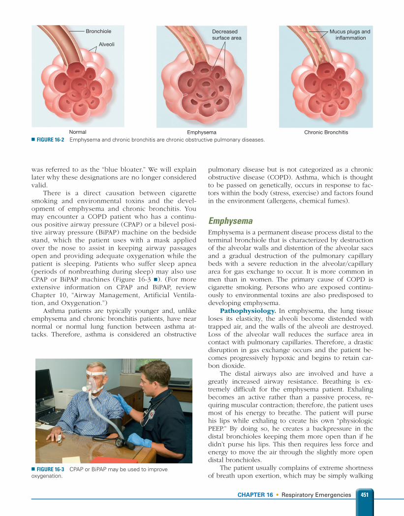
EmphysemaEmphysema is a permanent disease process distal to the terminal bronchiole that is characterized by destruction of the alveolar walls and distention of the alveolar sacs and a gradual destruction of the pulmonary capillary beds with a severe reduction in the alveolar/capillary area for gas exchange to occur. It is more common in men than in women. The primary cause of COPD is cigarette smoking. Persons who are exposed continu-ously to environmental toxins are also predisposed to developing emphysema.
Pathophysiology. In emphysema, the lung tissue loses its elasticity, the alveoli become distended with trapped air, and the walls of the alveoli are destroyed. Loss of the alveolar wall reduces the surface area in contact with pulmonary capillaries. Therefore, a drastic disruption in gas exchange occurs and the patient be-comes progressively hypoxic and begins to retain car-bon dioxide.
The distal airways also are involved and have a greatly increased airway resistance. Breathing is ex-tremely difficult for the emphysema patient. Exhaling becomes an active rather than a passive process, re-quiring muscular contraction; therefore, the patient uses most of his energy to breathe. The patient will purse his lips while exhaling to create his own “physiologic PEEP.” By doing so, he creates a backpressure in the distal bronchioles keeping them more open than if he didn’t purse his lips. This then requires less force and energy to move the air through the slightly more open distal bronchioles.
The patient usually complains of extreme shortness of breath upon exertion, which may be simply walking
was referred to as the “blue bloater.” We will explain later why these designations are no longer considered valid.
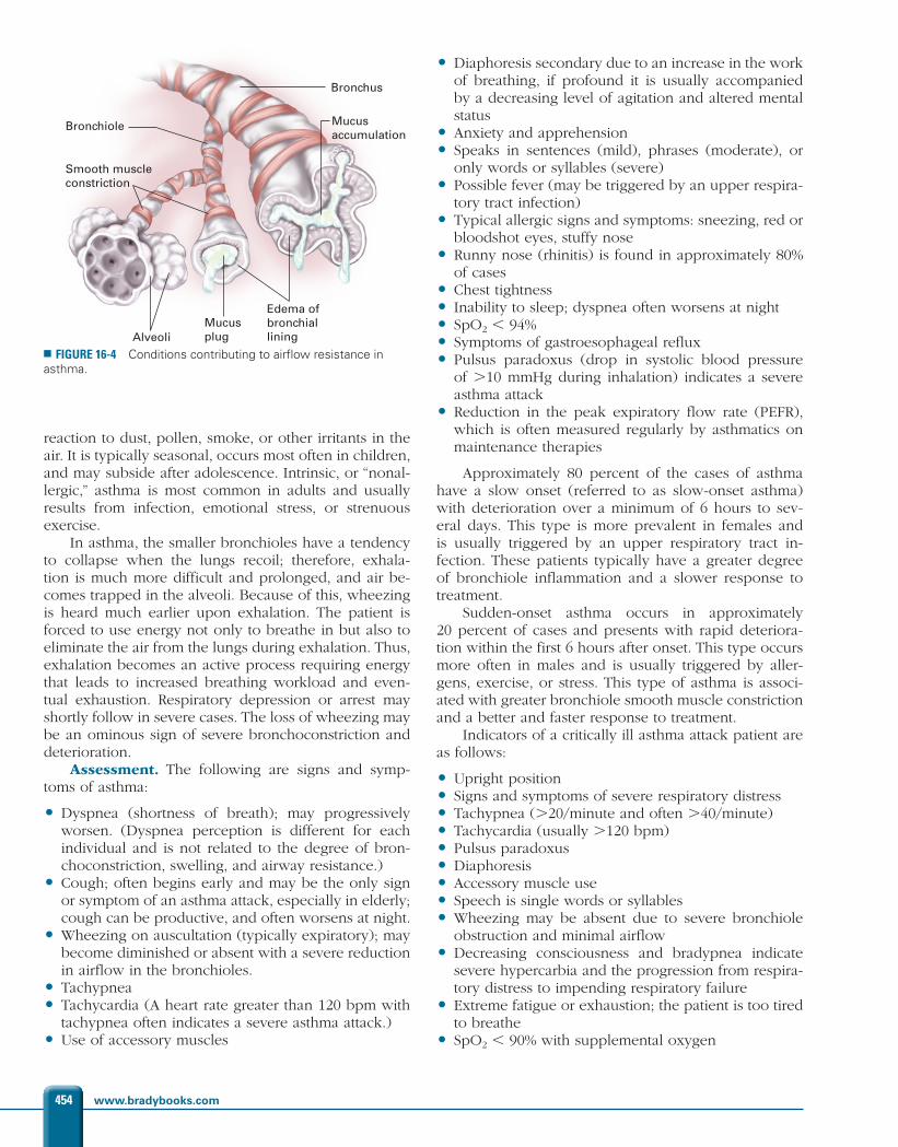
There is a direct causation between cigarette smoking and environmental toxins and the devel-opment of emphysema and chronic bronchitis. You may encounter a COPD patient who has a continu-ous positive airway pressure (CPAP) or a bilevel posi-tive airway pressure (BiPAP) machine on the bedside stand, which the patient uses with a mask applied over the nose to assist in keeping airway passages open and providing adequate oxygenation while the patient is sleeping. Patients who suffer sleep apnea (periods of nonbreathing during sleep) may also use CPAP or BiPAP machines (Figure 16-3 ■). (For more extensive information on CPAP and BiPAP, review Chapter 10, “Airway Management, Artificial Ventila-tion, and Oxygenation.”)
Asthma patients are typically younger and, unlike emphysema and chronic bronchitis patients, have near normal or normal lung function between asthma at-tacks. Therefore, asthma is considered an obstructive
Normal
Bronchiole
Alveoli
Emphysema
Decreased surface area
Chronic Bronchitis
Mucus plugs and inflammation
■ figure 16-2 Emphysema and chronic bronchitis are chronic obstructive pulmonary diseases.
■ figure 16-3 CPAP or BiPAP may be used to improve oxygenation.
M16_MIST9137_10_SE_CH16.indd 451 2/28/13 5:27 PM

www.bradybooks.com452
# 109912 Cust: Pearson Education / NJ / CHET Au: Mistovich Pg. No. 452 Title: Prehospital Emergency Care 10e Server:
C / M / Y / K Short / Normal
DESIGN SERVICES OF
S4carliSlePublishing Services
Pathophysiology. Chronic bronchitis involves in-flammation, swelling, and thickening of the lining of the bronchi and bronchioles and excessive mucus produc-tion. The alveoli remain unaffected by the disease; how-ever, the inflamed and swollen bronchioles and thick mucus restrict airflow to the alveoli so that they do not expand fully, causing respiratory distress and possible hypoxia.
With the swelling and thickening of the lining of the lower airways and the increase in mucus production in chronic bronchitis, the airways become very narrow, causing a high resistance to air movement and chronic difficulty in breathing. Recurrent infections leave scar tissue that further narrows the airways. Unlike in em-physema, the pulmonary capillary bed is not damaged. The body responds to the bronchiole obstruction by re-ducing ventilation and increasing cardiac output. The severe mismatch between the reduced amount of ven-tilation available in the alveoli and the increased blood flow through the pulmonary capillary leads to hypox-emia, hypercapnia (retention of carbon dioxide in the blood), and the production of an increased number of red blood cells (polycythemia).
Because of the increase in bronchiole obstruction, there is a reduction in the residual volume in the lungs that leads to “bloating” and a “blue” (cyanotic) appear-ance. Thus, the patient with chronic bronchitis was once referred to as a “blue bloater.” Again, this reference is outdated because of the combination of disease pro-cesses found in COPD patients.
Assessment. The following are signs and symp-toms of chronic bronchitis:
•Cough (hallmark sign) is prominent; vigorous cough-ing produces sputum
•Typically overweight, with prominent peripheral edema and chronic jugular vein distention
•Chronically cyanotic complexion (As already noted, chronic bronchitis patients were often called “blue bloaters,” but this is outdated because many COPD patients don’t conform to the description)
•Minimal difficulty in breathing and anxiety, unless in respiratory failure
•SpO2 reading of <94%, indicating chronic hypoxemia•Scattered rales and coarse rhonchi usually heard
upon auscultation of the lungs•Wheezes and, possibly, crackles at the bases of the
lungs•Asterixis (flapping of the extended wrists) may be
seen in respiratory failure
This patient frequently suffers from respiratory in-fections that lead to more acute episodes.
Emergency Medical Care for Emphysema and Chronic Bronchitis. Emergency care for the patient with emphysema and chronic bronchitis follows the same guidelines as for any patient suffering from dif-ficulty in breathing. Ensuring an open airway and
across a room. The thin appearance from weight loss and muscle wasting is associated with a reduction in cardiac output and the high energy demands of the re-spiratory muscles required for breathing. The loss of lung elasticity and trapping of air cause the chest to increase in diameter, which produces the barrel-chest appearance typical with this disease.
The emphysema patient compensates for the dis-ease process by hyperventilating. Because the patient hyperventilates, blood oxygen levels are maintained at a relatively normal level. This allows the patient to maintain a normal or “pink” appearance. The con-stant hyperventilation with purse-lip breathing makes the patient appear as if he is puffing. Thus, the name “pink puffer” was given to the emphysema patient. However, as stated earlier, the COPD patient often has a combination of disease processes; thus, the designa-tion of a patient with emphysema as a pink puffer is outdated.
Assessment. Many of the signs and symptoms of emphysema are similar to those listed earlier for respira-tory distress and may include the following:
•Anxious, alert and oriented•Dyspneic•Uses accessory muscles•Thin, barrel-chest appearance from chronic air trap-
ping in the alveoli causing the anterior–posterior di-ameter of the chest to increase
•Coughing, but with little sputum (material that is coughed up)
•Prolonged exhalation•Diminished breath sounds•Wheezing and rhonchi on auscultation•Pursed-lip breathing (physiologic PEEP)•Extreme difficulty of breathing on minimal exertion•Pink complexion from chronic hyperventilation
(emphysema patients were often called “pink puff-ers”; however, as already noted, this is outdated because many COPD patients don’t conform to the description)
•Tachypnea—breathing rate usually greater than 20 per minute at rest
•Tachycardia (increased heart rate)•Diaphoresis (sweating; moist skin)•SpO2 reading may be 94% or greater unless in respi-
ratory failure•Tripod or hunched-over position•May be on home oxygen
Chronic BronchitisChronic bronchitis is a disease process that affects pri-marily the bronchi and bronchioles. Like emphysema, chronic bronchitis is associated with cigarette smoking. By definition, chronic bronchitis is characterized by a productive cough that persists for at least three consecu-tive months a year for at least two consecutive years.
M16_MIST9137_10_SE_CH16.indd 452 2/28/13 5:27 PM

Chapter 16 • Respiratory Emergencies 453
# 109912 Cust: Pearson Education / NJ / CHET Au: Mistovich Pg. No. 453 Title: Prehospital Emergency Care 10e Server:
C / M / Y / K Short / Normal
DESIGN SERVICES OF
S4carliSlePublishing Services
AsthmaAsthma is a common respiratory condition that you may be called to the scene to manage. The most common complaint of the asthma patient is severe shortness of breath. Many asthma patients are aware of their condi-tion and have medication to manage the disease and its signs and symptoms. You may be called to the scene for a patient who is suffering an early-onset asthma attack or one in which the patient’s medication is not reversing the attack.
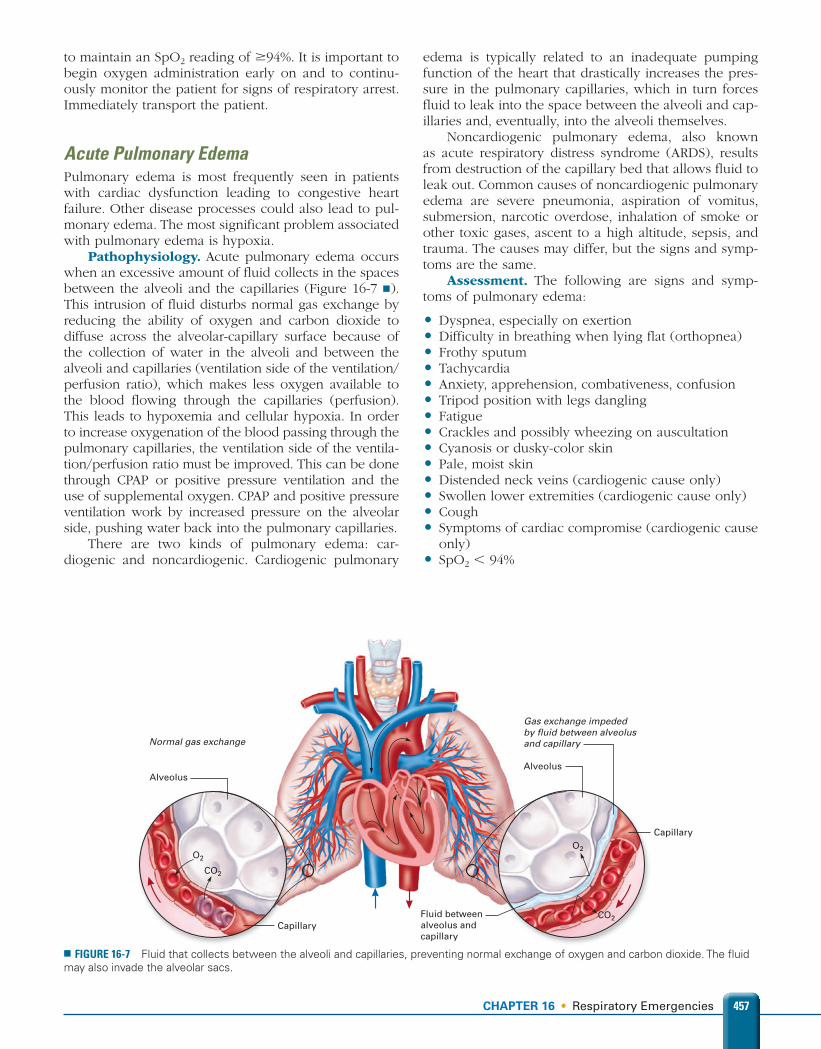
Pathophysiology. Asthma is characterized by an increased sensitivity of the lower airways to irritants and allergens, causing bronchospasm, which is a diffuse, re-versible narrowing of the bronchioles, as well as inflam-mation to the lining of the bronchioles. The following conditions in the asthma patient contribute to the in-creasing resistance to airflow and difficulty in breathing (Figure 16-4 ■):
•Bronchospasm (constriction of the smooth muscle in the bronchioles)
•Edema (swelling) of the inner lining in the airways•Increased secretion of mucus that causes plugging of
the smaller airways
Asthma patients usually suffer acute, irregular, peri-odic attacks, but between the attacks they usually have either no or very few signs or symptoms. A prolonged life-threatening attack that produces inadequate breathing and severe signs and symptoms is called status asthmati-cus. Status asthmaticus is a severe asthmatic attack that does not respond to either oxygen or medication. Patients in status asthmaticus require immediate and rapid trans-port to the hospital. Consider requesting ALS backup.
There are generally two kinds of asthma. Extrin-sic asthma, or “allergic” asthma, usually results from a
adequate breathing, position of comfort, and administra-tion of supplemental oxygen if necessary are key ele-ments in managing these patients. The patient may also have a prescribed metered-dose inhaler or small-volume nebulizer.
COPD patients may develop a hypoxic drive. Nor-mally, the body’s respiratory receptors respond to rising carbon dioxide levels to stimulate breathing. In some COPD patients, constantly high carbon dioxide levels in the blood from poor gas exchange cause the respi-ratory receptors to become insensitive to CO2 and to respond, instead, to low levels of oxygen. Theoretically, if high concentrations of oxygen are administered to the patient, the receptors pick up the increased oxygen level in the blood and send signals to the respiratory control center to reduce or even stop breathing. This usually occurs when high concentrations of oxygen are administered over a long period of time, but can occur over a short period of time, especially in the chronic bronchitis patient.
In the prehospital setting, this is a rare event and is not a major concern. Oxygen administration should take precedence over a concern about whether the hypoxic drive is going to be lost and cause the patient to stop breathing. (If this should happen, you would initiate positive pressure ventilation with supplemental oxygen, as for any patient with inadequate ventilation.)
If you have categorized the COPD patient as being a high priority, if respiratory distress is evident, and if trauma, shock, cardiac compromise, or other potentially life-threatening conditions exist, oxygen should be ad-ministered to maintain the SpO2 at or above 94%, which may be achieved with a nasal cannula at 2–6 lpm. Since many COPD patients are on home oxygen, you may be advised to apply a nasal cannula at the same liter flow or possibly 1 lpm higher than the home oxygen setting. Follow local protocol or medical direction’s order for oxygen administration in the COPD patient. As a gen-eral rule, never withhold oxygen from any patient who requires it.
If your protocol allows, in severe cases of respiratory distress consider the use of continuous positive airway pressure (CPAP). (See Chapter 10, “Airway Management, Artificial Ventilation, and Oxygenation.”) CPAP in COPD is indicated if one or more of the following is present:
•Moderate to severe dyspnea with the use of acces-sory muscles and paradoxical abdominal movement
•Respiratory rate >25 per minute
A nasal or mask device can be used to deliver the CPAP. A pressure of 5 to 10 cmH2O is usually required (follow your local protocol). Continuously assess for signs of improvement (Table 16-2) or deterioration (Table 16-3) during the use of CPAP.
If the signs of deterioration continue to progress or are not improving immediately, remove the CPAP device and begin bag-valve-mask ventilation. Be sure to deliver supplemental oxygen to the BVM device.
• Increasingrespiratoryrate• Lethargy• Patientisbecomingmoreexhaustedandfatigued• Speechlessness• Abdomenmovesinwardwithinhalationandoutward
with exhalation• DecreasingSpO2 reading
table 16-3 Signs of deterioration during the administration of Cpap
table 16-2 Signs of improvement during the administration of Cpap
• Reductioninthecomplaintofdyspnea• ImprovedSpO2 reading• Strongerrespiratoryeffort• Patientbecomesmorealert
M16_MIST9137_10_SE_CH16.indd 453 2/28/13 5:27 PM
www.bradybooks.com454
# 109912 Cust: Pearson Education / NJ / CHET Au: Mistovich Pg. No. 454 Title: Prehospital Emergency Care 10e Server:
C / M / Y / K Short / Normal
DESIGN SERVICES OF
S4carliSlePublishing Services
•Diaphoresis secondary due to an increase in the work of breathing, if profound it is usually accompanied by a decreasing level of agitation and altered mental status
•Anxiety and apprehension•Speaks in sentences (mild), phrases (moderate), or
only words or syllables (severe)•Possible fever (may be triggered by an upper respira-
tory tract infection)•Typical allergic signs and symptoms: sneezing, red or
bloodshot eyes, stuffy nose•Runny nose (rhinitis) is found in approximately 80%
of cases•Chest tightness•Inability to sleep; dyspnea often worsens at night•SpO2 < 94%•Symptoms of gastroesophageal reflux•Pulsus paradoxus (drop in systolic blood pressure
of >10 mmHg during inhalation) indicates a severe asthma attack
•Reduction in the peak expiratory flow rate (PEFR), which is often measured regularly by asthmatics on maintenance therapies
Approximately 80 percent of the cases of asthma have a slow onset (referred to as slow-onset asthma) with deterioration over a minimum of 6 hours to sev-eral days. This type is more prevalent in females and is usually triggered by an upper respiratory tract in-fection. These patients typically have a greater degree of bronchiole inflammation and a slower response to treatment.
Sudden-onset asthma occurs in approximately 20 percent of cases and presents with rapid deteriora-tion within the first 6 hours after onset. This type occurs more often in males and is usually triggered by aller-gens, exercise, or stress. This type of asthma is associ-ated with greater bronchiole smooth muscle constriction and a better and faster response to treatment.
Indicators of a critically ill asthma attack patient are as follows:
•Upright position•Signs and symptoms of severe respiratory distress•Tachypnea (>20/minute and often >40/minute)•Tachycardia (usually >120 bpm)•Pulsus paradoxus•Diaphoresis•Accessory muscle use•Speech is single words or syllables•Wheezing may be absent due to severe bronchiole
obstruction and minimal airflow•Decreasing consciousness and bradypnea indicate
severe hypercarbia and the progression from respira-tory distress to impending respiratory failure
•Extreme fatigue or exhaustion; the patient is too tired to breathe
•SpO2 < 90% with supplemental oxygen
reaction to dust, pollen, smoke, or other irritants in the air. It is typically seasonal, occurs most often in children, and may subside after adolescence. Intrinsic, or “nonal-lergic,” asthma is most common in adults and usually results from infection, emotional stress, or strenuous exercise.
In asthma, the smaller bronchioles have a tendency to collapse when the lungs recoil; therefore, exhala-tion is much more difficult and prolonged, and air be-comes trapped in the alveoli. Because of this, wheezing is heard much earlier upon exhalation. The patient is forced to use energy not only to breathe in but also to eliminate the air from the lungs during exhalation. Thus, exhalation becomes an active process requiring energy that leads to increased breathing workload and even-tual exhaustion. Respiratory depression or arrest may shortly follow in severe cases. The loss of wheezing may be an ominous sign of severe bronchoconstriction and deterioration.
Assessment. The following are signs and symp-toms of asthma:
•Dyspnea (shortness of breath); may progressively worsen. (Dyspnea perception is different for each individual and is not related to the degree of bron-choconstriction, swelling, and airway resistance.)
•Cough; often begins early and may be the only sign or symptom of an asthma attack, especially in elderly; cough can be productive, and often worsens at night.
•Wheezing on auscultation (typically expiratory); may become diminished or absent with a severe reduction in airflow in the bronchioles.
•Tachypnea•Tachycardia (A heart rate greater than 120 bpm with
tachypnea often indicates a severe asthma attack.)•Use of accessory muscles
Bronchus
Edema ofbronchiallining
Bronchiole
Smooth muscleconstriction
Mucusaccumulation
MucusplugAlveoli
■ figure 16-4 Conditions contributing to airflow resistance in asthma.
M16_MIST9137_10_SE_CH16.indd 454 2/28/13 5:27 PM

Chapter 16 • Respiratory Emergencies 455
# 109912 Cust: Pearson Education / NJ / CHET Au: Mistovich Pg. No. 455 Title: Prehospital Emergency Care 10e Server:
C / M / Y / K Short / Normal
DESIGN SERVICES OF
S4carliSlePublishing Services
other Conditions that Cause respiratory distressPneumoniaPneumonia is a common cause of death in the United States, especially in the elderly. Patients infected with the human immunodeficiency virus (HIV) and others who are on immunosuppressive drugs, such as trans-plant patients, are also very prone to pneumonia. Addi-tional risk factors include cigarette smoking, alcoholism, and exposure to cold temperatures.
Pathophysiology. Pneumonia is primarily an acute infectious disease caused by bacterium or a virus that affects the lower respiratory tract and causes lung inflam-mation and fluid- or pus-filled alveoli (Figure 16-5 ■). This leads to a ventilation disturbance in the alveoli with poor gas exchange, hypoxemia, and eventual cellular hypoxia. Pneumonia can also be caused by inhalation of toxic irritants or aspiration of vomitus and other substances.
Assessment. The signs and symptoms of pneumo-nia vary with the cause and the patient’s age. The patient generally appears ill and may complain of fever and se-vere chills. Look for the following signs and symptoms:
•Malaise and decreased appetite•Fever (may not occur in the elderly)•Cough—may be productive or nonproductive•Dyspnea (less frequent in the elderly)•Tachypnea and tachycardia•Chest pain—sharp and localized and usually made
worse when breathing deeply or coughing•Decreased chest wall movement and shallow respirations•Splinting of thorax by patient with his arm•Crackles, localized wheezing, and rhonchi heard on
auscultation•Altered mental status, especially in the elderly•Diaphoresis•Cyanosis•SpO2 < 94%
Emergency Medical Care. The pneumonia patient is managed no differently from any patient having dif-ficulty in breathing. Ensure the patient has an adequate
The asthma patient presenting with signs and symp-toms of being critically ill will require positive pressure ventilation and supplemental oxygen. If the patient is awake, alert, and able to obey commands, CPAP may be used. Watch for signs of deterioration and the need to begin bag-valve-mask ventilation.
Emergency Medical Care. During the primary as-sessment, you would have established and maintained an airway, applied oxygen or begun positive pressure ventilation with supplemental oxygen, and assessed the adequacy of circulation. Because the complaint of dys-pnea is characteristic of an asthma attack, all patients having an asthma attack should receive supplemental oxygen to maintain an SpO2 of 94% or greater. If signs of severe hypoxia are not present, this usually can be achieved by using a nasal cannula. In the pregnant pa-tient and those with preexisting cardiac disease, main-tain an SpO2 of 95%. Humidification of the oxygen is not necessary; however, it might be helpful in rehy-drating the airways. When providing positive pressure ventilation to a patient suffering a severe asthma attack in respiratory failure or arrest, the increase in resistance in the bronchioles will make ventilation more difficult to perform. The person operating the bag-valve mask will feel significant resistance when squeezing the bag. Watch for chest rise when providing ventilation to de-termine the necessary volume and pressure needed to effectively ventilate the patient. You must allow suffi-cient time for exhalation. Aggressive positive pressure ventilation may increase the amount of air trapped in the alveoli, increasing the pressure inside the chest and causing lung injury. High pressure will also result in reduced cardiac output. Deliver the ventilation with a bag-valve-mask device at a maximum rate of 10 to 12 times per minute.
CPAP may be beneficial in the acute asthma patient in respiratory distress or very early respiratory failure who is awake, alert, oriented, and able to obey com-mands (GCS > 10), is breathing on his own, is able to maintain his own airway, and has an SpO2 reading of <94%. CPAP administered to the asthma patient will improve oxygenation and decrease respiratory muscle fatigue by increasing the residual volume of air in the lungs, improving lung compliance, and reducing the work required to inflate the lungs during inhalation. (See Chapter 10, “Airway Management, Artificial Ventila-tion, and Oxygenation.”)
During the physical exam, it is necessary to calm the patient to reduce his workload of breathing and oxygen consumption. If the patient has a prescribed metered-dose inhaler or small-volume nebulizer, ad-ministration of the beta agonist medication should provide some relief of the breathing difficulty. Admin-istration of the beta2 agonist and CPAP could be done concurrently and with the proper equipment. Trans-port the patient, and continuously reassess the breath-ing status. ■ figure 16-5 Pathophysiology of pneumonia.
M16_MIST9137_10_SE_CH16.indd 455 2/28/13 5:28 PM

www.bradybooks.com456
# 109912 Cust: Pearson Education / NJ / CHET Au: Mistovich Pg. No. 456 Title: Prehospital Emergency Care 10e Server:
C / M / Y / K Short / Normal
DESIGN SERVICES OF
S4carliSlePublishing Services
creates cellular hypoxia through a disturbance on the perfusion side by blocking blood flow to ventilated and oxygenated alveoli. The degree of the perfusion distur-bance and the resulting cellular hypoxia is governed by the size of the embolus and the vessel that is blocked. A larger embolus blocks a larger vessel and reduces perfusion to a larger number of pulmonary capillaries, causing a greater severity of hypoxia.
Assessment. Signs and symptoms of pulmonary embolism depend on the size of the obstruction. If a clot obstructs a large artery, gas exchange will be se-verely impaired and signs and symptoms of respiratory distress will be evident. Suspect pulmonary embolism in any person with a sudden onset of unexplained dys-pnea and chest pain (typically sharp and localized to a specific area of the chest) and signs of hypoxia, but who has normal breath sounds and adequate volume. The following are signs and symptoms of pulmonary embolism. However, it is important to note that the signs and symptoms of pulmonary embolism are often non-specific and nondiagnostic.
•Sudden onset of unexplained dyspnea•Signs of difficulty in breathing or respiratory distress;
rapid breathing•Sudden onset of sharp, stabbing chest pain•Cough (may cough up blood)•Tachypnea•Tachycardia•Syncope (fainting)•Cool, moist skin•Restlessness, anxiety, or sense of doom•Decrease in blood pressure or hypotension (late sign)•Cyanosis (may be severe) (late sign)•Distended neck veins (late sign)•Crackles•Fever•SpO2 < 94%•Signs of complete circulatory collapse
It is important to note that not all of the signs and symptoms will always be present with pulmonary em-bolism. The three most common are chest pain, dys-pnea, and tachypnea (rapid breathing).
airway, ventilation, and oxygenation. Administer supple-mental oxygen via a nasal cannula at 2 to 4 lpm to maintain an SpO2 of 94%. In severe cases of hypoxia, a nonrebreather may be used. This is an acute infec-tious disease process that is not usually associated with severe bronchoconstriction, unless it occurs as a com-plication of asthma or COPD. Therefore, you would not expect the patient to have a metered-dose inhaler or small-volume nebulizer for this condition, nor would you necessarily consider their use unless indications of bronchoconstriction are present. Consult medical direc-tion and follow your local protocol for the use of the metered-dose inhaler or small-volume nebulizer and the administration of CPAP.
Pulmonary EmbolismIn pulmonary embolism, an obstruction of blood flow in the pulmonary arteries leads to hypoxia. Patients at risk for suffering a pulmonary embolism are those who ex-perience long periods of immobility (such as bedridden individuals, those who travel for a long period confined in one position, those with splints to extremities) as well as those with heart disease, recent surgery, long-bone fractures, venous pooling associated with pregnancy, cancer, deep vein thrombosis (development of clots in the veins, most commonly in the legs), estrogen therapy, clotting disorders, history of previous pulmonary embo-lism, and those who smoke.
Pathophysiology. Pulmonary embolism is a sud-den blockage of blood flow through a pulmonary artery or one of its branches. The embolism is usually caused by a blood clot, but it may also be caused by an air bubble, a fat particle, a foreign body, or amniotic fluid (Figure 16-6 ■). The embolism prevents blood from flowing to the lung. As a result, some areas of the lung have oxygen in the alveoli (adequate ventilation) but are not receiving any blood flow (reduced perfusion). Based on the ventilation/perfusion ratio, pulmonary embolism
■ figure 16-6 A blood clot, air bubble, fat particle, foreign body, or amniotic fluid can cause an embolism, blocking blood flow through a pulmonary artery.
In any patient complaining of shortness of breath, assess for pain, redness, increased warmth, and swelling to the lower leg, especially at the calf. These are signs of a deep vein thrombosis (DVT), a blood clot in a vein of the lower leg that may have broken free, traveled to the lungs, and caused a pulmonary embolism. ■
aSSeSSment tipS
Emergency Medical Care. During the primary assessment, you would have opened the airway and would have initiated positive pressure ventilation with supplemental oxygen or applied supplemental oxygen
M16_MIST9137_10_SE_CH16.indd 456 2/28/13 5:28 PM
Chapter 16 • Respiratory Emergencies 457
# 109912 Cust: Pearson Education / NJ / CHET Au: Mistovich Pg. No. 457 Title: Prehospital Emergency Care 10e Server:
C / M / Y / K Short / Normal
DESIGN SERVICES OF
S4carliSlePublishing Services
edema is typically related to an inadequate pumping function of the heart that drastically increases the pres-sure in the pulmonary capillaries, which in turn forces fluid to leak into the space between the alveoli and cap-illaries and, eventually, into the alveoli themselves.
Noncardiogenic pulmonary edema, also known as acute respiratory distress syndrome (ARDS), results from destruction of the capillary bed that allows fluid to leak out. Common causes of noncardiogenic pulmonary edema are severe pneumonia, aspiration of vomitus, submersion, narcotic overdose, inhalation of smoke or other toxic gases, ascent to a high altitude, sepsis, and trauma. The causes may differ, but the signs and symp-toms are the same.
Assessment. The following are signs and symp-toms of pulmonary edema:
•Dyspnea, especially on exertion•Difficulty in breathing when lying flat (orthopnea)•Frothy sputum•Tachycardia•Anxiety, apprehension, combativeness, confusion•Tripod position with legs dangling•Fatigue•Crackles and possibly wheezing on auscultation•Cyanosis or dusky-color skin•Pale, moist skin•Distended neck veins (cardiogenic cause only)•Swollen lower extremities (cardiogenic cause only)•Cough•Symptoms of cardiac compromise (cardiogenic cause
only)•SpO2 < 94%
Alveolus
CapillaryFluid betweenalveolus andcapillary
Alveolus
Gas exchange impededby fluid between alveolusand capillaryNormal gas exchange
CapillaryO2
CO2
CO2
O2
■ figure 16-7 Fluid that collects between the alveoli and capillaries, preventing normal exchange of oxygen and carbon dioxide. The fluid may also invade the alveolar sacs.
to maintain an SpO2 reading of 94%. It is important to begin oxygen administration early on and to continu-ously monitor the patient for signs of respiratory arrest. Immediately transport the patient.
Acute Pulmonary EdemaPulmonary edema is most frequently seen in patients with cardiac dysfunction leading to congestive heart failure. Other disease processes could also lead to pul-monary edema. The most significant problem associated with pulmonary edema is hypoxia.
Pathophysiology. Acute pulmonary edema occurs when an excessive amount of fluid collects in the spaces between the alveoli and the capillaries (Figure 16-7 ■). This intrusion of fluid disturbs normal gas exchange by reducing the ability of oxygen and carbon dioxide to diffuse across the alveolar-capillary surface because of the collection of water in the alveoli and between the alveoli and capillaries (ventilation side of the ventilation/ perfusion ratio), which makes less oxygen available to the blood flowing through the capillaries (perfusion). This leads to hypoxemia and cellular hypoxia. In order to increase oxygenation of the blood passing through the pulmonary capillaries, the ventilation side of the ventila-tion/perfusion ratio must be improved. This can be done through CPAP or positive pressure ventilation and the use of supplemental oxygen. CPAP and positive pressure ventilation work by increased pressure on the alveolar side, pushing water back into the pulmonary capillaries.
There are two kinds of pulmonary edema: car-diogenic and noncardiogenic. Cardiogenic pulmonary
M16_MIST9137_10_SE_CH16.indd 457 2/28/13 5:28 PM
www.bradybooks.com458
# 109912 Cust: Pearson Education / NJ / CHET Au: Mistovich Pg. No. 458 Title: Prehospital Emergency Care 10e Server:
C / M / Y / K Short / Normal
DESIGN SERVICES OF
S4carliSlePublishing Services
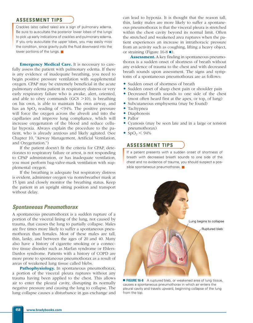
can lead to hypoxia. It is thought that the reason tall, thin, lanky males are more likely to suffer a spontane-ous pneumothorax is that the visceral pleura is stretched within the chest cavity beyond its normal limit. Often the stretched and weakened area ruptures when the pa-tient experiences an increase in intrathoracic pressure from an activity such as coughing, lifting a heavy object, or straining (Figure 16-8 ■).
Assessment. A key finding in spontaneous pneumo-thorax is a sudden onset of shortness of breath without any evidence of trauma to the chest and with decreased breath sounds upon assessment. The signs and symp-toms of a spontaneous pneumothorax are as follows:
•Sudden onset of shortness of breath•Sudden onset of sharp chest pain or shoulder pain•Decreased breath sounds to one side of the chest
(most often heard first at the apex, or top, of lung)•Subcutaneous emphysema (may be found)•Tachypnea•Diaphoresis•Pallor•Cyanosis (may be seen late and in a large or tension
pneumothorax)•SpO2 < 94%
Crackles (also called rales) are a sign of pulmonary edema. Be sure to auscultate the posterior lower lobes of the lungs to pick up early indications of crackles and pulmonary edema. If you only auscultate the upper lobes, you may easily miss the condition, since gravity pulls the fluid downward into the lower portions of the lungs. ■
aSSeSSment tipS
Emergency Medical Care. It is necessary to care-fully assess the patient with pulmonary edema. If there is any evidence of inadequate breathing, you need to begin positive pressure ventilation with supplemental oxygen. CPAP may be extremely beneficial in the acute pulmonary edema patient in respiratory distress or very early respiratory failure who is awake, alert, oriented, and able to obey commands (GCS >10), is breathing on his own, is able to maintain his own airway, and has an SpO2 reading of <94%. The positive pressure will force the oxygen across the alveoli and into the capillaries and improve lung compliance, which will increase oxygenation of the blood and reduce cellu-lar hypoxia. Always explain the procedure to the pa-tient, who is already anxious and likely agitated. (See Chapter 10, “Airway Management, Artificial Ventilation, and Oxygenation.”)
If the patient doesn’t fit the criteria for CPAP, dete-riorates to respiratory failure or arrest, is not responding to CPAP administration, or has inadequate ventilation, you must perform bag-valve-mask ventilation with sup-plemental oxygen.
If the breathing is adequate but respiratory distress is evident, administer oxygen via nonrebreather mask at 15 lpm and closely monitor the breathing status. Keep the patient in an upright sitting position and transport without delay.
Spontaneous PneumothoraxA spontaneous pneumothorax is a sudden rupture of a portion of the visceral lining of the lung, not caused by trauma, that causes the lung to partially collapse. Males are five times more likely to suffer a spontaneous pneu-mothorax than females. Most of these males are tall, thin, lanky, and between the ages of 20 and 40. Many also have a history of cigarette smoking or a connec-tive tissue disorder such as Marfan syndrome or Ehlers-Danlos syndrome. Patients with a history of COPD are more prone to spontaneous pneumothorax as a result of areas of weakened lung tissue called blebs.
Pathophysiology. In spontaneous pneumothorax, a portion of the visceral pleura ruptures without any trauma having been applied to the chest. This allows air to enter the pleural cavity, disrupting its normally negative pressure and causing the lung to collapse. The lung collapse causes a disturbance in gas exchange and
Lung begins to collapse
Ruptured bleb
■ figure 16-8 A ruptured bleb, or weakened area of lung tissue, causes a spontaneous pneumothorax in which air enters the pleural cavity and travels upward, beginning collapse of the lung from the top.
If a patient presents with a sudden onset of shortness of breath with decreased breath sounds to one side of the chest and no evidence of trauma, you should suspect a pos-sible spontaneous pneumothorax. ■
aSSeSSment tipS
M16_MIST9137_10_SE_CH16.indd 458 2/28/13 5:28 PM
Chapter 16 • Respiratory Emergencies 459
# 109912 Cust: Pearson Education / NJ / CHET Au: Mistovich Pg. No. 459 Title: Prehospital Emergency Care 10e Server:
C / M / Y / K Short / Normal
DESIGN SERVICES OF
S4carliSlePublishing Services
charged situation that is producing an anxious state in the patient. The following are signs and symptoms of hyperventilation syndrome:
•Fatigue•Nervousness and anxiety•Dizziness•Shortness of breath•Chest tightness•Numbness and tingling around the mouth, hands,
and feet•Tachypnea•Tachycardia•Spasms of the fingers and feet causing them to cramp
(carpopedal spasm)•May precipitate seizures in a patient with a seizure
disorder
Emergency Medical Care. The patient with a spontaneous pneumothorax may require supplemen-tal oxygen to maintain an SpO2 of 94% if the pa-tient presents with signs of respiratory distress, chest pain, or any other indicators for oxygen administration. If inadequate breathing is present, it is necessary to provide positive pressure ventilation. Positive pressure ventilation in a patient suffering from a pneumothorax must be performed with great care, since the pneumo-thorax could easily be converted into a tension pneu-mothorax (air entering the pleural cavity that cannot escape, eventually causing lung collapse). Use the most minimal tidal volume necessary to ventilate the patient effectively. If cyanosis, hypotension, and significant re-sistance to ventilation occur, suspect a tension pneu-mothorax. The pulse oximeter reading will also decline severely with the development of a tension pneumo-thorax. Contact ALS backup if you suspect a tension pneumothorax.
CPAP is contraindicated in a patient with a sus-pected pneumothorax regardless of the complaint of dyspnea and evidence of respiratory distress. The posi-tive pressure may increase the size of the pneumothorax and worsen the hypoxia.
Hyperventilation SyndromeHyperventilation syndrome is frequently encountered in the prehospital setting. It is commonly associated with situations in which the patient is emotionally upset or very excited. Patients suffering “panic at-tacks” will also suffer from hyperventilation syndrome. Although hyperventilation syndrome is most often as-sociated with an anxious patient, it is important to rec-ognize that hyperventilation syndrome can be caused by a serious medical problem. Therefore, always con-sider an underlying medical cause of hyperventilation syndrome when assessing and providing emergency medical care.
Pathophysiology. The hyperventilation syndrome patient is often anxious and experiences the feeling of not being able to catch his breath. The patient then begins to breathe faster and deeper, causing many of the signs and symptoms of hyperventilation to occur. The true hyperventilation syndrome patient begins to “blow off” excessive amounts of carbon dioxide. A cer-tain level of carbon dioxide is necessary for the body to function normally. When too much carbon dioxide has been eliminated through rapid breathing, the patient begins to experience worsened signs and symptoms of hyperventilation syndrome. The patient becomes more anxious because of the symptoms, and breathes even faster. One result is that the amount of calcium in the body decreases, causing the muscles of the feet and hands to cramp.
Assessment. Most often, the patient with true hy-perventilation syndrome will be found in an emotionally
The light-headedness, dizziness, or fainting experienced by the hyperventilating patient is caused by a drastic reduc-tion of carbon dioxide (the rapid breathing blows off exces-sive amounts of carbon dioxide). This causes the cerebral arteries to constrict excessively, reducing blood flow to the brain tissue, causing the light-headedness, dizziness, and fainting. ■
pathophySiology pearlS
Emergency Medical Care. The primary manage-ment is to get the patient to calm down and slow his breathing. Remove the patient from the source of anxiety or remove the source of anxiety from the scene, if pos-sible. For example, if the scene involves a domestic dis-pute, removing the other person involved in the dispute may calm the patient. Instruct the patient to consciously slow down his rate of breathing and the amount of air he is breathing. One technique is to have the patient close his mouth and breathe through his nose. You may need to coach the patient to help him slow his rate of breathing.
Do not have the patient breathe into a paper bag or oxygen mask not connected to oxygen to allow him to rebreathe carbon dioxide. These techniques can be fatal if the patient has a true underlying medical condi-tion that is causing the hyperventilation syndrome. Only use a carbon dioxide rebreathing technique if no under-lying medical conditions exist and you are specifically instructed by medical direction to do so. Keep in mind that conditions such as pulmonary embolism and myo-cardial infarction can present very similarly to hyperven-tilation syndrome. These are two of the conditions in which rebreathing carbon dioxide could be fatal.
If the patient has an SpO2 reading of <94% or any other signs of hypoxia or hypoxemia, administer sup-plemental oxygen. This can be initiated with a nasal cannula at 2 lpm and titrated upward to achieve an SpO2 reading of 94% or greater.
M16_MIST9137_10_SE_CH16.indd 459 2/28/13 5:28 PM

www.bradybooks.com460
# 109912 Cust: Pearson Education / NJ / CHET Au: Mistovich Pg. No. 460 Title: Prehospital Emergency Care 10e Server:
C / M / Y / K Short / Normal
DESIGN SERVICES OF
S4carliSlePublishing Services
EpiglottitisEpiglottitis, an inflammation affecting the upper airway, can be an acute, severe, life-threatening condition if left untreated. There is no age or season of prevalence asso-ciated with adult epiglottitis; however, males and smok-ers are more commonly affected. Although its incidence is low in the pediatric population, the incidence of adult epiglottitis has increased. The most common cause for epiglottitis in the adult population is Haemophilus influ-enzae type B. Due to the H. influenza vaccination, the incidence of epiglottitis in the pediatric patient popula-tion has significantly decreased. Adult epiglottitis can also occur as a result of a thermal injury.
Pathophysiology. As you recall, the epiglottis is a triangular cartilaginous structure that attaches at its base and closes over the glottic opening (opening to the larynx) during swallowing to prevent food or liquid from getting into the trachea. In epiglottitis, the epiglot-tis, area around the epiglottis, and base of the tongue become infected. As the condition progresses, the epi-glottis and the structures connected to or immediately surrounding it and the base of the tongue, become in-flamed and swollen, leading to a compromised airway and resultant respiratory compromise (Figure 16-9 ■). If untreated, this partial-to-complete airway obstruction leads to ineffective gas exchange in the lungs, hypoxia, acidosis, and eventually death.
Assessment. The following are signs and symptoms of epiglottitis:
•Upper respiratory tract infection, usually for 1 to 2 days prior to onset
•Dyspnea, usually with a more rapid onset•High fever (although it can occur with only mild
fevers)•Sore throat and pharyngeal pain•Inability to swallow with drooling (late sign of
impending failure)
Inflamed epiglottis
■ figure 16-9 Pathophysiology of epiglottitis.
Inspiratory stridor is an indication of an almost completely occluded airway. It is created when the patient breathes in sharply in order to draw air past the airway obstruction. As air passes through the narrowed glottic opening, airflow becomes turbulent and it creates the high-pitched sound. If the inspiratory stridor disappears and your patient’s mental status continues to deteriorate, it probably means that total airway occlusion has occurred. ■
aSSeSSment tipS
Emergency Medical Care. Treatment of epiglot-titis is focused on ensuring oxygenation and prevent-ing airway obstruction. If the patient’s breathing is still adequate, the first step is administration of high- concentration oxygen at 15 lpm to maximize oxygen-ation of the alveoli receiving airflow. In addition, and especially for the younger patient, maintaining a calm and quiet environment will help the patient to remain calm, and lessen the burden of respiratory distress. Keep the patient in a position that is comfortable to him, and expedite transport with ALS intercept if possible.
There is absolutely no need to force an inspection of the airway so long as the patient is adequately ex-changing air, and it should not be attempted. Any ad-ditional irritation to the inflamed epiglottis may result in additional swelling that totally occludes the airway. In fact, attempting airway maneuvers in a patient with epi-glottitis is only warranted in those extreme cases of re-spiratory occlusion from the swollen airway structures.
If the patient continues to deteriorate and requires as-sisted ventilations with a bag-valve-mask device, squeeze the bag slowly. This will help direct the air past the ob-struction and into the lungs rather than into the esophagus to inflate the stomach. If this is not effective in ventilating the patient, it is a situation of complete airway obstruc-tion at the level of the epiglottis, and an ALS provider may need to consider other advanced airway techniques.
PertussisPertussis (also known as “whooping cough”) is a re-spiratory disease that is characterized by uncontrolled coughing. It is a highly contagious disease that affects the respiratory system and is caused by bacteria that reside in the upper airway of an infected person. It is spread by respiratory droplets that are discharged from the nose and mouth during coughing. Pertussis has been found to occur in all age brackets, but it is most reported
•Anxiety and apprehension•Tripod position, usually with jaw jutted forward (late
sign of impending failure)•Fatigue•High-pitched inspiratory stridor•Cyanosis•Trouble or pain during speaking•SpO2 < 94%
M16_MIST9137_10_SE_CH16.indd 460 2/28/13 5:28 PM
Chapter 16 • Respiratory Emergencies 461
# 109912 Cust: Pearson Education / NJ / CHET Au: Mistovich Pg. No. 461 Title: Prehospital Emergency Care 10e Server:
C / M / Y / K Short / Normal
DESIGN SERVICES OF
S4carliSlePublishing Services
in children. Generally speaking, the younger the patient, the more severe the clinical condition that develops.
Pathophysiology. Pertussis typically starts out seeming very similar to a cold or a mild upper respiratory infection. Because of this, an older patient or the parents of an infant or child may try “waiting it out” before seek-ing medical care. Thus by the time the patient presents to EMS, the condition may be severe. Within 2 weeks or so of onset, the patient will develop episodes of rapid coughing (15 to 24 episodes in close sequence) as the body attempts to expel thick mucus from the airway, fol-lowed by a “crowing” or “whooping” sound made during inhalation as the patient breathes in deeply.
Complications of pertussis include pneumonia, dehy-dration, seizures, brain injuries, ear infections, and even death. Most deaths occur to younger patients who have not been immunized for this disease, or to those patients who are exposed before finishing the vaccination se-ries. In younger patients, the ongoing and uncontrolled coughing can severely disrupt normal breathing, dimin-ish gas exchange in the alveoli, and promote bacterial pneumonia.
Assessment. Signs and symptoms of pertussis are as follows:
•History of upper respiratory infection•Sneezing, runny nose, low-grade fever•General malaise (weakness, fatigue, not feeling well)•Increase in frequency and severity of coughing•Coughing fits, usually more common at night•Vomiting•Inspiratory “whoop” heard at the end of coughing
burst•Possible development of cyanosis during coughing
burst•Diminishing pulse oximetry finding•Exhaustion from expending energy during coughing
burst•Trouble speaking and breathing (dyspnea) during
burst
Pertussis actually has three stages. Stage 1 is characterized by findings consistent with a common cold or upper respira-tory infection. In stage 2, coughing continues to worsen to the point that medical care is sought (EMS is summoned), and thus the suspicion for pertussis (whooping cough) is formed. Stage 3 is the recovery stage, and recovery is usually gradual, taking several weeks until a resolution is reached. ■
aSSeSSment tipS
up with the coughing. The administration of humidified oxygen may help the mucus become less viscous and be expelled more easily. In addition, the patient will probably be anxious and/or scared, so the EMT should also try to ensure a quiet and calm environment. Expe-dite transport and consider an ALS intercept.
Finally, remember that this is a very contagious dis-ease, and the EMT should take all precautions neces-sary to prevent cross-contamination (to include putting a surgical mask on the patient to catch expelled airway droplets), so long as it does not impede the patient’s breathing. Following transport of a patient with known or suspected pertussis, consider totally disinfecting the patient compartment of the ambulance.
Cystic FibrosisCystic fibrosis (also known as CF, mucoviscidosis, or mu-covoidosis) is a hereditary disease. Although it commonly causes pulmonary dysfunction as a result of changes in the mucus-secreting glands of the lungs, it also affects the sweat glands, the pancreas, the liver, and the intestines.
The pulmonary complications are the most com-mon cause for a patient with this affliction to summon EMS. There is not yet a cure for CF, and many individu-als with this disease die at a young age (20s to 30s) as a result of pulmonary failure. In fact, CF is cited as one of the most common life-shortening genetic diseases. Because it is possible to detect CF in a patient at a very young age, it is common for the patient experiencing a crisis to already know of this diagnosis. Fortunately, medical research and treatment is lengthening the life span of some people to as high as 50 years. In termi-nal stages of the disease, the final medical recourse is lung transplantation when all other interventions have failed.
Pathophysiology. Lining most all of the respira-tory tree in the body is a layer of tissue that is coated with a mucous lining. This mucous lining is normally watery and helps to warm and humidify inspired air. It also serves to trap any inhaled particles. In cystic fibro-sis, however, an abnormal gene alters the functioning of the mucous glands lining the respiratory system and there is an overabundant production of mucus, which is very thick and sticky. As this thick mucus layer develops, there is blockage of the airways as well as an increase in the incidence of lung infections, since bacteria can read-ily grow in the thick mucus. Repeated lung infections in turn cause scarring of the lung tissue and promote ongo-ing pulmonary damage. As a result, there is progressive diminishment in the efficiency of respiratory function, which leads to eventual pulmonary failure and death.
Assessment. The following are signs and symp-toms of cystic fibrosis:
•Commonly a known history of the disease•Recurrent coughing•General malaise (weakness, fatigue, not feeling well)•Expectoration of thick mucus during coughing
Emergency Medical Care. Treatment of pertussis is similar to the treatment for many other respiratory problems. It is focused on ensuring oxygenation, revers-ing hypoxemia, and preventing airway obstruction. The patient should remain in a comfortable position, and the EMT should administer supplemental oxygen to main-tain an SpO2 of 94%. The EMT should also encourage the patient to expectorate any mucus that is brought
M16_MIST9137_10_SE_CH16.indd 461 2/28/13 5:28 PM

www.bradybooks.com462
# 109912 Cust: Pearson Education / NJ / CHET Au: Mistovich Pg. No. 462 Title: Prehospital Emergency Care 10e Server:
C / M / Y / K Short / Normal
DESIGN SERVICES OF
S4carliSlePublishing Services
•Recurrent episodes or history of pneumonia, bronchi-tis, and sinusitis
•Gastrointestinal complaints that may include diarrhea and greasy and/or foul-smelling bowel movements
•Abdominal pain from intestinal gas•Malnutrition or low weight despite a healthy appetite•Dehydration•Clubbing of the digits•Trouble speaking and breathing (dyspnea) with mu-
cus buildup•Signs of pneumonia
Emergency Medical Care. Emergency treatment of a patient suffering from exacerbation of CF is geared to-ward symptomatic relief of the respiratory distress. If the patient is breathing adequately, he should be provided with supplemental oxygen to maintain an SpO2 of 94% or greater. If the patient is expectorating thick mucus, humidification of the oxygen will help thin the secre-tions so that expelling them with coughing will become easier. The EMT may also consider administering normal saline through a small-volume nebulizer to aid in this. (Follow local protocol or medical direction.) The patient should remain in a sitting position for comfort and to aid in breathing. Establish ongoing pulse oximetry and, in severe cases, attempt to rendezvous with an ALS unit. If the patient’s condition is severe enough that breathing becomes inadequate, the EMT will have to deliver oxygen via positive pressure ventilation.
Poisonous ExposuresThousands of people die each year from exposures to poisonous substances (Figure 16-10 ■). Poisonous in-halation injury is an umbrella label for any type of inhalation injury that occurs secondary to exposure to toxic substances that can cause airway occlusion and/or pulmonary dysfunction by inhibiting the normal exchange of gases at the cellular level. For example, a patient may be exposed to a volatile chemical through an industrial accident, and this chemical could cause airway edema, alveolar lining damage, or displacement of oxygen. Any of these can result in respiratory failure and death of the patient unless there is immediate and effective treatment.
Pathophysiology. The inhalation of any vapors or fumes has the potential for causing some type of
Although the most common complaint patients with CF will have when they present to EMS is difficulty in breathing, many of the other findings are due to dysfunction of other organ systems. The abdominal findings such as dehydration, bowel changes, and poor weight gain are from the damage the disease inflicts on the gastrointestinal tract. Pancreatitis can also cause abdominal pain as does liver damage from the disease. ■
aSSeSSment tipS
compromise to the patient. The majority of toxic inhalation injuries, as will be discussed in Chapter 22, “Toxicologic Emergencies,” occur as the result of a fire. Sometimes, though, the toxic exposure may not be evident, since fumes may be present in the environment but undetected by the patient. The first indication of a toxic or poisonous inhalation exposure may be the patient’s collapse.
Commonly inhaled poisons that the EMT may be called on to treat include:
•Carbon monoxide•Cyanide•Natural gas•Chlorine gas•Liquid chemicals or sprays•Ammonia•Sulfur dioxide•Anesthetic gases•Solvents•Industrial gases•Hydrogen sulfide•Fumes/smoke from fires•Paints or Freon•Glue (Toluene)•Nitrous oxide•Amyl or butyl nitrate
These substances can have many effects on the body, as they all lead to cellular hypoxia by some mechanism. They may cause the soft tissue that lines the upper airway to swell to the point that airway occlusion occurs. Some can also cause the displacement of oxygen in the atmo-sphere and, hence, in the alveoli, also causing hypoxia. The chemicals may be caustic to the point that the alveo-lar lining is damaged and fluid starts to leak into the alve-oli, severely hampering gas exchange. And finally, some inhaled poisons may also exert an action on the body in and of themselves once they cross into the bloodstream. The end result of all these mechanisms, if treatment is de-layed, is inadequate cellular respiration, causing cellular death and, in turn, the death of the patient.
■ figure 16-10 Toxic exposure. (© Brendan McDermid/Reuters)
M16_MIST9137_10_SE_CH16.indd 462 2/28/13 5:28 PM

Chapter 16 • Respiratory Emergencies 463
# 109912 Cust: Pearson Education / NJ / CHET Au: Mistovich Pg. No. 463 Title: Prehospital Emergency Care 10e Server:
C / M / Y / K Short / Normal
DESIGN SERVICES OF
S4carliSlePublishing Services
Assessment. Signs and symptoms of poisonous in-halation injuries are as follows:
•History consistent with an inhalation injury (house fire, industrial accident, and so on)
•The presence of chemicals about the face from the exposure
•Findings of respiratory distress (consistent with either adequate or inadequate breathing)
•Cough, stridor, wheezing, or crackles•Oral or pharyngeal burns, possible hoarseness•Dizziness, feelings of malaise•Headache, confusion, altered mental status•Seizures•Cyanosis or other skin color changes•Nausea, vomiting, abdominal distress/pain•Copious secretions•Vital sign changes
Among the most important determinants for patient criti-cality is the length of exposure to the toxic substance and whether the patient was in an enclosed space. Additionally, exercise great personal caution when entering the scene of a known or potential toxic inhalation unless you are properly trained to do so and protected (i.e., wearing SCBA). ■
aSSeSSment tipS
Emergency Medical Care. Remember that symp-toms of respiratory distress are among the most common initial findings consistent with toxic inhalation injuries. The most important primary treatment is to limit the exposure if the patient is still in the toxic environment. Be sure, however, that you can rescue the patient in a manner that is not dangerous to you. If you can’t effect a rescue safely, your best action is to remain at a safe distance until properly trained and equipped providers can bring the patient to you.
After ensuring that there is no threat to you, your next goal is to ensure an open airway. If the patient is able to maintain his own airway and there is no as-sociated trauma, place him in a position of comfort. If he is traumatized or unresponsive, place the patient supine and immobilize as needed. Maximize oxygen-ation for the adequately breathing patient by providing oxygen at 15 lpm via nonrebreather mask. If you find the patient is breathing inadequately, provide positive pressure ventilation at a rate of 15 lpm per minute in the adult patient with supplemental oxygen attached. Be sure to properly treat any other injuries or abnormal findings, after ensuring that any compromises to the airway, breathing, and circulatory components are ap-propriately managed.
Prior to transport, try to ascertain as much informa-tion as possible about the inhaled poison for the receiv-ing facility. Because of the criticality of the patient or the potential for rapid deterioration, try to arrange an ALS intercept en route to the receiving facility. Provide early
notification to the staff at the receiving facility so they can prepare adequately for your arrival.
Viral Respiratory InfectionsAs its name indicates, a viral respiratory infection (VRI) is a condition of the respiratory system caused by a vi-rus. Common VRIs include bronchiolitis, colds, and the flu. In most situations for adults, VRIs are fairly mild, self-limiting, and confined to the upper respiratory sys-tem. In children, however, the infection has a greater propensity to spread into the lower airways where more significant infections can occur that will result in patient deterioration.
Pathophysiology. Viral respiratory infections are commonly referred to as upper respiratory infections (URIs) by the medical community because the majority of symptoms are found in the nose and throat. In small chil-dren, however, VRIs can also cause infections of the lower airway structures such as the trachea, bronchi, bronchioles, or lungs. When an infection involves these lower airway structures, depending on site, the patient may be diagnosed with croup, bronchiolitis, or pneumonia. (See Chapter 38, “Pediatrics,” for a discussion of these conditions.)
Known viruses that can cause VRIs include rhino-viruses, parainfluenza and influenza viruses, entero-viruses, respiratory syncytial virus (RSV), and some strains of the adenovirus. These viruses gain entry into the body by way of patient-to-patient contact or, to a lesser extent, through inhalation of respiratory droplets. The major pathophysiology of these viruses is exerted by triggering an inflammatory process with increased mucus production to the upper respiratory structures. There may also be a fever associated with the infection, coughing, runny nose, and findings of mild respiratory distress in the majority of cases. The VRI typically runs its course in about 14 days and, unless extenuating cir-cumstances are present (e.g., a secondary respiratory infection of the lower airways or severe respiratory dis-tress), rarely requires medical attention.
Assessment. It is, of course, nearly impossible (nor is it practical) for the EMT to determine if findings of respi-ratory distress are caused by a VRI or not. There are also no specific treatments for viral infections that the EMT can administer. The mainstay of emergency treatment for respiratory distress secondary to a VRI is supportive.
The following are signs and symptoms of viral re-spiratory infections:
•Nasal congestion•Sore or scratchy throat•Mild respiratory distress, coughing•Fever (usually around 101°F–102°F)•Malaise•Headaches and body aches•Irritability in infants and poor feeding habits•Tachypnea•Exacerbation of asthma if patient is asthmatic
M16_MIST9137_10_SE_CH16.indd 463 2/28/13 5:28 PM
www.bradybooks.com464
# 109912 Cust: Pearson Education / NJ / CHET Au: Mistovich Pg. No. 464 Title: Prehospital Emergency Care 10e Server:
C / M / Y / K Short / Normal
DESIGN SERVICES OF
S4carliSlePublishing Services
As stated previously, most people do not seek medical atten-tion or summon EMS for a typical VRI because the minimal clinical findings are usually easily treated with over-the-counter cold medications for symptomatic relief. However, if the pa-tient’s condition and findings of respiratory distress are severe enough to call EMS, chances are that the infection has spread into the lower airways and is starting to impinge on normal oxygenation by the lungs. ■
aSSeSSment tipS
Emergency Medical Care. The majority of cases of VRI do not present to EMS because the clinical presenta-tion is confined to nasal and pharyngeal discomfort. In the susceptible patient, however, there may be the pres-ence of concurrent lower tract infections that can cause some degree of respiratory distress. In all but the most severe (and uncommon) of cases, supportive treatment of positioning, oxygen therapy, emotional support, and gentle transport to the hospital is all that is necessary.
If the infection is allowed to persist without medical attention and it develops into a more serious viral infec-tion (especially for the very young or very old), supple-mental oxygen and, occasionally, mechanical ventilation may become warranted. Despite the often minimal pre-sentation, the EMT should always maintain a high index of suspicion for deterioration in a patient who does not respond favorably to supportive measures. As needed, contact ALS for potential medication administration in patients with obvious or potential deterioration.
metered-doSe inhalerS and Small-volume nebulizerS
A medication commonly prescribed for the patient with a chronic history of breathing problems is a beta2-specific bronchodilator that comes in a metered-dose inhaler (MDI) or that can, alternatively, be administered from a small-volume nebulizer (SVN). The medication is dis-pensed as an aerosol, or mist, that the patient inhales. If the patient has an MDI, the medication is already contained in the device, ready to be dispensed in aerosol form. If the patient, instead, uses an SVN, the liquid medication must be placed into the device where compressed air or oxygen converts it into an aerosol mist. Most people who use prescribed bronchodilator medications will have an MDI, rather than an SVN, because the MDI is more con-venient for the patient to use. SVNs are more commonly used in hospital settings, but some patients with chronic conditions will have an SVN for use at home. There are a variety of bronchodilator medications that can be pre-scribed. These medications can only be administered with the on-line or off-line approval of medical direction.
The most common bronchodilators that can be ad-ministered by MDI or SVN are listed in Figure 16-11 ■. (Note: All of the medications listed may be used in an MDI. Not all of them are suitable for nebulization in an
SVN.) They are considered to be beta2 agonists, which mimic the effects of the sympathetic nervous system. (The exception in the list is ipratropium, which is an anticholinergic bronchodilator rather than a beta2 bron-chodilator.) These drugs relax the bronchiole smooth muscle, which dilates the airways. This decreases re-sistance in the airways and improves the movement of air into the alveoli. Most bronchodilators begin to work almost immediately, and their effects may last up to 8 hours or more. Because of the swift relief they can provide, they are appropriate for prehospital administra-tion by the EMT with the approval of medical direction.
using a metered-dose inhalerThe MDI, also known as an “inhaler” or “puffer,” is a sim-ple device that consists of a metal canister and a plastic container with a mouthpiece and cap. The metal canister that contains the medication fits inside the plastic con-tainer. When the canister is depressed, it delivers a precise dose of medication for the patient to inhale. The medica-tion is directly deposited on the bronchioles at the site of bronchoconstriction. See the photos in EMT Skills 16-2, which illustrate administering a medication by MDI.
Some MDIs are connected to a device called a spacer. The spacer is a chamber that holds the medica-tion until it is inhaled, thus preventing any loss of med-ication to the outside and allowing a greater amount of the medication to be delivered to the patient. The spacer device is commonly used by patients who have difficulty using the MDI and is a very effective method of delivering the medication. Spacers are increasingly common as a standard in MDI administration. See the photos in EMT Skills 16-3, which illustrate how to ad-minister an MDI with a spacer.
If the patient is having breathing difficulty that is not related to trauma or a chest injury, and has one of the beta2-agonist bronchodilators in an MDI form prescribed to him by a physician, you should contact medical direc-tion for permission to administer the drug or follow lo-cal protocols. Instruct your patient as to what he should do, even if he claims to know how to use the MDI. During the administration, you must coach the patient to breathe in slowly and deeply, to hold his breath as long as he comfortably can, and to breathe out slowly through pursed lips. If the patient is not instructed or coached, the medication may not be effectively admin-istered. If the patient is unable to follow the procedure, even with coaching, you may need to administer the inhaler to the patient. Table 16-4 lists a series of “dos and don’ts” for administering medication from MDIs.
using a Small-volume nebulizerSome patients who are prescribed beta2 agonists may use a small-volume nebulizer rather than a metered-dose inhaler to dispense the medication. An SVN is a device that has a drug reservoir into which the patient
M16_MIST9137_10_SE_CH16.indd 464 2/28/13 5:28 PM

Chapter 16 • Respiratory Emergencies 465
# 109912 Cust: Pearson Education / NJ / CHET Au: Mistovich Pg. No. 465 Title: Prehospital Emergency Care 10e Server:
C / M / Y / K Short / Normal
DESIGN SERVICES OF
S4carliSlePublishing Services
Metered-Dose Inhaler (MDI)/Small-Volume Nebulizer (SVN)
Medication NameMetered-dose inhalers contain medications with a variety of generic and trade names, including the following. (Note: Not all drugs that are packaged as an MDI are available for nebulization.)
IndicationsAll of the following criteria must be met before an EMT administers a bronchodilator by metered-dose inhaler (MDI) or small-volume nebulizer (SVN) to a patient:• The patient exhibits signs and symptoms of breathing
difficulty (respiratory distress).• The patient has a physician-prescribed metered-dose
inhaler containing a medication specifically prepared to be delivered by nebulization.
• The EMT has received approval from medical direc-tion, whether on-line or off-line, to administer the medication.
ContraindicationsA bronchodilator by MDI or SVN should not be given if any of the following conditions exist:• The patient is not responsive enough to use the MDI
or SVN.• The MDI or SVN is not prescribed for the patient.• Medical direction has not granted permission.• The patient has already taken the maximum allowed
dose(s) prior to your arrival.
Medication FormsAerosolized medication in an MDI.Liquid medication packaged to be poured directly into the nebulizer chamber of an SVN (see EMT Skills 16-4C).
DosageEach time an MDI is depressed, it delivers a precise dose of medication to the patient. The total number of times the medication can be administered is determined by medical direction. When using an SVN, it usually takes
Meter-dosed inhaler.
Small-volume nebulizer.
5–10 minutes for the patient to inhale the medication, de-pending on the rate and depth of breathing. The medica-tion should be inhaled until the SVN no longer produces a mist.
AdministrationTo administer a bronchodilator by MDI (EMT Skills 16-2): 1. Ensure right patient, right medication, right dose,
right route, and right date. Determine if the patient is alert enough to use the inhaler and if any doses have already been administered prior to your arrival.
2. Obtain an order, either on-line or off-line, from medi-cal direction to assist with the administration of the medication.
3. Ensure that the inhaler is at room temperature or warmer. Shake the canister vigorously for at least 30 seconds.
4. Remove the nonrebreather mask from the patient. Instruct the patient to take the inhaler in his hand
Generic Name Trade Name
Albuterol Proventil®, Ventolin®
Metaproterenol Metaprel®, Alupent®
Isoetharine Bronkosol®
Bitolterol mesylate Tornalate®
Salmeterol xinafoate Serevent®
Ipratropium Atrovent®
Levalbuterol Xoponex
Pirbuterol Maxair
■ figure 16-11 Metered-dose inhaler and small-volume nebulizer.
continued
M16_MIST9137_10_SE_CH16.indd 465 2/28/13 5:28 PM
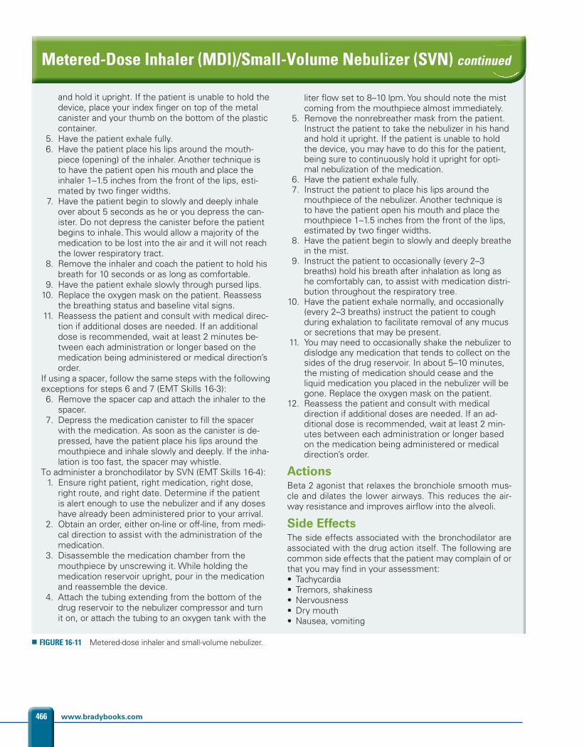
www.bradybooks.com466
# 109912 Cust: Pearson Education / NJ / CHET Au: Mistovich Pg. No. 466 Title: Prehospital Emergency Care 10e Server:
C / M / Y / K Short / Normal
DESIGN SERVICES OF
S4carliSlePublishing Services
Metered-Dose Inhaler (MDI)/Small-Volume Nebulizer (SVN) continued
and hold it upright. If the patient is unable to hold the device, place your index finger on top of the metal canister and your thumb on the bottom of the plastic container.
5. Have the patient exhale fully. 6. Have the patient place his lips around the mouth-
piece (opening) of the inhaler. Another technique is to have the patient open his mouth and place the inhaler 1–1.5 inches from the front of the lips, esti-mated by two finger widths.
7. Have the patient begin to slowly and deeply inhale over about 5 seconds as he or you depress the can-ister. Do not depress the canister before the patient begins to inhale. This would allow a majority of the medication to be lost into the air and it will not reach the lower respiratory tract.
8. Remove the inhaler and coach the patient to hold his breath for 10 seconds or as long as comfortable.
9. Have the patient exhale slowly through pursed lips. 10. Replace the oxygen mask on the patient. Reassess
the breathing status and baseline vital signs. 11. Reassess the patient and consult with medical direc-
tion if additional doses are needed. If an additional dose is recommended, wait at least 2 minutes be-tween each administration or longer based on the medication being administered or medical direction’s order.
If using a spacer, follow the same steps with the following exceptions for steps 6 and 7 (EMT Skills 16-3): 6. Remove the spacer cap and attach the inhaler to the
spacer. 7. Depress the medication canister to fill the spacer
with the medication. As soon as the canister is de-pressed, have the patient place his lips around the mouthpiece and inhale slowly and deeply. If the inha-lation is too fast, the spacer may whistle.
To administer a bronchodilator by SVN (EMT Skills 16-4): 1. Ensure right patient, right medication, right dose,
right route, and right date. Determine if the patient is alert enough to use the nebulizer and if any doses have already been administered prior to your arrival.
2. Obtain an order, either on-line or off-line, from medi-cal direction to assist with the administration of the medication.
3. Disassemble the medication chamber from the mouthpiece by unscrewing it. While holding the medication reservoir upright, pour in the medication and reassemble the device.
4. Attach the tubing extending from the bottom of the drug reservoir to the nebulizer compressor and turn it on, or attach the tubing to an oxygen tank with the
liter flow set to 8–10 lpm. You should note the mist coming from the mouthpiece almost immediately.
5. Remove the nonrebreather mask from the patient. Instruct the patient to take the nebulizer in his hand and hold it upright. If the patient is unable to hold the device, you may have to do this for the patient, being sure to continuously hold it upright for opti-mal nebulization of the medication.
6. Have the patient exhale fully. 7. Instruct the patient to place his lips around the
mouthpiece of the nebulizer. Another technique is to have the patient open his mouth and place the mouthpiece 1–1.5 inches from the front of the lips, estimated by two finger widths.
8. Have the patient begin to slowly and deeply breathe in the mist.
9. Instruct the patient to occasionally (every 2–3 breaths) hold his breath after inhalation as long as he comfortably can, to assist with medication distri-bution throughout the respiratory tree.
10. Have the patient exhale normally, and occasionally (every 2–3 breaths) instruct the patient to cough during exhalation to facilitate removal of any mucus or secretions that may be present.
11. You may need to occasionally shake the nebulizer to dislodge any medication that tends to collect on the sides of the drug reservoir. In about 5–10 minutes, the misting of medication should cease and the liquid medication you placed in the nebulizer will be gone. Replace the oxygen mask on the patient.
12. Reassess the patient and consult with medical direction if additional doses are needed. If an ad-ditional dose is recommended, wait at least 2 min-utes between each administration or longer based on the medication being administered or medical direction’s order.
ActionsBeta 2 agonist that relaxes the bronchiole smooth mus-cle and dilates the lower airways. This reduces the air-way resistance and improves airflow into the alveoli.
Side EffectsThe side effects associated with the bronchodilator are associated with the drug action itself. The following are common side effects that the patient may complain of or that you may find in your assessment:• Tachycardia• Tremors, shakiness• Nervousness• Dry mouth• Nausea, vomiting
■ figure 16-11 Metered-dose inhaler and small-volume nebulizer.
M16_MIST9137_10_SE_CH16.indd 466 2/28/13 5:28 PM

Chapter 16 • Respiratory Emergencies 467
# 109912 Cust: Pearson Education / NJ / CHET Au: Mistovich Pg. No. 467 Title: Prehospital Emergency Care 10e Server:
C / M / Y / K Short / Normal
DESIGN SERVICES OF
S4carliSlePublishing Services
Metered-Dose Inhaler (MDI)/Small-Volume Nebulizer (SVN)
ReassessmentWhenever you administer a bronchodilator to a patient, you must perform a reassessment. The following steps must be included:• Reassess the vital signs.• Question the patient about the effect of the medication
on the relief of the difficulty in breathing.• Perform a focused physical exam if changes in the con-
dition or new complaints occur.
• Constantly monitor the airway and breathing status; if the breathing becomes inadequate, begin positive pressure ventilation with supplemental oxygen.
• If the medication has had no or little effect, consult medical direction to consider another dose.
• Document any findings during the reassessment.
■ figure 16-11 Metered-dose inhaler and small-volume nebulizer.
table 16-4 mdi administration dos and don’ts
When administering a metered-dose inhaler, follow these tips:
Do
Instruct the patient to breathe in slowly and deeply.
Be sure the patient is breathing in through his mouth.
Shake the canister for at least 30 seconds before removing the cap.
Depress the canister as the patient begins to inhale.
Coach the patient to hold his breath as long as possible.
Use a spacer device if available and the patient is used to it.
Don’t
Allow the patient to breathe in too quickly.
Allow the patient to breathe in through his nose.
Administer the medication before shaking the canister.
Depress the canister before the patient begins to inhale.
Forget to coach the patient to hold his breath as long as possible.
The patient may experience a variety of side effects from the medication. The most common are an increased heart rate, tremors, and nervousness. More detailed information about bronchodilators and other side effects are listed in Figure 16-11.
places the beta2 medication in liquid form. The device is then attached to a small electrical compressor that delivers compressed air to the nebulizer by tubing. As an alternative, the supply tubing for the nebulizer can be attached to an oxygen source with the regulator set at 8–10 lpm. As the compressed air or oxygen enters the SVN, it nebulizes the medication (creates a mist) that
the patient inhales by way of a mouthpiece attached to the top of the device. (In hospitals and some home settings, the mist may be delivered through a face mask instead of a mouthpiece.)
With an SVN, the patient will continue to inhale the fine mist laden with the medication over a period of time, until the misting stops, rather than in a short mea-sured burst as occurs with the MDI. As with the MDI, the inhaled medication is deposited directly on the bronchi-ole tissue to promote relaxation. This method of deliv-ery is neither better nor worse than an MDI; it is simply an alternative way of inhaling the medication. Refer to the photos in EMT Skills 16-4 to review the procedure for administration by SVN. The indications for admin-istration, how to coach the patient during medication administration, and how you reassess the patient are identical to those used with a metered-dose inhaler. Fol-lowing administration, after the misting stops, remove the nebulizer and place the patient back on oxygen if it was removed during drug administration.
advair: not for emergency useA drug that is commonly prescribed for patients with un-controlled asthma is the Advair Diskus (Figure 16-12 ■). Unlike the short-acting beta2 agonists (for example, alb-uterol, Xopenex, Bronkosol) that are delivered via an MDI or SVN, Advair is a long-acting beta2-specific drug (salme-terol xinafoate) that also contains a steroid (fluticasone propionate) that is used as a maintenance drug. The drug comes in a rotodisk or discus delivery device that requires a different method of administration than the MDI or SVN. Even though Advair is used to treat asthma, it is not to be used as a rescue inhaler for the patient experiencing an acute asthma attack. Only a short-acting beta2-agonist drug as listed in Figure 16-11 should be used in the emergency care of the patient experiencing an acute asthma attack.
M16_MIST9137_10_SE_CH16.indd 467 2/28/13 5:29 PM

# 109912 Cust: Pearson Education / NJ / CHET Au: Mistovich Pg. No. 468 Title: Prehospital Emergency Care 10e Server:
C / M / Y / K Short / Normal
DESIGN SERVICES OF
S4carliSlePublishing Services
468 www.bradybooks.com
age-related variationS: pediatriCS and geriatriCS
In almost all emergency situations, the EMT must remain acutely aware of idiosyncrasies of patients at both age extremes, the very young and the very old. The incidence and presentation of respiratory disorders in the pediatric patient can be unique because of the differences in anat-omy and physiology; similarly, the changes that occur to the anatomy and physiology of the body during aging can alter the incidence and presentation of respiratory distress in the geriatric patient. The goal of the following section is to introduce some of the idiosyncrasies that may be present in patients at the age extremes.
pediatric patientsSince infants and children generally have healthy hearts, respiratory failure is the most likely cause of both respi-ratory arrest and cardiac arrest. This may be prevented if you can recognize the early signs of respiratory dis-tress or respiratory failure and provide the appropriate emergency care.
Respiratory failure for the pediatric patient is de-fined as inadequate oxygenation of the blood and an in-adequate elimination of carbon dioxide from the body. It is usually the result of an inadequate respiratory rate and/or inadequate tidal volume, either of which consti-tutes inadequate breathing and can lead to respiratory arrest. The root cause is most likely either an upper airway blockage or a lower airway disease. Respiratory conditions in infants and children are discussed in more detail in Chapter 38, “Pediatrics.”
respiratory distress in the pediatric patient: assessment and CareDispatch to an emergency involving an infant or child may provide indications that the patient is suffering from
respiratory distress or failure. The time it takes you to ar-rive at the patient’s location should be used to mentally process the points discussed in this section.
Scene Size-Up and Primary AssessmentDuring scene size-up with the infant or child, as with the adult patient, look for clues to help rule out trauma as a cause of the problem.
Many of the signs and symptoms of breathing dif-ficulty can be spotted as you form your general impres-sion during the primary assessment. Labored or noisy breathing, and a child who is sitting up in a tripod po-sition, lying limply, or unresponsive can be detected “from the doorway,” even before you approach the pa-tient. Additional signs and symptoms will be discovered as you contact the infant or child to assess mental status, airway, breathing, and circulation.
Secondary AssessmentOther signs and symptoms may be noted during the secondary assessment of the young patient. (Assessing the infant or child patient will be covered in detail in Chapter 38, “Pediatrics.”) Typically, signs and symptoms of respiratory distress will precede respiratory failure in the infant or child. These signs and symptoms will be indications that the body is attempting to compensate for the poor oxygen and carbon dioxide exchange by increasing the work of breathing.
Early Signs of Breathing Difficulty (Respira-tory Distress) in the Infant or Child. Because re-spiratory distress may quickly proceed to respiratory failure in the infant or child, it is vital that you recognize any of these early signs of breathing difficulty:
•Increased use of accessory muscles to breathe•Sternal and intercostal retractions during inspiration•Tachypnea (increased breathing rate)•Tachycardia (increased heart rate)•Nasal flaring•Prolonged exhalation•Frequent coughing—may be present rather than
wheezing in some children•Cyanosis to the extremities•Anxiety
The chest wall is extremely pliable in infants and young children, and the intercostal muscles (muscles between the ribs) are not well developed. Therefore, the child relies heavily on the diaphragm and abdomi-nal muscles to breathe and does not rely very much on the intercostal muscles. Obvious abdominal move-ment is expected during normal breathing in an infant or young child. In a sense, the intercostal muscles in the infant and young child are viewed more as ac-cessory muscles to breathing. Thus, in the infant and young child, retractions appear to be more prominent early in respiratory distress when they begin to use the
■ figure 16-12 The Advair Diskus is commonly prescribed to asthma patients but should not be used as a rescue inhaler for the patient experiencing an acute asthma attack.
M16_MIST9137_10_SE_CH16.indd 468 2/28/13 5:29 PM

Chapter 16 • Respiratory Emergencies
# 109912 Cust: Pearson Education / NJ / CHET Au: Mistovich Pg. No. 469 Title: Prehospital Emergency Care 10e Server:
C / M / Y / K Short / Normal
DESIGN SERVICES OF
S4carliSlePublishing Services
469
intercostal muscles to assist in breathing. This is very different from the adult who has a stiffer chest and well-developed intercostal muscles. Retractions in an adult respiratory distress patient are a significant sign of se-vere respiratory distress.
Signs of Inadequate Breathing (Respiratory Failure) in the Infant or Child. Signs of respiratory failure, which may be similar to those of inadequate breathing, are an indication that the cells are not receiv-ing an adequate oxygen supply. Respiratory compro-mise, leading to the need to provide positive pressure ventilation, should be recognized in patients who have signs and symptoms of respiratory distress with in-creased efforts to breathe who continue to deteriorate, or in patients who have inadequate respiratory effort, even with no signs or symptoms of distress.
Respiratory arrest is a condition with no respirations or respiratory effort; however, a pulse is still present.
The signs listed here occur late in a respiratory emergency and are an indication that you must imme-diately intervene and begin positive pressure ventilation with supplemental oxygen:
•Altered mental status—the patient may be listless or completely unresponsive
•Bradycardia (slow heart rate) (Bradycardia would be an initial response in a newborn, a late response in an infant.)
•Hypotension (low blood pressure)•Extremely fast, slow, or irregular breathing pattern•Cyanosis to the core of the body and mucous
membranes—a late and inconsistent sign•Loss of muscle tone (limp appearance)•Diminished or absent breath sounds•Head bobbing—bobbing of the head with each
breath•Grunting—heard in infants and children during exha-
lation, indicating diseases that produce lung collapse•Seesaw or rocky breathing—the chest is drawn in-
ward and the abdomen moves outward, indicating extreme inspiratory efforts
•Decreased response to pain•Inadequate tidal volume (poor chest rise and fall)
Emergency Medical CareThe emergency medical care for the infant and child is similar to that for the adult. Your goal should be to promptly and efficiently care for the infant or child and minimize the amount of stress. An increased stress level will increase the work of breathing and the body’s ox-ygen demand. Because of the danger that respiratory distress will deteriorate into respiratory failure, prompt intervention and transport is especially critical for the infant or child.
For a child who is experiencing difficulty in breath-ing with adequate breathing (respiratory distress), take the following steps:
1. Allow the child to assume a position of comfort to re-duce the work of breathing and to maintain a more patent airway. Do not remove the infant or child from his parent (or other caretaker). Allowing the parent to hold the child will reduce the apprehension and stress levels, thereby reducing the breathing work-load and oxygen demand.
2. Apply supplemental oxygen to a child who is sitting up in his parent’s lap. If the child does not tolerate an oxygen mask, have the parent hold it near the child’s face (Figure 16-13 ■).
3. If at any time the infant or child’s breathing becomes inadequate (respiratory failure), remove him from the parent, establish an open airway, and begin posi-tive pressure ventilation with supplemental oxygen. It will be necessary to repeat the physical exam and vital signs.
Just as with adults, a child may also have an MDI or SVN prescribed for respiratory problems associated with the lower airway. These children may present with audible wheezing; diminished breath sounds bilaterally; pale, cool, clammy skin; cyanosis; poor chest rise and fall; and other signs of breathing difficulty. If the child is experiencing breathing difficulty and has a prescribed inhaler or nebulizer, follow the same emergency care procedures for administration of the medication via MDI or SVN as for the adult.
It is important to bear in mind that the upper air-way can be obstructed by foreign bodies or from swell-ing associated with certain diseases, medical conditions, burns, or toxic inhalations. Stridor and crowing are typi-cal sounds made when the upper airway is partially ob-structed by a foreign body or swelling. If a foreign body obstruction is suspected, and the airway is completely blocked, perform foreign body airway obstruction (FBAO) maneuvers to attempt to relieve the obstruc-tion. (Refer to Chapter 38, “Pediatrics,” to review these techniques for the infant and child.) If the airway is partially blocked, place the patient on a nonrebreather mask at 15 lpm and immediately begin transport. Be
■ figure 16-13 If the child does not tolerate the mask, have the parent hold the mask near the child’s face.
M16_MIST9137_10_SE_CH16.indd 469 2/28/13 5:29 PM

www.bradybooks.com470
# 109912 Cust: Pearson Education / NJ / CHET Au: Mistovich Pg. No. 470 Title: Prehospital Emergency Care 10e Server:
C / M / Y / K Short / Normal
DESIGN SERVICES OF
S4carliSlePublishing Services
alert to begin FBAO maneuvers if the partial obstruction becomes complete.
It is important to distinguish blockage caused by a foreign body in the airway from blockage caused by disease. FBAO maneuvers may involve inserting suction devices or the fingers into the airway to remove foreign materials. With some airway diseases, inserting anything into the airway will cause dangerous spasms along the airway, making the condition worse.
Croup (laryngotracheobronchitis), commonly seen in children, involves the swelling of the larynx, trachea, and bronchi, causing breathing difficulty. The child typi-cally does not feel well, has a sore or hoarse throat, and has a fever. At night, the condition usually worsens. You might hear a hallmark sign of croup, a cough that sounds like a barking seal. Provide oxygen to the pa-tient, humidified if possible, and begin transport. Usu-ally cool night air will reduce the signs and symptoms somewhat; therefore, the condition may subside slightly after the child is taken outside for transfer to the ambu-lance. Inadequate breathing can result from croup, so continuously monitor the breathing status.
Gathering a history is especially important when airway blockage is suspected, since it may identify ei-ther preexisting diseases that may be causing the airway closure or events that may have led to a foreign body obstruction. (For example, someone witnessed the child choking on food or saw the child put an object into his mouth, or there were small objects around the child that he could have swallowed.) If the blockage was sudden and there was something around that the child could have swallowed and there is no history or other sign of disease, treat the patient for an upper airway foreign body obstruction. If the blockage came on gradually, the child has other signs of being ill, or the child has a his-tory of respiratory or other disease, and no one saw the child swallow anything, avoid inserting anything into the airway. Instead, provide oxygen or positive pressure ventilation with supplemental oxygen as necessary.
ReassessmentTransport any infant or child with difficulty breathing or signs of inadequate breathing or airway blockage. Pro-vide reassessment en route. Be prepared to intervene more aggressively if the condition deteriorates.
geriatric patientsRespiratory distress can result from any of a number of conditions occurring in the geriatric patient. It can be the primary symptom of a pulmonary problem, or it can be a symptom secondary to failure of a different body system (congestive heart failure, for example, can cause difficulty in breathing). Therefore, difficulty breathing or “shortness of breath” (dyspnea) is one of the more common complaints noted in the elderly. It is impor-tant to realize that the elderly already have diminished
respiratory function. Therefore, any additional burden can easily overwhelm the respiratory system and lead to inadequate breathing.
respiratory distress in the geriatric patient: assessment and CareThe EMT may have indication that the geriatric patient is suffering from respiratory distress or failure from in-formation received by dispatch. Hints could include the known presence of dyspnea by the caller, but it may also be manifested in complaints such as weakness, in-ability to ambulate, confusion, chest/abdominal pain, or falls. The time it takes you to arrive at the patient’s location should be used to mentally process the points discussed in this section.
Scene Size-Up and Primary AssessmentDuring scene size-up with the geriatric patient, look for clues to help rule out trauma as a cause of the problem. Many of the signs and symptoms of breathing difficulty can be spotted as you form your general impression dur-ing the primary assessment. Labored or noisy breathing, a patient in a tripod position, a patient lying in bed with multiple pillows behind his head, or unresponsiveness can be detected even before you approach the patient. Additional signs and symptoms will be discovered as you contact the patient to assess mental status, airway, breathing, and circulation.
Secondary AssessmentOther signs and symptoms may be noted during the secondary assessment of the geriatric patient. (Assessing the geriatric patient will be covered in detail in Chap-ter 39, “Geriatrics.”) The geriatric patient may not ex-hibit the typical response to hypoxia and hypercapnea; additionally, this patient may not develop fever in re-sponse to infection. The signs and symptoms of respira-tory distress usually briefly precede respiratory failure in geriatric patients. Older patients do not have the com-pensatory mechanisms a younger adult has, and they typically decompensate much more rapidly. Whenever you see indications of respiratory distress, remember that these indicate the body is attempting to compensate for the poor oxygen and carbon dioxide exchange by increasing the work of breathing, but this compensatory mechanism may be short-lived.
Early Signs of Breathing Difficulty (Respiratory Distress) in the Geriatric Patient. Because respiratory distress may quickly proceed to respiratory failure in the geriatric patient, it is vital that you recognize these early signs of breathing difficulty:
•Increased use of accessory muscles to breathe•Sternal and intercostal retractions during inspiration
M16_MIST9137_10_SE_CH16.indd 470 2/28/13 5:29 PM
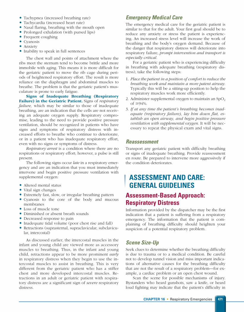
Chapter 16 • Respiratory Emergencies
# 109912 Cust: Pearson Education / NJ / CHET Au: Mistovich Pg. No. 471 Title: Prehospital Emergency Care 10e Server:
C / M / Y / K Short / Normal
DESIGN SERVICES OF
S4carliSlePublishing Services
•Tachypnea (increased breathing rate)•Tachycardia (increased heart rate)•Nasal flaring, breathing with the mouth open•Prolonged exhalation (with pursed lips)•Frequent coughing•Cyanosis•Anxiety•Inability to speak in full sentences
The chest wall and points of attachment where the ribs meet the sternum tend to become brittle and more immobile with aging. This means it is more difficult for the geriatric patient to move the rib cage during peri-ods of heightened respiratory effort. The result is more reliance on the diaphragm and abdominal muscles to breathe. The problem is that the geriatric patient’s mus-culature is prone to early fatigue.
Signs of Inadequate Breathing (Respiratory Failure) in the Geriatric Patient. Signs of respiratory failure, which may be similar to those of inadequate breathing, are an indication that the cells are not receiv-ing an adequate oxygen supply. Respiratory compro-mise, leading to the need to provide positive pressure ventilation, should be recognized in patients who have signs and symptoms of respiratory distress with in-creased efforts to breathe who continue to deteriorate, or in a patient who has inadequate respiratory effort, even with no signs or symptoms of distress.
Respiratory arrest is a condition where there are no respirations or respiratory effort; however, a pulse is still present.
The following signs occur late in a respiratory emer-gency and are an indication that you must immediately intervene and begin positive pressure ventilation with supplemental oxygen:
•Altered mental status•Vital sign changes•Extremely fast, slow, or irregular breathing pattern•Cyanosis to the core of the body and mucous
membranes•Loss of muscle tone•Diminished or absent breath sounds•Decreased response to pain•Inadequate tidal volume (poor chest rise and fall)•Retractions (suprasternal, supraclavicular, subclavicu-
lar, intercostal)
As discussed earlier, the intercostal muscles in the infant and young child are viewed more as accessory muscles to breathing. Thus, in the infant and young child, retractions appear to be more prominent early in respiratory distress when they begin to use the in-tercostal muscles to assist in breathing. This is very different from the geriatric patient who has a stiffer chest and more developed intercostal muscles. Re-tractions in an adult or geriatric patient with respira-tory distress are a significant sign of severe respiratory distress.
471
Emergency Medical CareThe emergency medical care for the geriatric patient is similar to that for the adult. Your first goal should be to reduce any anxiety or stress the patient is experienc-ing. An increased stress level will increase the work of breathing and the body’s oxygen demand. Because of the danger that respiratory distress will deteriorate into respiratory failure, prompt intervention and transport is especially critical.
For a geriatric patient who is experiencing difficulty in breathing with adequate breathing (respiratory dis-tress), take the following steps:
1. Place the patient in a position of comfort to reduce the breathing work and maintain a more patent airway. Typically this will be a sitting-up position to help the respiratory muscles work more efficiently.
2. Administer supplemental oxygen to maintain an SpO2 of 94%.
3. If at any time the patient’s breathing becomes inad-equate (respiratory failure), lay him down flat, es-tablish an open airway, and begin positive pressure ventilation with supplemental oxygen. It will be nec-essary to repeat the physical exam and vital signs.
ReassessmentTransport any geriatric patient with difficulty breathing or signs of inadequate breathing. Provide reassessment en route. Be prepared to intervene more aggressively if the condition deteriorates.
aSSeSSment and Care: general guidelineS
assessment-based approach: respiratory distressInformation provided by the dispatcher may be the first indication that a patient is suffering from a respiratory emergency. The information that the patient is com-plaining of breathing difficulty should heighten your suspicion of a potential respiratory problem.
Scene Size-UpSeek clues to determine whether the breathing difficulty is due to trauma or to a medical condition. Be careful not to develop tunnel vision and miss important indica-tions of alternative causes for the breathing difficulty that are not the result of a respiratory problem—for ex-ample, a cardiac problem or an open chest wound.
Scan the scene for possible mechanisms of injury. Bystanders who heard gunshots, saw a knife, or heard loud fighting may indicate that the patient’s difficulty in
M16_MIST9137_10_SE_CH16.indd 471 2/28/13 5:29 PM

www.bradybooks.com
# 109912 Cust: Pearson Education / NJ / CHET Au: Mistovich Pg. No. 472 Title: Prehospital Emergency Care 10e Server:
C / M / Y / K Short / Normal
DESIGN SERVICES OF
S4carliSlePublishing Services
breathing may be trauma related. The patient who is found in his house in the middle of the living room may have fallen, struck his chest against the coffee table, and fractured some ribs, causing the lung to collapse. A tall, lanky male patient may have been moving a heavy ob-ject or coughing when he experienced a sudden onset of shortness of breath that progressively worsened. Look for oxygen tanks, oxygen tubing, or oxygen concentra-tors, or medication inhalers or nebulizers at the scene. They usually indicate a chronic respiratory disease. Also scan the scene for alcohol, which is a common contribu-tor to choking and upper airway obstruction and aspira-tion of vomitus.
Primary AssessmentForm a general impression and assess the mental status, airway, breathing, and circulation.
General Impression. Several clues can help you form an impression of a patient who is suffering respira-tory distress. These include:
•The patient’s position. Most frequently, in severe cases of respiratory distress, patients sit upright and lean slightly forward, supporting themselves with their arms, elbows locked in place in front of them be-tween their dangling legs, holding onto the seat. This is referred to as a tripod position (Figure 16-14 ■).
The patient in a reclining or supine position could indicate two possible scenarios: (1) the patient is only in mild distress, or (2) the patient is in such severe respiratory distress that he is too exhausted from trying to breathe to hold himself up. This pa-tient requires immediate intervention since respira-tory arrest usually follows shortly after development of severe fatigue.
•The patient’s face. An agitated or confused facial ex-pression may indicate inadequate breathing, hypoxia, or hypercarbia.
472
■ figure 16-14 A patient in respiratory distress is commonly found in a “tripod” position.
Hypoxia causes the patient to become agitated and aggressive. Hypercarbia causes confusion, disorientation, and lethargy. ■
pathophySiology pearlS
begin to droop and the head bobs with each res-piration, as if the patient is beginning to fall asleep while sitting upright. This is a sign that the patient needs positive pressure ventilation immediately.
•Use of the muscles in the neck and retractions of the muscles between the ribs (intercostal muscles). Ac-cessory muscle use and retractions are an indication of a significant increased inhalation effort with each breath.
•Cyanosis. Cyanosis (bluish gray skin color) is a clear indication of hypoxia but also a sign that may occur late. Look at the area around the nose and mouth when getting the general impression. You will ex-amine many other areas for cyanosis in the physical exam.
•Diaphoresis. A patient with respiratory distress com-monly is diaphoretic (having sweaty and moist skin that is inappropriate for the temperature or activity). The patient becomes more diaphoretic the harder he works to breathe.
•The patient’s speech. If the speech is normal, assume that the airway is open and clear and the distress is minimal. If the patient is alert and makes eye contact but is unable to speak, consider a severe condition. The patient may speak one or two words and then pause to gasp for a breath. The number of words the patient can speak during one breath usually corre-lates with the severity of the breathing difficulty.
•Altered mental status. A change in the mental sta-tus, such as confusion, is a clear indication of inad-equate oxygenation of the brain (cerebral hypoxia) and a buildup of carbon dioxide. A sign of immi-nent respiratory failure is when the patient’s eyelids
M16_MIST9137_10_SE_CH16.indd 472 2/28/13 5:29 PM

Chapter 16 • Respiratory Emergencies 473
# 109912 Cust: Pearson Education / NJ / CHET Au: Mistovich Pg. No. 473 Title: Prehospital Emergency Care 10e Server:
C / M / Y / K Short / Normal
DESIGN SERVICES OF
S4carliSlePublishing Services
Pale, cool, clammy skin is an early sign of hypoxia in the pa-tient. Cyanosis is a clear but late sign of hypoxia. ■
aSSeSSment tipS
•Nasal flaring. Flaring of the nostrils with inhalation is another indication that the patient is working hard to breathe in.
•Pursed lips. Pursed-lip breathing is when the patient puts his lips together during exhalation as if he is going to whistle. Patients with some chronic respira-tory diseases do this subconsciously. This is done to keep the airway pressure in the smaller bronchi-oles higher during exhalation so they don’t collapse but remain open, making the next inhalation a little easier.
Mental Status. Restlessness, agitation, confusion, and unresponsiveness are frequently associated with breathing difficulty because the brain is not getting enough oxygen.
Airway. Assess the airway for any indication of a complete or partial obstruction from secretions, blood, vomitus, or a foreign body. Listen for snoring, stridor, gurgling, or crowing. Each indicates partial airway obstruction. However, keep in mind that obstructed breathing is not always noisy. Clear the airway with suction, manual maneuvers, and airway adjuncts as needed.
Breathing. Carefully assess the breathing. Look at the chest rise and fall, listen and feel for air flowing in and out of the mouth and nose, and quickly auscul-tate the lungs. Be aware of a chest that is moving up and down upon inspection but produces very little or no air movement from the mouth and nose. Efforts to breathe are being made but are not effective. This pa-tient needs positive pressure ventilation with supple-mental oxygen no matter how well the chest is rising and falling. Auscultate the breath sounds on both sides of the chest. Absent or diminished breath sounds are an indication that very little air is moving in and out of the lungs.
Determine an approximate respiratory rate. If the chest is not rising adequately with each breath or you do not hear or feel an adequate volume of air escaping on exhalation, begin positive pressure ventilation with supplemental oxygen.
Look for respiratory rates outside the ranges of 8–24 breaths per minute for adults, 15–30 per minute for chil-dren, and 25–50 per minute for infants. A respiratory rate that is too slow will not allow enough air to be transported to the alveoli for adequate gas exchange, leading to hypoxia. This patient must be provided posi-tive pressure ventilation.
The average range of normal respiration for an adult patient is 12–20 breaths per minute; however, it is possible for the patient with lower or higher rates to be breathing adequately. Lookattheentirepatientandassessthetidalvolumeclosely.If the tidal volume (assess chest rise and air movement) is in-adequate in high rates, immediately begin ventilation. Elderly patients will have higher resting respiratory rates, typically 20 per minute. ■
aSSeSSment tipS•Pallor. Pale skin color is a sign of hypoxia and severe respiratory distress. Pallor will be seen earlier and more often than cyanosis.
On the other hand, a respiratory rate that is too fast does not always compensate for a poor volume of air the patient is breathing. In many cases, the respira-tory rate becomes so fast (adult > 40/minute, infant > 60/minute) that it does not allow the patient to take a full enough breath before having to exhale, leading to ineffective volumes of air during inhalation. The respi-ratory rate will appear faster than normal; however, the chest will not rise adequately or very little air move-ment will be felt. You must immediately begin positive pressure ventilation. The inadequate chest rise and fall or little movement of air is commonly referred to as shallow breathing. Shallow breathing is an indication of inadequate breathing.
In order to have adequate breathing, you must have both an adequate rate and an adequate tidal volume (amount of air moving in and out with one breath). If either the rate or the tidal volume is inadequate, the pa-tient must be provided positive pressure ventilation. If both the rate and the tidal volume are inadequate, you must provide positive pressure ventilation. If the rate and tidal volume are both adequate, you can administer oxygen to maintain an SpO2 of 94%.
You must be aggressive when managing the airway and ventilation status of the patient. Remember that any indication of poor or inadequate ventilation must be managed with positive pressure ventilation.
During your assessment, you can summarize the pa-tient’s respiratory status as follows:
One inadequate (either rate or tidal volume) = inad-equate breathing.Two inadequates (both rate and tidal volume) = in-adequate breathing.Two adequates (both rate and tidal volume) = ad-equate breathing.
Remember that a patient who complains of breath-ing difficulty or presents with signs of hypoxia or hy-percarbia may have either adequate or inadequate breathing, as just defined. The patient with inadequate breathing will be treated differently from the patient with adequate breathing, as follows:
Inadequate breathing—Provide positive pressure ven-tilation with a bag-valve mask or pocket mask with oxygen connected to the device.Adequate breathing—Administer oxygen to maintain an SpO2 of 94% (Figure 16-15 ■).
M16_MIST9137_10_SE_CH16.indd 473 2/28/13 5:29 PM
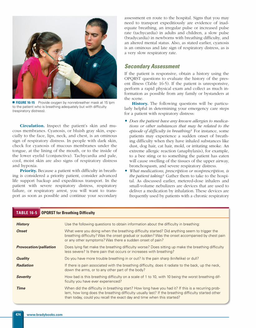
www.bradybooks.com474
# 109912 Cust: Pearson Education / NJ / CHET Au: Mistovich Pg. No. 474 Title: Prehospital Emergency Care 10e Server:
C / M / Y / K Short / Normal
DESIGN SERVICES OF
S4carliSlePublishing Services
Circulation. Inspect the patient’s skin and mu-cous membranes. Cyanosis, or bluish gray skin, espe-cially to the face, lips, neck, and chest, is an ominous sign of respiratory distress. In people with dark skin, check for cyanosis of mucous membranes under the tongue, at the lining of the mouth, or to the inside of the lower eyelid (conjunctiva). Tachycardia and pale, cool, moist skin are also signs of respiratory distress and hypoxia.
Priority. Because a patient with difficulty in breath-ing is considered a priority patient, consider advanced life support backup and expeditious transport. In the patient with severe respiratory distress, respiratory failure, or respiratory arrest, you will want to trans-port as soon as possible and continue your secondary
■ figure 16-15 Provide oxygen by nonrebreather mask at 15 lpm to the patient who is breathing adequately but with difficulty (respiratory distress).
table 16-5 opQrSt for breathing difficulty
History Use the following questions to obtain information about the difficulty in breathing:
Onset What were you doing when the breathing difficulty started? Did anything seem to trigger the breathing difficulty? Was the onset gradual or sudden? Was the onset accompanied by chest pain or any other symptoms? Was there a sudden onset of pain?
Provocation/palliation Does lying flat make the breathing difficulty worse? Does sitting up make the breathing difficulty less severe? Is there pain that occurs or increases with breathing?
Quality Do you have more trouble breathing in or out? Is the pain sharp (knifelike) or dull?
Radiation If there is pain associated with the breathing difficulty, does it radiate to the back, up the neck, down the arms, or to any other part of the body?
Severity How bad is this breathing difficulty on a scale of 1 to 10, with 10 being the worst breathing dif-ficulty you have ever experienced?
Time When did the difficulty in breathing start? How long have you had it? If this is a recurring prob-lem, how long does the breathing difficulty usually last? If the breathing difficulty started other than today, could you recall the exact day and time when this started?
assessment en route to the hospital. Signs that you may need to transport expeditiously are evidence of inad-equate breathing, an irregular pulse or increased pulse rate (tachycardia) in adults and children, a slow pulse (bradycardia) in newborns with breathing difficulty, and an altered mental status. Also, as stated earlier, cyanosis is an ominous and late sign of respiratory distress, as is a very slow respiratory rate.
Secondary AssessmentIf the patient is responsive, obtain a history using the OPQRST questions to evaluate the history of the pres-ent illness (Table 16-5). If the patient is unresponsive, perform a rapid physical exam and collect as much in-formation as possible from any family or bystanders at the scene.
History. The following questions will be particu-larly helpful in determining your emergency care steps for a patient with respiratory distress:
•Does the patient have any known allergies to medica-tions or other substances that may be related to the episode of difficulty in breathing? For instance, some patients may experience a sudden onset of breath-ing difficulty when they have inhaled substances like dust, dog hair, cat hair, mold, or irritating smoke. An extreme allergic reaction (anaphylaxis), for example, to a bee sting or to something the patient has eaten will cause swelling of the tissues of the upper airway, bronchospasm, and severe respiratory distress.
•What medications, prescription or nonprescription, is the patient taking? Gather them to take to the hospi-tal. As discussed earlier, metered-dose inhalers and small-volume nebulizers are devices that are used to deliver a medication by inhalation. These devices are frequently used by patients with a chronic respiratory
M16_MIST9137_10_SE_CH16.indd 474 2/28/13 5:29 PM

Chapter 16 • Respiratory Emergencies 475
# 109912 Cust: Pearson Education / NJ / CHET Au: Mistovich Pg. No. 475 Title: Prehospital Emergency Care 10e Server:
C / M / Y / K Short / Normal
DESIGN SERVICES OF
S4carliSlePublishing Services
table 16-6 medications Commonly used for respiratory problems
Bronchodilators Albuterol (Proventil, Ventolin)
Bitolterol mesylate (Tornalate)
Ipratropium bromide (Atrovent)
Isoetharine (Bronkosol)
Metaproterenol (Metaprel, Alupent)
Salmeterol xinafoate (Serevent)
Montelukast (Singulair)
Levalbuterol(Xopenex)
Pirbuterol (Maxair)
Potential side effects: increased heart rate, nervous-ness, shakiness, nausea, vomiting, sleeplessness, dry mouth, and allergic skin rash
Mucolytics Acetylcysteine (Mucomyst) Potential side effects: nausea, increased wheezing, and altered sense of taste
Steroids Beclomethasone (Vanceril Inhaler, Beclovent®)
Flunisolide (AeroBid)
Triamcinolone acetonide (Azmacort)
Potential side effects: dry mouth and increased wheezing
disease or recurring breathing problems. Occasion-ally you might find oral medications used specifically for respiratory problems. Common medications that might be found on the patient or at the scene are found in Table 16-6. It is important to recognize these since they might provide you with a clue that the pa-tient has a history of respiratory problems, and also, medical direction may instruct you to administer one of them. Ask if the patient has already taken any of the medications prior to your arrival, and if so, how many times. Report this information to medical direc-tion or the receiving hospital.Note: Medications used by patients with chronic respi-ratory disease have a variety of side effects. Because many patients self-administer their medication prior to your arrival, this may confuse your assessment slightly since the signs and symptoms now exhibited may result from the medication and not necessarily from the respiratory condition. These side effects are also listed in Table 16-6.
•Does the patient have a preexisting respiratory or car-diac disease?
•Has the patient ever been hospitalized for a chronic condition that produces recurring episodes of diffi-culty in breathing? If so, did he have an endotracheal tube placed down his throat to breathe or require admission to an intensive care unit? This usually indi-cates that the patient may have a tendency to deterio-rate much more rapidly and may require quicker and more aggressive intervention.
Physical Exam. The physical exam will give you further information that may indicate the severity of the breathing distress and help you determine whether to simply apply high-concentration oxygen by non-rebreather mask or to proceed with positive pressure
Typically, inhalation is an active process requiring energy, whereas exhalation is a passive process requiring no energy. The patient with respiratory distress may have difficulty not only breathing in but also breathing out. He moves to active in-halation and active exhalation, both requiring energy. This may lead to faster muscle fatigue and early respiratory failure. ■
pathophySiology pearlS
ventilation with supplemental oxygen. If the patient is unresponsive, perform a rapid assessment. In the re-sponsive patient, focus the exam on the areas that might provide you with clues as to the severity of the condition.
The posture of the patient is important. As the pa-tient becomes exhausted, you will notice his posture relaxing. This is an indication that he may require arti-ficial ventilation very shortly. Alterations in mental sta-tus, combativeness, agitation, and confusion indicate a decreasing level of oxygen getting to the brain and an increasing level of carbon dioxide. A continuous decline would be an indication of the need for aggressive emer-gency care to include positive pressure ventilation with supplemental oxygen.
•Inspect the lips and around the nose and inside the mouth for cyanosis.
•Assess the neck for jugular vein distention, which might indicate an extreme increase in pressure in the chest or venous system. Inspect and palpate for an indrawing of the trachea and tracheal deviation. The trachea pulls inward during inhalation when constricted airflow is present. Tracheal deviation, which is a late sign, is the result of an extreme amount of pressure built up on one side of the chest, collapsing the lung and pushing the mediastinum (the tissues and organs between the lungs, including the heart) and the trachea to the oppo-site side. This is a sign of a life-threatening emergency.
M16_MIST9137_10_SE_CH16.indd 475 2/28/13 5:29 PM

www.bradybooks.com476
# 109912 Cust: Pearson Education / NJ / CHET Au: Mistovich Pg. No. 476 Title: Prehospital Emergency Care 10e Server:
C / M / Y / K Short / Normal
DESIGN SERVICES OF
S4carliSlePublishing Services
to reverse the bronchospasm and allow for greater air movement. Inspiratory crackles (rales) are indicative of fluid accumulation in the alveoli from a failing heart. This fluid decreases the gas exchange across the alveo-lar membrane and can lead to respiratory distress or failure. Finally, rhonchi are sounds heard in the larger airways and represent mucus accumulation.
Vital Signs. The systolic blood pressure may drop during inhalation. This is related to a drastic increase in pressure inside the chest. When taking the blood pres-sure by auscultation, watch the needle when obtaining the systolic pressure. If the needle suddenly drops more than 10 mmHg when the patient inhales, it is a significant finding of a severe respiratory condition such as obstruc-tive lung disease. This finding is referred to as pulsus paradoxus. You may also note this as a sudden decrease in the amplitude (strength) of the pulse when the patient inhales. As the patient exhales, the pulse strength returns.
The heart rate may be increased (tachycardia) or de-creased (bradycardia). Bradycardia in adults, infants, and children is a grave sign of extremely poor oxygenation, impending respiratory failure, and possible cardiac arrest. The skin is usually moist, pale, and cool (lung infections associated with breathing difficulty may produce warm, dry, or moist skin). The breathing rate is typically increased (tachypnea); however, it may decrease as the patient be-comes tired and the oxygen levels drop significantly. Cya-nosis may be noted in severely hypoxic patients.
Subcutaneous emphysema is an indication that an air leak is present somewhere in an air-containing structure in the neck or chest. Gravity pulls heavier substances downward, forc-ing the air to travel upward toward the neck and head in the patient who is in a seated position. ■
pathophySiology pearlS
Also inspect for retractions, or pulling of the tissue inward, involving the muscles of the neck, at the supra-sternal notch, and behind the clavicles. This indicates that the patient is making an extreme effort to breathe and is another situation in which positive pressure venti-lation with supplemental oxygen should be considered.
When assessing the neck, look specifically for jugular vein distention during the inhalation phase of respiration. If the jugular veins distend during inhalation, and return to normal during exhalation, this is an indication of a severely increased pressure in the chest or around the heart. It is referred to as Kussmaul sign. ■
aSSeSSment tipS
Subcutaneous emphysema can be felt much more easily than it can be seen on inspection. ■
aSSeSSment tipS
•Inspect and palpate the chest for retraction of the muscles between the ribs, asymmetrical chest wall movement, and also for subcutaneous emphysema, which is air trapped under the subcutaneous layer of the skin. It is felt as a crackling sensation, often described as “Rice Krispies” under the fingertips. Un-equal chest wall movement may be a sign that air is trapped in one side of the chest cavity and prevent-ing adequate ventilation. Subcutaneous emphysema is a common result of trauma to the neck and chest, indicating a hole in the lung, trachea, bronchus, or esophagus. Inspect for any evidence of trauma.
•Auscultate the lungs to determine whether the breath sounds are equal on both sides of the chest. Diminished or absent breath sounds on one side of the chest mean that the lung is not being adequately ventilated because of obstruction, collapse, or surrounding air or fluid. If both lungs have diminished or absent breath sounds, it is an indication that the breathing is inadequate and the patient needs immediate ventilation with supplemental oxygen connected to the ventilation device.
An increase in the pressure inside the chest will cause a de-crease in blood flowing through the veins back to the right side of the heart. This, in turn, will decrease the volume of blood filling the left ventricle, causing a drop in the amount of blood being ejected into the aorta and arteries during the heart’s con-traction. You will see this as a sudden decrease in systolic blood pressure and pulse amplitude (strength) during inhalation. ■
pathophySiology pearlS
Early application of a pulse oximeter, if available, is important in a patient with any evidence of respiratory distress, complaint of breathing difficulty, or signs of in-adequate breathing. A pulse oximeter reading (SpO2) of less than 94% in a patient with any breathing difficulty is a concern. This is an indication of hypoxemia (low oxygen levels in the blood). If the pulse oximeter reading is below 90%, this is a significant indication of severe hypoxemia.
Closely evaluate the ventilatory status (respiratory rate and tidal volume). If both are adequate, administer oxy-gen to maintain an SpO2 of 94%. If either rate or tidal volume is inadequate, begin positive pressure ventilation. You would expect the pulse oximeter reading to increase once the patient is provided oxygen in adequate breath-ing or following positive pressure ventilation if the patient was breathing inadequately. Therefore, if a pulse oximeter reading can be obtained prior to any administration of oxygen or ventilation, it would provide a baseline reading for you to assess the effectiveness of your treatment.
Wheezing is a musical whistling sound that is heard in all lung fields upon auscultation of the chest. It is caused by narrowing of the lower airways, primarily the bronchioles, from bronchospasm and edema, or swell-ing. Wheezing is heard primarily during exhalation. You can expect to hear it most frequently in patients with a history of asthma or emphysema, but it may also be heard when fluid builds up in the lungs. These patients commonly carry medications that can be administered
M16_MIST9137_10_SE_CH16.indd 476 2/28/13 5:29 PM

Chapter 16 • Respiratory Emergencies 477
# 109912 Cust: Pearson Education / NJ / CHET Au: Mistovich Pg. No. 477 Title: Prehospital Emergency Care 10e Server:
C / M / Y / K Short / Normal
DESIGN SERVICES OF
S4carliSlePublishing Services
If the SpO2 reading continues to remain at 94% or less after the patient has been on supplemental oxygen for a period of time, it may indicate that the patient is still hy-poxic and a disturbance in gas exchange is still present. If this patient were taken off the oxygen, you would expect the pulse oximeter reading to fall below a normal level, indicating severe hypoxemia. Normally, once oxygen has been applied or the patient is being ventilated, the SpO2 should read >94% or higher. You may still experience a decline once the oxygen is removed. If the SpO2 reading does not increase to >94% or higher after oxygen admin-istration, be concerned that a hypoxemic state still exists.
At any time you suspect that the patient is not breathing adequately, you must immediately begin to ventilate using a bag-valve mask or other ventilation de-vice with oxygen connected to the device. It is impor-tant to note that even though the patient is breathing, he may need to be ventilated. You would integrate your ventilation into the patient’s own spontaneous pattern and rate of breathing to produce an effective rate and tidal volume. Do not hesitate to ventilate a spontane-ously breathing patient who has an inadequate rate or tidal volume. A poor SpO2 reading may assist you in making the decision to ventilate. For example, if a pa-tient were breathing at 38 times per minute with a shal-low tidal volume and had an SpO2 reading of 88%, you would immediately make the decision to ventilate the patient with a bag-valve-mask device. You would venti-late at 10–12 times per minute, adding your ventilation to the patient’s spontaneous breathing.
Signs and Symptoms. A wide variety of signs and symptoms may be associated with breathing difficulty, depending on the location of the obstruction or disease process, the mental status of the patient, and the severity of the respiratory distress. A large number of respiratory conditions, including both medical illness and traumatic injuries, cause signs and symptoms of breathing diffi-culty. Not all of the signs or symptoms will be present with each patient, nor will you find two cases that are exactly alike. The degree of difficulty in breathing can vary widely from minor to severe.
Respiratory distress may be produced by both medical ill-nesses and traumatic injuries. Do not assume that a patient in respiratory distress is suffering a medical illness, but expose the patient and inspect for any evidence of traumatic injuries, especially in the patient with a sudden onset of symptoms. ■
aSSeSSment tipS
The severity of shortness of breath does not directly corre-late with the level of hypoxia. A severely hypoxic patient may not complain of extreme shortness of breath. Pay close at-tention to your assessment findings. ■
aSSeSSment tipS
hypoxic than the one who perceives the level 10 sever-ity. A pulse oximeter and a good physical exam would provide the evidence to determine the hypoxia levels of the two patients. Also, remember that hypoxic patients may experience an altered mental status in which they do not complain of the shortness of breath as readily as patients who are not as short of breath.
The key is to recognize the patient who is having any difficulty in breathing, perform an accurate assess-ment, and manage any immediate life threats. The fol-lowing are common signs of breathing difficulty:
•Shortness of breath (dyspnea)•Restlessness, agitation, and anxiety•Increased heart rate (tachycardia) or irregular heart
rate in adults and children and a sudden decrease in heart rate (bradycardia) in newborns
•Faster-than-normal breathing rates (tachypnea)•Slower-than-normal breathing rates (bradypnea)•Cyanosis to the core of the body, usually seen in the
face, neck, and upper chest (This is a late sign of hy-poxia. Pale skin is seen earlier than cyanosis as a sign of hypoxia. Flushed, or red, skin may indicate an allergic reaction. With any of these discolorations, the skin is typically moist.)
•Abnormal upper airway sounds: crowing, gurgling, snoring, and stridor
•Audible wheezing upon inhalation and exhalation (In some conditions, wheezing on exhalation will develop before wheezing on inhalation. Auscultation with a stethoscope may reveal wheezing and crackles that cannot be heard by just listening with the ear.)
•Diminished ability or inability to speak•Retractions from the use of accessory muscles in the
upper chest and between the ribs and use of the mus-cles of the neck in breathing
•Excessive use of the diaphragm to breathe, produc-ing abdominal breathing in which the abdomen is moving significantly during the breathing effort
•Shallow breathing, identified by very little chest rise and fall, and poor movement of air in and out of the mouth
•Coughing, especially if it is a productive cough that produces mucus
•Irregular breathing patterns•Tripod position•Barrel chest (Figure 16-16 ■) indicating emphysema,
a chronic respiratory condition•Altered mental status—from disorientation to
unresponsiveness•Nasal flaring, when the nostrils widen and flare out
upon inhalation
It is important to recognize that the degree of short-ness of breath or the severity of the complaint of shortness of breath does not correlate with the level of hypoxia. In other words, a patient who states that his severity of dyspnea is a “10” on a scale of 1 to 10, with 10 being the worst, is not necessarily more hypoxic than the patient who complains of “a little shortness of breath” with a severity of “3” on the same scale. It is possible that the patient who perceives the level 3 severity is much more
M16_MIST9137_10_SE_CH16.indd 477 2/28/13 5:29 PM
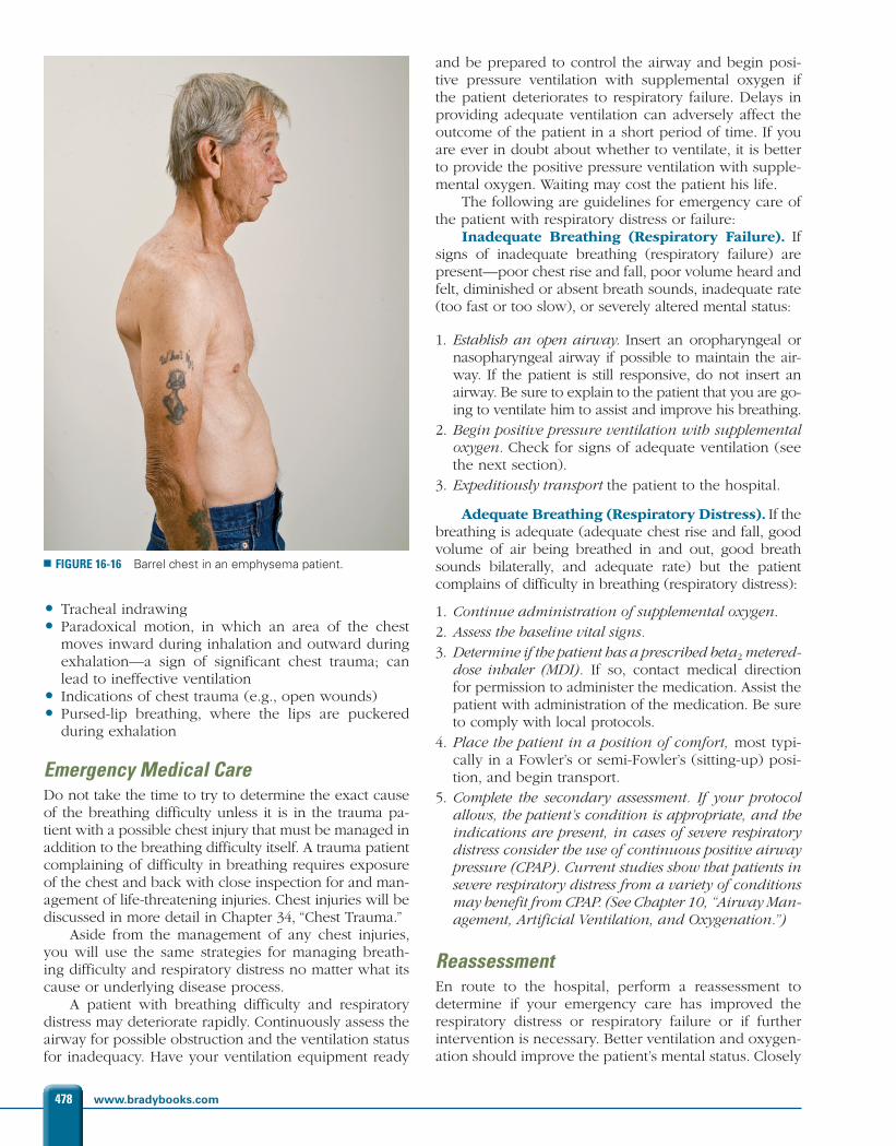
www.bradybooks.com478
# 109912 Cust: Pearson Education / NJ / CHET Au: Mistovich Pg. No. 478 Title: Prehospital Emergency Care 10e Server:
C / M / Y / K Short / Normal
DESIGN SERVICES OF
S4carliSlePublishing Services
•Tracheal indrawing•Paradoxical motion, in which an area of the chest
moves inward during inhalation and outward during exhalation—a sign of significant chest trauma; can lead to ineffective ventilation
•Indications of chest trauma (e.g., open wounds)•Pursed-lip breathing, where the lips are puckered
during exhalation
Emergency Medical CareDo not take the time to try to determine the exact cause of the breathing difficulty unless it is in the trauma pa-tient with a possible chest injury that must be managed in addition to the breathing difficulty itself. A trauma patient complaining of difficulty in breathing requires exposure of the chest and back with close inspection for and man-agement of life-threatening injuries. Chest injuries will be discussed in more detail in Chapter 34, “Chest Trauma.”
Aside from the management of any chest injuries, you will use the same strategies for managing breath-ing difficulty and respiratory distress no matter what its cause or underlying disease process.
A patient with breathing difficulty and respiratory distress may deteriorate rapidly. Continuously assess the airway for possible obstruction and the ventilation status for inadequacy. Have your ventilation equipment ready
■ figure 16-16 Barrel chest in an emphysema patient.
and be prepared to control the airway and begin posi-tive pressure ventilation with supplemental oxygen if the patient deteriorates to respiratory failure. Delays in providing adequate ventilation can adversely affect the outcome of the patient in a short period of time. If you are ever in doubt about whether to ventilate, it is better to provide the positive pressure ventilation with supple-mental oxygen. Waiting may cost the patient his life.
The following are guidelines for emergency care of the patient with respiratory distress or failure:
Inadequate Breathing (Respiratory Failure). If signs of inadequate breathing (respiratory failure) are present—poor chest rise and fall, poor volume heard and felt, diminished or absent breath sounds, inadequate rate (too fast or too slow), or severely altered mental status:
1. Establish an open airway. Insert an oropharyngeal or nasopharyngeal airway if possible to maintain the air-way. If the patient is still responsive, do not insert an airway. Be sure to explain to the patient that you are go-ing to ventilate him to assist and improve his breathing.
2. Begin positive pressure ventilation with supplemental oxygen. Check for signs of adequate ventilation (see the next section).
3. Expeditiously transport the patient to the hospital.
Adequate Breathing (Respiratory Distress). If the breathing is adequate (adequate chest rise and fall, good volume of air being breathed in and out, good breath sounds bilaterally, and adequate rate) but the patient complains of difficulty in breathing (respiratory distress):
1. Continue administration of supplemental oxygen. 2. Assess the baseline vital signs. 3. Determine if the patient has a prescribed beta2 metered-
dose inhaler (MDI). If so, contact medical direction for permission to administer the medication. Assist the patient with administration of the medication. Be sure to comply with local protocols.
4. Place the patient in a position of comfort, most typi-cally in a Fowler’s or semi-Fowler’s (sitting-up) posi-tion, and begin transport.
5. Complete the secondary assessment. If your protocol allows, the patient’s condition is appropriate, and the indications are present, in cases of severe respiratory distress consider the use of continuous positive airway pressure (CPAP). Current studies show that patients in severe respiratory distress from a variety of conditions may benefit from CPAP. (See Chapter 10, “Airway Man-agement, Artificial Ventilation, and Oxygenation.”)
ReassessmentEn route to the hospital, perform a reassessment to determine if your emergency care has improved the respiratory distress or respiratory failure or if further intervention is necessary. Better ventilation and oxygen-ation should improve the patient’s mental status. Closely
M16_MIST9137_10_SE_CH16.indd 478 2/28/13 5:29 PM

Chapter 16 • Respiratory Emergencies 479
# 109912 Cust: Pearson Education / NJ / CHET Au: Mistovich Pg. No. 479 Title: Prehospital Emergency Care 10e Server:
C / M / Y / K Short / Normal
DESIGN SERVICES OF
S4carliSlePublishing Services
monitor the patient’s airway for possible occlusion and the ventilation status for signs of inadequate breathing. If the patient continues to deteriorate, it may be neces-sary to begin positive pressure ventilation with supple-mental oxygen. Monitor the SpO2 reading. An increase in the SpO2 indicates improvement in the condition, whereas a decrease may indicate a worsening condition with further hypoxia. Always assess the respiratory rate and tidal volume when there is a decrease in the SpO2 reading.
Monitor the pulse for changes in the heart rate and regularity. A decreasing heart rate in a patient who has tachycardia may indicate improvement if the mental status is also improving and the respiratory distress is subsiding. If the heart rate is declining along with the mental status, and the respiratory distress is worsening, this is an omi-nous sign of impending respiratory failure. Increases in the heart rate may be seen with the administration of many of the metered-dose inhalers. These medications mimic the actions of the sympathetic nervous system; therefore, a slight increase in heart rate may be anticipated. This tachy-cardia would be expected to decrease once the condition improves and the medication begins to wear off.
Beta2 metered-dose inhalers also contain trace beta1 proper-ties. Beta1 properties increase the heart rate and the force of contraction. An increase in the heart rate following the administration of a beta2 MDI is often a side effect from the trace beta1 properties. ■
pathophySiology pearlS
Assessment Summary
RESPIRATORY DISTRESSThe following are findings that may be associated with breathing difficulty.
Scene Size-UpIs breathing difficulty due to a medical or a traumatic cause? Look for evidence of:
Mechanism of injury—collision, fall, guns, knives, bruis-ing on chest
Home or portable oxygen tanks or concentrators indi-cating chronic respiratory problems
Alcohol or food that may indicate choking
Primary AssessmentGeneral ImpressionPosition of patient:
TripodLying flat
Facial expression:Agitated or confused
Speech:Patient may gasp for breath between words.
Mental StatusAlert to unresponsiveRestlessnessAgitationDisorientation
AirwayInspect for incomplete or partial obstructionCrowing and stridor (indicate partial obstruction)Gurgling (indicates fluid in the airway; suction required)
BreathingSigns of inadequate breathing, including poor chest
rise and fall, poor volume heard and felt, diminished or absent breath sounds
Wheezing heard on auscultation
CirculationTachycardia (more typical in adult with hypoxia)Bradycardia (more typical in infant or young child with
hypoxia)Cyanosis to mucous membranes, around nose and
mouth, nail beds, chest, and neckStatus: Priority Patient
History and Secondary AssessmentHistorySigns and symptoms:
Shortness of breathRestlessness and anxietyDifficulty in breathing while lying flatDiaphoresis
Known allergies to medication or other substancesMedications for respiratory conditionsHome oxygen
Prescribed metered-dose inhaler (MDI)Pre-existing respiratory or cardiac diseaseHospitalized for respiratory condition
Physical ExamHead, neck, and face:
Cyanosis to face, neck, and mucous membranesJugular venous distention (may indicate heart failure or
lung injury [tension pneumothorax])Jugular vein engorgement on inhalation (Kussmaul sign)
Nasal flaringPursed-lip breathing
Chest:RetractionsAccessory muscle useWheezingProductive coughBarrel chest (indicates emphysema, a chronic respira-
tory condition)Abdomen:
Use of abdominal muscles when breathingExtremities:
Cyanosis to fingers and nail bedsPale skinDiaphoresis
Baseline Vital SignsBP: normalBP: sudden decrease in the systolic BP by 10 mmHg or
greater with inhalationHR: increased in adults; slow in infants and young
childrenPulse: sudden decrease in the pulse amplitude with
inhalation RR: increased; may decrease with greater hypoxiaSkin: cyanosis, paleness, diaphoresisPupils: dilated; sluggish to respond to lightSpO2: 94%
continued
continued
Moist skin (diaphoresis) is a result of the sympa-thetic nervous system response in the patient with breathing difficulty. An increase in diaphoresis would correlate with a worsening condition.
Reassess and record the blood pressure. Reassess the breath sounds. Improved air movement in the lungs will produce clearer and louder breath sounds on both sides of the chest. Conversely, as the condition dete-riorates, the breath sounds become diminished to ab-sent. Note that decreased wheezing may not indicate improvement; it may actually indicate severe broncho-constriction with less air movement.
The patient with breathing difficulty is considered a priority patient, especially if the condition does not re-spond to your emergency care. You should consider ad-vanced life support backup.
If the patient’s complaint changes, repeat the physi-cal exam and vital signs. Ensure that the oxygen is ap-plied properly and flowing adequately. Continuously assess the status of the breathing.
If positive pressure ventilation with supplemental oxygen has been initiated, continuously assess its ef-fectiveness. Ensure that oxygen is connected and ad-equately flowing to the pocket mask or the reservoir of the bag-valve-mask device.
Summary: assessment and CareTo review assessment findings that may be associated with breathing difficulty and emergency care for breath-ing difficulty, see Figures 16-17 ■ and 16-18 ■.
■ figure 16-17a Assessment summary: respiratory distress.
M16_MIST9137_10_SE_CH16.indd 479 2/28/13 5:29 PM

www.bradybooks.com480
# 109912 Cust: Pearson Education / NJ / CHET Au: Mistovich Pg. No. 480 Title: Prehospital Emergency Care 10e Server:
C / M / Y / K Short / Normal
DESIGN SERVICES OF
S4carliSlePublishing Services
Assessment Summary
RESPIRATORY DISTRESSThe following are findings that may be associated with breathing difficulty.
Scene Size-UpIs breathing difficulty due to a medical or a traumatic cause? Look for evidence of:
Mechanism of injury—collision, fall, guns, knives, bruis-ing on chest
Home or portable oxygen tanks or concentrators indi-cating chronic respiratory problems
Alcohol or food that may indicate choking
Primary AssessmentGeneral ImpressionPosition of patient:
TripodLying flat
Facial expression:Agitated or confused
Speech:Patient may gasp for breath between words.
Mental StatusAlert to unresponsiveRestlessnessAgitationDisorientation
AirwayInspect for incomplete or partial obstructionCrowing and stridor (indicate partial obstruction)Gurgling (indicates fluid in the airway; suction required)
BreathingSigns of inadequate breathing, including poor chest
rise and fall, poor volume heard and felt, diminished or absent breath sounds
Wheezing heard on auscultation
CirculationTachycardia (more typical in adult with hypoxia)Bradycardia (more typical in infant or young child with
hypoxia)Cyanosis to mucous membranes, around nose and
mouth, nail beds, chest, and neckStatus: Priority Patient
History and Secondary AssessmentHistorySigns and symptoms:
Shortness of breathRestlessness and anxietyDifficulty in breathing while lying flatDiaphoresis
Known allergies to medication or other substancesMedications for respiratory conditionsHome oxygen
Prescribed metered-dose inhaler (MDI)Pre-existing respiratory or cardiac diseaseHospitalized for respiratory condition
Physical ExamHead, neck, and face:
Cyanosis to face, neck, and mucous membranesJugular venous distention (may indicate heart failure or
lung injury [tension pneumothorax])Jugular vein engorgement on inhalation (Kussmaul sign)
Nasal flaringPursed-lip breathing
Chest:RetractionsAccessory muscle useWheezingProductive coughBarrel chest (indicates emphysema, a chronic respira-
tory condition)Abdomen:
Use of abdominal muscles when breathingExtremities:
Cyanosis to fingers and nail bedsPale skinDiaphoresis
Baseline Vital SignsBP: normalBP: sudden decrease in the systolic BP by 10 mmHg or
greater with inhalationHR: increased in adults; slow in infants and young
childrenPulse: sudden decrease in the pulse amplitude with
inhalation RR: increased; may decrease with greater hypoxiaSkin: cyanosis, paleness, diaphoresisPupils: dilated; sluggish to respond to lightSpO2: 94%
continued
continued
■ figure 16-17a Assessment summary: respiratory distress.
Emergency Care Protocol
RESPIRATORY DISTRESS 1. Establish and maintain an open airway.2. Suction secretions as necessary.3. If breathing is inadequate, provide positive pressure
ventilation with supplemental oxygen at a minimum rate of 10–12 ventilations/minute for an adult and 12–20 ventilations/minute for an infant or child.
4. Apply a pulse oximeter. If breathing is adequate, ad-minister oxygen to maintain an SpO2 of � 94%.
5. If the patient has signs and symptoms of breathing difficulty and has a prescribed metered-dose inhaler or a nebulization device, administer the beta 2-specific drug as appropriate according to medical direction:• Beta 2-specific drugs mimic the sympathetic nervous
system and cause bronchodilation.• Dose is precisely delivered by the device.• Obtain an order from medical direction.• Ensure the “five rights” of medication administration.• Assemble the equipment (MDI or nebulizer) as
discussed previously.
• Place the device, and instruct the patient how to breathe.
• Replace nonrebreather mask on patient.• Record time and reassess patient.• Beta 2 side effects: tachycardia tremors nervousness dry mouth nausea
6. Consider advanced life support if the condition does not improve.
7. Transport in a position of comfort. 8. Perform a reassessment of the patient’s status ev-
ery 5 minutes. 9. Complete the secondary assessment.If your protocol
allows, in cases of severe respiratory distress con-sider the use of continuous positive airway pressure (CPAP). Current studies show that patients in severe respiratory distress from a variety of conditions may benefit from CPAP. (See Chapter 10, “Airway Manage-ment, Artificial Ventilation, and Oxygenation.”
■ figure 16-17b Emergency care protocol: respiratory distress.
M16_MIST9137_10_SE_CH16.indd 480 2/28/13 5:29 PM
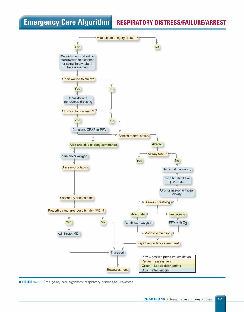
Chapter 16 • Respiratory Emergencies 481
# 109912 Cust: Pearson Education / NJ / CHET Au: Mistovich Pg. No. 481 Title: Prehospital Emergency Care 10e Server:
C / M / Y / K Short / Normal
DESIGN SERVICES OF
S4carliSlePublishing Services
Mechanism of injury present?
Yes
Yes
No
No
Yes No
Yes
Administer MDI
No
Yes No
Consider manual in-linestabilization and assessfor spinal injury later in
the assessment
Open wound to chest?
Occlude withnonporous dressing
Obvious flail segment?
Consider, CPAP or PPV
Assess mental status
Alert and able to obey commands Altered
Airway open?
Assess breathing
Assess circulation
Assess circulation
Secondary assessment
Prescribed metered dose inhaler (MDI)?
Rapid secondary assessment
Suction if necessary
Head tilt-chin lift orjaw thrust
Oro- or nasopharyngealairway
Adequate Inadequate
Administer oxygen
Administer oxygen
PPV with O2
Transport
Reassessment
Yellow = assessmentGreen = key decision pointsBlue = interventions
PPV = positive pressure ventilation
■ figure 16-18 Emergency care algorithm: respiratory distress/failure/arrest.
emergency Care algorithm reSpiratory diStreSS/failure/arreSt
M16_MIST9137_10_SE_CH16.indd 481 2/28/13 5:29 PM

# 109912 Cust: Pearson Education / NJ / CHET Au: Mistovich Pg. No. 482 Title: Prehospital Emergency Care 10e Server:
C / M / Y / K Short / Normal
DESIGN SERVICES OF
S4carliSlePublishing Services
www.bradybooks.com
emt SKillS16-1 Auscultating the Chest
midclavicularlines
■ 16-1a Auscultate the anterior chest at the second intercostal space at each midclavicular line.
midaxillaryline
■ 16-1b Auscultate the lateral chest at the fourth to fifth intercostal space at each midaxillary line.
midscapularlines
■ 16-1C Auscultate the posterior chest below the tip of the scapula on each midscapular line.
482482
M16_MIST9137_10_SE_CH16.indd 482 2/28/13 5:30 PM

# 109912 Cust: Pearson Education / NJ / CHET Au: Mistovich Pg. No. 483 Title: Prehospital Emergency Care 10e Server:
C / M / Y / K Short / Normal
DESIGN SERVICES OF
S4carliSlePublishing Services
Chapter 16 • Respiratory Emergencies 483
emt SKillS16-2 Administering Medication by Metered-Dose Inhaler
■ 16-2a Consult with medical direction for an order to administer the medication.
■ 16-2b Check to make sure the medication is for the patient, that it is the proper one to administer, and that it has not reached its expiration date.
■ 16-2C Shake the inhaler vigorously for at least 30 seconds. ■ 16-2d Instruct the patient to inhale slowly and deeply for about 5 seconds. As the patient begins to inhale, depress the canister.
■ 16-2f Instruct the patient to exhale slowly through pursed lips.■ 16-2e Remove the inhaler and instruct the patient to hold the breath for 10 seconds or for as long as is comfortable.
continued
M16_MIST9137_10_SE_CH16.indd 483 2/28/13 5:30 PM

# 109912 Cust: Pearson Education / NJ / CHET Au: Mistovich Pg. No. 484 Title: Prehospital Emergency Care 10e Server:
C / M / Y / K Short / Normal
DESIGN SERVICES OF
S4carliSlePublishing Services
www.bradybooks.com484
■ 16-2g Replace the oxygen on the patient. Reassess the breathing status and vital signs.
emt SKillS16-3 Administering a Metered-Dose Inhaler with a Spacer
■ 16-3a Remove the spacer cap. Attach the spacer to the inhaler mouthpiece.
■ 16-3b Depress the medication canister to fill the spacer with medication.
■ 16-3C Instruct the patient to inhale slowly and deeply. The spacer may whistle if the patient is inhaling too quickly.
continued
M16_MIST9137_10_SE_CH16.indd 484 2/28/13 5:30 PM
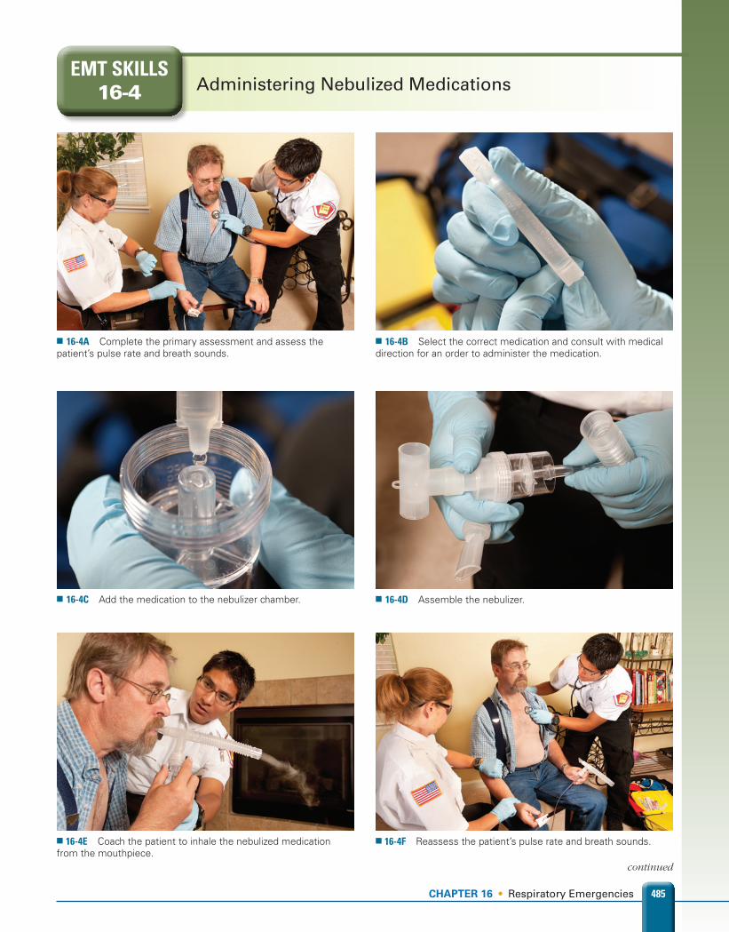
# 109912 Cust: Pearson Education / NJ / CHET Au: Mistovich Pg. No. 485 Title: Prehospital Emergency Care 10e Server:
C / M / Y / K Short / Normal
DESIGN SERVICES OF
S4carliSlePublishing Services
Chapter 16 • Respiratory Emergencies 485
emt SKillS16-4 Administering Nebulized Medications
■ 16-4a Complete the primary assessment and assess the patient’s pulse rate and breath sounds.
■ 16-4b Select the correct medication and consult with medical direction for an order to administer the medication.
■ 16-4C Add the medication to the nebulizer chamber. ■ 16-4d Assemble the nebulizer.
■ 16-4e Coach the patient to inhale the nebulized medication from the mouthpiece.
■ 16-4f Reassess the patient’s pulse rate and breath sounds.
continued
M16_MIST9137_10_SE_CH16.indd 485 2/28/13 5:31 PM
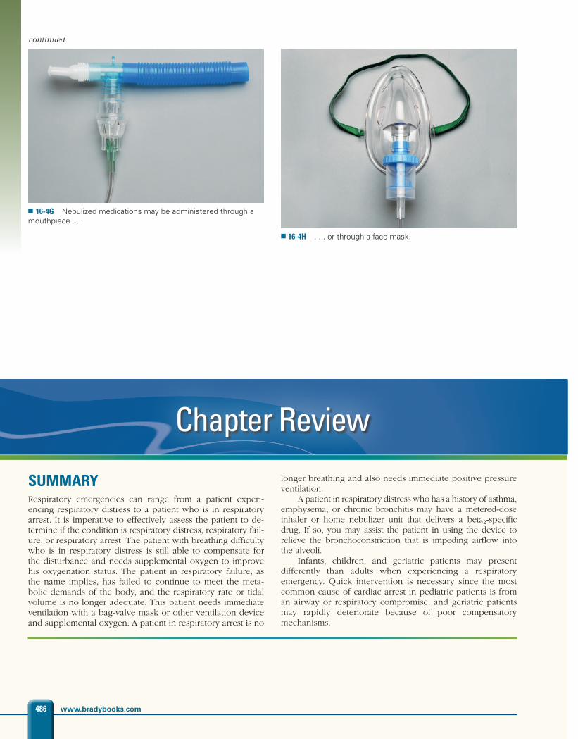
# 109912 Cust: Pearson Education / NJ / CHET Au: Mistovich Pg. No. 486 Title: Prehospital Emergency Care 10e Server:
C / M / Y / K Short / Normal
DESIGN SERVICES OF
S4carliSlePublishing Services
www.bradybooks.com486
SummaryRespiratory emergencies can range from a patient experi-encing respiratory distress to a patient who is in respiratory arrest. It is imperative to effectively assess the patient to de-termine if the condition is respiratory distress, respiratory fail-ure, or respiratory arrest. The patient with breathing difficulty who is in respiratory distress is still able to compensate for the disturbance and needs supplemental oxygen to improve his oxygenation status. The patient in respiratory failure, as the name implies, has failed to continue to meet the meta-bolic demands of the body, and the respiratory rate or tidal volume is no longer adequate. This patient needs immediate ventilation with a bag-valve mask or other ventilation device and supplemental oxygen. A patient in respiratory arrest is no
longer breathing and also needs immediate positive pressure ventilation.
A patient in respiratory distress who has a history of asthma, emphysema, or chronic bronchitis may have a metered-dose inhaler or home nebulizer unit that delivers a beta2-specific drug. If so, you may assist the patient in using the device to relieve the bronchoconstriction that is impeding airflow into the alveoli.
Infants, children, and geriatric patients may present differently than adults when experiencing a respiratory emergency. Quick intervention is necessary since the most common cause of cardiac arrest in pediatric patients is from an airway or respiratory compromise, and geriatric patients may rapidly deteriorate because of poor compensatory mechanisms.
Chapter Review
■ 16-4g Nebulized medications may be administered through a mouthpiece . . .
■ 16-4h . . . or through a face mask.
www.bradybooks.com486
continued
M16_MIST9137_10_SE_CH16.indd 486 2/28/13 5:31 PM

# 109912 Cust: Pearson Education / NJ / CHET Au: Mistovich Pg. No. 487 Title: Prehospital Emergency Care 10e Server:
C / M / Y / K Short / Normal
DESIGN SERVICES OF
S4carliSlePublishing Services
Case Study Follow-up
Chapter 16 • Respiratory Emergencies 487
Scene Size-upYou have been dispatched to a 31-year-old female patient complaining of difficulty in breathing. A man nervously greets you at the curb as you gather your equipment. He indicates that the patient is his wife, Anna Sanders, who is having an extremely hard time breathing. You are led up to the third floor of an apart-ment complex. You do not note any possible hazards, but are looking at how difficult the extrication might be. Upon walking into the apartment you note a young female patient sitting in a tripod position next to the kitchen table.
primary assessmentAs you start to introduce yourself, the patient begins to speak, gasping for her breath after each word. With great difficulty she states, “I—can’t—breathe.” Based on Mrs. Sanders’s facial expression and posture, she appears to be in a great deal of distress. Her airway is open and her breathing is rapid and labored at a rate of 34 per minute. There are audiblewheezeswhensheexhales.TheSpO2 is 78% on room air; thus, you decide to immediately apply oxygen via a nonrebreather mask at 15 lpm to maximize oxygenation of the patient. Her radial pulse is about 110 per minute. The skin is moist and slightly pale. You recognize the patient as a priority and signal your partner to get the stretcher while you continue with the secondary assessment.
Secondary assessmentYou begin to evaluate the difficulty in breathing using the OPQRSTmnemonic.You ask Anna questions shecan answer with a nod or a shake of her head to reduce her need to respond by speaking. Some questions you direct to her husband. You ascertain that the breathing difficulty began gradually about 2 hours ago and got pro-gressively worse. She is unable to lie down because this causes her breathing to get much worse, although sitting up is not much better. She has had similar episodes in thepast,butnoneseemtohavebeenthissevere.Onascale of 1 to 10, Mrs. Sanders indicates that her difficulty in breathing is about an 8 or 9.
You continue to obtain a history. The primary symptom is severe difficulty in breathing. Mrs. Sanders has an al-lergy to penicillin. When asked about medications that she takes, Mr. Sanders brings you a prescription of al buterol in a metered-dose inhaler. She is on no other medication. When asked if she has taken any of the albuterol, her husband says, “She took one puff about 15 minutes ago.” She has a past medical history of asthma and suffers these attacks maybe once every four or five months. She has had nothing to eat for about 3 hours but drank a small glass of orange juice about an hour ago. She was cleaning the kitchen when the episode began.
You quickly perform a physical exam. You assess her neck for jugular vein distention. Inspection of her chest and abdomen reveals significant use of the abdominal muscles when exhaling. The breath sounds are dimin-ished bilaterally and you hear wheezing even without using your stethoscope. Her fingertips are slightly cya-notic. You assess the baseline vital signs and find a blood pressure of 134/86; pulse of 118 per minute and regular; respirations at 32 per minute and labored with audible wheezing; the skin moist and slightly pale. Her SpO2 reading is 78% prior to oxygen administration.
The patient meets the criteria for CPAP administra-tion and does not present with any contraindications. You explain the procedure and equipment to the patient and initiate and increase the pressure to 5 cmH2O.Youcon-tact your medical director, Dr. Maxwell, for an order to administer the albuterol by a small-volume nebulizer in conjunction with the CPAP device. You check the medica-tion to ensure it is prescribed to Mrs. Sanders, that it is the correct medication, and that it has not expired. You report your physical findings and history to Dr. Maxwell. He gives you an order to administer one dose. If there is no relief of the symptoms, he instructs you to contact him for further orders. You proceed with administration of albuterol with the CPAP.
reassessmentYou reassess the vital signs following administration of the albuterol. The blood pressure is 130/84, pulse rate de-creases to 90 per minute, and respirations are now 18 per minuteandmuchlesslabored.HerSpO2 reading is 96%. The audible wheezes are minimal. The skin is not as moist and both skin and fingernails begin to return to a normal color. You secure Mrs. Sanders in a Fowler’s position on a stair chair, and you and your partner transport her down to a stretcher your partner has placed on the first floor.
You reassess the difficulty in breathing. Mrs. Sanders is now able to talk in complete sentences and indicates that the shortness of breath is much less severe. She is now only slightly short of breath. As a result of the excellent response of the patient to the treatment, you switch the oxygen therapy to a nasal cannula at 2 lpm, document your findings and emergency care, and radio the hospital with a report.
Upon arrival at the hospital, you provide the nursing staff with an oral report. You write a prehospital care re-port form as your partner restocks the ambulance. Be-fore leaving the hospital, you check in on Mrs. Sanders and find her to be relaxed and breathing well. She thanks you for your prompt response and emergency care. You then mark back in service and prepare for the next call.
An author podcast is available by going to www.bradybooks .com, Author Podcasts.
M16_MIST9137_10_SE_CH16.indd 487 2/28/13 5:31 PM

CritiCal thinKingYou arrive on the scene and find a 72-year-old female patient sitting up in her recliner in the living room of her home. She looks very fatigued and appears to be in severe respiratory distress. As you approach her, she appears extremely pale and diaphoretic with circumoral cyanosis. Her head is bobbing with each breath. As you ask her name, she can barely say it. She is gasping with each breath she takes. Her respiratory rate is 36 per minute with a shallow tidal volume. Her radial pulse is weak and rapid. Her skin is pale, very cool, and extremely moist. Her nail beds and fingertips are cyanotic. Her SpO2 reading is 82%. Her blood pressure is 92/70 mmHg. She has a history of congestive heart failure, two previous heart attacks, and hypertension.
1. What would be the immediate emergency care provided during the primary assessment?
2. What is the respiratory status of the patient? 3. How would you manage the respiratory status of the
patient? 4. What would you expect to find upon auscultation of the
lungs? 5. What areas of the lungs would be most important to
auscultate? 6. What would be the most effective method to increase
oxygenation in the patient?
www.bradybooks.com
# 109912 Cust: Pearson Education / NJ / CHET Au: Mistovich Pg. No. 488 Title: Prehospital Emergency Care 10e Server:
C / M / Y / K Short / Normal
DESIGN SERVICES OF
S4carliSlePublishing Services
in review 1. List the major signs and symptoms of breathing difficulty. 2. List the signs of adequate breathing. 3. List the signs of inadequate breathing. 4. List the steps of emergency care for a patient who is ex-
hibiting signs and symptoms of breathing difficulty but is breathing adequately (respiratory distress).
5. List the steps of emergency care for a patient who is in respiratory failure.
6. List the signs of adequate positive pressure ventilation and the steps to take if ventilation is inadequate.
7. Explain the steps to administer a medication by metered-dose inhaler and by small-volume nebulizer.
8. List the indications and contraindications for the use of a beta-agonist drug.
9. Describe the early signs of breathing difficulty in the in-fant or child; list the signs of inadequate breathing and respiratory failure in the infant or child.
10. Explain how to distinguish airway obstruction in the infant or child patient caused by disease, from airway obstruction caused by a foreign body; explain how treatment would differ for the two types of airway obstruction.
488
M16_MIST9137_10_SE_CH16.indd 488 2/28/13 5:31 PM


![Community management of chronic obstructive … · chronic bronchitis and emphysema (National Institute for Health and Care Excellence [NICE], 2010). It ... Community management of](https://static.fdocuments.us/doc/165x107/5b92d91d09d3f27f5d8c0d7a/community-management-of-chronic-obstructive-chronic-bronchitis-and-emphysema.jpg)

