The Nervous System. Types Central Nervous System (CNS)Peripheral Nervous System (PNS)
Received 08/26/2019 Review began 09/02/2019 Review ended 09/02/2019 Published 09/17/2019
© Copyright 2019Bordoni et al. This is an open accessarticle distributed under the terms ofthe Creative Commons AttributionLicense CC-BY 3.0., which permitsunrestricted use, distribution, andreproduction in any medium, providedthe original author and source arecredited.
Tentorium Cerebelli: the Bridge Betweenthe Central and Peripheral Nervous System,Part 2Bruno Bordoni , Marta Simonelli , Maria Marcella Lagana
1. Cardiology, Foundation Don Carlo Gnocchi, Milan, ITA 2. Osteopathy, French-Italian School ofOsteopathy, Pisa, ITA 3. Radiology, IRCCS Fondazione Don Carlo Gnocchi Onlus, Milan, ITA
Corresponding author: Bruno Bordoni, [email protected] Disclosures can be found in Additional Information at the end of the article
AbstractThe tentorium cerebelli is a meningeal portion in relation to the skull, the nervous system, andthe cervical tract. In this second part, the article discusses the systematic tentorialrelationships, such as the central and cervical neurological connections, the venous circulationand highlights possible clinical alterations that could cause pain. To understand the function ofanatomy, we should always remember that every area of the human body is never a segment,but a functional continuum.
Categories: Physical Medicine & Rehabilitation, Anatomy, Osteopathic MedicineKeywords: tentorium cerebelli, fascia, pain, venous circulation, neurological connections, cranio
Introduction And BackgroundCervical neurological connectionsThe ansa cervicalis characterizes the first cervical roots and connects all anterior cervical nerveexits with the inferior floor of the oral cavity, the trigeminal system, the respiratory controlsystem, and the sympathetic system. The descending branch of the hypoglossal nerveanastomoses with C1, forming the ansa hypoglossi or ansa cervicalis superior [1]. The inferiorroot of the ansa cervicalis, also known as descendens cervicalis, is formed by ascendant fibersfrom spinal nerves C2-C3 and occasionally fibers C4, lying anteriorly to the common carotidartery (it passes laterally or medially to the internal jugular vein upon anatomicalvariations) [1]. Some authors suggest an additional anastomosis with posterior cervicalnerves [2]. The ansa cervicalis communicates directly with the nodose ganglion of the vagusnerve, particularly from C1-C2, or with a branch originating directly from the vague with spinalnerves [2]. A variant of this connection to the spinal nerves and the vague is the directcommunication with the hypoglossal nerve [2]. The inferior roots of the ansa cervicalisexchange fibers with the phrenic nerve, most likely with its accessory fibers, affecting phrenicinnervation areas; a direct intervention of the first cervical roots would support the phrenicaction [3]. Sympathetic ganglia are connected to the ansa cervicalis, as well as the spinaltrigeminal ganglion [3-4]. According to some authors the dorsal root C1, or occipital nerve, maybe absent in 8% of the population, and only a low percentage has an anastomosis with the CNXI, through a branch of C1, known as the nerve of McKenzie [5]. The cervical spinal nerve 1does not show meningeal or articular branches to the vertebra. Throughout most of thepopulation, the nerve has a small ganglion, close to the intervertebral foramen and medial tothe dural tissue, functions of which we have few reliable researches about; according to someauthors, it might have a role in the perception of chemoreception variations [5]. This means
1 2 3
Open Access ReviewArticle DOI: 10.7759/cureus.5679
How to cite this articleBordoni B, Simonelli M, Lagana M (September 17, 2019) Tentorium Cerebelli: the Bridge Between theCentral and Peripheral Nervous System, Part 2. Cureus 11(9): e5679. DOI 10.7759/cureus.5679

that the ganglion might perceive inflammatory substances if they accumulated in themyofascial tissue, causing occipital pain [6]. The dorsal branch is strictly connected to thevertebral artery and is posterior to the arch of atlas, aiming to innervate sub-occipital musclesand semispinalis capitis; C1 also carries the scalp sensibility [6-7]. An ascending branch reachesthe skin of the upper-posterior portion of the neck and the anterior region of the scalp,travelling with the occipital artery [6]. This branch communicates with C2 and C3. Thedenticulate ligaments link root C1 with C2 [7]. The nerve C2 or great occipital nerve (GON),originates from the medial branch of the dorsal ramus of C2, travelling posteriorly through thefirst and second vertebra, communicating with the dorsal root of C3 [8-9]. It ascends towardsthe scalp at about 4 cm from Inion and between the inferior oblique muscle and thesemispinalis capitis. In 45% of the population, it crosses the trapezius or its inferolateralaponeurosis near Inion, where it branches [8-9]. It ascends underneath the skin in a verticalpathway with the occipital artery (the artery is often medial to the nerve), until it reaches thevertex. In this area, it shares its innervation area with the trochlear nerve of the trigeminal.This cervical branch interacts with the nuchal ligament of the trapezius, which relates directlyto the dura mater in the atlantooccipital space [10]. Studies demonstrate how a branch of C2 isinvolved with innervation of trapezius [11]. Within anterior branches of C2 the cutaneous ones,such as the transverse nerve, the lesser occipital nerve and the great auricular nerve, carry thesensation from the anterolateral cervical skin and can cause temporomandibular pain [12]. Thetransverse cervical nerve, also known as superficial cervical or cutaneous cervical (C2-C3),turns around the posterior border of the edge of sternocleidomastoid (SCM) about halfway, and,passing obliquely forward to the anterior border of the muscle, it perforates the superficialcervical fascia, and divides beneath the platysma to innervate the anterolateral part of theneck [13]. The lesser occipital nerve (C2-C3) ascends along the posterior border of the SCM tospread within the occipital and mastoidal cutaneous area [14]. The great auricular nerve arisesfrom the second and third cervical nerves (C2-C3), winds around the posterior border of theSCM (as the previous two), and runs transversally towards the auricle upon the muscle. Thethird occipital nerve (C3) is the superficial medial branch of the third cervical dorsal ramus, andit supplies the C2-C3 zygapophysial joint while crossing the joint laterally. Also, it supplies partof the semispinalis capitis muscle by travelling deeply along the muscle course, piercing thesplenius and trapezius, before sending anastomotic branches to C2. It becomes cutaneous onceexiting the nuchal ligament involving sensitive innervation of a small skin area just underneaththe nuchal line [15]. The ramus of C3 receives anastomosis from the superior cervical ganglionof the sympathetic system, and is closely related to the vertebral artery, which sends branchesright in proximity of the intervertebral foramen; it also receives anastomosis with C4 nerveroot [16].
ReviewCentral neurological connectionsThe fifth cranial nerve or trigeminal nerve is the largest of the cranial nerves. It is responsiblefor sensation and motor functions in the face, most of the scalp, teeth, oral and nasal cavity,masticatory muscle motor activity and other muscle motor activities [16-17]. It containsproprioceptive fibers from the masticatory and extraocular muscle districts [16]. The trigeminalconsists of four mesencephalic nuclei. The mesencephalic nucleus is responsible fortransmitting the proprioceptive fibers from the extraocular and masticatory muscles, regulatingthe bite strength [16]. The afferent fibers to the nucleus carry the pressure and kinestheticsensations from the teeth, the periodontium, the hard palate, and the temporomandibular joint(TMJ). This nucleus is located in the lower end of the mesencephalon and on the superior pons,laterally to the cerebral aqueduct of sylvius, and along the margins of the periaqueductal grayand anterolateral to the fourth ventricle, medial to the sensory nucleus [16]. The main sensorynucleus carries impulses for the tactile and pressure senses; it is located laterally of thetrigeminal root entry zone on the superior pons; its fibers cross the ventral posteromedialnucleus of the thalamus, composing the ventral and dorsal trigeminothalamic tracts [16]. The
2019 Bordoni et al. Cureus 11(9): e5679. DOI 10.7759/cureus.5679 2 of 7
motor nucleus is medial compared to the sensory nucleus; its fibers exit the brainstem, beingthen incorporated into the mandibular division, passing below the trigeminal ganglion withoutcreating synapses [16]. The spinal nucleus manages pain and temperature sense modalities andinvolves the C2-C4 cervical tract until it reaches the anterolateral area of the fourth ventricle. Itreceives information from the oral and nasal cavity, the facial cutaneous zones, like the cheeks,the forehead, and the jaw [16]. The trigeminal root, also called cisternal segment, is locatedbefore entering the Meckel cave and it mainly consists of two-thirds of sensory fibers. Itcontinues forward and below the tentorial border and the superior petrosal sinus, between themeningeal and periosteal layer of the dura mater, finally entering the Meckel cave [16]. Thisspace is a dural fold located on the petrous apex of the temporal bone and closes to thecavernous sinus, containing the trigeminal ganglion (Figure 1). This cave "lays" on the internalcarotid [18-19]. The motor and sensory portions entering the Meckel cave are in anastomoticcommunication, and finally, form the trigeminal ganglion or Gasser’s ganglion [19]. The XIInerve or hypoglossal nerve is considered entirely motor and plays an important role for thetongue [20]. It controls the intrinsic and extrinsic muscles of the tongue (genioglossus,hyoglossus, and styloglossus), and the infrahyoid region through neurological connections withthe ansa cervicalis [20]. The XII nerve can be divided based on its anatomical area: intracranial,cisternal, skull base, and extracranial [21]. The first segment is the hypoglossal nucleus, locatedin the dorsal spinal cord between the midline and the dorsal nucleus of the X cranial nerve; itcontinues as a thin nucleus with a caudocranial direction to the medulla [21]. The hypoglossalnucleus receives fibers from the glossopharyngeal nerve, the vague and the trigeminal system,in order to mediate a wide variety of stimuli and reflexes coming from the tongue and thepharyngeal mucosa, necessary for swallowing and phonating. Circulation comes from anteriorspinal and vertebral arteries [21]. The cisternal segment is formed by the coalescence of rootscoming out the spinal cord, generating the XII nerve; it can blend with some fibers of the Xnerve, and its fibers pass behind the vertebral artery, before entering the hypoglossal canal [21-22]. The skull base area covered by the XII nerve is the hypoglossal canal, which is a foramen inthe occipital bone located below the jugular foramen; it runs obliquely forwards (anterolateral)superiorly to each occipital condyle, while the jugular foramen is lateral from the condyle [21].The extracranial segment can be further divided into a superior carotid space and anteriorspace [21]. When the hypoglossal nerve exits the hypoglossal canal, near the atlas, itanastomoses with some branches of the superior cervical ganglion of the sympathetic system,and with a fiber that connects to the first and second ascending cervical branches (C1-C4 oransa cervicalis or hypoglossal ansa). A descending branch of the hypoglossal nerve (or superiorroot of the ansa cervicalis) initiates from this loop, and it innervates the infrahyoid muscles,such as the thyrohyoid and geniohyoid muscles [21, 23]. This branch may enter the thoraxengaging the parasympathetic and sympathetic trunk and the visceral function of themediastinum [23]. Within the ansa cervicalis area, the XII nerve communicates with theglossopharyngeal nerve, with the possibility of anastomosis between the two branches of thehypoglossal nerve, prior to the hyoid bone, between the genioglossus muscle above and thegeniohyoid muscle below; this anastomosis is known with different names, such as thesuprahyoid loop of the hypoglossal nerve or Hyrtl’s suprahyoid ansa [23]. The descendingbranch of the hypoglossal nerve can anastomose with the phrenic nerve/ansa cervicalis,innervating the sternothyroid muscle, known as Valentin’s anastomosis [23].
2019 Bordoni et al. Cureus 11(9): e5679. DOI 10.7759/cureus.5679 3 of 7

FIGURE 1: The image shows the dural connections and thevenous sinuses.
Venous circulationThe tentorium cerebelli influences the posterior cranial venous circulation (Figure 2). Thesuperior and inferior sagittal sinuses, and the sinus rectus converge to the torcular herophili,while the occipital sinus can arise from the internal occipital protuberance, which is theinsertion of the posterior tentorium cerebelli [24]. A dural structural alteration of the tentoriumcould cause venous diseases, which may affect the central nervous system and the cervicalnervous system [25-28].
2019 Bordoni et al. Cureus 11(9): e5679. DOI 10.7759/cureus.5679 4 of 7
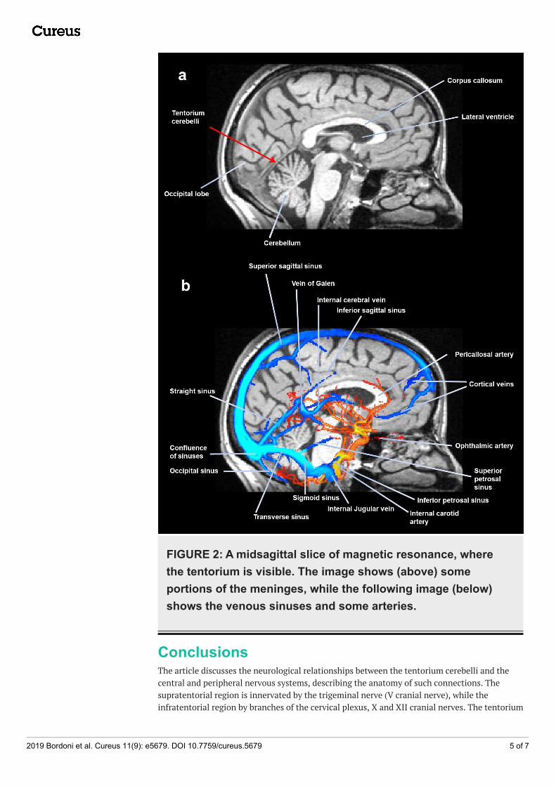
FIGURE 2: A midsagittal slice of magnetic resonance, wherethe tentorium is visible. The image shows (above) someportions of the meninges, while the following image (below)shows the venous sinuses and some arteries.
ConclusionsThe article discusses the neurological relationships between the tentorium cerebelli and thecentral and peripheral nervous systems, describing the anatomy of such connections. Thesupratentorial region is innervated by the trigeminal nerve (V cranial nerve), while theinfratentorial region by branches of the cervical plexus, X and XII cranial nerves. The tentorium
2019 Bordoni et al. Cureus 11(9): e5679. DOI 10.7759/cureus.5679 5 of 7

has fascial relationships with the cervical tract thanks to connections with the sub-occipitalmuscle, the nuchal and yellow ligaments, and the Hoffman ligaments. The tentorium affects thecerebrospinal fluid and venous circulation.
Additional InformationDisclosuresConflicts of interest: In compliance with the ICMJE uniform disclosure form, all authorsdeclare the following: Payment/services info: All authors have declared that no financialsupport was received from any organization for the submitted work. Financial relationships:All authors have declared that they have no financial relationships at present or within theprevious three years with any organizations that might have an interest in the submitted work.Other relationships: All authors have declared that there are no other relationships oractivities that could appear to have influenced the submitted work.
References1. Banneheka S: Anatomy of the ansa cervicalis: nerve fiber analysis . Anat Sci Int. 2008, 83:61-
67. 10.1111/j.1447-073X.2007.00202.x2. Diamond M, Wartmann CT, Tubbs RS, Shoja MM, Cohen-Gadol AA, Loukas M: Peripheral
facial nerve communications and their clinical implications. Clin Anat. 2011, 24:10-18.10.1002/ca.21072
3. Banneheka S: Morphological study of the ansa cervicalis and the phrenic nerve . Anat Sci Int.2008, 83:31-44. 10.1111/j.1447-073X.2007.00201.x
4. Bordoni B, Zanier E: The continuity of the body: hypothesis of treatment of the fivediaphragms. J Altern Complement Med. 2015, 21:237-242.
5. Bordoni B, Zanier E: Anatomic connections of the diaphragm: influence of respiration on thebody system. J Multidiscip Healthc. 2013, 6:281-291. 10.2147/JMDH.S45443
6. Tubbs RS, Loukas M, Yalçin B, Shoja MM, Cohen-Gadol AA: Classification and clinicalanatomy of the first spinal nerve: surgical implications. J Neurosurg Spine. 2009, 10:390-394.10.3171/2008.12.SPINE08661
7. Tubbs RS, Loukas M, Slappey JB, Shoja MM, Oakes WJ, Salter EG: Clinical anatomy of the C1dorsal root, ganglion, and ramus: a review and anatomical study. Clin Anat. 2007, 20:624-627.10.1002/ca.20472
8. Tubbs RS, Watanabe K, Loukas M, Cohen-Gadol AA: The intramuscular course of the greateroccipital nerve: novel findings with potential implications for operative interventions andoccipital neuralgia. Surg Neurol Int. 2014, 5:155. 10.4103/2152-7806.143743
9. Kemp WJ 3rd, Tubbs RS, Cohen-Gadol AA: The innervation of the scalp: a comprehensivereview including anatomy, pathology, and neurosurgical correlates. Surg Neurol Int. 2011,2:178.
10. Dean NA, Mitchell BS: Anatomic relation between the nuchal ligament (ligamentum nuchae)and the spinal dura mater in the craniocervical region. Clin Anat. 2002, 15:182-185.10.1002/ca.10001
11. Tubbs RS, Shoja MM, Loukas M, Lancaster J, Mortazavi MM, Hattab EM, Cohen-Gadol AA:Study of the cervical plexus innervation of the trapezius muscle . J Neurosurg Spine. 2011,14:626-629. 10.3171/2011.1.SPINE10717
12. Gupta C, D'souza AS, Raythe B: Anatomical variations in the emergence of the cutaneousnerves from the nerve point in the neck and identification of the landmarks to locate thenerve point with its clinical implications: a cadaveric study on South Indian human foetuses. JClin Diagn Res. 2013, 7:413-417. 10.7860/JCDR/2013/4598.2787
13. Standring S: Head and neck. Gray’s Anatomy: The Anatomical Basis of Clinical Practice.Fortieth edition. Churchill Livingstone, London, UK; 2008. pp. 435-466.
14. Cao J, Fu D, Li S: A three-dimensional digital visualization model of cervical nerves in ahealthy person. Neural Regen Res. 2013, 8:1829-1836. 10.3969/j.issn.1673-5374.2013.20.001
15. Tubbs RS, Mortazavi MM, Loukas M, D'Antoni AV, Shoja MM, Chern JJ, Cohen-Gadol AA:Anatomical study of the third occipital nerve and its potential role in occipital headache/neck
2019 Bordoni et al. Cureus 11(9): e5679. DOI 10.7759/cureus.5679 6 of 7

pain following midline dissections of the craniocervical junction. J Neurosurg Spine. 2011,15:71-75. 10.3171/2011.3.SPINE10854
16. Joo W, Yoshioka F, Funaki T, Mizokami K, Rhoton AL Jr: Microsurgical anatomy of thetrigeminal nerve. Clin Anat. 2014, 27:61-88. 10.1002/ca.22330
17. Bathla G, Hegde AN: The trigeminal nerve: an illustrated review of its imaging anatomy andpathology. Clin Radiol. 2013, 68:203-213. 10.1016/j.crad.2012.05.019
18. Arslan M, Deda H, Avci E, et al.: Anatomy of Meckel's cave and the trigeminal ganglion:anatomical landmarks for a safer approach to them. Turk Neurosurg. 2012, 22:317-323.10.5137/1019-5149.JTN.5213-11.1
19. Sabancı PA, Batay F, Civelek E, Al Mefty O, Husain M, Abdulrauf SI, Karasu A: Meckel's cave.World Neurosurg. 2011, 76:335-341. 10.1016/j.wneu.2011.03.037
20. Bordoni B, Marelli F, Morabito B: The tongue after whiplash: case report and osteopathictreatment. Int Med Case Rep J. 2016, 9:179-182. 10.2147/IMCRJ.S111147
21. Alves P: Imaging the hypoglossal nerve. Eur J Radiol. 2010, 74:368-377.10.1016/j.ejrad.2009.08.028
22. Bademci G, Yaşargil MG: Microsurgical anatomy of the hypoglossal nerve . J Clin Neurosci.2006, 13:841-847. 10.1016/j.jocn.2005.12.028
23. Shoja MM, Oyesiku NM, Shokouhi G, et al.: A comprehensive review with potentialsignificance during skull base and neck operations, Part II: glossopharyngeal, vagus,accessory, and hypoglossal nerves and cervical spinal nerves 1-4. Clin Anat. 2014, 27:131-144.10.1002/ca.22342
24. Kiliç T, Akakin A: Anatomy of cerebral veins and sinuses . Front Neurol Neurosci. 2008, 23:4-15. 10.1159/000111256
25. Kortman HG, Boukrab I, Bloemsma G, et al.: Tentorial dural arteriovenous fistulas: a single-center cohort of 12 patients. J Cerebrovasc Endovasc Neurosurg. 2017, 19:284-290.10.7461/jcen.2017.19.4.284
26. Gioppo A, Faragò G, Caldiera V, Caputi L, Cusin A, Ciceri E: Medial tentorial duralarteriovenous fistula embolization: single experience with embolic liquid polymer SQUID andreview of the literature. World Neurosurg. 2017, 107:1050-1051. 10.1016/j.wneu.2017.08.050
27. Kim WY, Kim JB, Nam TK, Kim YB, Park SW: Cervical myelopathy caused by intracranial duralarteriovenous fistula. Korean J Spine. 2016, 13:67-70. 10.14245/kjs.2016.13.2.67
28. Santillan A, Safdieh JE, Gobin YP, Patsalides A: Neurological picture. Bilateral thalamicvenous hypertension caused by a tentorial dural arteriovenous fistula: endovasculartreatment. J Neurol Neurosurg Psychiatry. 2011, 82:749-750. 10.1136/jnnp.2010.231969
2019 Bordoni et al. Cureus 11(9): e5679. DOI 10.7759/cureus.5679 7 of 7