Parathyroid gland
61
Diseases of parathyroid gland
Transcript of Parathyroid gland
Diseases of parathyroid gland


Parathyroid Hormone:
• parathyroid Hormone Structure and Synthesis PTH is an 84-amino-acid single-chain peptide. The amino acid portion, PTH(1–34), is highly conserved and is critical for the biologic actions of the molecule. The PTH gene is located on chromosome 11.
• PTH is initially synthesized as a larger molecule ( preproparathyroidhormone, consisting of 115 amino acids), which is then reduced in size by a second cleavage ( proparathyroid hormone, 90 amino acids) before secretion as the 84-amino-acid peptide.
• Transcriptional suppression of the PTH gene by calcium is nearly maximal at physiologic calcium concentrations.
• Hypocalcemia increases transcriptional activity within hours. 1,25(OH) 2 D 3 strongly suppresses PTH gene transcription.
Parathyroid Hormone
• Regulation of PTH Secretion
• PTH secretion increases steeply to a maximum value of five times the basal rate of secretion as calcium concentration falls from normal to the range of 7.5–8.0 mg/ dL .
• The ionized fraction of blood calcium is the important determinant of hormone secretion.
• ECF calcium controls PTH secretion by interaction with a calcium sensor, a G protein–coupled receptor (GPCR) for which Ca 2+ ions act as the ligand .
• Stimulation of the receptor by high calcium levels suppresses PTH secretion. The receptor is present in parathyroid glands.
• Point mutations associated with loss of function cause a syndrome FHH resembling hyperparathyroidism but with hypocalciuria . On the other hand, gain-of-function mutations cause a form of hypocalcemia resembling hypoparathyroidism .
• Metabolism -
• Secreted PTH has a half-life of 2 to 4 minutes. In the liver, PTH is metabolized into the active N-terminal component and the relatively inactive C-terminal fraction. The C-terminal component is excreted by the kidneys .Much of the proteolysis of hormone occurs in the liver and kidney.

Calcitonin and Vitamin D• Calcitonin and Vitamin D Calcitonin is produced by thyroid C cells and functions as
an antihypercalcemic hormone by inhibiting osteoclast -mediated bone resorption• . Calcitonin production is stimulated by calcium and catecholamines ,
cholecystokinin , and glucagon.• At the kidney, calcitonin increases phosphate excretion by inhibiting its
reabsorption .• Calcitonin plays a minimal, if any, role in the regulation of calcium levels in
humans. However, it is very useful as a marker of MTC and in treating acute hypercalcemic crisis.
• Vitamin D refers to vitamin D2 and vitamin D3 , both of which are produced by photolysis of naturally occurring sterol precursors.
• Vitamin D 2 is available commercially, whereas vitamin D 3 is the most important physiologic compound .
• Vitamin D is metabolized in the liver to its primary circulating form, 25-hydroxyvitamin D. Further hydroxylation in the kidney results in 1,25-dihydroxy vitamin D, which is the most metabolically active form of vitamin D.
• Vitamin D stimulates the absorption of calcium and phosphate from the gut and the resorption of calcium from the bone.
Hyperfunction of the parathyroid glands may be classified as primary, secondary, or tertiary.
• PHPT arises from increased PTH production from abnormal parathyroid glands and results from a disturbance of normal feedback control exerted by serum calcium.
• Elevated PTH levels may also occur as a compensatory response to hypocalcemic states resulting from chronic renal failure or GI malabsorption of calcium.
• This secondary HPT can be reversed by correction of the underlying problem (e.g., kidney transplantation for chronic renal failure).
• However, chronically stimulated glands may occasionally become autonomous, resulting in persistence or recurrence of hypercalcemia after successful renal transplantation, resulting in tertairy HPT.
Hyperparathyroidism

Primary Hyperparathyroidism
• PHPT is a common disorder, affecting 100,000 individuals annually in the United States.
• PHPT occurs in 0.1 to 0.3% of the general population and is more common in women (1:500) than in men (1:2000).
• Increased PTH production leads to hypercalcemia via increased GI absorption of calcium, increased production of vitamin D3 , and reduced renal calcium clearance.
• PHPT is characterized by increased parathyroid cell proliferation and PTH secretion that is independent of calcium levels.

Etiology
• The exact cause of PHPT is unknown-• 1 Exposure to low-dose therapeutic ionizing radiation .• 2 Familial predisposition account for some cases. • 3 Declining renal function with age as well as alteration in the sensitivity of parathyroid glands to
suppression by calcium. • 4 Lithium therapy has been known to shift the set point for PTH secretion in parathyroid cells,
thereby resulting in elevated PTH levels and mild hypercalcemia . Lithium stimulates the growth of abnormal parathyroid glands in vitro and also in susceptible patients in vivo.
• 5 PHPT results from the enlargement of a single gland or parathyroid adenoma in approximately 80% of cases, multiple adenomas or hyperplasia in 15 to 20% of patients, and parathyroid carcinoma in 1% of patients.
• - When more than one abnormal parathyroid gland is identified preoperatively or intraoperatively , the patient has hyperplasia (all glands abnormal) until proven otherwise.
• -Genetics-Most cases of PHPT are sporadic. However, PHPT also occurs within the spectrum of a number of inherited disorders such as MEN1, MEN2A, isolated familial HPT, and familial HPT with jaw- tumor syndrome. All of these syndromes are inherited in an autosomal dominant fashion.

MEN-1
•PHPT is the earliest and most common manifestation of MEN1 and develops in 80 to 100% of patients by age 40 years old.
• These patients also are prone to pancreatic neuroendocrine tumors and pituitary adenomas and, less commonly, to adrenocorticaltumors , lipomas , skin angiomas , and carcinoid tumors of the bronchus, thymus, or stomach.
• Prolactinomas occur in 10 to 50% of MEN1 patients and constitute the most common pituitary lesion.
• MEN1 has been shown to result from germline mutations in the MEN1 gene, a tumor -suppressor gene located on chromosome 11q12-13 which encodes menin , a protein that is postulated to interact with the transcription factors .

MEN2A and other syndromes:
• HPT develops in about 20% of patients with MEN2A and generally is less severe.
• MEN2A ( Sipple syndrome) , is characterized by pheochromocytoma , medullary carcinoma, and parathyroid hyperplasia. MEN2A is caused by germlinemutations of the RET proto-oncogene located on chromosome 10.
• Patients with the familial HPT with jaw- tumor syndrome have an increased predisposition to parathyroid carcinoma. This syndrome maps to a tumor -suppressor locus HRPT2 , on chromosome 1.
• Patients belonging to isolated HPT kindreds also appear to demonstrate linkage to HRPT2 .l]

Genetics
• Approximately 25 to 40% of sporadic parathyroid adenomas and some hyperplastic parathyroid glands have mutation at 11q 13, the site of the MEN1 gene.
• The parathyroid adenoma 1 oncogene ( PRAD1 ), which encodes cyclin D1, a cell cycle control protein, is overexpressed in about 18% of parathyroid adenomas.
• Other chromosomal regions deleted in parathyroid adenomas include 1p, 6q, 15q, 16p and 19p.
• Sporadic parathyroid cancers are characterized by uniform loss of the tumor -suppressor gene RB , which is involved in cell cycle regulation, and 60% have HRPT2 mutations.
• These alterations are rare in benign parathyroid tumors and may have implications for diagnosis. The p53 tumor -suppressor gene is also inactivated in a subset (30%) of parathyroid carcinomas.

Clinical Manifestations
• Patients with PHPT formerly presented with the "classic" pentad of symptoms (i.e., kidney stones, painful bones, abdominal groans, psychic moans, and fatigue overtones).
• Currently, most patients present with weakness, fatigue, polydipsia , polyuria , nocturia , bone and joint pain, constipation, decreased appetite, nausea, heartburn, pruritus , depression, and memory loss.
• Furthermore, these symptoms and signs improve in most, but certainly not all, patients after parathyroidectomy . Truly "asymptomatic" PHPT appears to be rare, occurring in <5% of patients.

Renal Disease
• Approximately 80% of patients with PHPT have some degree of renal dysfunction or symptoms.
• Kidney stones were previously reported in up to 80% of patients but now occur in about 20 to 25%.
• The calculi are typically composed of calcium phosphate or oxalate. • Nephrocalcinosis , which refers to renal parenchymal calcification, is found
in <5% of patients and is more likely to lead to renal dysfunction. • Chronic hypercalcemia also can impair concentrating ability, thereby
resulting in polyuria , polydipsia , and nocturia . Hypertension is reported to occur in up to 50% of patients with PHPT.
• Hypertension appears to be more common in older patients and correlates with the magnitude of renal dysfunction and, in contrast to other symptoms, is least likely to improve after parathyroidectomy

Bone Disease
• Bone disease, including osteopenia , osteoporosis, and osteitis fibrosacystica , is found in about 15% of patients with PHPT.
• Increased bone turnover, as found in patients with osteitis fibrosa cystica , can be determined by an elevated blood alkaline phosphatase level.
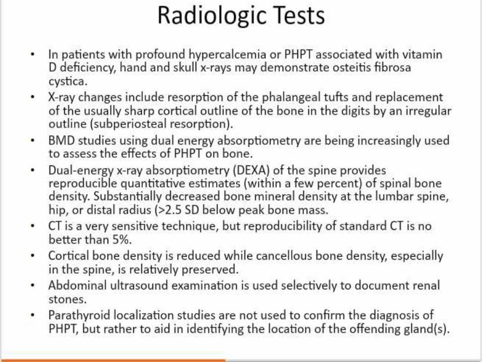
• Advanced PHPT with osteitis fibrosa cystica now occurs in <5% of patients. • It has pathognomonic radiologic findings, which are best seen on x-rays of
the hands and are characterized by subperiosteal resorption (most apparent on the radial aspect of the middle phalanx of the second and third fingers), bone cysts, and tufting of the distal phalanges .
• The skull also may be affected and appears mottled with a loss of definition of the inner and outer cortices.
• Brown or osteoclastic tumors and bone cysts also may be present.• Severe bone disease, resulting in bone pain and tenderness and/or
pathologic fractures, is rarely observed nowaday

Bone Disease
• Reductions of bone mineral density (BMD) with osteopenia and osteoporosis are more common.
• Patients with normal serum alkaline phosphatase levels almost never have clinically apparent osteitis fibrosa cystica .
• HPT typically results in a loss of bone mass at sites of cortical bone such as the radius and relative preservation of cancellous bone such as that located at the vertebral bodies.
• Patients with PHPT, however, also may have osteoporosis of the lumbar spine that improves dramatically following parathyroidectomy .
• Fractures also occur more frequently in patients with PHPT, and the incidence of fractures also decreases after parathyroidectomy .
• Bone disease correlates with serum PTH and vitamin D levels.
Gastrointestinal Complications
• PHPT has been associated with peptic ulcer disease.
• An increased incidence of pancreatitis also has been reported in patients with PHPT, although this appears to occur only in patients with profound hypercalcemia (Ca 2+ ≥12.5 mg/ dL ).
• Patients with PHPT also have an increased incidence of cholelithiasis , presumably due to an increase in biliary calcium, which leads to the formation of calcium bilirubinate stones.

Neuropsychiatric Complications
• Severe hypercalcemia may lead to various neuropsychiatric manifestations such as florid psychosis, obtundation , or coma.
• In mild hypercalcemia symptoms such as depression, anxiety, and fatigue are more commonly observed.
• The etiology of these symptoms is not known. Studies demonstrate that levels of certain neurotransmitters (monoamine metabolites 5-hydroxyindoleacetic acid and homovanillic acid) are reduced in the cerebrospinal fluid of patients with PHPT when compared to controls.
• Electroencephalogram abnormalities also occur in patients with primary and secondary HPT and normalize following parathyroidectomy .

Other Features
• PHPT also can lead to fatigue and muscle weakness, which is prominent in the proximal muscle groups. Although the exact etiology of this finding is not known, muscle biopsy studies show that weakness results from a neuropathy, rather than a primary myopathic abnormality.
• Patients with HPT also have an increased incidence of chondrocalcinosis , gout, and pseudogout , with deposition of uric acid, calcium, pyrophosphate crystals in the joints.
• Calcification at ectopic sites such as blood vessels, cardiac valves, and skin also has been reported, as has hypertrophy of the left ventricle independent of the presence of hypertension

Physical Findings
• Parathyroid tumors are seldom palpable, except in patients with profound hypercalcemia .
• A palpable neck mass in a patient with PHPT is more likely to be thyroid in origin or a parathyroid cancer.
• Patients also may demonstrate evidence of band keratopathy , a deposition of calcium in Bowman's membrane just inside the iris of the eye.
• This nonspecific condition generally is caused by chronic eye diseases such as uveitis , and glaucoma, but also may occur in the presence of conditions associated with high calcium or phosphate levels.
• Fibro-osseous jaw tumors , and or the presence of familial disease in patients with PHPT and jaw tumors , if present, should alert the physician to the possibility of parathyroid carcinoma.

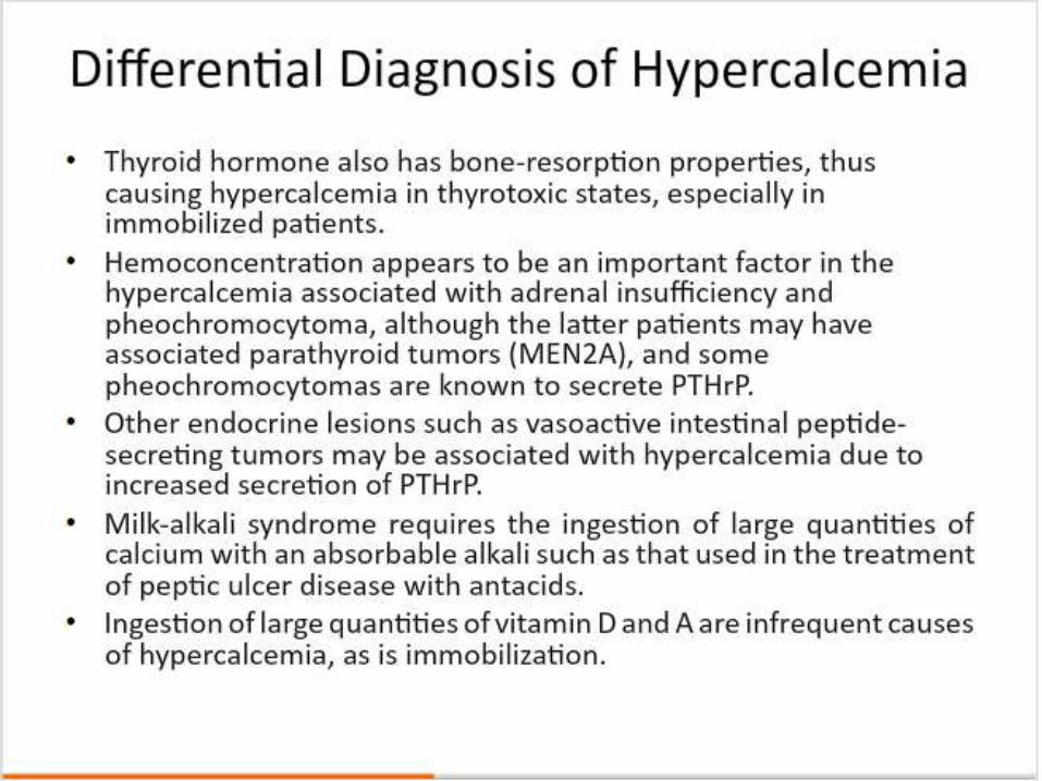
Differential Diagnosis of Hypercalcemia
• primary, secondary, tertiary • Malignancy—hematologic (multiple myeloma), solid
tumors (due to PTHrP )• Endocrine diseases—hyperthyroidism, addisonian crisis,
VIPoma• Granulomatous diseases— sarcoidosis , tuberculosis,
berylliosis , histoplasmosis• Milk-alkali syndrome Drugs— thiazide diuretics, lithium, • vitamin A or D intoxication • Familial hypocalciuric hypercalcemia• Paget's disease• Immobilization


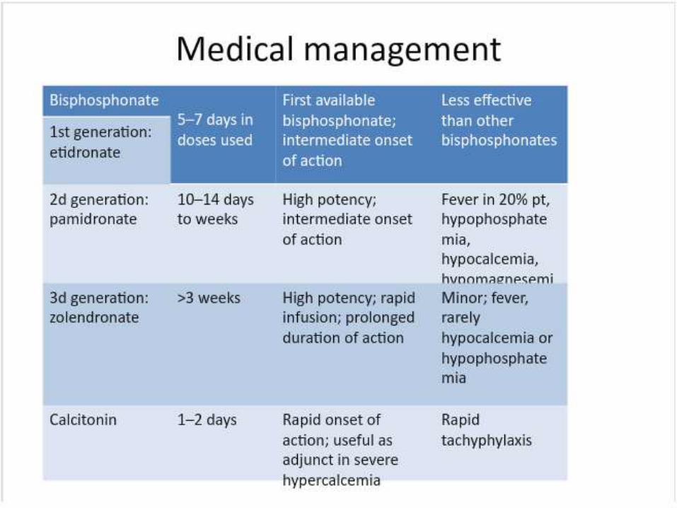
Medical management
• A number of medical therapies such as selective estrogen receptor modifiers and bisphosphonates have been used to successfully lower serum calcium and increase BMD in patients with PHPT.
• More recently, calcimimetics (modifiers of the sensitivity of the CASR) have been used in randomized, multicenter controlled trials and have been shown to decrease both serum calcium and PTH levels in both symptomatic and asymptomatic PHPT patients. Although these therapies show promise, long-term outcome data are lacking, and their routine use is not advocated at this time.


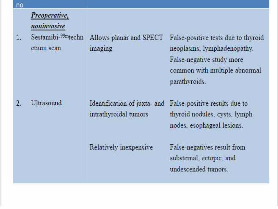
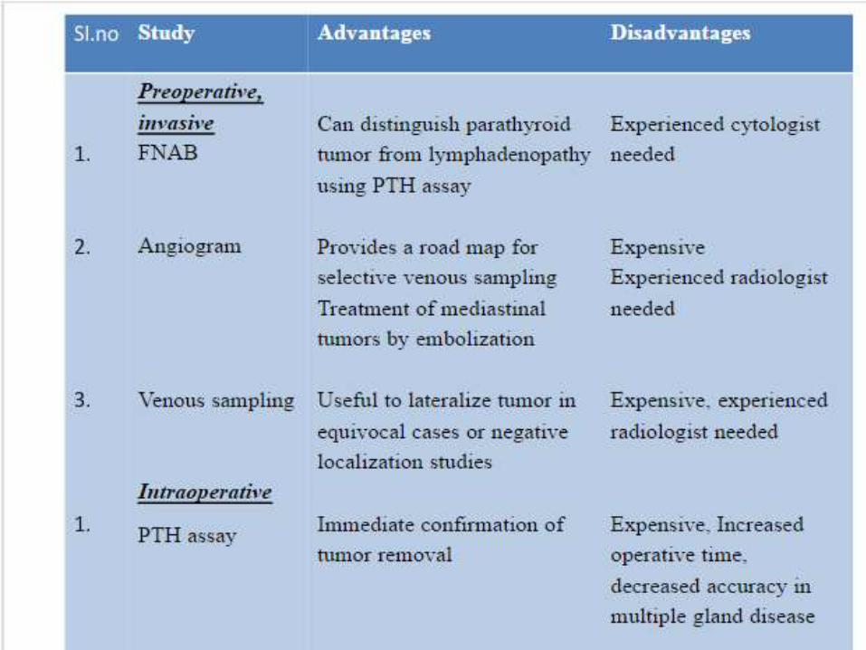
Localization studies:
• Single-photon emission computed tomography(SPECT), particularly when used with CT, has been shown to be superior to other nuclear medicine–based imaging

Parathyroidectomy
• Parathyroidectomy
• Unilateral parathyroid exploration
• Radio-guided parathyroidectomy
• Total endoscopic parathyroidectomy
• Minimally invasive parathyroidectomy
• Autotransplantation For patients with hyperplasia, a titanium clip is placed across the most normal gland.
• Sternotomy- A sternotomy is usually not recommended at the initial operation, unless the calcium level is >13 mg/ dL

Complications of Parathyroid Surgery
• Transient and permanent vocal cord palsy and hypoparathyroidism.
• Patients with symptomatic hypocalcemia or those with calcium levels <8 mg/ dL are treated with oral calcium supplementation (up to 1 to 2 g every 4 hours). 1,25-dihydroxy vitamin D [ calcitriol ( Rocaltrol ) 0.25 to 0.5 g bid] .
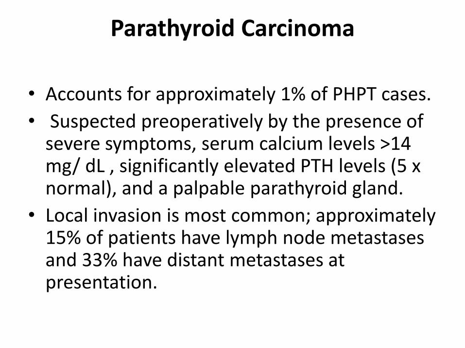
Parathyroid Carcinoma
• Accounts for approximately 1% of PHPT cases.
• Suspected preoperatively by the presence of severe symptoms, serum calcium levels >14 mg/ dL , significantly elevated PTH levels (5 x normal), and a palpable parathyroid gland.
• Local invasion is most common; approximately 15% of patients have lymph node metastases and 33% have distant metastases at presentation.

Parathyroid Carcinoma
• Prophylactic neck dissection is not advised because it is associated with an increased risk of complications and does not appear to have a significant impact on survival.
• Reoperation is indicated for locally recurrent or metastatic disease to control hypercalcemia .
• Radiation and chemotherapy may be considered in patients with unresectable disease.

Familial Hyperparathyroidism
• Generally have a higher incidence of multiglandular disease, supernumerary glands, and recurrent or persistent disease.
• Warrant a more aggressive approach and are not candidates for various focused surgical approaches.
• Preoperative sestamibi scan and ultrasound can be obtained in patients with inherited HPT to identify potential ectopic glands.
• A bilateral cervical thymectomy , regardless of the results of localization studies.
• Both subtotal parathyroidectomy and total parathyroidectomy with autotransplantation are appropriate and parathyroid tissue also should be cryopreserved .

Parathyromatosis
• Parathyromatosis is a rare condition characterized by the finding of multiple nodules of hyper-functioning parathyroid tissue throughout the neck and mediastinum , usually following a previous parathyroidectomy .
• The true etiology of parathyromatosis is not known. It is postulated to arise either from overgrowth of congenital parathyroid rests or seeding at surgery from rupture of parathyroid tumors or subtotal resection of hyperplasticglands.
• Parathyromatosis represents a rare cause of persistent or recurrent HPT and can be identified intraoperatively .
• Aggressive local resection of these deposits can result in normocalcemia but is rarely curative.

Secondary Hyperparathyroidism
• Secondary HPT commonly occurs in patients with chronic renal failure but also may occur in those with hypocalcemia secondary to inadequate calcium or vitamin D intake, or malabsorption .
• The pathophysiology of HPT in chronic renal failure is complex and appears to be related to hyperphosphatemia (and resultant hypocalcemia), deficiency of 1,25-dihydroxy vitamin D due to loss of renal tissue, low calcium intake, decreased calcium absorption. Patients generally are hypocalcemic or normocalcemic . Aluminum hydroxide, which often was used as a phosphate binder, has been shown to contribute to the osteomalacia observed in this disease. These patients generally are treated medically with a low-phosphate diet, phosphate binders, adequate intake of calcium and 1,25-dihydroxy vitamin D and a high calcium, low- aluminum dialysis bath.
• Calcimimetics have been shown to control parathyroid hyperplasia and osteitis fibrosa cystica associated with secondary HPT and to decrease plasma PTH and total and ionized calcium levels in humans.

Secondary Hyperparathyroidism
• Surgical treatment was traditionally recommended for,
1 patients with bone pain, pruritus , and
2 a calcium-phosphate product ≥70,
3 calcium >11 mg/ dL with markedly elevated PTH,
4 calciphylaxis ,
5 progressive renal osteodystrophy , and soft tissue calcification and tumoral calcinosis , despite maximal medical therapy.
Tertiary Hyperparathyroidism
• Generally, renal transplantation is an excellent method of treating secondary HPT, but some patients develop autonomous parathyroid gland function i,e . Tertiary HPT.
• Tertiary HPT can cause problems similar to PHPT, such as pathologic fractures, bone pain, renal stones, peptic ulcer disease, pancreatitis, and mental status changes.
• The transplanted kidney is also at risk.• Operative intervention is indicated for symptomatic disease or if
autonomous PTH secretion persists for >1 year after a successful transplant.
• Subtotal or total parathyroidectomy with autotransplantation and an upper thymectomy .
• All parathyroid glands be identified and subtotal parathyroidectomy be performed as long-term follow-up studies show that limited excisions in these patients are associated with an up to fivefold increased risk of recurrent or persistent disease.

Hypoparathyroidism
• Is an endocrine disorder in which hypocalcemia and hyperphosphatemia are the result of a deficiency in PTH secretion or action.
• The parathyroid glands may be congenitally absent in DiGeorge syndrome, an autosomal dominant form involving microdeletions of chromosome 22q11.2 has been described which also is characterized by lack of thymic development and, cardiovascular, facial, and other developmental defects, and most patients die in early childhood with severe infections, hypocalcemiaand seizures, or cardiovascular complications
Hypoparathyroidism
• It can occur as part of a multiglandular endocrine deficiency syndrome (type 1) characterized most commonly by hypoparathyroidism , adrenal insufficiency, and mucocutaneous candidiasis .
• By far, the most common cause of hypoparathyroidism is thyroid surgery , particularly total thyroidectomy with a concomitant central neck dissection. Patients often develop transient hypocalcemia due to ischemia of the parathyroid glands; permanent hypoparathyroidism is rare.
• Hypoparathyroidism also may occur after parathyroid surgery, which is more likely if patients undergo a subtotal resection or total parathyroidectomy with parathyroid autotransplantation.

Conditions Causing Hypocalcemia
• Hypoparathyroidism -Surgical Neonatal FamilialHeavy metal deposition Magnesium depletion
Resistance to the action of PH PseudohypoparathyroidimRenal failure Medications— calcitonin , bisphosphonates , mithramycin
Conditions Causing Hypocalcemia
• Failure of normal 1,25-dihydroxy vitamin D production
• Resistance to the action of 1,25-dihydroxy vitamin D
• Acute complex
formation or deposition of calcium
Acute hyperphosphatemia
Acute pancreatitis
Massive blood transfusion (citrate overload)

Clinical features
• Patients initially develop circumoral and fingertip numbness and tingling.
• Mental symptoms include anxiety, confusion, and depression.• Physical examination reveals positive Chvostek's sign (contraction
of facial muscles elicited by tapping on the facial nerve anterior to tragus of the ear) and Trousseau's sign ( carpopedal spasm which is elicited by occluding blood flow to the forearm with a blood pressure cuff for 2 to 3 minutes).
• Erb`s sign – increased electric excitability of the muscles. • Tetany , which is characterized by tonic- clonic seizures, carpopedal
spasm, and laryngeal stridor , may prove fatal and should be prevented.

Treatment
• Most patients with postoperative hypocalcemia can be treated with oral calcium and vitamin D supplements;
• IV calcium infusion is rarely required except in patients with preoperative osteitis fibrosacystica .
• IV calcium gluconate infusion for the treatment of moderate to severe hypocalcemia
Pseudohypoparathyroidism
• PHP is a hereditary disorder characterized by symptoms and signs of hypoparathyroidism , typically in association with distinctive skeletal and developmental defects.
• The hypoparathyroidism is due to a deficient end-organ response to PTH.
• Hyperplasia of the parathyroids , a response to hormone resistance, causes elevation of PTH levels.

Pseudohypoparathyroidism
• Patients have low calcium and high phosphate levels, as with true hypoparathyroidism . PTH levels, however, are elevated, reflecting resistance to hormone action.
• AHO, consisting of short stature, round face, skeletal anomalies ( brachydactyly ), and heterotopic calcification is seen in PHP-Ia.
• Response of Urinary cAMP to PTH is low in Type-l and normal in Type 2.

Genetics
• Multiple defects have now been identified in the GNAS-1 gene in PHP- Ia and PPHP patients.
• This gene, which is located on chromosome 20q13, encodes the stimulatory G-protein subunit G s α .
• PHP- Ia and PPHP, have an inheritance pattern consistent with gene imprinting —only females, can transmit the full disease with hypocalcemia .
• In the renal cortex, it is postulated that only the maternal allele is normally active (independent of any mutation).
• Maternal gene imprinting- PHP-la. • Paternal gene imprinting- PPHP.

Pseudohypoparathyroidism
• Ellsworth-Howard test- Measurement of serum and urinaryphosphorus after intravenous administration of parathyroid extract (200U); used in the diagnosis
of Pseudohypoparathyroidism .• Normally -5 fold increase in urinary phosphorus excretion.• Hypoparathyroidism -10 fold increase in urinary
phosphorus excretion.• Pseudohypoparathyroidism - <2 fold increase in urinary
phosphorus excretion.• Treatment Similar to that of hypoparathyroidism , except
that the doses of vitamin D and calcium are usually lower than those required in true hypoparathyroidism .