Parasite Ecology of Fish with Black Spot Disease
30
Running head: PARASITE ECOLOGY 1 Parasite Ecology of Fish with Black Spot Disease Sarah C. Kirse A Senior Thesis submitted in partial fulfillment of the requirements for graduation in the Honors Program Liberty University Spring 2010
Transcript of Parasite Ecology of Fish with Black Spot Disease
Running head: PARASITE ECOLOGY 1
Parasite Ecology of Fish with Black Spot Disease
Sarah C. Kirse
A Senior Thesis submitted in partial fulfillment
of the requirements for graduation in the Honors Program
Liberty University Spring 2010
PARASITE ECOLOGY 2
Acceptance of Senior Honors Thesis
This Senior Honors Thesis is accepted in partial fulfillment of the requirements for graduation from the
Honors Program of Liberty University.
______________________________ J. Douglas Oliver, Ph.D.
Thesis Chair
______________________________
Alan Gillen, Ed.D. Committee Member
______________________________ Timothy Van Voorhis, Ph.D.
Committee Member
______________________________
Brenda Ayres, Ph.D. Honors Director
______________________________
Date
PARASITE ECOLOGY 3
Abstract
The purpose of this thesis is to obtain an understanding of parasite ecology and the incidence of
black spot disease in freshwater fish. Parasite morphology, transmission, and life cycles are
important to understand before being able to control a parasite. Uvulifer ambloplitis is a larval
trematode worm that infects freshwater fish and causes a black pigment to be produced in
response to its metacercariae stage. Other immune responses and effects on the intermediate
hosts, fish and snails, are examined. A study was done on the fish of Rutledge Creek, Virginia to
take a closer look at the epidemiology of this parasite. A flourishing parasite community at
Rutledge Creek can be an indicator of good stream health and quality. Typically, the prevention
and control of trematode parasites focuses on molluscicidal chemicals and plants.
PARASITE ECOLOGY 4
Running Head: PARASITE ECOLOGY
Parasite Ecology of Fish with Black Spot Disease
Ecology is defined as the study of the relationship between organisms, their
environments, and each other. Therefore, the way an organism affects and regulates its
environment can be a part of ecology. Organisms that infect other organisms, such as parasites,
need to be studied from a range of perspectives. Although a parasite’s environment is primarily
the host organism, the latter is only a small part of the overall larger habitat of the parasite.
Parasites are a significant part of a typical functioning ecosystem. Just as every other
organism has a role within the ecosystem, parasites have an ecological niche. Their niche
includes the resources and space of the host organism’s body and the abiotic conditions they
survive in while completing their life cycle. As a parasite develops an ecological association
with a particular host, there may be host specificity but also an immune reaction by the host. Site
specificity within the host indicates parasitic adaptation to its environment.
Since parasites are a functional part of an ecosystem, they are involved in food webs and
are part of at least least one trophic feeding level. They are always found at a trophic level above
their intended hosts, because they use the resources of their hosts to reproduce. Heterotrophic
organisms cannot create their own energy; parasites rely on already existing organic molecules to
fuel their development and longevity. Although parasites obtain energy in the same way that any
other heterotrophic organism does, they often have simple digestive tracts that allow for quick
and simple absorption.
Epidemiology is the “study of all ecological aspects of a disease to explain its
transmission, distribution, prevalence, and incidence in a population” (Roberts & Janovy, 2005).
There are many factors that affect the prevalence of a parasite: host specificity, ecological
PARASITE ECOLOGY 5
climate, and the ability to reproduce and transmit effectively (Roberts & Janovy, 2005).
Medically, parasites are also a burden on many third world countries. As a result, methods of
parasite infection and control are frequently and extensively studied.
One particular disease caused by parasites is schistosomiasis. This common infection is
caused by a schistosome parasitic flatworm. Schistosomiasis has received a great deal of study
because it affects and kills annually 50 million people (Roberts & Janovy, 2005). An analogous
parasite in fish is Uvulifer ambloplitis. This parasite heavily infects numerous fish resulting in
greater predation on them and early death.
This U. ambloplitis infection, known as black spot disease, is common in freshwater fish.
The larval Trematode which burrows into the fish’s skin causes the fish to form a black pigment.
This trematode lives within the tissues of the host; it is classified a histozoic parasite. Its life
cycle is very complex, requiring fish-eating birds, snails, and fish at various stages in order to
survive. The definitive host, the organism in which this parasite reaches maturity, is the
kingfisher, while the two intermediate hosts are a snail and a fish.
This disease does not pose a threat to humans and thus does not require extensive
prevention. However, understanding the infection, transmission, and morphology of a fish
parasite may lead to greater general knowledge of how parasites function. There are control
methods in development that may reduce parasitism in general. Typically, these controls target a
crucial intermediate host and attempt to reduce it in the environment. The best way to prevent
black spot disease in fish is to disrupt this parasite’s life cycle by decreasing the number of
intermediate host snails.
PARASITE ECOLOGY 6
Life Cycle of Uvulifer ambloplitis
Uvulifer ambloplitis is considered to be a schistosome-like parasite; such a parasite is
classified as a Digenean, a type of flatworm. Schistosome parasites also have “numerous long-
range host switches among both vertebrates and snail hosts” (Loker & Brant, 2005). For
example, U. ambloplitis can host in fish, birds, and snails, which is a typical schistosome-like
quality. At least six different species of schistosome-like flukes (flatworms) have been found to
cause a black-spot response in freshwater fish. Of these six, only two closely related species
(Uvulifer ambloplitis and Crassiphiala bulboglossa) are well-studied (Hoffman, 1955).
Crassiphiala bulboglossa is related to U. ambloplitis and has a similar life cycle. It produces
black spots in 11 species of fish (Olsen, 1996).
In the life cycle of these parasites, an infected fish is eaten by a bird, the final host; there
thse parasites attach to the intestinal mucosa. Within twenty-seven days, they become adults and
produce eggs. The unembryonated eggs are passed through the feces of the kingfisher. The eggs
are released with the bird’s feces, and under favorable temperatures, hatch in water in about 21
days (Roberts & Janovy, 2005). This hatching produces a juvenile stage known as miracidia. The
miracidia penetrate ram's horn snails, shed their ciliated epithelia, and transform into mother
sporocysts, which are characterized by retaining the eyespots of the miracidia. Sporocysts
produced by the mother sporocyst infect the digestive gland and liver. When mature, each
sporocyst is approximately 2 mm long when it constricts and forms a birth pore. Cercariae
appear in sporocysts, about 6 weeks after infection of the snails. The snails are then filled with
germ balls and cercariae; the cercariae are in a variety of developmental stages (Olsen, 1996).
When mature, the fork tailed, free-swimming cercariae escape into the water. They
frequently swim for short intervals and then rest, hanging in the water, "with the furcae spread
PARASITE ECOLOGY 7
and the fore part of the body folded upon itself" (Olsen, 1996). When coming in contact with
bass, perch, or sunfish, the cercariae attach to the fish and penetrate the skin, dropping the tail
during the penetration process. The cercariae then enter the muscles and scales of the fish (Au &
Berra, 1978). After entering the muscle and scales, they transform into black spot metacercariae.
These metacercariae secrete a roomy hyaline cyst around themselves. By the end of week 3, in
response to the parasite, the host has deposited black pigment around the cysts. The parasite is
visible as black spots, also called black grubs. Infections which are heavy may cause the eyes of
the infected fish to bulge. This condition is known as popeye. Heavy infections in small fish fry
are commonly fatal (Olsen, 1996).
Unlike many protozoan parasites where tissue and cell invasion is very common, most
helminths are not intracellular. Although many juvenile or cystic stages of helminths invade
tissues of an organism, they are not intracellular (Chappell & Secombes, 1996). The black spots
result only from the penetrating metacercariae. This does not mean the parasite itself is colored
black, just that the fish produces a black pigment to surround the encysted metacercariae. This is
simply a physiological reaction to the presence of the larval parasite (Davis, 1967).
It is important to note that the final host in these parasites must be a kingfisher in order to
complete the life cycle. If a different animal eats a diseased fish, there is no danger of infection
(Meyer & Hoffman, 1976). Therefore, the completion of the parasites lifecycle is dependent on
the feeding habits of kingfishers as well as the prevalence of infective snails in their habitat.
Without both of these factors the parasite cannot propagate (Blankespoor, 2008). Kingfishers are
relatively small, about the size of a pigeon. Because of their size, their diet consists mostly of
small fish that easily fit into their beaks. This means that the persistence of Uvulifer ambloplitis
PARASITE ECOLOGY 8
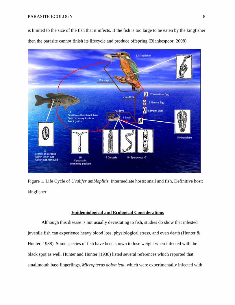
is limited to the size of the fish that it infects. If the fish is too large to be eaten by the kingfisher
then the parasite cannot finish its lifecycle and produce offspring (Blankespoor, 2008).
Figure 1. Life Cycle of Uvulifer ambloplitis. Intermediate hosts: snail and fish, Definitive host:
kingfisher.
Epidemiological and Ecological Considerations
Although this disease is not usually devastating to fish, studies do show that infested
juvenile fish can experience heavy blood loss, physiological stress, and even death (Hunter &
Hunter, 1938). Some species of fish have been shown to lose weight when infected with the
black spot as well. Hunter and Hunter (1938) listed several references which reported that
smallmouth bass fingerlings, Micropterus dolomieui, which were experimentally infected with

PARASITE ECOLOGY 9
U. ambloplitis showed slight but statistically significant weight loss compared with the control
fish.
The possibility of this parasite infecting a human is very minute. These parasites are very
specific in needing the final host, the Belted Kingfisher, to reach the adult stage. Although these
black spots do not have a healthy, appetizing appearance, infected fish are still able to be eaten.
However, some precautions must be taken. Since the cysts and black pigment are found on the
skin of the fish, simply skinning the fish can be a proper removal technique. This may remove
almost all of the cysts, so follow this procedure with normal cooking. A well cooked fish will
have no grubs present and can be safely eaten (Les, 1975).
An organism infested with parasites is considered to be sickly; however, a rich parasitic
community can indicate a thriving stream environment. Rutledge Creek Waste Water Treatment
Plant has been examined for pollution by biology students. The Waste Water Treatment Plant
underwent an upgrade within the last few years, somewhat improving the species richness of the
stream. The increase in the fish, snail, and bird population allowed the life cycle of U.
ambloplitis to thrive within Rutledge Creek. This increased prevalence of parasites can be
considered an indicator of a healthy, restored environment.
One particular study done by Huspeni and Lafferty (2004) examined the effects of
environmental restoration on larval Digenean parasites with an intermediate snail host. It was
found that parasite prevalence was increased as the site was restored. Species diversity was also
increased, leading to an increased avian population in the ecosystem (Huspeni & Lafferty, 2004).
The improved ecosystem health and increase of intermediate and final hosts led to an increased
parasite community. Parasites are not consistently indicators of pollution in a stream; if moderate
PARASITE ECOLOGY 10
levels of pollution are present, the level of parasites is less helpful in indicating ecosystem health
(Huspeni & Lafferty, 2004).
Incorporating the analysis of parasites into the assessment of an ecosystem is
recommended. Parasites select hosts discriminately and an increase in a particular parasitic
species can be a sign of another organism thriving within the ecosystem. U. ambloplitis will
thrive in a community where fish, snails, and birds are large contributors in the food web and
population (Au & Berra, 1978). If the parasite is put under particular environmental stress or
species richness begins to decline, the parasite community will being to degrade (Marcogliese,
2005). Because parasites are integral parts to any ecosystem, knowledge of the parasite that is
most prevalent in the stream community will be the most helpful in studying the ecosystem.
Although helpful, parasitic indicators should be only a part of a larger assessment of stream
health (Marcogliese, 2005).
Morphology of Specimens
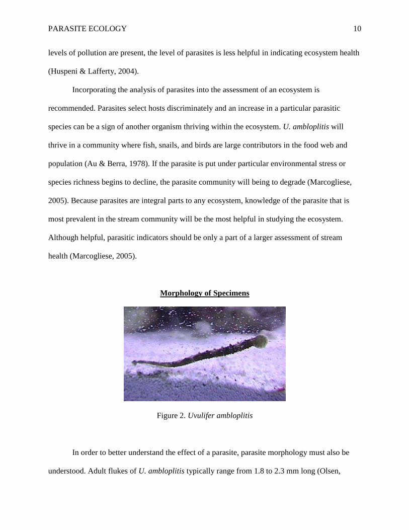
Figure 2. Uvulifer ambloplitis
In order to better understand the effect of a parasite, parasite morphology must also be
understood. Adult flukes of U. ambloplitis typically range from 1.8 to 2.3 mm long (Olsen,
PARASITE ECOLOGY 11
1996). It can be split into two sections named the forebody and the hindbody. The posterior half
of the fluke's forebody forms a necklike structure. The forebody is bowl-shaped with the oral
sucker inside but near the anterior margin (Olsen, 1996).
Behind the oral sucker is where the tribocytic organ is found; this organ secretes enzymes
that digest the mucus of the host and functions as an additional adhesive structure (Roberts &
Janovy, 2005). The ventral sucker is near the center, and this sucker is anterior to the holdfast
organ. The hindbody is about 3 times the length of the forebody. It is also about 4.5 times as long
as the diameter of the reproductive organs, the small testis. Vitelline follicles extend posteriorly
about to the level of the copulatory bursa. The eggs measure 90 to 99 micrometers long by 56 to
66 micrometers wide (Olsen, 1996).
In response to this schistosome-like parasite, the immune system of a fish has numerous
offensive and defensive responses. The first response that is broken by a parasite is the barrier of
the scales and skin of the fish. The cercariae of U. ambloplitis burrow through this layer before
gaining access to their host (Au & Berra, 1978). Once this physical defense has been defeated,
cellular defenses must respond to the invasion. These responses are initiated when the host
organism recognizes that a substance is foreign and does not belong. These defenses include
antibodies, B and T lymphocytes, and inflammation (Chappell & Secombes, 1996). In order to
be successful, this parasite uses developed host immune response evasion tactics.
The first evasion tactic of U. ambloplitis is that the metacercariae secrete a hyaline cyst
wall around them when they enter a fish host. This allows the parasite to be isolated from the
intermediate host response. In most fish the response is a small capsule, “characterized by the
presence of macrophages surrounded by a fibrous layer” (Matthews & Wood, 2006, 180). In
other fish, the reaction is also a capsule with 3 different layers. The inner layer is a large area of
PARASITE ECOLOGY 12
dead macrophages surrounding the parasite, followed by a thinner layer of living macrophages,
and then an outer layer of vascular fibroblasts and fibers. In the fish studied by Matthews and
Wood, the capsule formed by the fish included the deposit of melanin around the cyst, creating
the visible black spot (1996).
Wood and Matthews also examined the differing mortality of stages of the parasite’s life
cycle that was due to a fish immune serum. It was shown that free cercariae were killed, but the
encapsulated metacercariae were unaffected. In fact, when metacercariae induce a strong
pigmentation reaction around them, their infectivity is maintained within a fish (Matthews &
Wood, 1996). Once ingested in the intestines of the final host, the metacercariae end their
dormancy and develop into adults.
Although the “black spot” is evidence of an immunological response, many other cell
mediated responses are also taking place once a fish is infected. One paper established that “in
vivo macrophages, neutrophils, eosinophils, and lymphocytes can all be involved in the host
response to nematode, digenea, and cestode infections” (Chappel & Secombes, 1996, 169).
Secombes and Chappel showed that the parasitic larval forms can be encapsulated by eosinophils
and neutrophils in order to minimize damage done to the integument of the fish. Although fish
can moderate the parasite, U. ambloplitis can “release a large array of immunogenic molecules
into their surrounding environments, many of which give rise to ineffectual immune responses
and may be regarded as immune evasion molecules” (Chappel & Secombes, 1996, 172).
Infection of the intermediary host, snails, can be an indicator of a lack of stream
pollution. Molluscs are infected by the first larval stage, miracidia, which hatches from an egg.
The mollusc then aids the parasite in developing from miracidia into the swimming cercariae
stage. When the cercariae are fully developed, they leave the mollusc and move toward the next

PARASITE ECOLOGY 13
host in the life cycle. Molluscs present in polluted streams with Trematode infections have
markedly different physiology. The combined stress of a pollutant and a parasite infection can
decrease cardiac activity, food consumption, and respiration of the snail (Morley, 2006). Waste
water pollution may also cause a change in the metabolism of the molluscs. Morley studied the
changes in molluscs affected by Trematodes and pollution. He found that there were significant
changes in “haemolymh density, protein content, pH, and residual nitrogen” (Morley, 2006, 31).
Morley also concluded that the black spot infected molluscs inhibited the development from
miracidia to cercariae in the Trematode life cycle (2006). Snails that exhibited high density
Trematode infection levels were distressed to the point that cercariae development was
completely repressed. Even if infected molluscs did remain healthy enough to produce cercariae,
the biology and functions of the swimming parasite was greatly altered (Morley, 2006). Waste
water pollution increased the mortality rate of this fish parasite, a positive environment effect.
Trematode infections also affect the endocrine system. The endocrine system is
responsible for the release of hormones that signal growth and development and regulate
homeostasis throughout the snail’s body. As a parasite draws on the nutrients of the host, the
endocrine system will therefore be suppressed and unable to regulate hormones as usual. A
Trematode parasite will put biological stress on the mollusc host and make it more susceptible to
environmental pollution (Morley, 2006). Chemical pollution, in particular, causes a disruption in
the endocrine system of snails. Snails found in heavily polluted areas are reproductively inhibited
by abnormal growth of their reproductive organs. Morley noticed the intertwining relationship
that Trematodes parasites and chemical pollution have in disrupting the natural functioning of
the endocrine system in snails (2006).
PARASITE ECOLOGY 14
U. ambloplitis has both direct and indirect effects on the intermediate hosts. A direct
interaction of this trematode on the snail is the competition for resources. Food and space are
absorbed by the parasite for development that is needed by the snail. The indirect effect of a
parasite on its host is sometimes a change in the host’s phenotype. Parasites with complex life
cycles were able to manipulate the host’s phenotype to make transmission to the next host more
likely (Poulin, 1998). This change in phenotype results in lowering the hosts’ immunity and
facilitates the invasion of other parasites. One experiment was done studying the indirect effects
of the trematode parasite, Microphallus papillorobustus, on amphipods. The infected amphipods
exhibited an abnormal behavior that made them more vulnerable to bird predators. The avian
predators were the definitive host of the Trematode parasite life cycle; this abnormal behavior
created by the parasite increased transmission to the final host (Poulin, 1998).
Altered behavior of fish due to trematode infections was studied extensively in killfish.
Twelve unparasitized killfish were captured and 30 parasitized killfish were obtained. These 42
fish were placed in a glass aquarium and observed over a period of a few days (Lafferty &
Morris, 1996). Over this period, an observer noticed and recorded several abnormal behaviors in
the parasitized fish that made them more noticeable. These included: contorting, shimmying, and
jerking. Shimmying fish vibrated for a few moments, flashing fish turned the lateral side of their
body towards the top of the tank, and jerking fish moved suddenly forward a few centimeters.
Fish that were contorted bent their tail and head in opposite directions.
When the fish behavior was recorded, it was not known whether each fish was parasitized
or free of infection. After the fishes’ behaviors were accurately recorded, they were euthanized
and dissected to determine the degree of infection. As hypothesized, parasitized killfish showed
these conspicuous behaviors four times more than unparasitized fish. Also, the heavier the
PARASITE ECOLOGY 15
trematode infection, the more often these fish exhibited altered behavior (Lafferty & Morris,
1996).
In 1996, Lafferty and Morris took this experiment one step further to see whether such
abnormal behaviors increased predation on parasitized fish. Parasitized and unparasitized killfish
were placed in each of two pens. The control pen contained a netted covering big enough for
these killfish to jump through, but small enough to keep birds from being able to catch and eat
them. After 20 days, the fish were recollected and dissected. It was found that parasitized fish
were attacked by birds at a substantially higher rate than unparasitized killfish. After making the
fish vulnerable to bird predators, population sizes dropped. In the experimental pen, the
parasitized fish population dropped from 95 to 44 while the unparasitized population dropped
from 95 to 91. The intensity of infection was also shown to increase vulnerability to prey; the
majority of heavily infected fish were captured by birds (Lafferty & Morris, 1996).
The study supported the hypothesis that parasites “modify the behavior of their
intermediate hosts and make them more susceptible to predation” (Lafferty & Morris, 1996,
1395). Lafferty and Morris also concluded that parasite intensity was necessary for increased
behavior modification; a group of parasites is able to more effectively turn “a small behavior
modification into a large increase in predation” (1996, 1396) than one parasite. This work shows
that even as a larval cyst, parasites can inflict changes that greatly increase transmission rates.
Experimental Work by Others
A study done by Ray-Jean Au and Tim M. Berra (1978) on the incidence of black spot
disease in fishes in Cedar Fork Creek, Ohio, is helpful in understanding what areas of a river will
have a higher prevalence than others of black spot disease. Their purpose was to report the
PARASITE ECOLOGY 16
natural incidence of infection of black spot disease among the fish in a small North Central Ohio
stream, Cedar Fork Creek. The creek was reported to be a clean and clear, unpolluted, gravel-
bottomed, rapid flowing stream with a population of thirty different species. The stream was also
a good mix of pools and riffles which allowed for proper analysis of incidence of the disease in
different locations. The Belted kingfisher, the final host of U. amploplitis is common in the area,
and snails are present as well.
Since the entire life cycle could be completed in this stream, it was a prime environment
for the fish collection. In order to save time allow for increased collection of fish, only the left
side of each fish was counted for black spots. The assumption made was that cysts are equally
likely on each side of the fish. They recorded the total length of each fish and the number of
cysts formed. They were able to identify the parasite by dissecting the cysts from freshly caught
fish and then examining the live metacercariae under a microscope. They admit that since every
cyst could not possibly be examined, there was a possibility that their study could include
another agent of black spot disease as well.
Berra and Au (1978) examined a total of 4175 specimens of fish belonging to 29 taxa in 6
families. From this total, 3698 (89%) were infected with one or more metacercariae on the left
side. Their figures were in conjunction with the findings of Evans and Mackiewicz (1958) for
New York stream fishes. Interestingly, Vinikour (1977) indicated that there was a greater
incidence of infection in a stream with a higher nutrient load than in a less productive stream.
Berra and Au found no evidence of immunity to the black spot parasite. Their data
suggested that fish found in pools and slow moving water were more highly infected than fish
from runs and ripples. This can be correlated to the fact that the intermediate snail host,
Helisoma, is found more commonly in pools. Also, any cercariae that are present in riffles are
PARASITE ECOLOGY 17
easily swept away in the rapids leaving little chance of infecting a fish (Au & Berra, 1978). They
concluded the study by commenting that other factors such as: host specificity, immunity, and
skin thickness are also involved in infection levels (Au & Berra, 1978).
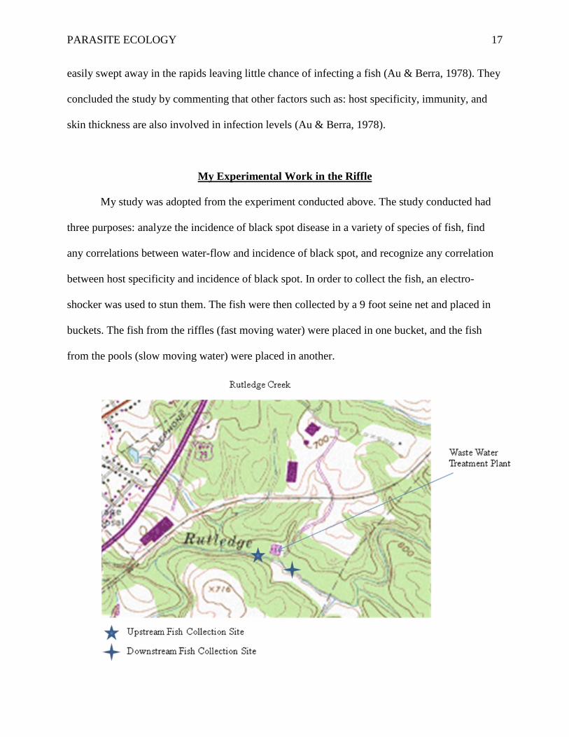
My Experimental Work in the Riffle
My study was adopted from the experiment conducted above. The study conducted had
three purposes: analyze the incidence of black spot disease in a variety of species of fish, find
any correlations between water-flow and incidence of black spot, and recognize any correlation
between host specificity and incidence of black spot. In order to collect the fish, an electro-
shocker was used to stun them. The fish were then collected by a 9 foot seine net and placed in
buckets. The fish from the riffles (fast moving water) were placed in one bucket, and the fish
from the pools (slow moving water) were placed in another.
PARASITE ECOLOGY 18
The two sites where fish were collected, one upstream and one downstream of a waste
water treatment plant are shown in the map above. A total of seven species were examined from
Rutledge Creek, north of Lynchburg, Virginia. They were as follows: Etheostoma flabellare (fan
tail darter), Rhinichthys atratulus (black nosed dace), Thoburnia rhothoeca (torrent sucker),
Rhinichthys cataractae (long nose dace), Ethostoma nigrum (johnny darter), Phoxinus oreas
(mountain redbelly dace), and Luxilus cornutus (common shiner).
From the riffles, a total of 175 fish were collected. The species that had the most success
collection numbers was the fan tail darter; they comprised 102 of the 175 fish (Table 1). The next
two largest collections were the black nosed dace (31 fish) and the torrent sucker (23 fish).
Following these were long nose dace (8 fish), the johnny darter (5 fish), the mountain redbelly
dace (4), and the common shiner (2). The percent infection with the black spot parasite ranged
from 0% to 100%. Of fan tail darters, 90.2% were infected, 80.65% of the black nosed dace were
infected, 0% of the torrent suckers, 25% of the long nose dace, 20% of the johnny darters, 50%
of the mountain redbelly dace, and 100% of the common shiners were infected. The average total
tail lengths of the fish were as follows: fan tail darter, 4.6 cm, black nosed dace, 5.3 cm, long
nose dace, 7.3 cm, johnny darter, 5.6cm, mountain redbelly dace, 3.4 cm, and common shiner,
7.1 cm.
The number of black spots on the left side of each fish was counted, and it appeared that
the average numbers varied amongst the various species. The highest average number of black
spots was found in the fan tail darter with a mean of 20 spots per fish. This was followed by the
black nosed dace which had 15.2 spots per fish, the johnny darter with 11 spots per fish, the long

PARASITE ECOLOGY 19
nose dace with 10.5 spots per fish, and the mountain redbelly dace and the common shiner which
both averaged 3 spots per fish.
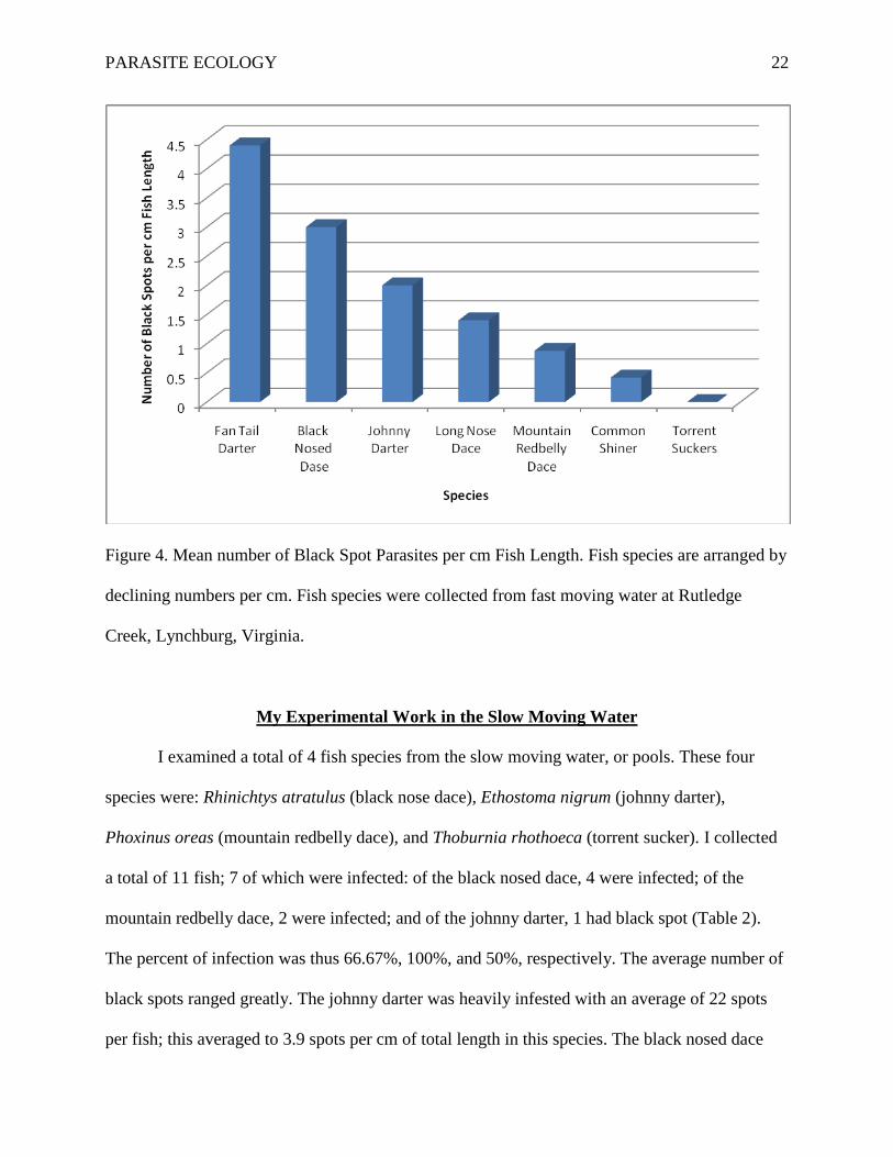
Upon further calculation, I deduced the mean number of black spots per cm total length
in each species of fish. Our data is as follows: fan tail darter, 4.4 spots/cm, black nosed dace, 3
spots/cm, johnny darter, 2 spots/cm, long nose dace, 1.4 spots/cm, mountain redbelly dace, 0.88
spots/cm, and the common shiner, 0.42 spots/cm. There was no obvious pattern in the length of
the fish versus the intensity of black spot infection.
Species Common Name
Total No.
No. infected
% Infected
Avg. TL*
Range TL
Avg. NBS**
Range NBS
NBS** per cm
TL Etheostoma flabellare
Fan Tail Darter 102 92 90.20% 4.6 3.0-
7.5 20 3.0-61 4.4
Rhinichthys atratulus
Black Nosed Dace
31 25 80.65% 5.2 3.0-7.1 15.2 3.0-
42 3
Thoburnia rhothoeca
Torrent Sucker 23 0 0% N/A N/A N/A N/A N/A
Rhinichthys cataractae
Long Nose Dace
8 2 25% 7.3 7.1-7.5 10.5 10.0-
11.0 1.4
Ethostoma nigrum
Johnny Darter 5 1 20% 5.6 N/A 11 N/A 2
Phoxinus oreas
Mountain Redbelly
Dace 4 2 50% 3.4 3.0-
3.8 3 2.0-4.0 0.88
Luxilus cornutus
Common Shiner 2 2 100% 7.1 6.7-
7.5 3 4.0-2.0 0.42
Table 1. Riffle Data and Averages of Collected Fish
*Total length of the fishes (TL) are given in cm.
**NBS=number of black spots on left side of each specimen.
PARASITE ECOLOGY 20
Interpretation of Riffle Data
I began by examining the data collected on the species for which we had the largest
catch— Fantail Darter. Of the 102 collected, 92 were infected (90.2%, see Table 1). An
explanation for this can be seen in the feeding habits of this species. The fan tail darter feeds
primarily on snails, worms, and mosquito larvae. Snails are the first intermediate host in the
black spot life cycle. Fan tail free embryos are benthic and rarely feed elsewhere. This provides
strong support for a correlation between the intensity of black spot infection and living near and
feeding on snails. The fan tail darter was the fish species that carried the heaviest parasite
infection. The relationship between this fish species and snails greatly increases the chance of
black spot infection.
In contrast with the fan tail darter living habits is the johnny darter; this type of darter is
typically a pool dwelling species. It was expected to find low population numbers of johnny
darters in the riffle; only 5 were found. Only 20% of riffle johnny darters were infected
compared to 90.2% of fan tails. Johnny darters are also benthic fish. However, their feeding
pattern is not primarily snails as is the fan tail darter. Johnny darters feed on zooplankton, midge
larvae, mayflies, other small insects, and small snails. Fan tails feed on the intermediate host,
snails, more than the johnny darter, leading to a higher parasite load in the fan tail than the
johnny darter.
In continuing my examination of data, I noticed that the black nose dace also have a
strikingly high infection rate, 80.65%. This species of dace is also a benthic species. They feed
on larvae, small crustaceans, small worms, and plant material. The cercariae are the black spot’s
infective larvae that swim along the bottom of the creek; I hypothesized that there may be a
greater amount of cercariae that swim along the bottom. As the ceracariae desire to maximize
PARASITE ECOLOGY 21
transmission rate, they swim along the bottom where either snail or fish can be found, possibly
increasing the chance of burrowing into a host. Au and Berra (1978) shared that “rapidly moving
water over an unstable bottom is not good snail habitat and promotes rapid dilution of the
cercariae” (321). This means that cercariae need to use other strategies to infect riffle fish such as
swimming in locations with the highest host population.
We found a 0% infection rate among only one species of fish, the torrent sucker. These
fish are known to be pelagic; they are also much larger fish than the darters. Their feeding
characteristics make them less likely to obtain an infection from a bottom dwelling snail. Suckers
are also not the type of species that Kingfishers prey upon (Olsen, 1996); this makes it very
difficult for an infection or completion of the life cycle to occur in this species. Parasites develop
characteristics that make completing the life cycle more probable. This can be accomplished by
avoiding hosting in larger fish that are more vulnerable to predation by the kingfisher. Figure 4,
shown below, gives a graph showing numbers of spots per cm in each species of fish collected
from the riffles.
PARASITE ECOLOGY 22
Figure 4. Mean number of Black Spot Parasites per cm Fish Length. Fish species are arranged by
declining numbers per cm. Fish species were collected from fast moving water at Rutledge
Creek, Lynchburg, Virginia.
My Experimental Work in the Slow Moving Water
I examined a total of 4 fish species from the slow moving water, or pools. These four
species were: Rhinichtys atratulus (black nose dace), Ethostoma nigrum (johnny darter),
Phoxinus oreas (mountain redbelly dace), and Thoburnia rhothoeca (torrent sucker). I collected
a total of 11 fish; 7 of which were infected: of the black nosed dace, 4 were infected; of the
mountain redbelly dace, 2 were infected; and of the johnny darter, 1 had black spot (Table 2).
The percent of infection was thus 66.67%, 100%, and 50%, respectively. The average number of
black spots ranged greatly. The johnny darter was heavily infested with an average of 22 spots
per fish; this averaged to 3.9 spots per cm of total length in this species. The black nosed dace
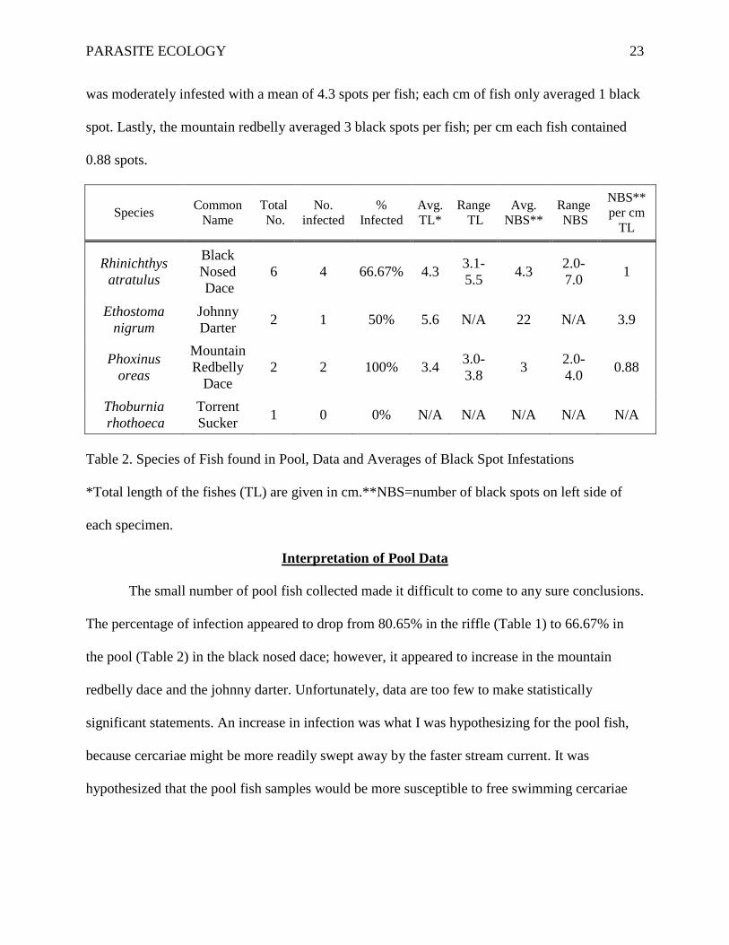
PARASITE ECOLOGY 23
was moderately infested with a mean of 4.3 spots per fish; each cm of fish only averaged 1 black
spot. Lastly, the mountain redbelly averaged 3 black spots per fish; per cm each fish contained
0.88 spots.
Species Common Name
Total No.
No. infected
% Infected
Avg. TL*
Range TL
Avg. NBS**
Range NBS
NBS** per cm
TL
Rhinichthys atratulus
Black Nosed Dace
6 4 66.67% 4.3 3.1-5.5 4.3 2.0-
7.0 1
Ethostoma nigrum
Johnny Darter 2 1 50% 5.6 N/A 22 N/A 3.9
Phoxinus oreas
Mountain Redbelly
Dace 2 2 100% 3.4 3.0-
3.8 3 2.0-4.0 0.88
Thoburnia rhothoeca
Torrent Sucker 1 0 0% N/A N/A N/A N/A N/A
Table 2. Species of Fish found in Pool, Data and Averages of Black Spot Infestations
*Total length of the fishes (TL) are given in cm.**NBS=number of black spots on left side of
each specimen.
Interpretation of Pool Data
The small number of pool fish collected made it difficult to come to any sure conclusions.
The percentage of infection appeared to drop from 80.65% in the riffle (Table 1) to 66.67% in
the pool (Table 2) in the black nosed dace; however, it appeared to increase in the mountain
redbelly dace and the johnny darter. Unfortunately, data are too few to make statistically
significant statements. An increase in infection was what I was hypothesizing for the pool fish,
because cercariae might be more readily swept away by the faster stream current. It was
hypothesized that the pool fish samples would be more susceptible to free swimming cercariae
PARASITE ECOLOGY 24
and therefore more parasitized. In contrast, despite the decrease in stream velocity, the number of
visible black spots only appeared to increase in two of the species.
Reading other similar results suggests a pattern of infection among riffle fish and pool
fish. Such experiments suggest that pool fish typically experienced higher parasite loading than
riffle fish. It is hypothesized that this is due to an increase in the density of fish parasites in a
pool area over a riffle. For example, Godin, Krause, and Ruxton (1999) studied the prevalence of
Crassiphiala bubloglossa, a trematode parasite strikingly similar to U. ambloplitis, in fish in
shallow, slow moving water. They found that a sucker population that was commonly found in
pool areas was heavily parasitized (Godin, Krause, & Ruxton, 1999).
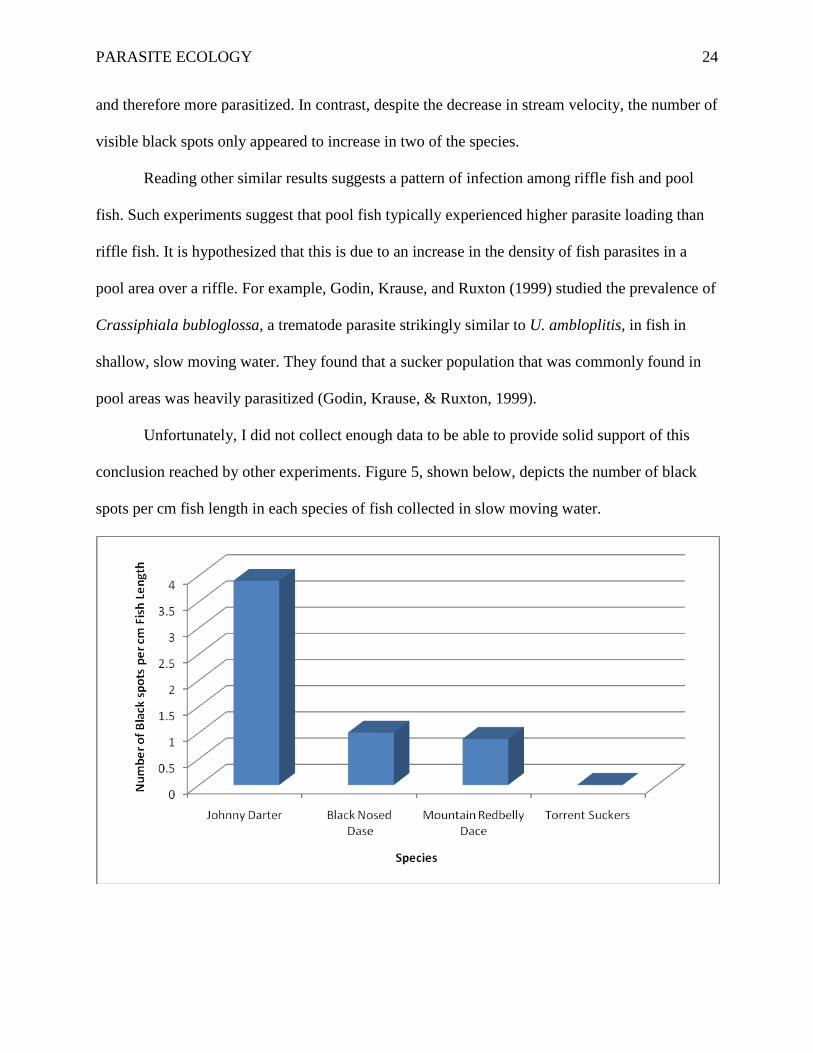
Unfortunately, I did not collect enough data to be able to provide solid support of this
conclusion reached by other experiments. Figure 5, shown below, depicts the number of black
spots per cm fish length in each species of fish collected in slow moving water.
PARASITE ECOLOGY 25
Figure 5. Mean number of Black Spot Parasites per cm Fish Length. Fish species are arranged by
declining numbers per cm. Fish species were collected from slow moving water at Rutledge
Creek, north of Lynchburg, Virginia.
Prevention, Treatment, and Control of Black Spot
A common method of prevention of parasitism of fish is the addition of synthetic
molluscicides. This approach destroys the intermediate snail host, interrupting the life cycle of
the parasite. If the cercariae are unable to develop within the snails, the metacercariae are unable
to form, and the life cycle is halted. However, the molluscicides that are presently available may
disturb an ecosystem because of the possible effect on the many non-target organisms. Just as
pollution affects numerous parts of an environment, so do synthetic molluscicides.
Synthetic molluscicides may not only disrupt an ecosystem but may also be extremely
expensive. Examples of such molluscicides are: Copper Sulfate, Niclosamide, and
Bromoacetimide. Niclosamide is currently the molluscicide of choice among the environmental
community. It is active at reducing snail populations at all stages of the life cycle and is effective
at killing parasite larvae. Niclosamide is not toxic to humans or nearby vegetation although it is
highly toxic to fish. A study of the effects of Niclosamide showed that it was “costly and highly
toxic to fish” and “did not prevent the recolonization of sites by remaining snails” (Dissous &
Lardens, 1998). This molluscicde only offers short term snail reduction and could potentially
lead to snails resistant to chemical molluscicides.
A recent more natural alternative is available-- the use of plants with molluscicidal
properties. Many plants have natural molluscicidal properties, and this may provide advantages
economically and ecologically. These plants are less harmful to the non-target organisms in the
ecosystem (Brielmann & Cseke, et al., 2006). One molluscidial plant, Phytolacca dodencandra,
PARASITE ECOLOGY 26
produces a chemical compound called saponin that kills snails; another plant produces
isoflavonoids that are also harmful to snail populations. Molluscicides like Niclosamide are
expensive to synthesize while plants with molluscicidal characteristics can be grown. The
limiting factor to these plants is that large scale production has been difficult to develop (Dissous
& Lardens, 1998).
Typically the control used for a parasite like Black Spot is a molluscicide, but other
interesting avenues are being explored. These new controls include the use of Malayan Snail-
Eating Turtles (Malayemys subtrijuga) and Redear Sunfish (Lepomis miscrolophus). These two
species have a diet that consists primarily of snails. About 95% of the diet of the Redear Sunfish
is snails (Goodman, Marschall, & Stein, 1984). This characteristic makes this fish particularly
fatal to the Black Spot life cycle.
In artificial situations, certain trematode parasites have been used to sterilize snails,
reducing the population over a longer amount of time. The larval trematodes damage the
reproductive organs of the snail and effectually castrate the mollusc. However, when applied in a
field study, the trematode additions were much less effective than molluscicides (Dissous &
Lardens, 1998). Vegetation removal is also used in artificial situations were snail population
needs to be controlled (Berra 1978). After all methods have been reviewed, the most cost
effective and ecologically safe molluscicide is the use of snail predators or competitors in the
ecosystem according to Dissous and Lardens (1998).
Conclusions
Black spot disease in fish is caused by the parasite Uvulifer ambloplitis and other
digenetic trematodes of the family Strigeidae. This parasite has a life cycle which requires three
hosts. The two intermediate hosts are the snails and fish, while the definitive host is the
PARASITE ECOLOGY 27
kingfisher or other avian host. This particular disease is not infective to humans and has a
limited, but visible effect on the fish. Altered behavior and numerous immune responses occur in
the snail and fish to reduce damage done by the parasite. Prevention and control can be found in
a variety of forms, but increasingly, control has focused on decreasing the intermediary snail
population.
Although parasites are harmful to their hosts, parasitism is a common occurrence within
ecosystems, and can be an accurate indicator of pollution. As pollution decreases, parasite
population can actually increase, as my and many other studies have shown. Since parasites
generally choose healthy organisms to infect, the prevalence of the trematode parasite may
ironically be a small indicator of a healthy stream in terms of its low levels of pollution (Roberts
& Janovy, 2005).
My and many other workers’ parasite studies show how common parasitism is in some
environments. Since parasites continue to infect humans and other organisms, parasite ecology
and epidemiology still needs to be studied. It is especially important to understand the
interactions of parasites and their environments in order to learn how to better control them.
PARASITE ECOLOGY 28
References
Au, R.J., & Berra, T. (1978). Incidence of black spot disease in fishes in Cedar Fork Creek,
Ohio. Ohio Journal of Science, 78(6), 318-322.
Blankespoor, C., & Blankespoor, H. (2008). Host-parasite relationships of Uvulifer ambloplitis
in fish of Douglas Lake. Biological Station, University of Michigan.
Brielmann, H., Cseke, L., et al. (2006). Natural products from plants. 2nd Ed. CRC Press.
Chappell, L.H., & Secombes, C.J. (1996). Fish immune responses to experimental and natural
infection with helminth parasites. Annual Review of Fish Diseases, 6, 167-177.
doi:10.1016/S0959-8030(96)90012-5.
Davis, H.S. (1967). Culture and diseases of game fishes. Berkley, CA: University of California
Press.
Dissous, C., & Lardans, V. (1988). Snail Control Strategies for Reduction of Schistosomiasis
Transmission. Parasitology Today, 14(10), 413-417. doi:10.1016/S0169-4758(98)01320-
9.
Godin, J., Krause, J., Ruxton, G. (1999). Distribution of Crassiphiala bulboglossa, a Parasitic
Worm, in Shoaling Fish. Journal of Animal Ecology, 68(1), 27-33. Retrieved from
http://www.jstor.org/stable/2647296
Goodman, C., Marschall, E., & Stein, R. (1984). Using time and energetic measures of cost in
estimating prey value for fish predators. Ecology, 65(3), 702-715. Retrieved from
http://www.jstor.org/stable/1938042
Hoffman, G.L. (1955). Neascus Nolfi N. Sp. (Trematoda: Strigeida) from the cyprinid minnows
with notes on the artificial digest recovery of helminths. American Midland Naturalist,
53(1), 198-204. Retrieved from http://www.jstor.org/stable/2422310
PARASITE ECOLOGY 29
Hoggman, G.L., & Meyer, F.P. (1976). Parasites and diseases of warmwater fishes. Stutgart,
AK: U.S. Dept. of Interior, Fish and Wildlife Service.
Lafferty, K., Morris, A. (1996). Altered Behavior of Parasitized Killifish Increases Susceptibility
to Predation by Bird Final Hosts. Ecology, 77(5), 1390-1397.
Les, B. (1975). Common parasites of freshwater fish. Madison, WI: Department of Natural
Resources.
Loker, E., & Brant, S. (2005). Diversification, dioecy and dimorphism in schistosomes. Trends
in Parasitology, 22(11), 521-528. doi:10.1016/j.pt.2006.09.001
Marcogliese, D. (2005, January). Parasites of the superorganism: Are they indicators of
ecosystem health?. International Journal for Parasitology, 35(7), 705-716.
doi:10.1016/j.ijpara.2005.01.015
Matthews, R., & Wood, B. (1996). The immune response of the thick-lipped grey mullet, Chelon
labrosus (Risso, 1826), to metacercarial infections of Cryptocotyle lingua (Creplin,
1825). Journal of Fish Biology, 31(sA), 175-183. doi:10.1111/j.1095-8649.1987.tb05310.
Morley, N.J. (2010). Interactive effects of infectious disease and pollution in aquatic molluscs.
Aquatic Toxicology, 96(1), 27-36. doi:10.1016/j.aquatox.2009.09.017.
Olsen, O. (1974). Animal parasites: their life cycles an ecology. Baltimore, MD: University Park
Press.
Poulin, R. (1999). The functional importance of parasites in animal communities: many roles at
many levels? International Journal for Parasitology, 29(6), 903-914. doi:10.1016/S0020-
7519(99)00045-4.
Roberts, L., Janovy, R., & Schimidt, G. (2005). Foundations of Parasitology. New York, NY:
McGraw Hill Publishers.
PARASITE ECOLOGY 30
Vinikour, W.S. (1977). Incidence of Neascus rhinighthysi (Trematoda: Diplostomatidae) on
longnose dace, Rhinichthys cataractae (Pisces: Cyprinidae), related to fish size and
capture location. Transactions of American Fish Society, 106(1), 83-88.
Walker, A. (2006). Do trematode parasites disrupt defence-cell signaling in their snail hosts?
Trends in Parasitology, 22(4), 154-159. doi:10.1016/j.pt.2006.02.003