Parallel Muscles Figure 11–1a. Skeletal Motion Skeletal muscles attach to skeleton, produce...
67
Parallel Muscles Parallel Muscles Figure 11–1a
-
Upload
patricia-jackson -
Category
Documents
-
view
218 -
download
0
Transcript of Parallel Muscles Figure 11–1a. Skeletal Motion Skeletal muscles attach to skeleton, produce...
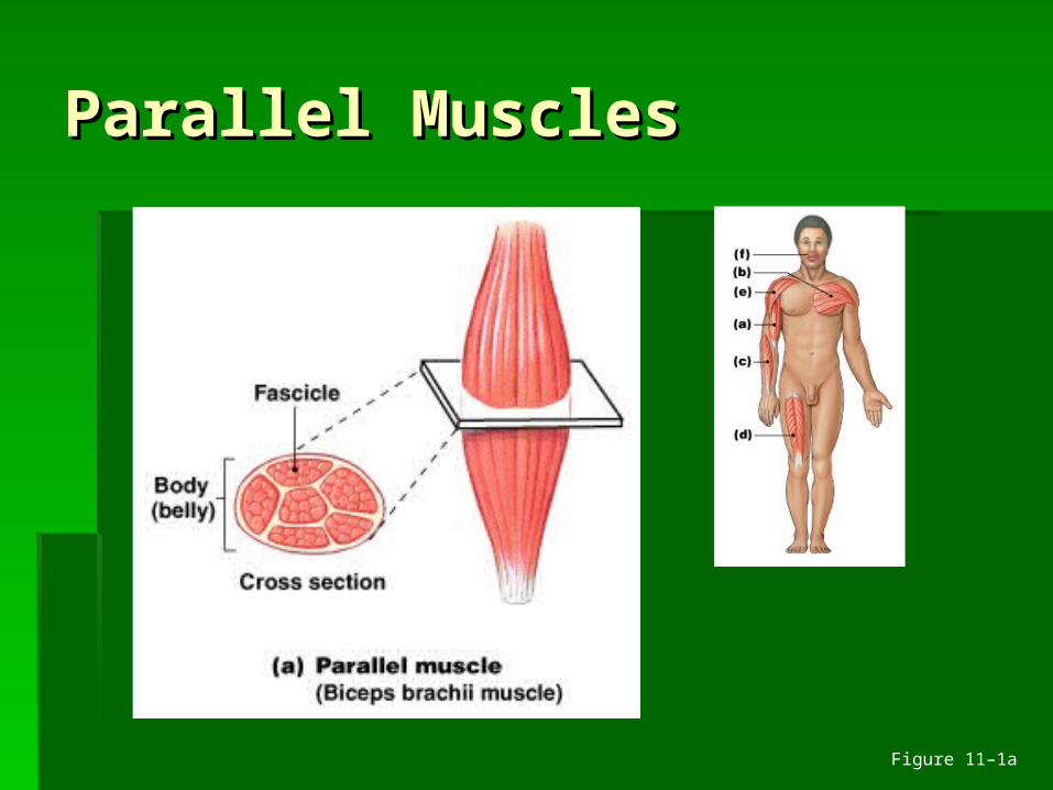
Parallel MusclesParallel Muscles
Figure 11–1a
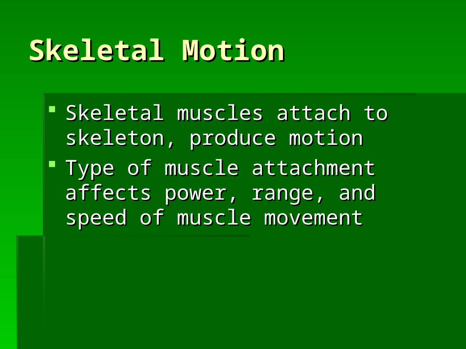
Skeletal MotionSkeletal Motion
Skeletal muscles attach to skeleton, Skeletal muscles attach to skeleton, produce motionproduce motion
Type of muscle attachment affects Type of muscle attachment affects power, range, and speed of muscle power, range, and speed of muscle movementmovement
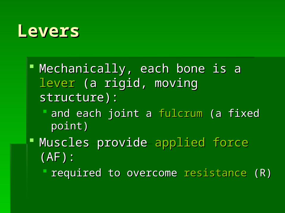
LeversLevers
Mechanically, each bone is a Mechanically, each bone is a leverlever (a (a rigid, moving structure):rigid, moving structure): and each joint a and each joint a fulcrumfulcrum (a fixed point) (a fixed point)
Muscles provide Muscles provide applied forceapplied force (AF): (AF): required to overcome required to overcome resistanceresistance (R) (R)
Functions of a LeverFunctions of a Lever
To change:To change: direction of an AFdirection of an AF distance and speed of movement produced distance and speed of movement produced
by an AFby an AF effective strength of an AFeffective strength of an AF
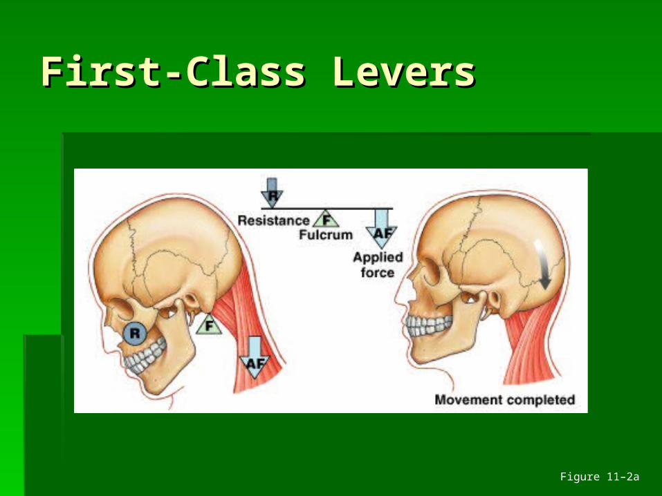
First-Class LeversFirst-Class Levers
Figure 11–2a
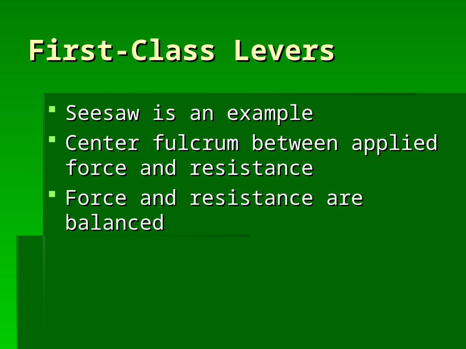
First-Class LeversFirst-Class Levers
Seesaw is an exampleSeesaw is an example Center fulcrum between applied force Center fulcrum between applied force
and resistanceand resistance Force and resistance are balancedForce and resistance are balanced
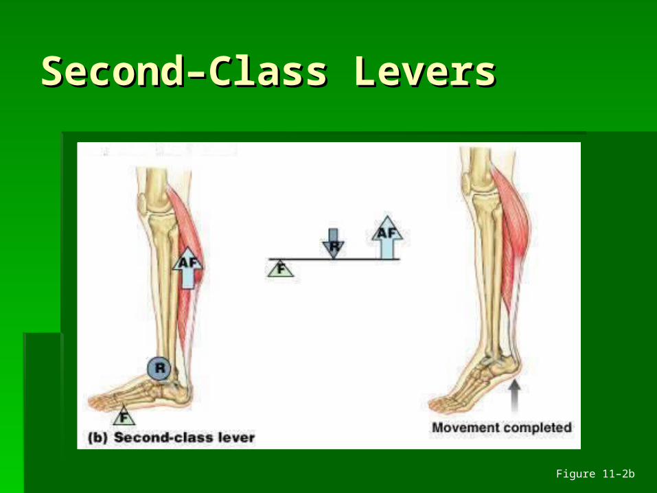
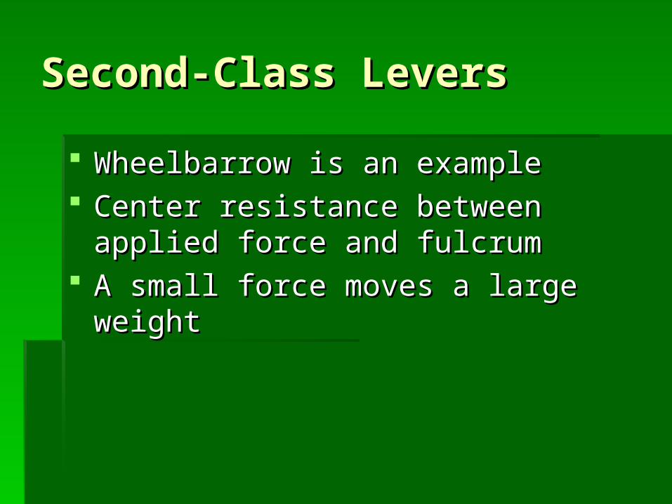
Second–Class LeversSecond–Class Levers
Figure 11–2b
Second-Class LeversSecond-Class Levers
Wheelbarrow is an exampleWheelbarrow is an example Center resistance between applied force Center resistance between applied force
and fulcrumand fulcrum A small force moves a large weightA small force moves a large weight
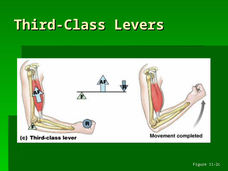
Third-Class LeversThird-Class Levers
Figure 11–2c
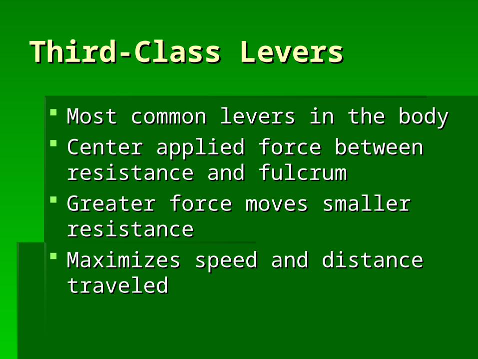
Third-Class LeversThird-Class Levers
Most common levers in the bodyMost common levers in the body Center applied force between resistance Center applied force between resistance
and fulcrumand fulcrum Greater force moves smaller resistanceGreater force moves smaller resistance Maximizes speed and distance traveledMaximizes speed and distance traveled
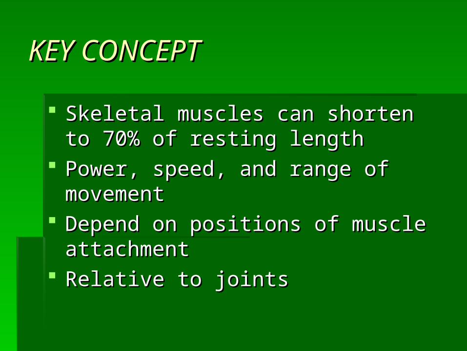
KEY CONCEPTKEY CONCEPT
Skeletal muscles can shorten to 70% of Skeletal muscles can shorten to 70% of resting length resting length
Power, speed, and range of movement Power, speed, and range of movement Depend on positions of muscle Depend on positions of muscle
attachment attachment Relative to jointsRelative to joints
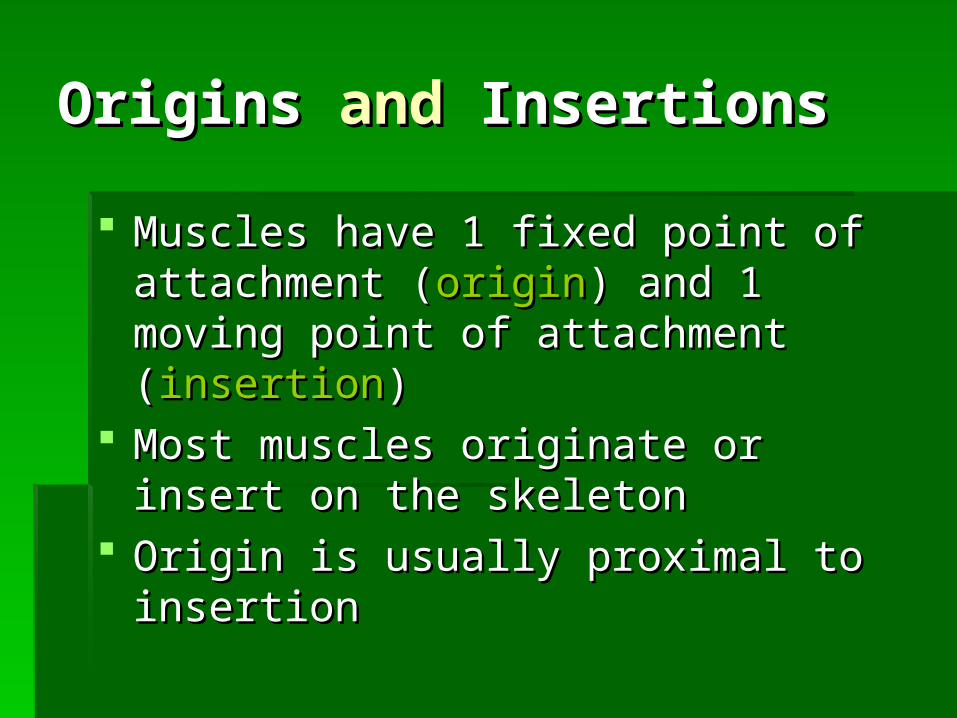
OriginsOrigins and and InsertionsInsertions
Muscles have 1 fixed point of attachment Muscles have 1 fixed point of attachment ((originorigin) and 1 moving point of attachment ) and 1 moving point of attachment ((insertioninsertion) )
Most muscles originate or insert on the Most muscles originate or insert on the skeletonskeleton
Origin is usually proximal to insertionOrigin is usually proximal to insertion
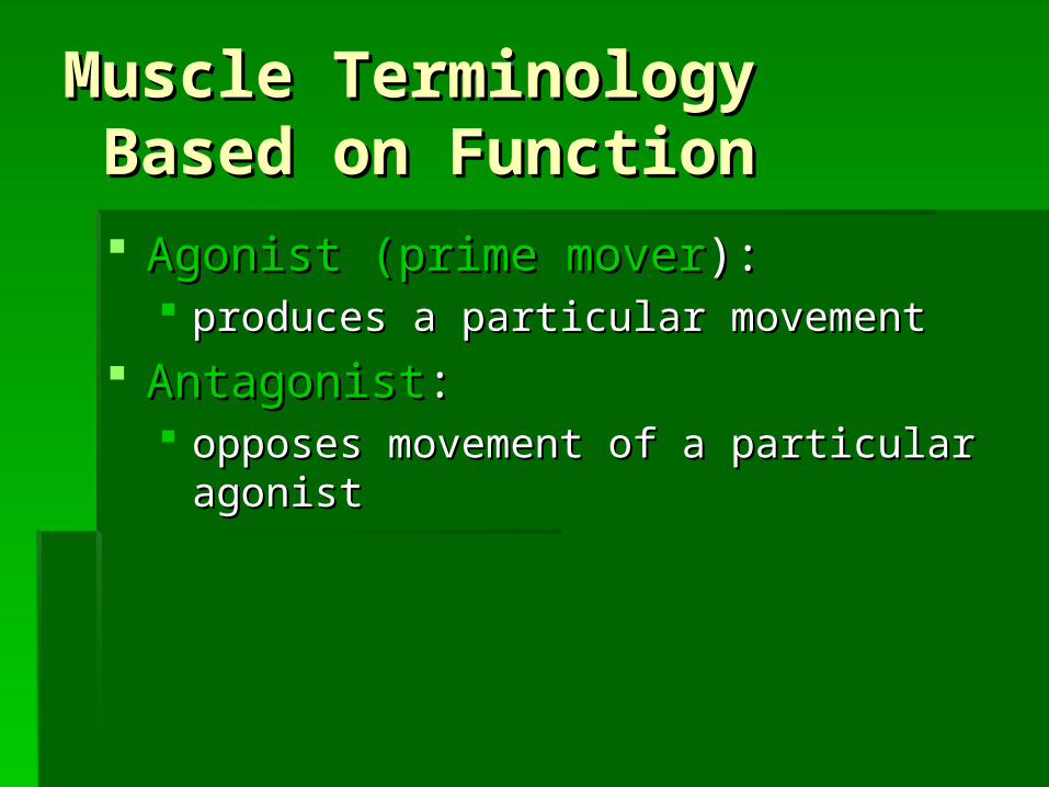
Muscle TerminologyMuscle Terminology Based on Function Based on Function
Agonist (prime moverAgonist (prime mover):): produces a particular movementproduces a particular movement
AntagonistAntagonist:: opposes movement of a particular agonistopposes movement of a particular agonist
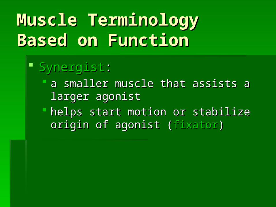
Muscle Terminology Muscle Terminology Based on FunctionBased on Function
SynergistSynergist:: a smaller muscle that assists a larger a smaller muscle that assists a larger
agonistagonist helps start motion or stabilize origin of helps start motion or stabilize origin of
agonist (agonist (fixatorfixator))
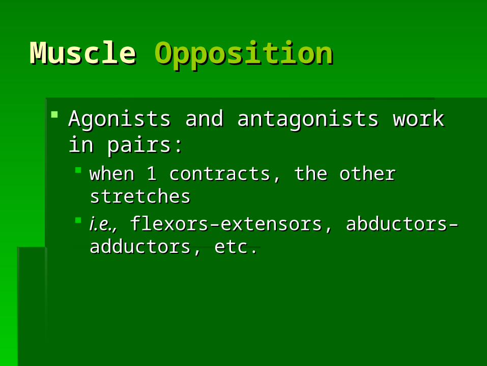
Muscle Muscle OppositionOpposition
Agonists and antagonists work in pairs:Agonists and antagonists work in pairs: when 1 contracts, the other stretcheswhen 1 contracts, the other stretches i.e.,i.e., flexors–extensors, abductors– flexors–extensors, abductors–
adductors, etc.adductors, etc.
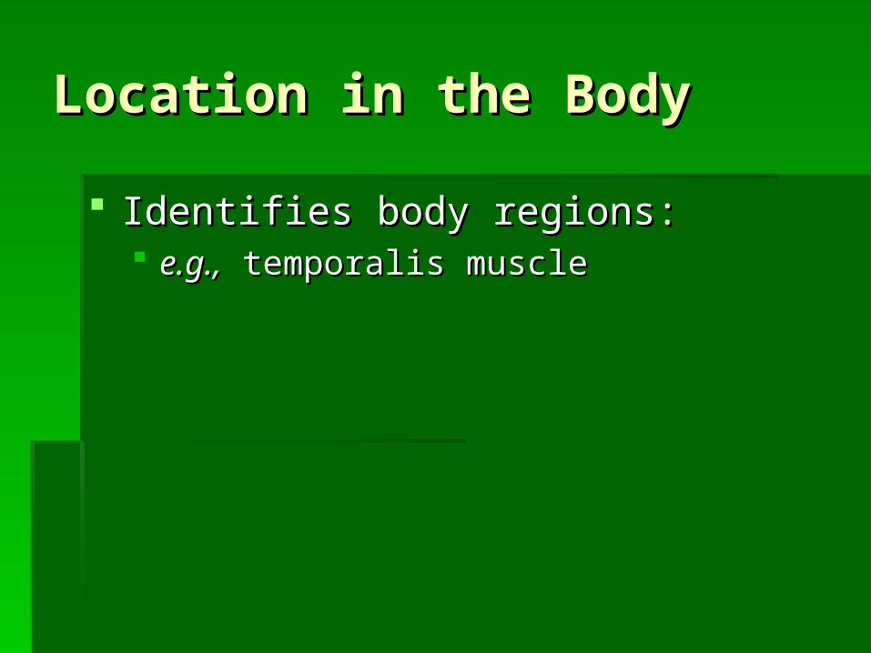
Location in the BodyLocation in the Body
Identifies body regions:Identifies body regions: e.g.,e.g., temporalis muscle temporalis muscle
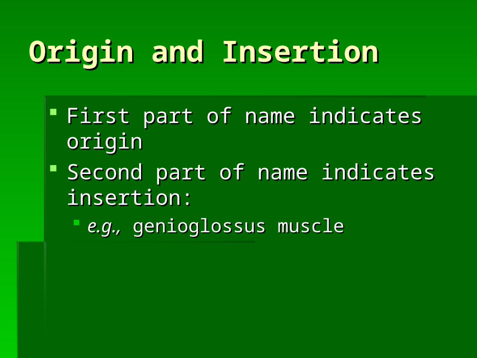
Origin and InsertionOrigin and Insertion
First part of name indicates originFirst part of name indicates origin Second part of name indicates insertion:Second part of name indicates insertion:
e.g.,e.g., genioglossus muscle genioglossus muscle
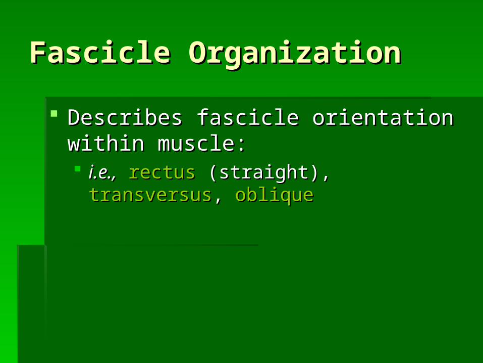
Fascicle OrganizationFascicle Organization
Describes fascicle orientation within Describes fascicle orientation within muscle:muscle: i.e.,i.e., rectusrectus (straight), (straight), transversustransversus, , obliqueoblique
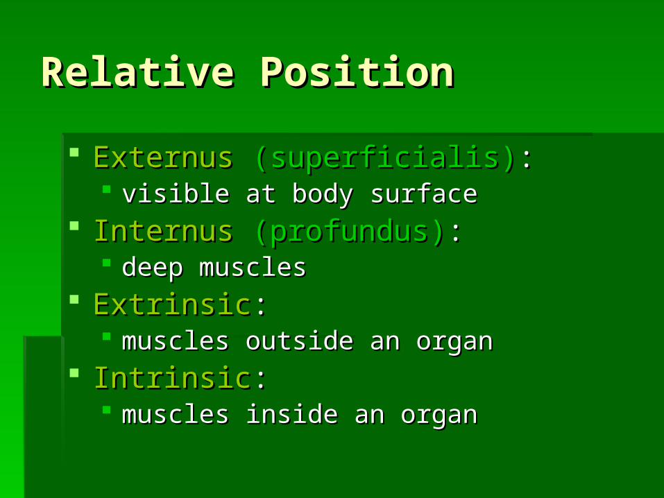
Relative PositionRelative Position
ExternusExternus (superficialis)(superficialis):: visible at body surfacevisible at body surface
InternusInternus (profundus)(profundus):: deep musclesdeep muscles
ExtrinsicExtrinsic:: muscles outside an organmuscles outside an organ
IntrinsicIntrinsic:: muscles inside an organmuscles inside an organ
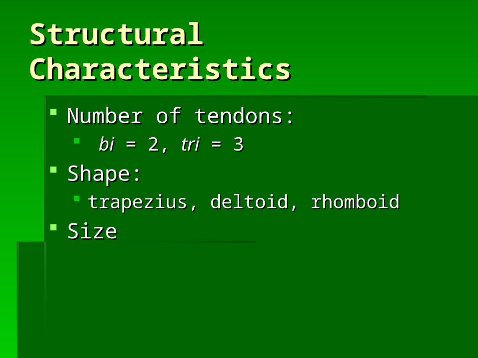
Structural CharacteristicsStructural Characteristics
Number of tendons:Number of tendons: bibi = 2, = 2, tritri = 3 = 3
Shape: Shape: trapezius, deltoid, rhomboidtrapezius, deltoid, rhomboid
SizeSize
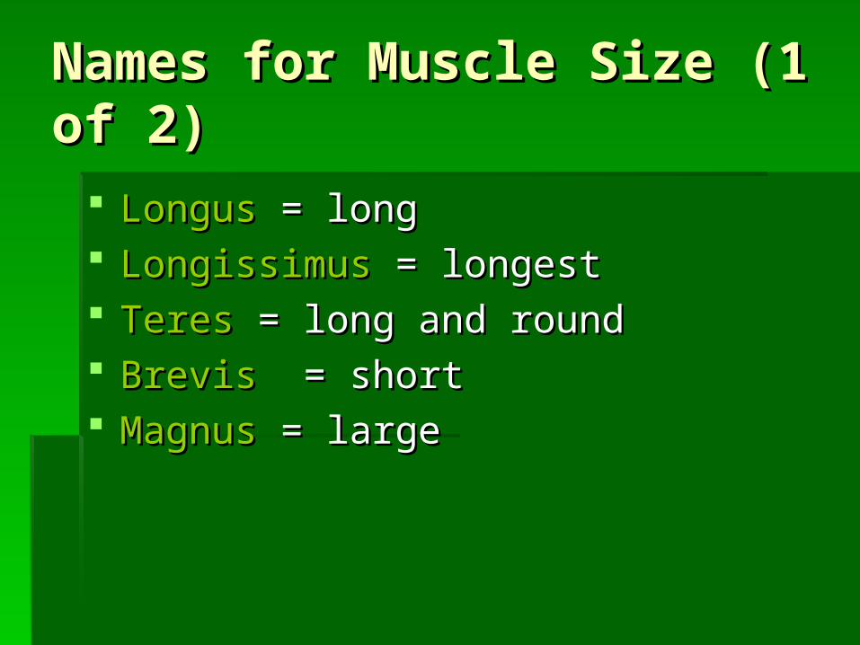
Names for Muscle Size (1 Names for Muscle Size (1 of 2)of 2)
LongusLongus = long = long LongissimusLongissimus = longest = longest TeresTeres = long and round = long and round BrevisBrevis = short = short MagnusMagnus = large = large
Names For Muscle Size (2 Names For Muscle Size (2 of 2)of 2)
MajorMajor = larger = larger MaximusMaximus = largest = largest MinorMinor = small = small MinimusMinimus = smallest = smallest

ActionAction
Movements: Movements: e.g.,e.g., flexor, extensor, retractor flexor, extensor, retractor
Occupations or habits: Occupations or habits: e.g., risore.g., risor = laughter = laughter
Divisions of the Muscular Divisions of the Muscular SystemSystem
1.1. Axial musclesAxial muscles:: position head and spinal columnposition head and spinal column move rib cagemove rib cage 60% of skeletal muscles60% of skeletal muscles
2.2. Appendicular musclesAppendicular muscles:: support pectoral and pelvic girdlessupport pectoral and pelvic girdles support limbssupport limbs 40% of skeletal muscles40% of skeletal muscles
The Axial MusclesThe Axial Muscles
Divisions based on location and function:Divisions based on location and function: muscles of head and neckmuscles of head and neck muscles of vertebral columnmuscles of vertebral column oblique and rectus musclesoblique and rectus muscles muscles of pelvic floormuscles of pelvic floor
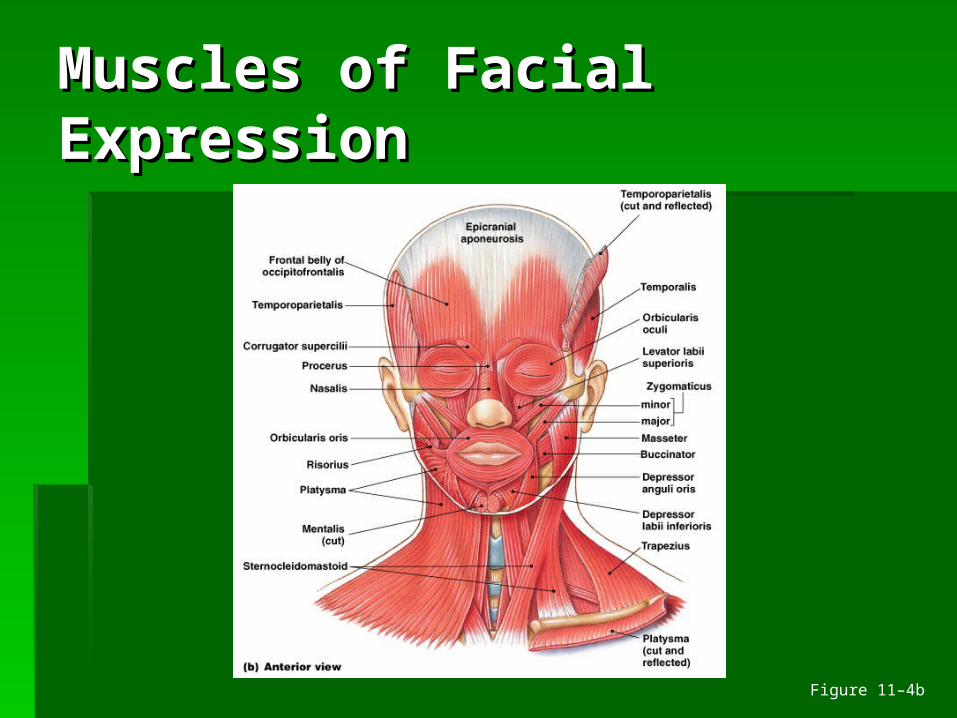
Muscles of Facial Muscles of Facial ExpressionExpression
Figure 11–4b
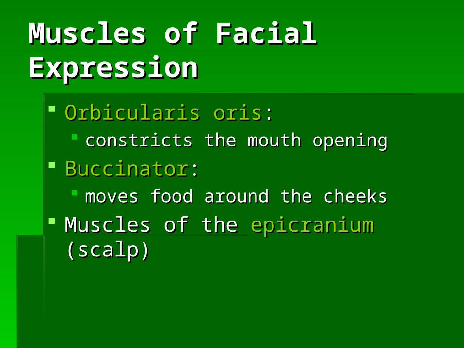
Muscles of Facial Muscles of Facial ExpressionExpression
Orbicularis orisOrbicularis oris:: constricts the mouth openingconstricts the mouth opening
BuccinatorBuccinator:: moves food around the cheeksmoves food around the cheeks
Muscles of the Muscles of the epicraniumepicranium (scalp) (scalp)
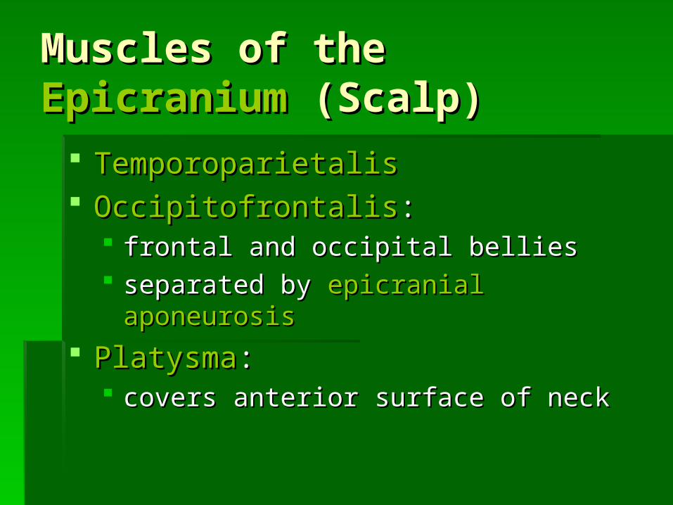
Muscles of the Muscles of the EpicraniumEpicranium (Scalp) (Scalp)
TemporoparietalisTemporoparietalis OccipitofrontalisOccipitofrontalis::
frontal and occipital belliesfrontal and occipital bellies separated by separated by epicranial aponeurosisepicranial aponeurosis
PlatysmaPlatysma:: covers anterior surface of neckcovers anterior surface of neck
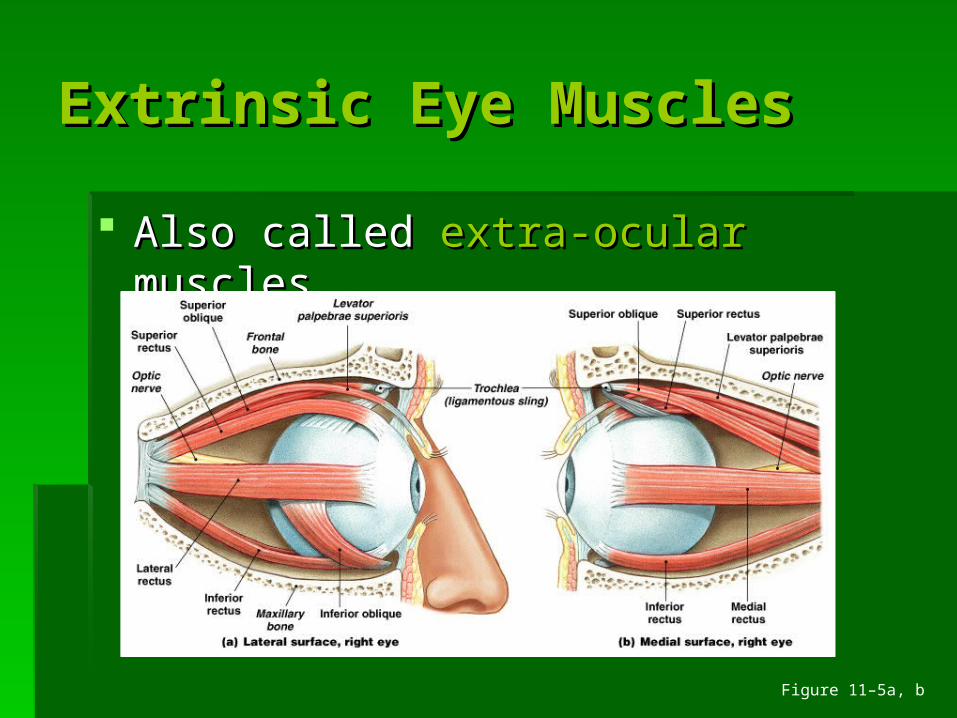
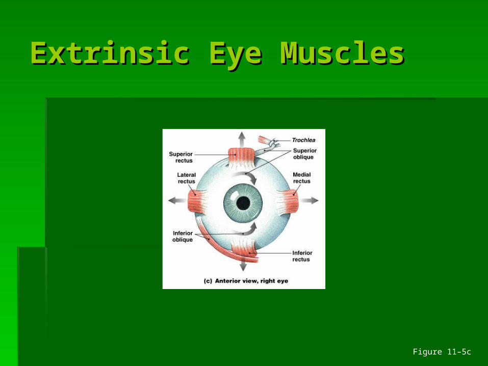
Extrinsic Eye MusclesExtrinsic Eye Muscles
Also called Also called extra-ocularextra-ocular muscles muscles
Figure 11–5a, b
Extrinsic Eye MusclesExtrinsic Eye Muscles
Figure 11–5c
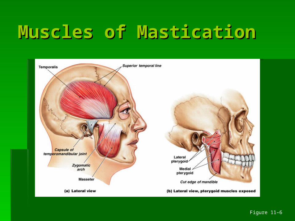
Muscles of MasticationMuscles of Mastication
Figure 11–6
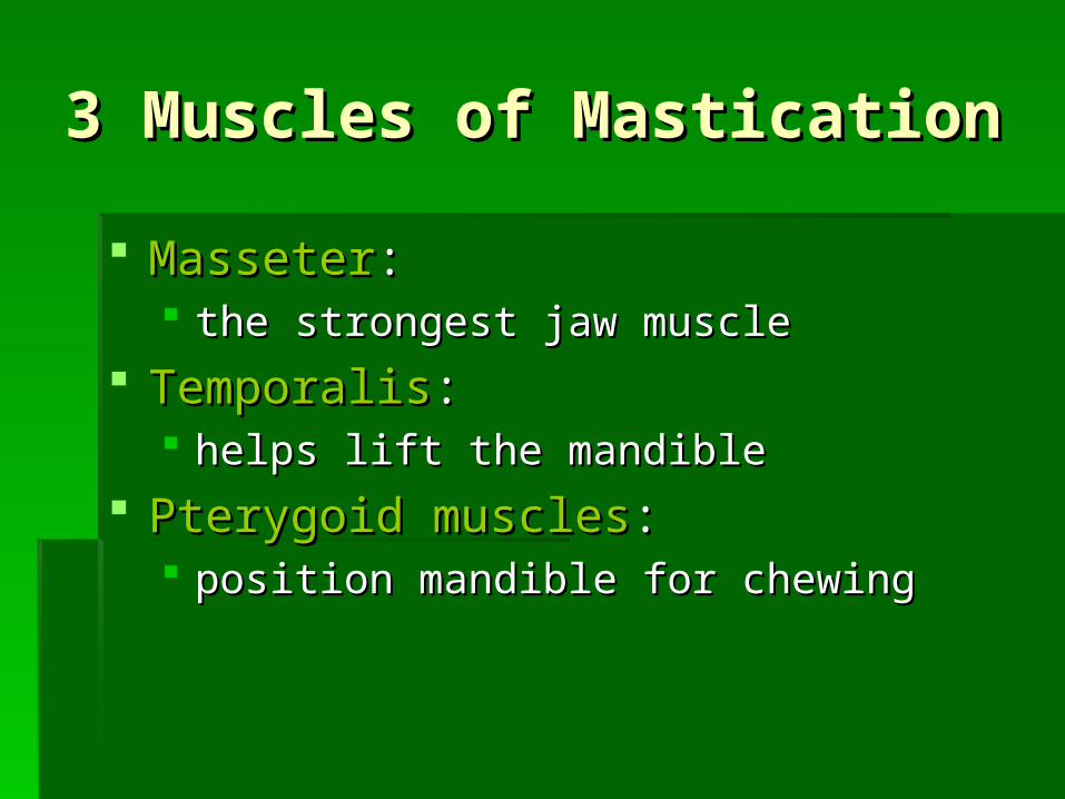
3 Muscles of Mastication 3 Muscles of Mastication
MasseterMasseter:: the strongest jaw musclethe strongest jaw muscle
TemporalisTemporalis:: helps lift the mandiblehelps lift the mandible
Pterygoid musclesPterygoid muscles:: position mandible for chewingposition mandible for chewing
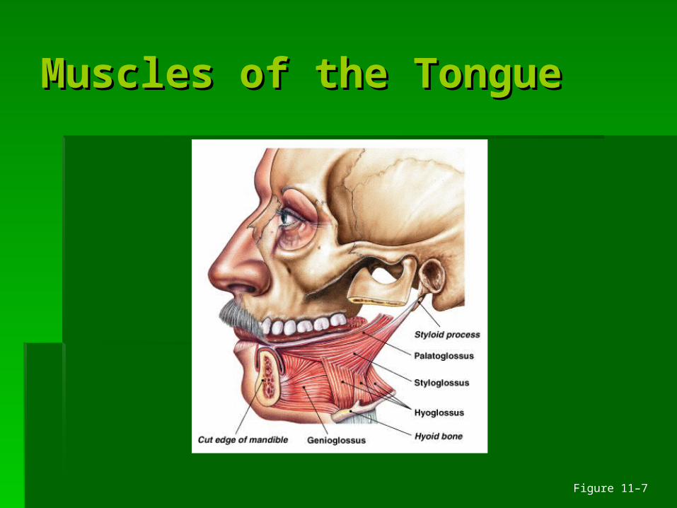
Muscles of the TongueMuscles of the Tongue
Figure 11–7
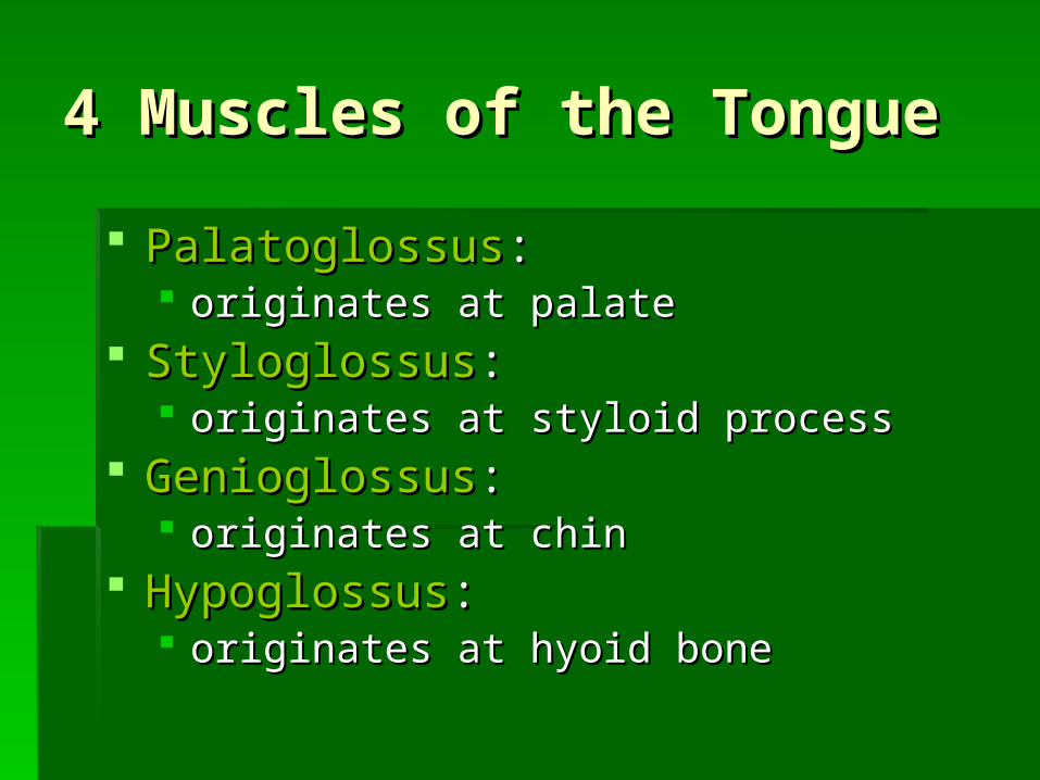
4 Muscles of the Tongue 4 Muscles of the Tongue
PalatoglossusPalatoglossus:: originates at palateoriginates at palate
StyloglossusStyloglossus:: originates at styloid processoriginates at styloid process
GenioglossusGenioglossus:: originates at chinoriginates at chin
HypoglossusHypoglossus:: originates at hyoid boneoriginates at hyoid bone
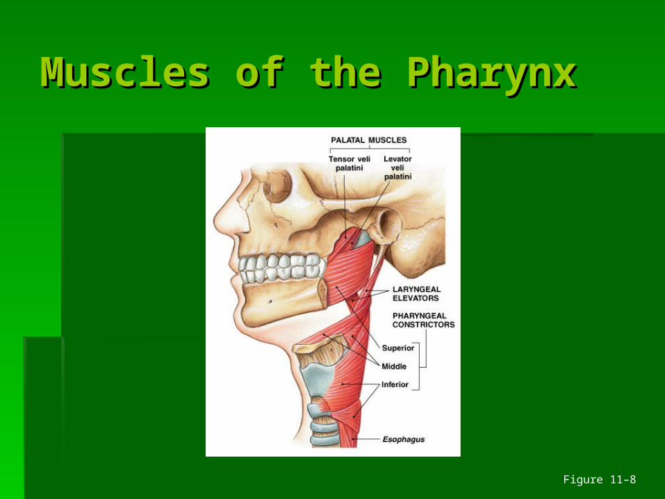
Muscles of the PharynxMuscles of the Pharynx
Figure 11–8
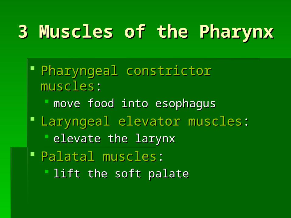
3 Muscles of the Pharynx 3 Muscles of the Pharynx
Pharyngeal constrictor musclesPharyngeal constrictor muscles:: move food into esophagusmove food into esophagus
Laryngeal elevator musclesLaryngeal elevator muscles:: elevate the larynxelevate the larynx
Palatal musclesPalatal muscles:: lift the soft palatelift the soft palate
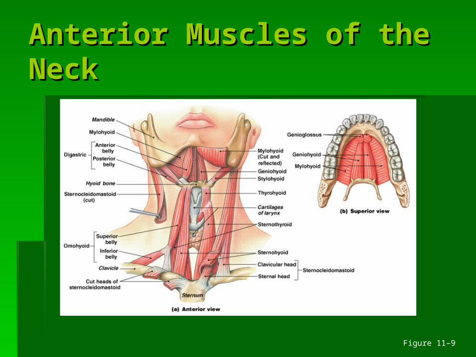
Anterior Muscles of the Anterior Muscles of the NeckNeck
Figure 11–9
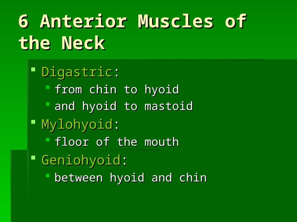
6 Anterior Muscles of the 6 Anterior Muscles of the Neck Neck
DigastricDigastric:: from chin to hyoidfrom chin to hyoid and hyoid to mastoidand hyoid to mastoid
MylohyoidMylohyoid:: floor of the mouthfloor of the mouth
GeniohyoidGeniohyoid:: between hyoid and chinbetween hyoid and chin
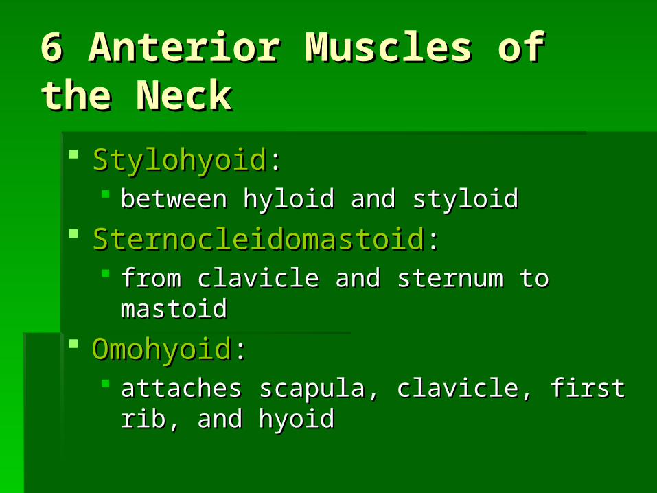
6 Anterior Muscles of the 6 Anterior Muscles of the Neck Neck
StylohyoidStylohyoid:: between hyloid and styloidbetween hyloid and styloid
SternocleidomastoidSternocleidomastoid:: from clavicle and sternum to mastoidfrom clavicle and sternum to mastoid
OmohyoidOmohyoid:: attaches scapula, clavicle, first rib, and hyoidattaches scapula, clavicle, first rib, and hyoid
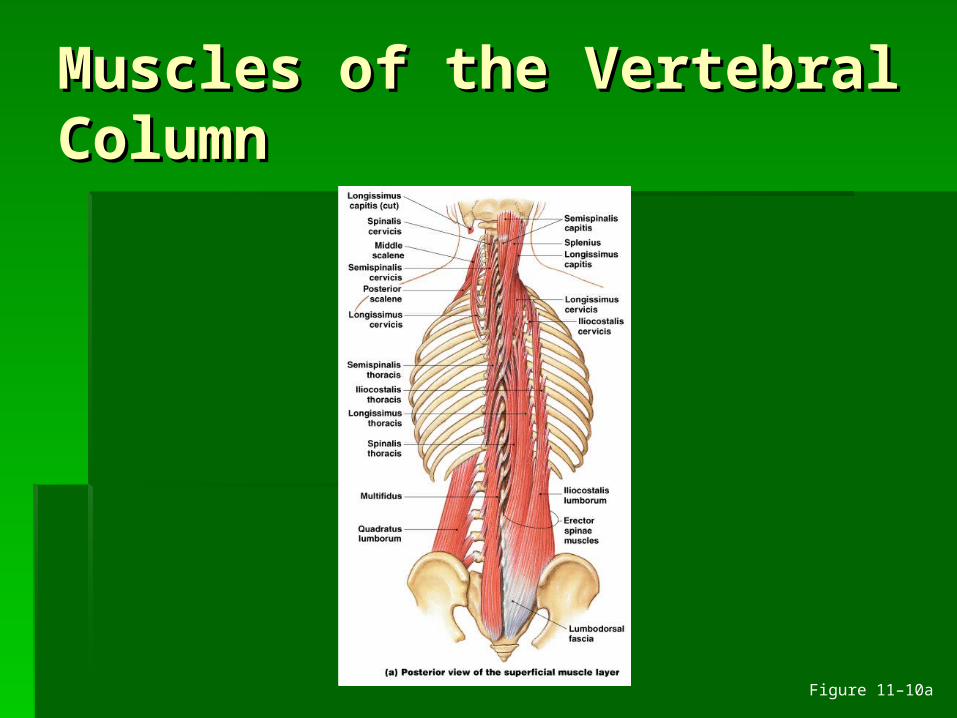
Muscles of the Vertebral Muscles of the Vertebral ColumnColumn
Figure 11–10a
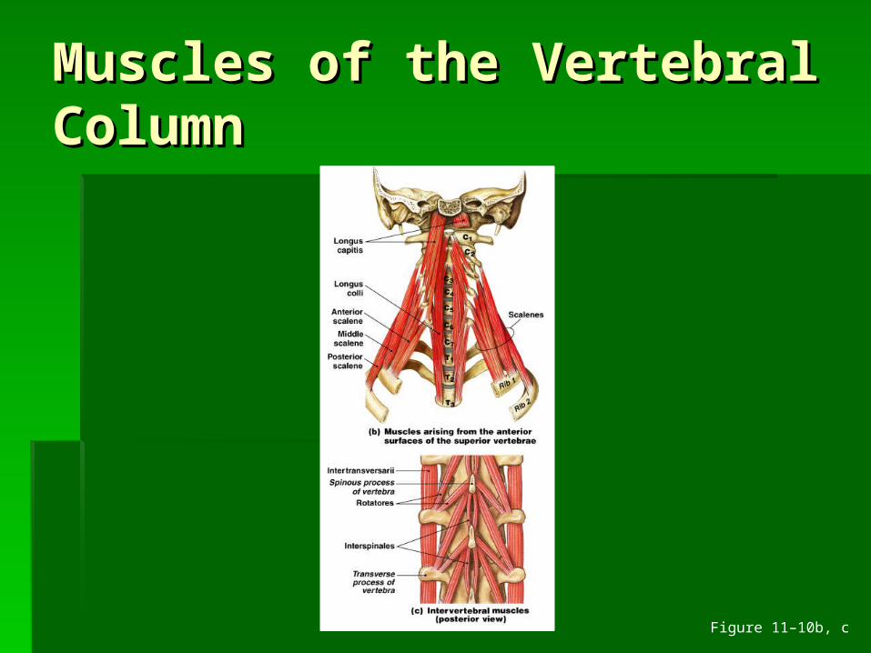
Muscles of the Vertebral Muscles of the Vertebral ColumnColumn
Figure 11–10b, c
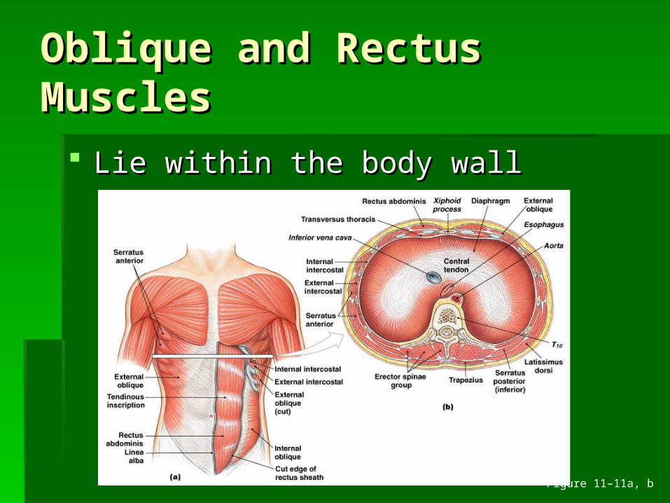
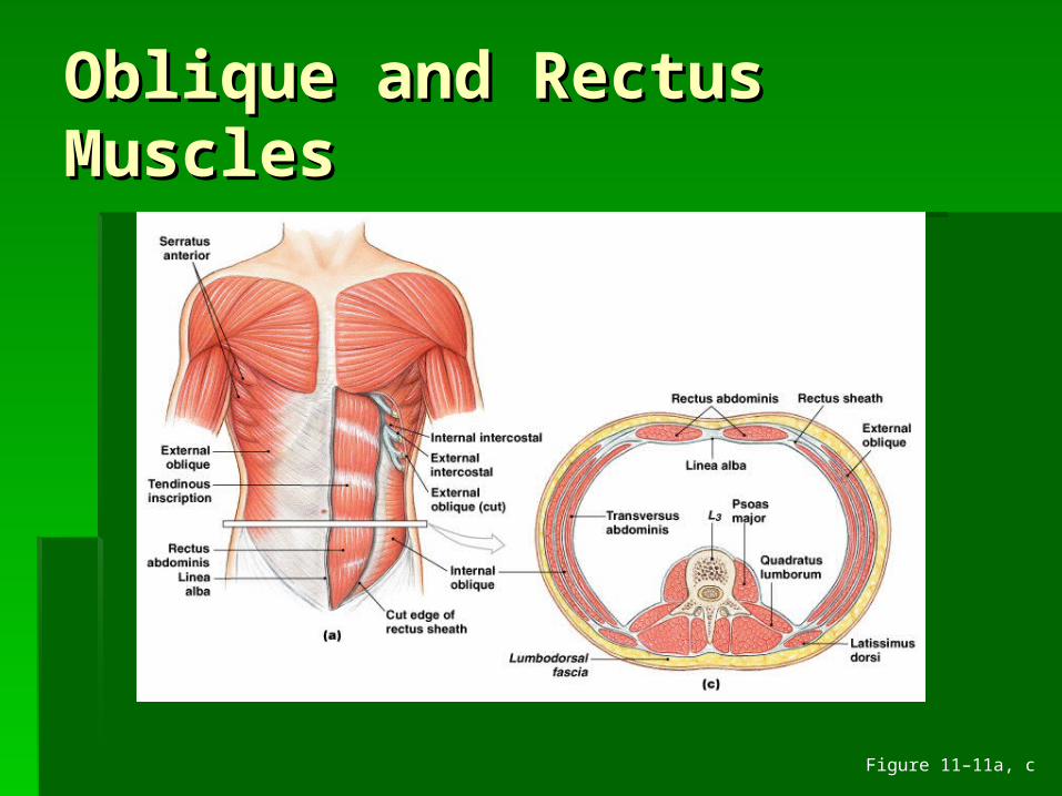
Oblique and Rectus Oblique and Rectus MusclesMuscles
Lie within the body wallLie within the body wall
Figure 11–11a, b
Oblique and Rectus Oblique and Rectus MusclesMuscles
Figure 11–11a, c
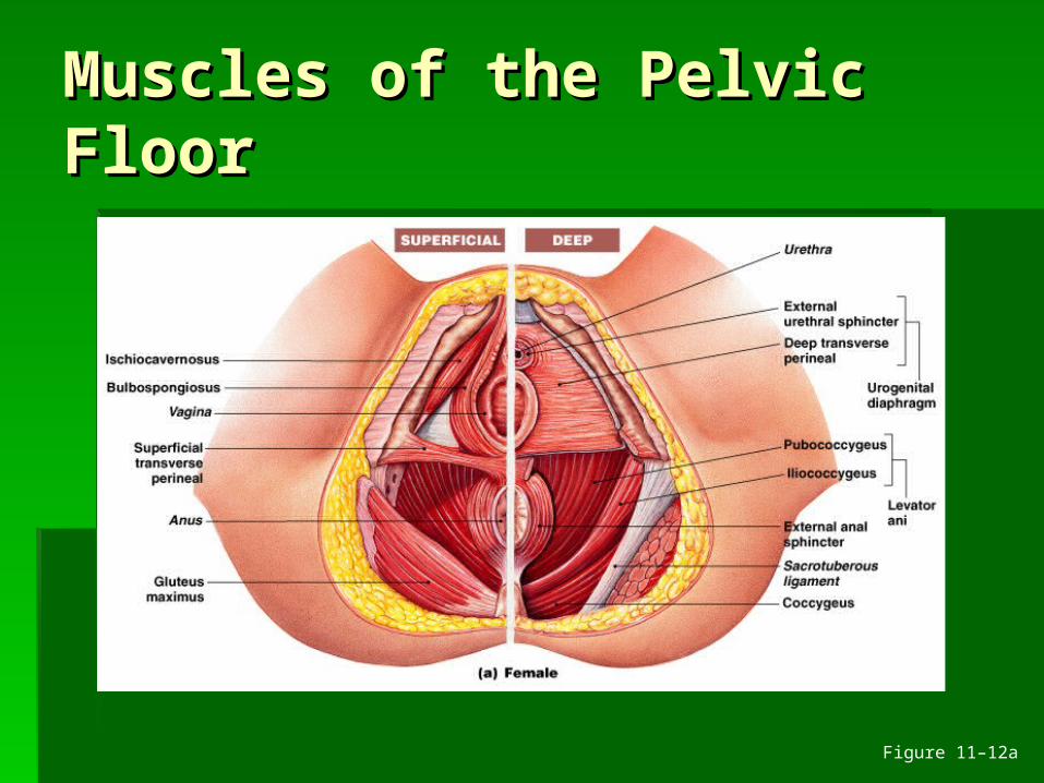
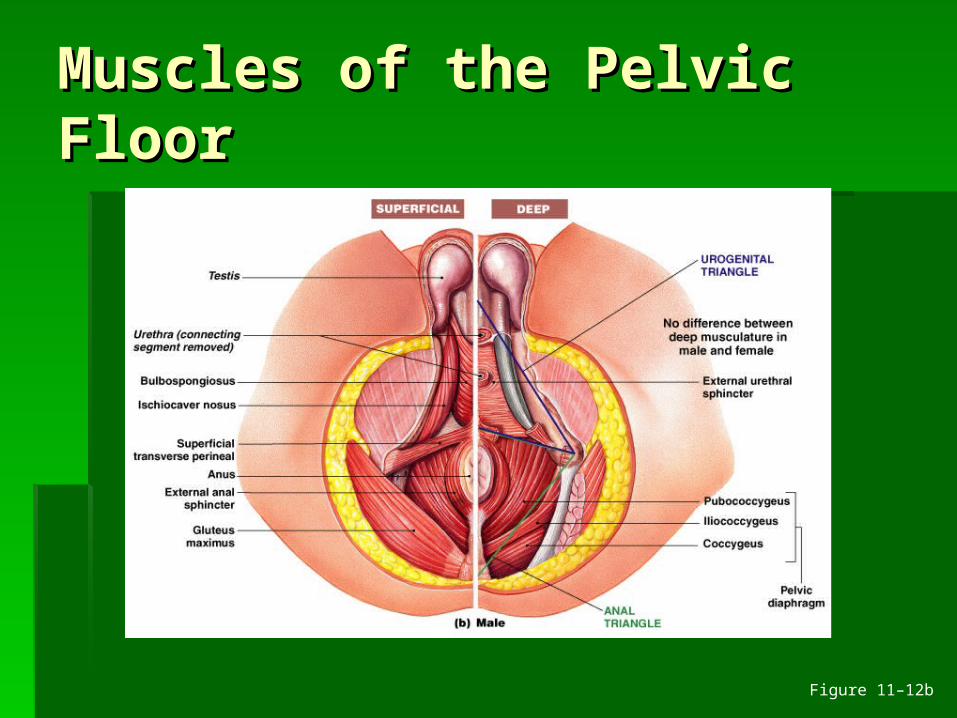
Muscles of the Pelvic Muscles of the Pelvic FloorFloor
Figure 11–12a
Muscles of the Pelvic Muscles of the Pelvic FloorFloor
Figure 11–12b

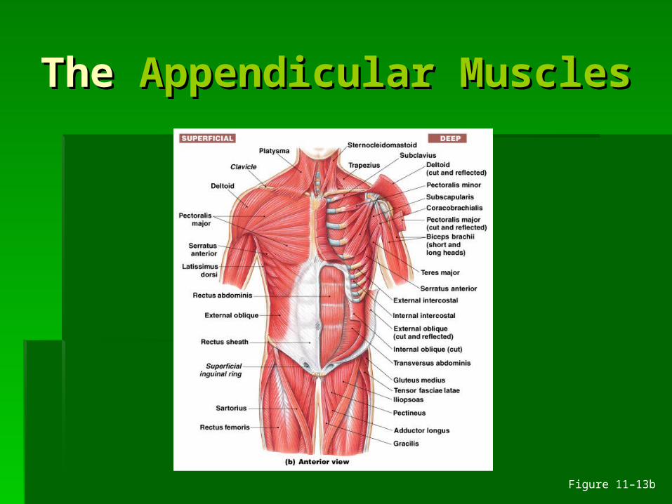
The The Appendicular Appendicular MusclesMuscles
Figure 11–13a
The The Appendicular Appendicular MusclesMuscles
Figure 11–13b
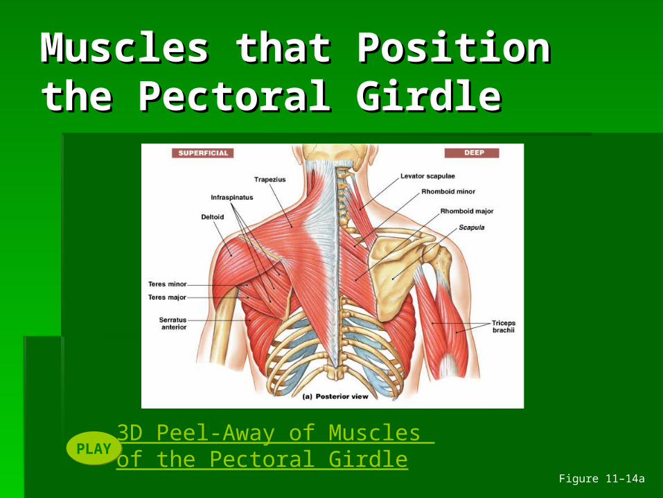
3D Peel-Away of Muscles of the Pectoral GirdlePLAYPLAY
Muscles that Position Muscles that Position the Pectoral Girdlethe Pectoral Girdle
Figure 11–14a
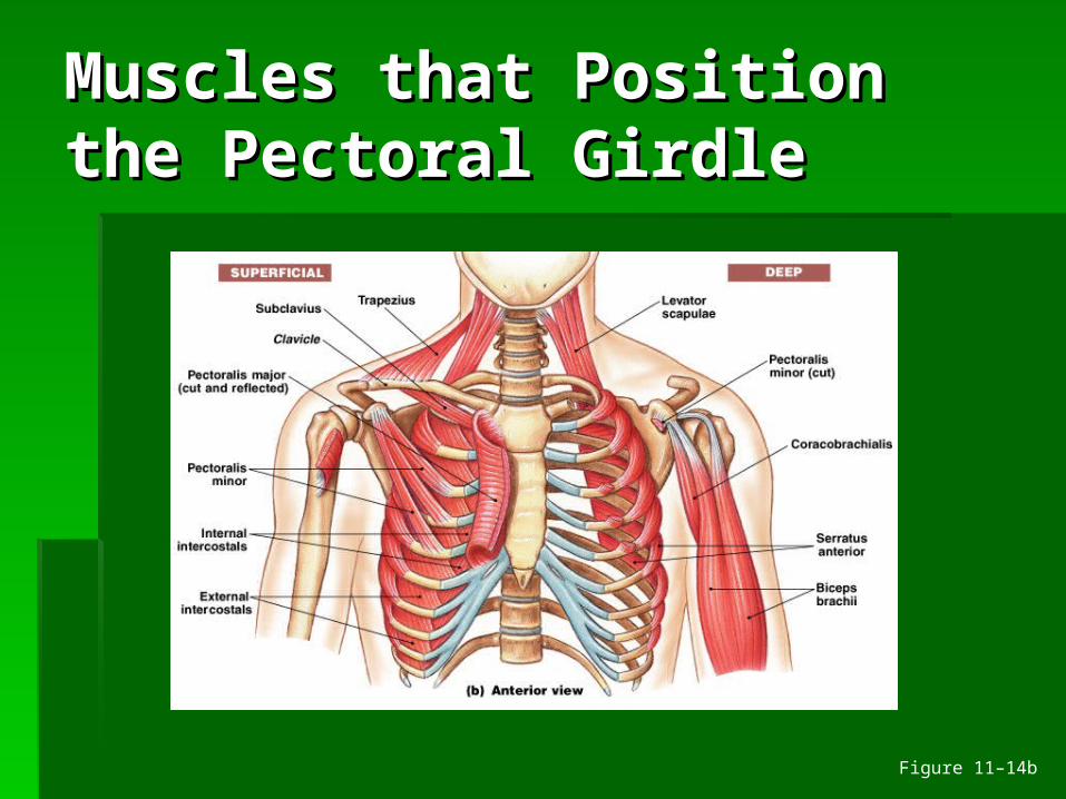
Muscles that Position Muscles that Position the Pectoral Girdlethe Pectoral Girdle
Figure 11–14b
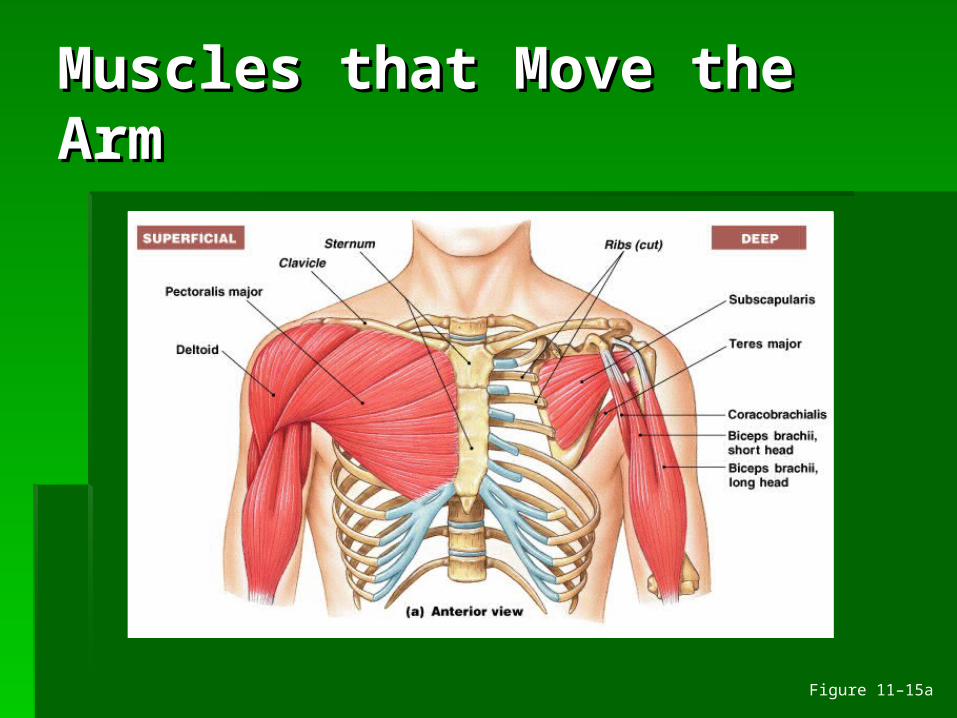
Muscles that Move the Muscles that Move the ArmArm
Figure 11–15a
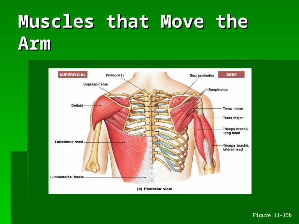
Muscles that Move the Muscles that Move the ArmArm
Figure 11–15b
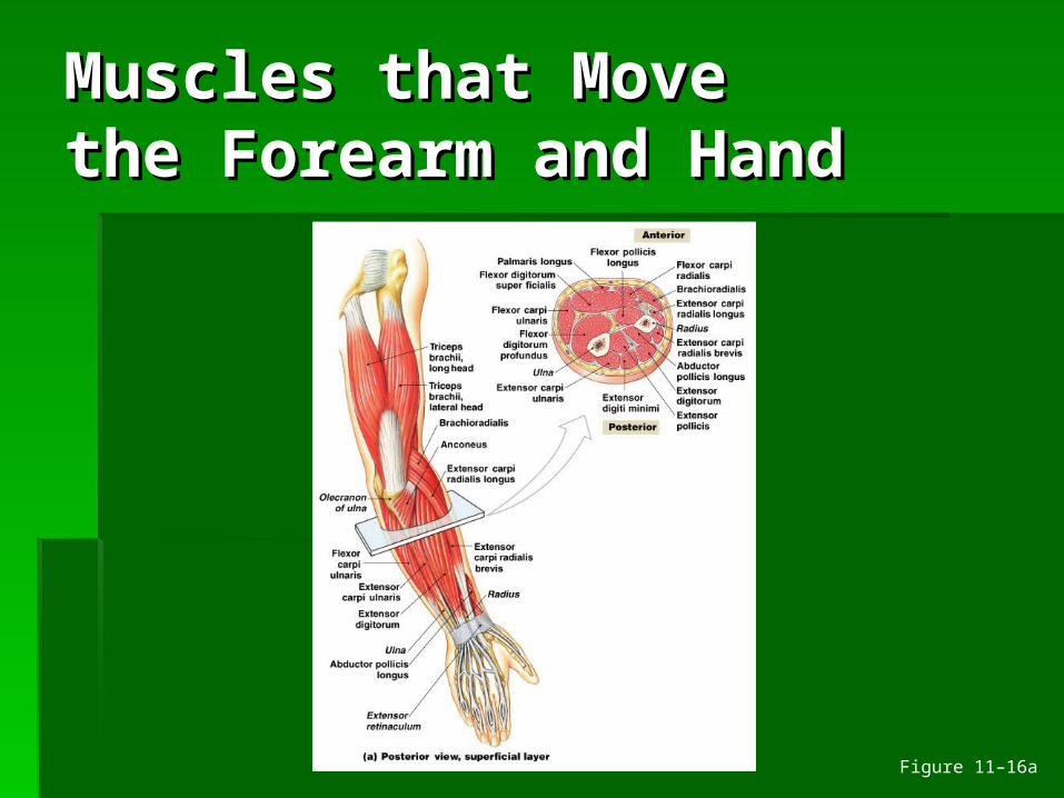
Muscles that Move Muscles that Move the Forearm and Handthe Forearm and Hand
Figure 11–16a

Muscles that Move Muscles that Move the Forearm and Handthe Forearm and Hand
Figure 11–16b
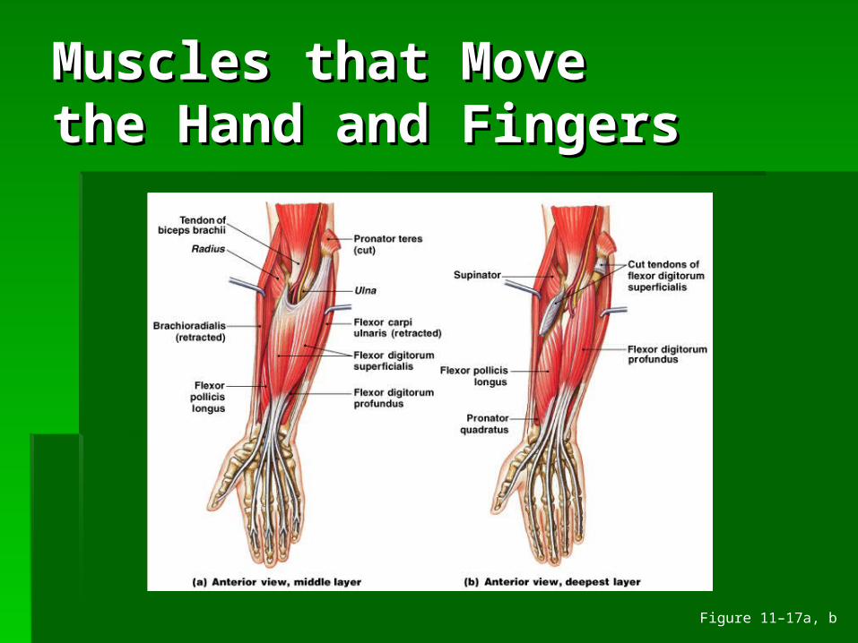
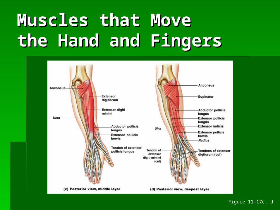
Muscles that Move Muscles that Move the Hand and Fingersthe Hand and Fingers
Figure 11–17a, b
Muscles that Move Muscles that Move the Hand and Fingersthe Hand and Fingers
Figure 11–17c, d
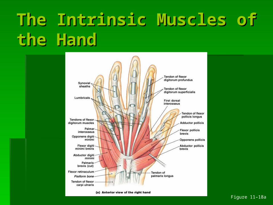
The Intrinsic Muscles of The Intrinsic Muscles of the Handthe Hand
Figure 11–18a
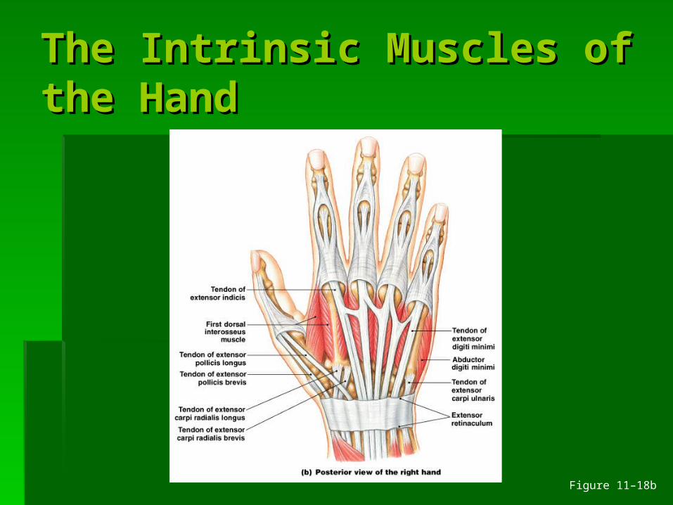
The Intrinsic Muscles of The Intrinsic Muscles of the Handthe Hand
Figure 11–18b
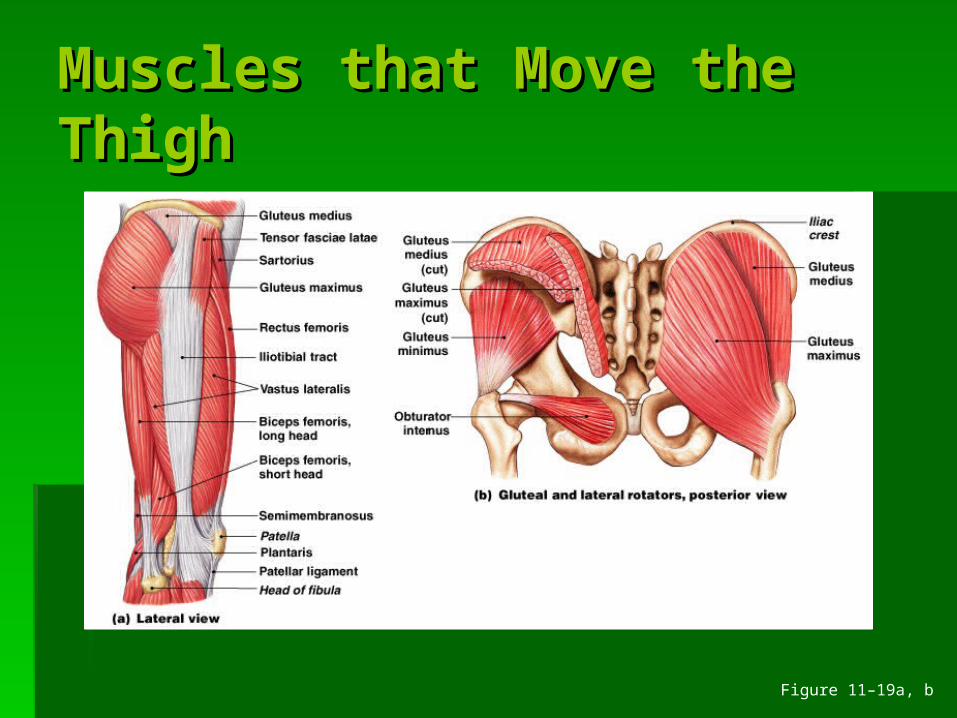
Muscles that Move the Muscles that Move the ThighThigh
Figure 11–19a, b
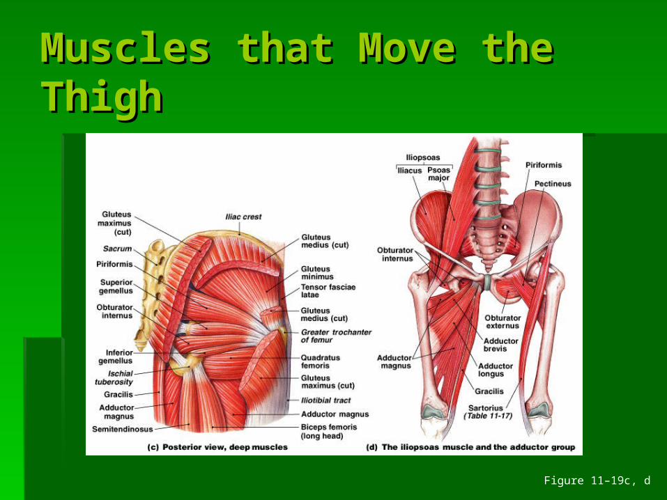
Muscles that Move the Muscles that Move the ThighThigh
Figure 11–19c, d
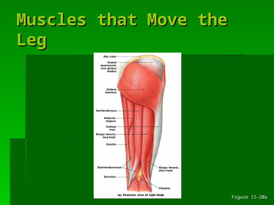
Muscles that Move the Muscles that Move the LegLeg
Figure 11–20a
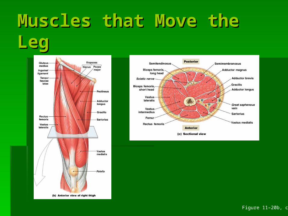
Muscles that Move the Muscles that Move the LegLeg
Figure 11–20b, c
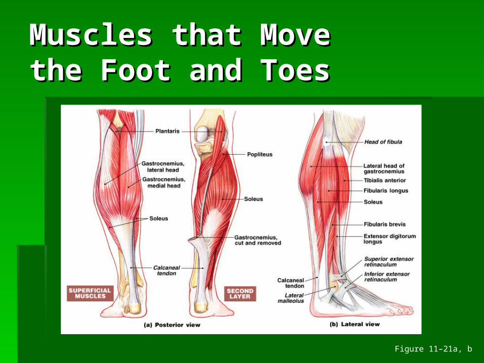
Muscles that Move Muscles that Move the Foot and Toesthe Foot and Toes
Figure 11–21a, b
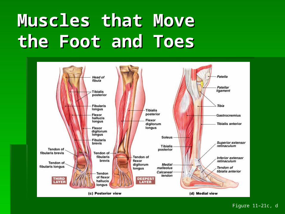
Muscles that Move Muscles that Move the Foot and Toesthe Foot and Toes
Figure 11–21c, d
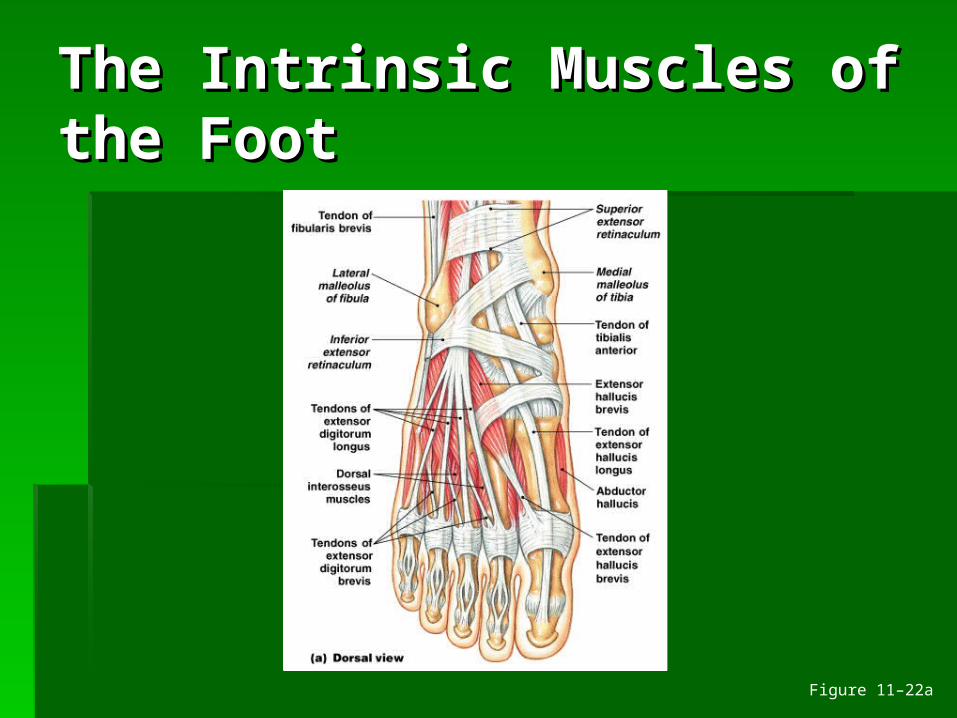
The Intrinsic Muscles of The Intrinsic Muscles of the Footthe Foot
Figure 11–22a
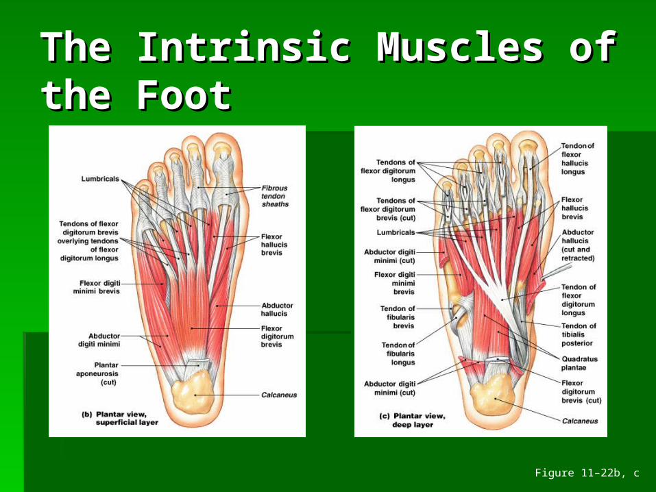
The Intrinsic Muscles of The Intrinsic Muscles of the Footthe Foot
Figure 11–22b, c
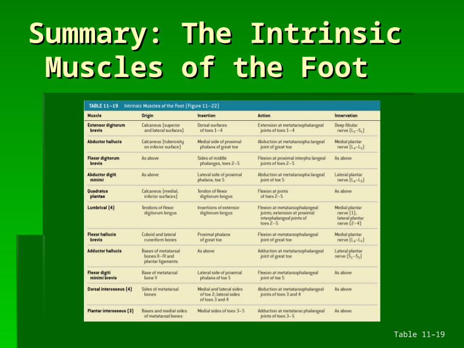
Summary: The IntrinsicSummary: The Intrinsic Muscles of the Foot Muscles of the Foot
Table 11–19
Integration with Integration with Other Systems (1 of 2)Other Systems (1 of 2)
Cardiovascular system:Cardiovascular system: delivers oxygen and fueldelivers oxygen and fuel removes carbon dioxide and wastesremoves carbon dioxide and wastes
Respiratory system:Respiratory system: responds to oxygen demand of musclesresponds to oxygen demand of muscles