Panoramic Technique Errors
36
The following slides describe the basic errors seen in Panoramic Radiography. Panoramic Technique Errors In navigating through the slides, you should click on the left mouse button when you see the mouse holding an x-ray tubehead or you are done reading a slide. Hitting “Enter” or “Page Down” will also work. To go back to the previous slide, hit “backspace” or “page up”.
Transcript of Panoramic Technique Errors

The following slides describe the basic errors seen in Panoramic Radiography.
Panoramic Technique Errors
In navigating through the slides, you should click on the left mouse button when you see the mouse holding an x-ray tubehead or you are done reading a slide. Hitting “Enter” or “Page Down” will also work. To go back to the previous slide, hit “backspace” or “page up”.

Teeth Too AnteriorIf the teeth are positioned in front of the notches in the bitestick (see diagram below left), the anterior teeth will appear narrower and will be blurred (less sharp than normal). If the teeth are in front of the notches, they are closer to the film, resulting in less magnification horizontally (narrowing). Being out of the focal trough makes the images less sharp.

Teeth Too Anterior
This film shows the blurring and narrowing of the anterior teeth.
R L

Teeth Too Anterior
R L
R L

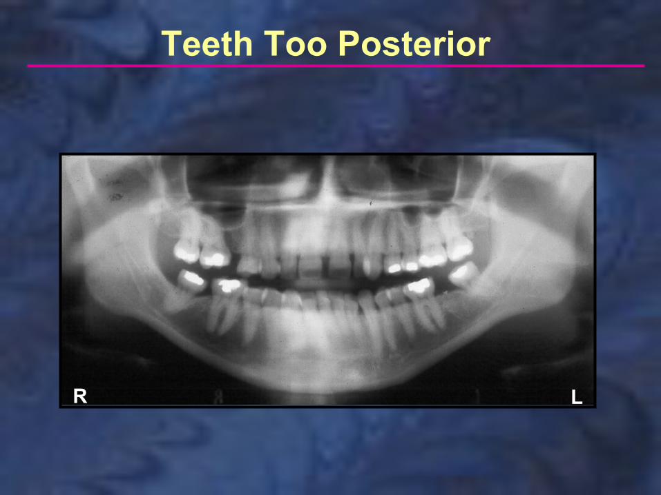
Teeth Too PosteriorIf the teeth are positioned behind the notches in the bitestick (see diagram below left), the anterior teeth will appear wider and will be blurred (less sharp than normal). If the teeth are behind the notches, they are farther from the film, resulting in more magnification horizontally (widening). Being out of the focal trough makes the images less sharp.
Teeth Too Posterior
R L

Teeth Too Posterior
R L

If the head is turned slightly to the side (not centered on the bitestick), the structures on one side will be closer to the film and the structures on the other side will be farther from the film. In the diagram below, the head was turned to the right and the teeth are closer to the film on that side.
Head Turned

The teeth are smaller on the side to which the head is turned. (When the teeth are closer to the film, there is less magnification horizontally). The teeth that are farther from the film are wider because there is increased magnification horizontally.
Head Turned
Head Turned
R L
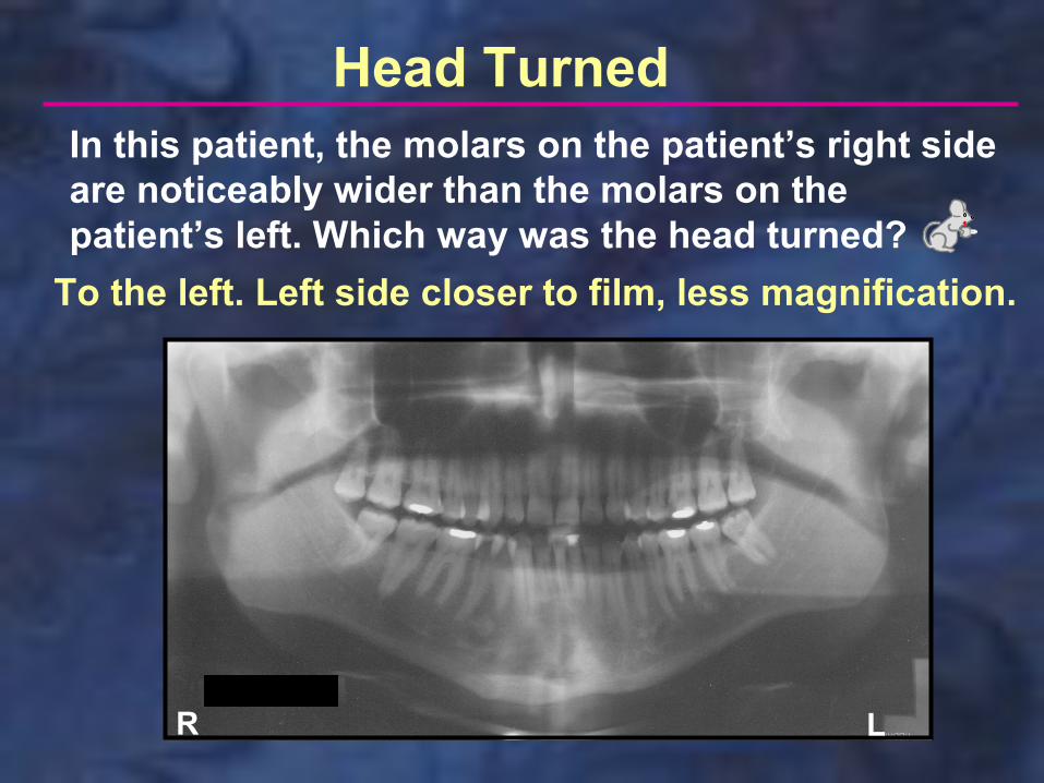
In this patient, the molars on the patient’s right side are noticeably wider than the molars on the patient’s left. Which way was the head turned?
To the left. Left side closer to film, less magnification.

Head Turned
R L
On this film, not only are the teeth wider on one side, but the ramus is also wider on that side. (The black arrows are the same length on both sides). Which side was farthest from the film?
The patient’s right side; farther from the film, more magnification.

Head Turned
R
In this film, the patient’s head was turned to the right, resulting in a widening of the teeth and ramus on the patient’s left side.

If the head is positioned so that the Frankfort Plane is inclined downward (see diagram below left), the mandibular incisors will appear shortened and the mandible will be V-shaped (Exaggerated smile).
Head Tipped Down

Head Tipped Down
R L
Notice how short the mandibular incisors appear. The rest of the teeth are relatively normal.

Head Tipped Down
R
Again we see shortened mandibular incisors, V-shaped mandible.
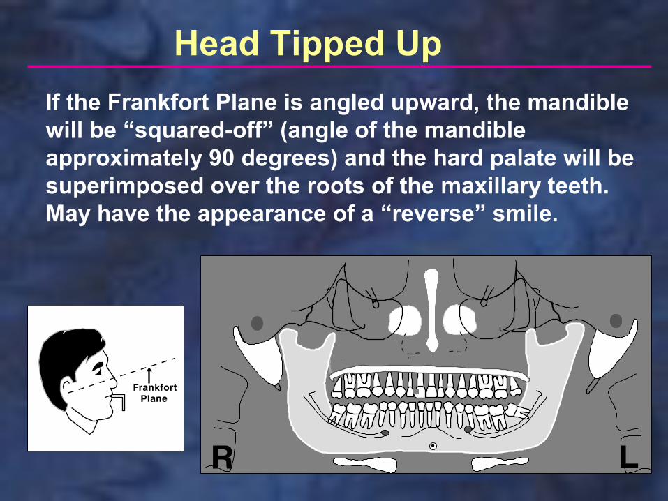
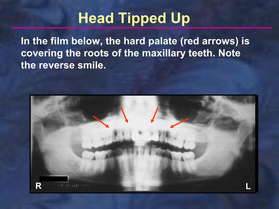
If the Frankfort Plane is angled upward, the mandible will be “squared-off” (angle of the mandible approximately 90 degrees) and the hard palate will be superimposed over the roots of the maxillary teeth. May have the appearance of a “reverse” smile.
Head Tipped Up
Head Tipped Up
R L
In the film below, the hard palate (red arrows) is covering the roots of the maxillary teeth. Note the reverse smile.
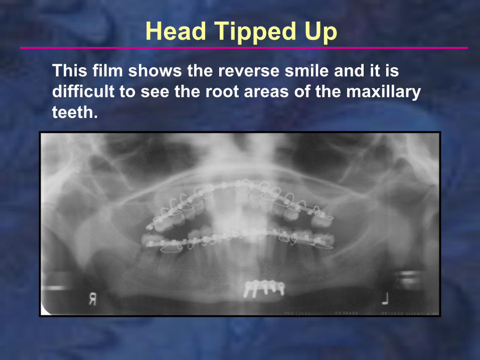
Head Tipped Up
This film shows the reverse smile and it is difficult to see the root areas of the maxillary teeth.
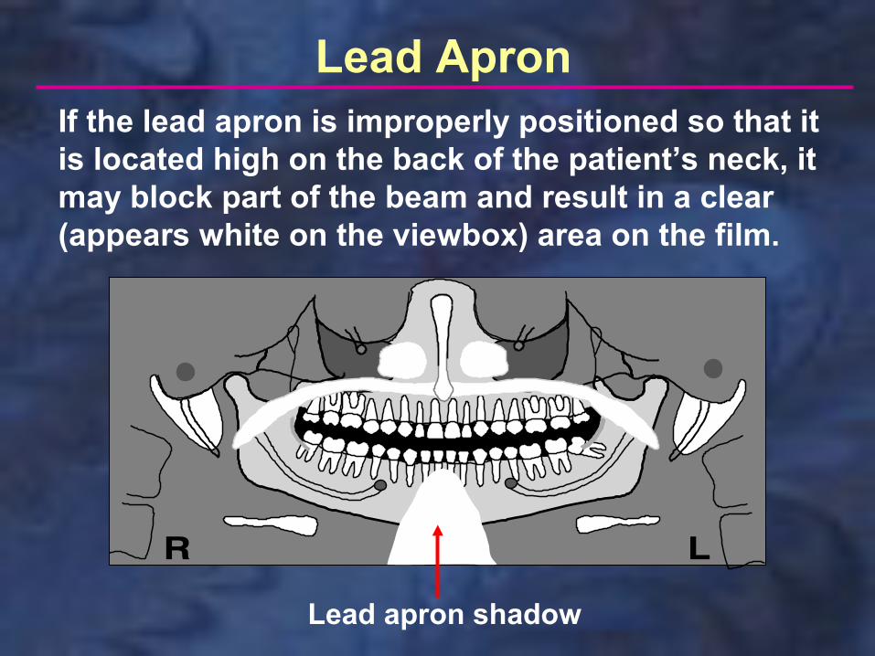
Lead apron shadow
Lead ApronIf the lead apron is improperly positioned so that it is located high on the back of the patient’s neck, it may block part of the beam and result in a clear (appears white on the viewbox) area on the film.
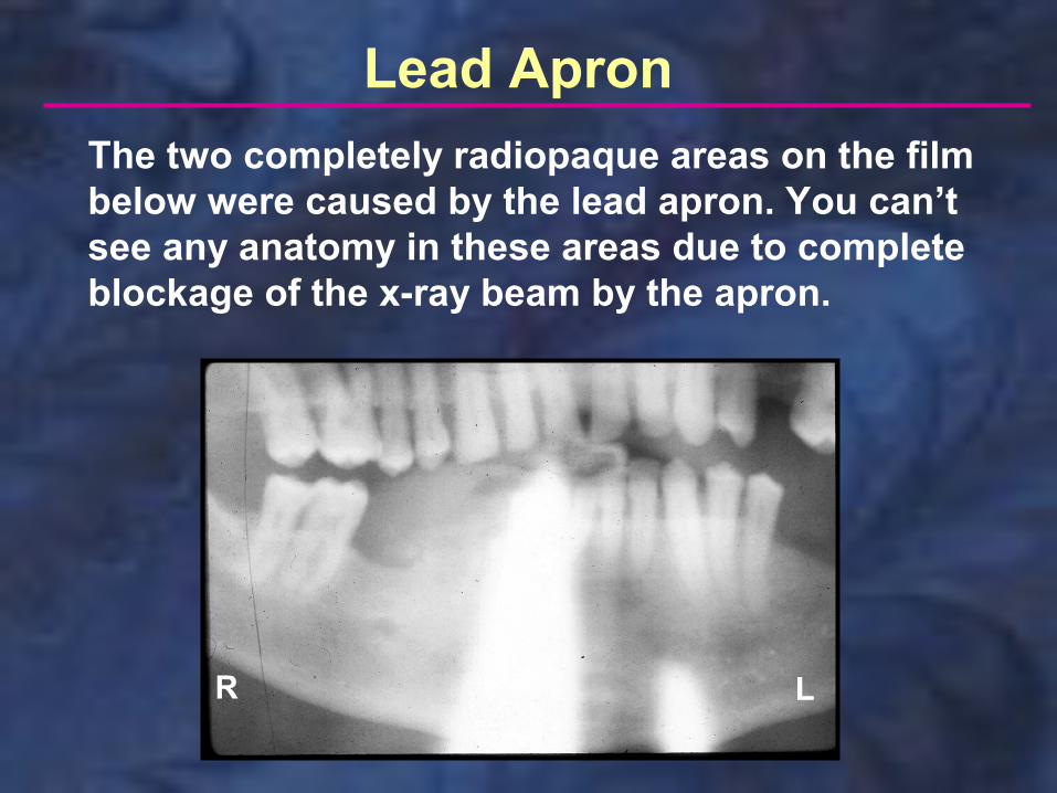
Lead Apron
R L
The two completely radiopaque areas on the film below were caused by the lead apron. You can’t see any anatomy in these areas due to complete blockage of the x-ray beam by the apron.

Lead Apron
R L
The film below shows an extensive white area caused by the lead apron. Note the black dots (arrows) that represent the stitching on the apron. The thyroid collar should never be used for panoramic radiography since it would routinely cause this same problem.

Cervical Vertebrae (Spine) If the patient is not standing straight, the cervical vertebrae may block the x-ray beam as the tubehead travels behind the patient at an upward angle. This results in a radiopaque area that extends up through the middle of the film (arrows below). The teeth/bone are faintly visible in the radiopaque area (not completely blocked out as with the lead apron).

This film shows the radiopaque “shadow” caused by the cervical vertebrae in a patient that is not standing straight. Note that the edges of this radiopaque area are not as sharp as those produced by the lead apron; here the radiopacity blends in with the surrounding bone.
Cervical Vertebrae (Spine)

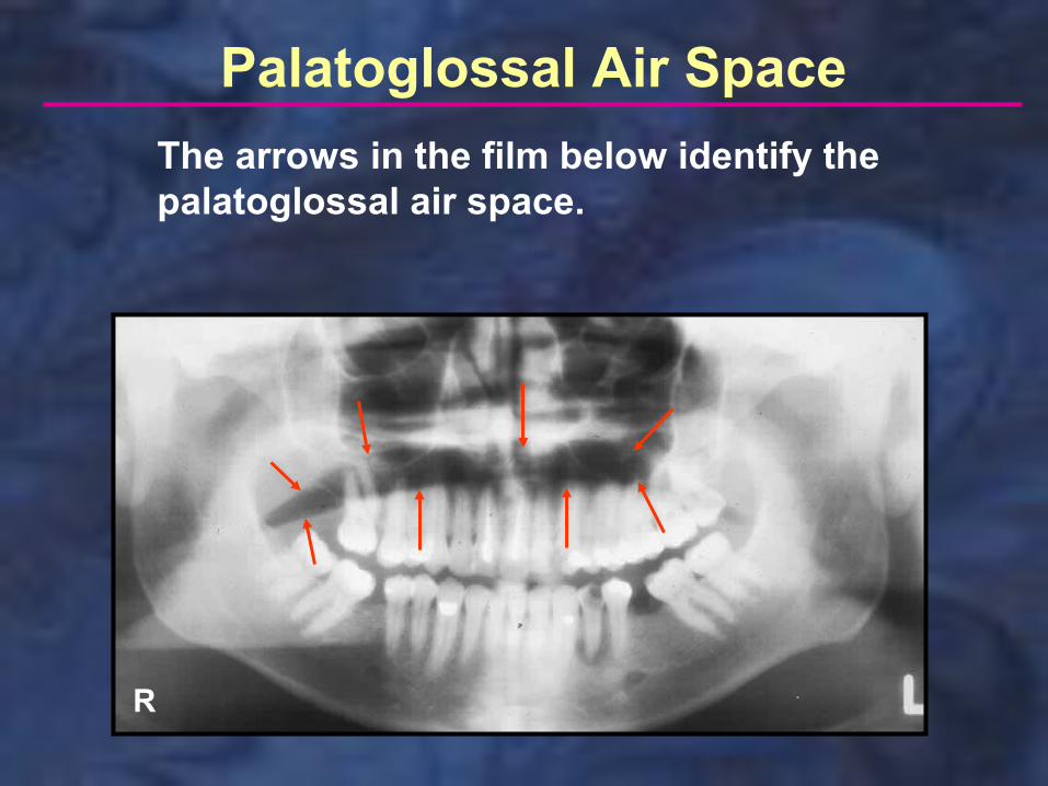
The Palatoglossal Air Space (black area below) is caused by failure to keep the tongue against the palate during exposure. This makes it difficult to diagnose periapical pathology, which also is dark, in the maxillary area. Many patients have difficulty complying with the instructions to keep the tongue against the palate during exposure and this space is often seen. As long as you instruct the patient, this would not be considered an error.
Palatoglossal Air Space
Palatoglossal Air Space
R
The arrows in the film below identify the palatoglossal air space.

Static electricity appears as black lines or dots on the film, often having a tree-branch appearance. It is caused by removing the film from the box or cassette too quickly, creating static discharge.
Static Electricity

Static Electricity
R L
This film shows two major errors, one of which is the interesting aliigator-like static electricity at the top of the film. The small black circles at the bottom of the film are also caused by static. What is the other error seen on this film?The film is extremely underexposed. This was probably caused by placing the film outside the intensifying screens (not between them) in a flexible cassette.

Failure to Remove Appliances
R L
As part of patient preparation, appliances should be removed from the mouth. In this patient, the complete upper denture was left in the mouth. This would not require a retake, since the acrylic of the denture base allows x-rays to pass through and the bone is clearly visible.

Failure to Remove Appliances
R L
In this patient, both upper and lower removable partial dentures were left in the mouth. In this case the metal frameworks obscure large areas of the teeth and the film should be retaken.
Failure to Remove Tongue Ring
R L
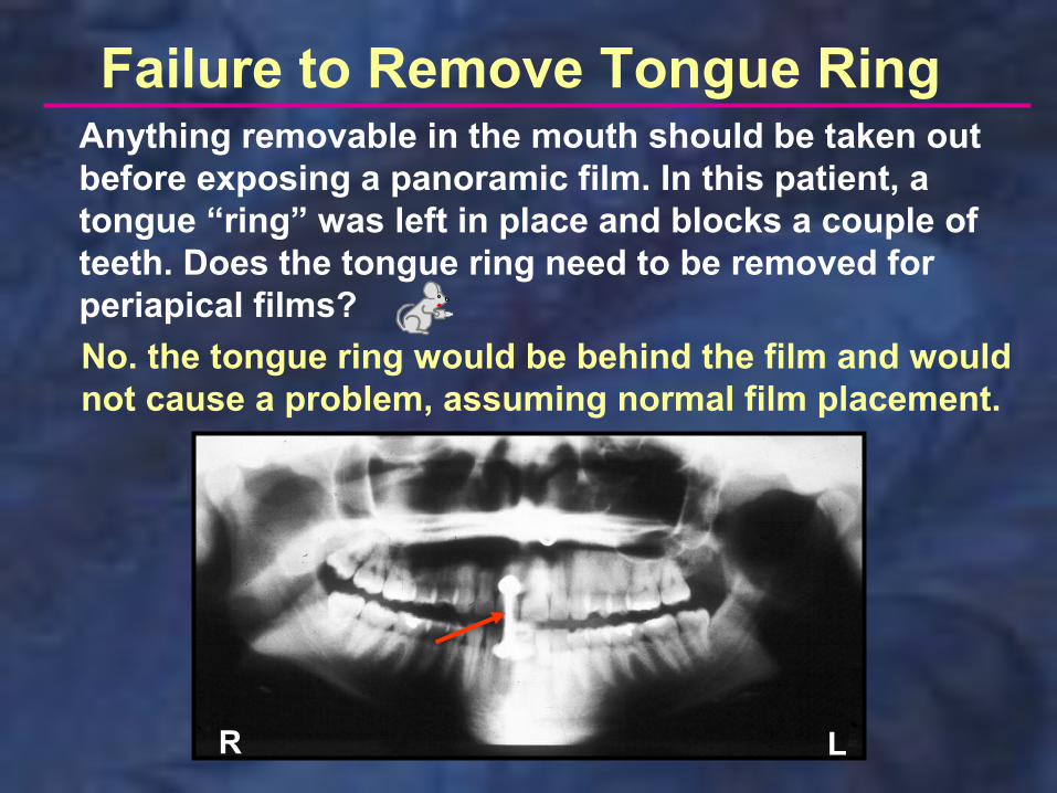
Anything removable in the mouth should be taken out before exposing a panoramic film. In this patient, a tongue “ring” was left in place and blocks a couple of teeth. Does the tongue ring need to be removed for periapical films?
No. the tongue ring would be behind the film and would not cause a problem, assuming normal film placement.

Glasses
R L
Glasses should routinely be removed for panoramic exposures. The bottom part of the frame/lenses may obscure the periapical area of the maxillary anterior teeth. What other error is evident on this film?
The head is tipped up too much. Notice the reverse smile and the proximity of the hard palate to the roots of the maxillary teeth.

Patient Movement
R L
It is important for the patient to remain still during a panoramic exposure. This film shows excessive patient movement (unknown cause) and must be retaken.
Patient Movement
R
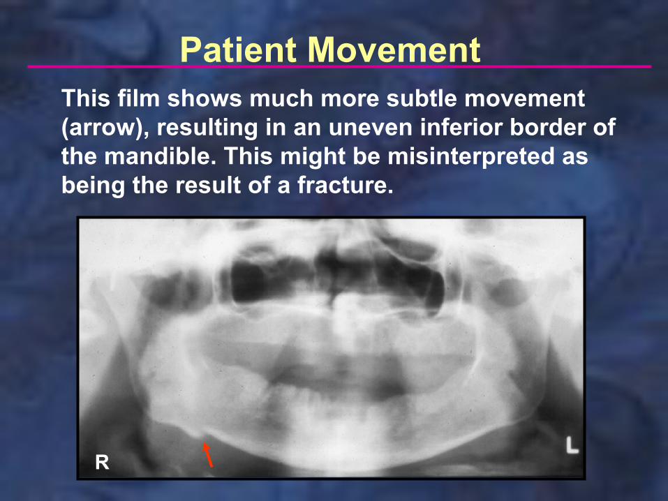
This film shows much more subtle movement (arrow), resulting in an uneven inferior border of the mandible. This might be misinterpreted as being the result of a fracture.

Double Exposure
R
It is preferable to process films immediately after exposure. If cassettes are laid aside for later processing, the operator may inadvertently pick up a cassette that has already been exposed and use it again. This results in a double exposure as seen below. What other error is evident on this film?
Static electricity at the bottom of the film.
Incorrect Exposure Settings
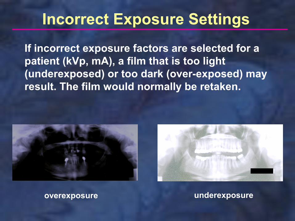
overexposure underexposure
If incorrect exposure factors are selected for a patient (kVp, mA), a film that is too light (underexposed) or too dark (over-exposed) may result. The film would normally be retaken.
This concludes the section on Panoramic Technique Errors. Additional self-study modules are available at: http://dent.osu.edu/radiology/resources.htm
If you have any questions, you may e-mail me at: [email protected]
Robert M. Jaynes, DDS, MSDirector, Radiology GroupCollege of DentistryOhio State University