PAINLESS LUMP IN THE BREAST–AN UNUSUAL PRESENTATION …
6
34 JUMDC, Vol. 1, No. 2, Jul-Dec 2010 Case Report PAINLESS LUMP IN THE BREAST–AN UNUSUAL PRESENTATION OF CHLOROMA 1 Mubashar Ahmad, 2 Saira Bilal, 3 Arif Hussain, 4 Sadia Hameed 1 Demonatrator Pathology, UMDC, Faisalabad. 2 Assistant Professor Pathology, UMDC, Faisalabad. 3 Assistant Professor Pathology, PMC, Faisalabad. 4 Professor Pathology, UMDC, Faisalabad. INTRODUCTION Chloroma (also known as granulocytic sarcoma or myeloid sarcoma) is a rare solid extramedullary tumour composed of immature granulocytic cells. It was first described in 1811 by Burns 1 and named “chloroma” by King 2 in 1853 because of its green color. Its relationship with leukemia was later established in 1893 3 . This is an uncommon complication of acute and chronic myeloid leukemia and may appear at any time during the course of the disease. Sometimes patients of acute myeloid leukemia can present with tumours in other areas like the spinal cord, maxilla, eye etc. We are reporting a case of chloroma who presented with a painless lump in the breast and was diagnosed when she was referred for fine needle aspiration cytology (FNAC). CASE REPORT A 22 year old female presented with a lump in her right breast in the subareolar region. It had been present for the past 6 months. The patient had noticed it accidentally and she did not bother about it for a few months, but the lump had gradually increased in size. She consulted a surgeon who referred her for FNAC of the swelling. The lump measured 4.0×3.0 cm. The skin over it was normal. It was mobile and non tender. FNAC was done. An enlarged lymph node was also palpable in her right axilla. It measured 1.5×1.5 cm. FNAC of this was also done. A lymph node was also palpable in the inguinal region but was small in size. On account of enlargement of multiple lymph nodes a peripheral blood smear was also taken. Smears from the breast lump (Figure 1 and 2) and from the axillary nodule revealed large cells with abundant cytoplasm and some of these cells contained granules. The peripheral smear (Figure 3) revealed the presence of blast cells which were 70% of the total. Some of the blast cells showed auer rods (Figure 4). Platelets were also reduced on the smear. As the peripheral film showed blast cells a bone marrow was advised. The bone marrow (Figure 5) was aspirated from posterior iliac crest and showed hypercellular smears and cell trails. Erythropoeisis, myelopoeisis and megakaryopoesis were suppressed. The smear was diffusely infiltrated by blast cells which comprised of 90% of the nucleated cells. Most of the blast cells had abundant cytoplasm showing scanty granules. Some of the blast cells showed Aeur rods. Sudan B black (Figure 6) was done for confirmation of the suspected diagnosis. The picture was consistent with acute myeloid leukemia. It was graded to be M1 according to the FAB classification. DISCUSSION The word chloroma is derived from the Greek word choloros, as the tumours had a green tinge due to the presence of myeloperoxidase. It is not essential that all chloromas are green. The other synonyms are granulocytic sarcoma, extramedullary myeloid tumour and myeloblastoma. The tumour may rarely precede the onset of leukemia by many months 4 but usually it is found during the course of the disease or even during the remission. Chloromas are rare lesions with an incidence of 3-4.7% of the chronic myeloproliferative disorders 5 . Corresponding Author: Dr. Sadia Hameed, 1-B, Staff Colony, Punjab Medical College, Faisalabad. E-mail: [email protected]
Transcript of PAINLESS LUMP IN THE BREAST–AN UNUSUAL PRESENTATION …
34 JUMDC, Vol. 1, No. 2, Jul-Dec 2010
Case Report
PAINLESS LUMP IN THE BREAST–AN UNUSUAL PRESENTATION OF CHLOROMA 1Mubashar Ahmad, 2Saira Bilal, 3Arif Hussain, 4Sadia Hameed 1Demonatrator Pathology, UMDC, Faisalabad. 2Assistant Professor Pathology, UMDC, Faisalabad. 3Assistant Professor Pathology, PMC, Faisalabad. 4Professor Pathology, UMDC, Faisalabad. INTRODUCTION Chloroma (also known as granulocytic sarcoma or myeloid sarcoma) is a rare solid extramedullary tumour composed of immature granulocytic cells. It was first described in 1811 by Burns1 and named “chloroma” by King2 in 1853 because of its green color. Its relationship with leukemia was later established in 18933. This is an uncommon complication of acute and chronic myeloid leukemia and may appear at any time during the course of the disease. Sometimes patients of acute myeloid leukemia can present with tumours in other areas like the spinal cord, maxilla, eye etc. We are reporting a case of chloroma who presented with a painless lump in the breast and was diagnosed when she was referred for fine needle aspiration cytology (FNAC). CASE REPORT A 22 year old female presented with a lump in her right breast in the subareolar region. It had been present for the past 6 months. The patient had noticed it accidentally and she did not bother about it for a few months, but the lump had gradually increased in size. She
consulted a surgeon who referred her for FNAC of the swelling. The lump measured 4.0×3.0 cm. The skin over it was normal. It was mobile and non tender. FNAC was done. An enlarged lymph node was also palpable in her right axilla. It measured 1.5×1.5 cm. FNAC of this was also done. A lymph node was also palpable in the
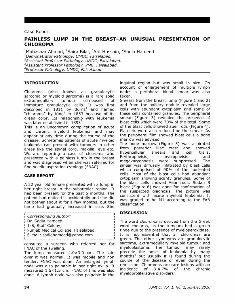
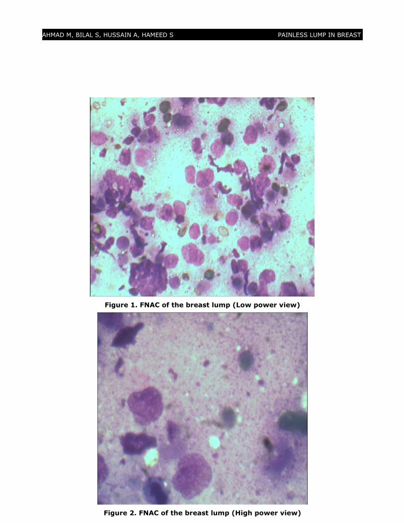
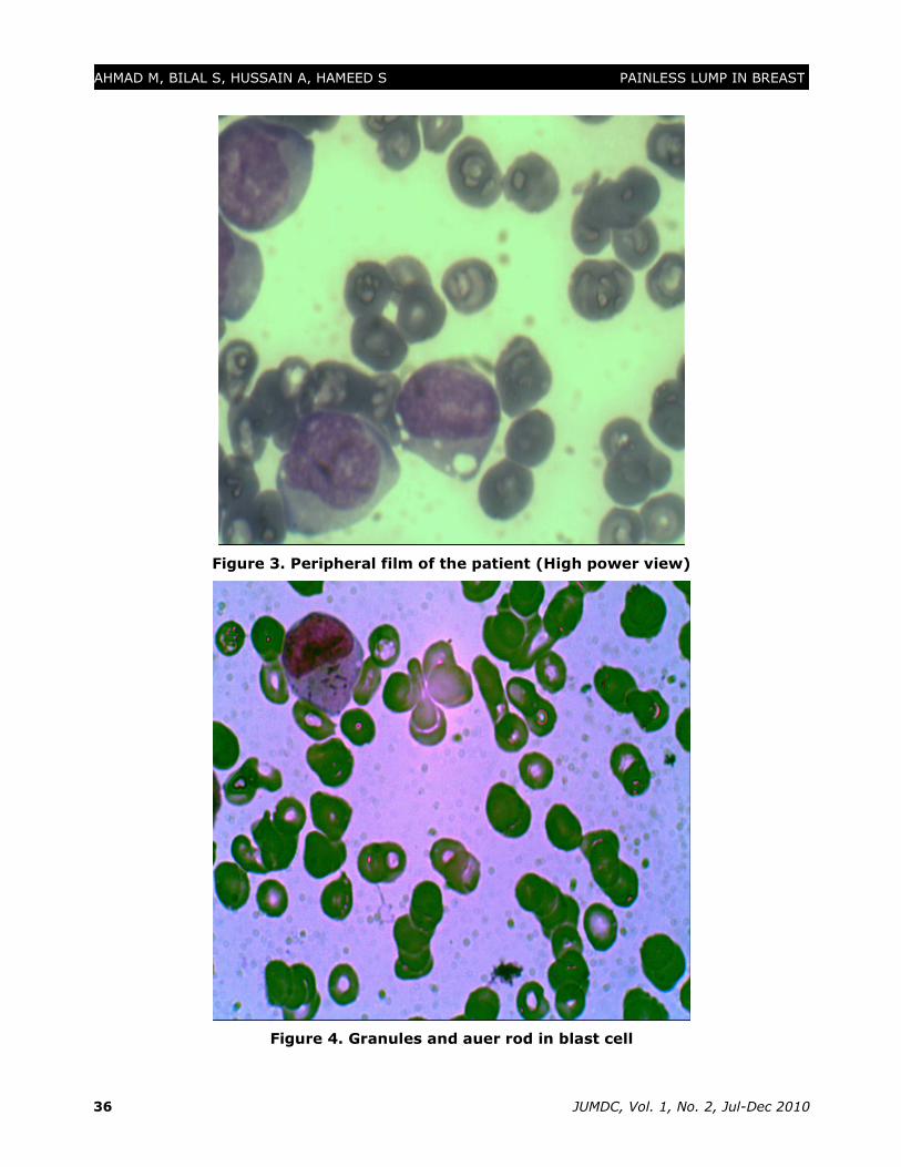
inguinal region but was small in size. On account of enlargement of multiple lymph nodes a peripheral blood smear was also taken. Smears from the breast lump (Figure 1 and 2) and from the axillary nodule revealed large cells with abundant cytoplasm and some of these cells contained granules. The peripheral smear (Figure 3) revealed the presence of blast cells which were 70% of the total. Some of the blast cells showed auer rods (Figure 4). Platelets were also reduced on the smear. As the peripheral film showed blast cells a bone marrow was advised. The bone marrow (Figure 5) was aspirated from posterior iliac crest and showed hypercellular smears and cell trails. Erythropoeisis, myelopoeisis and megakaryopoesis were suppressed. The smear was diffusely infiltrated by blast cells which comprised of 90% of the nucleated cells. Most of the blast cells had abundant cytoplasm showing scanty granules. Some of the blast cells showed Aeur rods. Sudan B black (Figure 6) was done for confirmation of the suspected diagnosis. The picture was consistent with acute myeloid leukemia. It was graded to be M1 according to the FAB classification. DISCUSSION The word chloroma is derived from the Greek word choloros, as the tumours had a green tinge due to the presence of myeloperoxidase. It is not essential that all chloromas are green. The other synonyms are granulocytic sarcoma, extramedullary myeloid tumour and myeloblastoma. The tumour may rarely precede the onset of leukemia by many months4 but usually it is found during the course of the disease or even during the remission. Chloromas are rare lesions with an incidence of 3-4.7% of the chronic myeloproliferative disorders5.
Corresponding Author:
Dr. Sadia Hameed,
1-B, Staff Colony,
Punjab Medical College, Faisalabad.
E-mail: [email protected]
JUMDC, Vol. 1, No. 2, Jul-Dec 2010 35
AHMAD M, BILAL S, HUSSAIN A, HAMEED S PAINLESS LUMP IN BREAST
Figure 1. FNAC of the breast lump (Low power view)
Figure 2. FNAC of the breast lump (High power view)
JUMDC, Vol. 1, No. 2, Jul-Dec 2010 36
AHMAD M, BILAL S, HUSSAIN A, HAMEED S PAINLESS LUMP IN BREAST
Figure 3. Peripheral film of the patient (High power view)
Figure 4. Granules and auer rod in blast cell

JUMDC, Vol. 1, No. 2, Jul-Dec 2010 37
AHMAD M, BILAL S, HUSSAIN A, HAMEED S PAINLESS LUMP IN BREAST
Figure 5. Bone marrow of the patient
Figure 6. Sudan B black on the bone marrow
JUMDC, Vol. 1, No. 2, Jul-Dec 2010 38
AHMAD M, BILAL S, HUSSAIN A, HAMEED S PAINLESS LUMP IN BREAST
Chloromas can develop in any part of the
body6. The tumour can spread from the bone
marrow through the Haversian canals and can
infiltrate the periosteum, particularly of the
skull, sternum, ribs, spine, sacrum and
proximal portions of the long bones. From
here, the chloroma cells spread to the blood
invading any organ. The most common sites
of tumour invasion are the peritoneum,
pericardium, bronchus, bladder, mediastinum,
kidneys and lung7-11. In addition chloromas
can also develop in the soft palate, the
rhinopharynx, orbit, salivary glands, scalp and
face.12 Uncommon sites of chloroma
localization are: the jaw13,14, facial nerve15,
lips16, nasal cavity17, maxilla18 and temporal
bone.19
As the chloromas can occur in different areas
of the body the clinical effects will be
dependent on the site of the lesions. In our
case patient presented with a lump in the
breast. She never had any tests done. After
she was diagnosed as a case of acute
myeloblastic leukemia (AML) a review of her
symptoms revealed that she had been having
progressive weakness which was associated
with fever. Had the finger prick not being
performed the diagnosis of AML would have
not been possible in this case.
The tumours that can be confused with
chloroma are histiocytic lymphoma, poorly
differentiated lymphoblastic lymphoma,
lymphoma with large cells, Ewing sarcoma,
some acute lymphocytic leukaemia as well as
primitive neuroepithelial tumours.20
CONCLUSION
Peripheral smear of a patient with multiple
lymph nodes is very helpful in establishing the
diagnosis especially leukemia.
REFERENCES
1. Burns, Observations of Surgical Anatomy in Head and Neck, Thomas Royce,
Edinburgh, UK, 1811.
2. King. “A case of Chloroma.” Monthly
Journal of the Medical Society, vol. 17,
p.97, 1853.
3. Dock G. “Chloroma and it's relation to leukemia.” The American Journal of the
Medical Sciences, vol. 106, pp.152–157,
1893.
4. Mason TE, Dermaree RS, Margolis CL. Granulocytic sarcoma (chloroma) two
years preceding myelogenous leukemia.
Cancer 1973; 31:423-432.
5. Uyesugi WI, Watabe J, Petermann G. Orbital and facial granulocytic sarcoma
(chloroma): a case report. Pediatr Radiol
2000; 30: 276-8.
6. Mazur EM, Mertino JR. Myeloblastoma and acute myelogenous leukemia: Presentation
with cervical neuropathy and complete
response to chemotherapy. Cancer 1982;
49: 637-639.
7. Harris DW, Ostlere LS, Rustin MHA. Cutaneous granulocytic sarcoma (chloroma)
presenting as the first sign of relapse
following autologous bone marrow
transplantation for acute myeloid
leukaemia. Br J Dermatol 1992; 127:182-4.
8. Menasce LP, Banerjee SS, Beckett E. Extra-medullary myeloid tumor (granulocytic
sarcoma) is often misdiagnosed: a study
of 26 cases. Histopathol 1999; 34: 391-8.
9. Nounou R, Al-Zahrani HH, Ajarim DS, Martin J, Iqbal A, Naufal R, et al.
Extramedullary myeloid cell tumours
localised to the mediastinum. A rare
clinicopathological entity with unique
karyotypic features. J Clin Pathol 2002;
55: 221-5.
10. De Paz R, Canales MA, Hernandez-Navarro F. Granulocytic sarcoma (chloroma) of the
lung. Br J Haematol 2003; 120: 176.
11. Park HJ, Jeong DH, Song HG, Lee GK, Han GS, Cha SH, et al. Myeloid sarcoma of
both kidney, the brain, and multiple bones
in a nonleucemic child. Yonsei Med J 2003;
44: 740-3.
12. Nieman RS, Barcos M, Berard C.
Granulocytic sarcoma: A clinicopathologic
study of 61 biopsied cases. Cancer 1981;
48: 1426-37.
13. Conran MJC, Keohane C, Kearney PJ. Chloroma of the mandible: a problem of
diagnosis and management. Acta Paediatr
Scand 1982; 71: 1041-3.
JUMDC, Vol. 1, No. 2, Jul-Dec 2010 39
AHMAD M, BILAL S, HUSSAIN A, HAMEED S PAINLESS LUMP IN BREAST
14. Reichart PA, Roemeling R, Krech R. Mandibular myelosarcoma (chloroma):
Primary oral manifestation of
promyelocytic leukemia. Oral Surg 1984;
58: 424-6.
15. Chapman P, Johnson SAN. Mastoid
chloroma as relapse in acute myeloid
leukemia. J Laryngol Otol 1980; 94: 1423-7.
16. Sadick N, Edlin D, Myskowski PL.
Granulocytic sarcoma. Arch Dermatol
1984; 120: 1341-3.
17. Prades JM, Alaani A, Mosnier JF,
Dummollard JM, Martin C. Granulocytic
sarcoma of the nasal cavity. Rhinology
2002; 40: 159-61.
18. Takaue Y, Culbert SJ, van Eys J, Dalton WT Jr, Cork A, Trujillo JM. Spontaneous cure of end-stage acute nonlymphocytic leukemia complicated with chloroma (granulocytic sarcoma). Cancer 1986; 58: 1101-5.
19. Ferri E, Minotto C, Ianniello F, Cavaleri S, Armato E, Capuzzo P. Maxillo-ethmoidal chloroma in acute myeloid leukaemia: case report. Acta Otorhinolaryngol Ital. 2005 June; 25(3): 195–199.
20. Fellbaum C, Hansmann ML. Immuno-histochemical differential diagnosis of granulocytic sarcoma and malignant lymphomas on formalin fixed material. Virchows Arch A PatholnAnat Histopathol, 1990; 416(4): 351-5.