Pain is not always a bleed? - Haemophilia Foundation … 201… · Pain is not always a bleed?...
35
Pain is not always a bleed? Recognising other causes of pain across the lifespan Alison Morris Abi Polus Senior Musculoskeletal Physiotherapist Senior Clinical Physiotherapist Princess Margaret Hospital for Children The Alfred
Transcript of Pain is not always a bleed? - Haemophilia Foundation … 201… · Pain is not always a bleed?...

Pain is not always a bleed? Recognising other causes of pain across the lifespan
Alison Morris Abi Polus
Senior Musculoskeletal Physiotherapist Senior Clinical Physiotherapist
Princess Margaret Hospital for Children The Alfred

Children and Adolescents Are Not Mini Adults!
Many injuries that occur are due to physiological differences in the growing skeleton.
Growth Plates
Responsible for longitudinal bone growth
Injuries can produce growth disturbance at the associated growth plate resulting in deformity
Prior to skeletal maturity – physis 2-5x weaker than surrounding fibrous tissue – children often have fractures & avulsions rather than ligament & tendon injuries.
The same mechanism of injury may result in a different pathological condition in children from adults.

X Ray Comparison in Children & Adults

Slipped Upper Femoral Epiphysis (SUFE)
Age – usually between 9 and 15 with a peak approx 12-13yrs
Historically seen in overweight boys.
Most commonly gradual onset but may occur suddenly and 7% bilateral presentation
Clinical Presentation
Antalgic Gait
Vague pain in groin, thigh or knee (acute pain in sudden presentations eg post fall)
On examination – shortened, externally rotated limb
Obligatory external rotation with passive hip flexion
Kasper 2007; Herngren 2017 ;Perry 2017

Slipped Upper Femoral Epiphysis (SUFE)
Imaging
Plain X Ray
Management
Immediate referral to Orthopaedics for surgical pinning.
Prophylactic pinning of non affected side controversial
Complications
Compromise of vascular supply to femoral head leading to avascular necrosis and early osteoarthritis
Chondrolysis – often due to pin penetration. Incidence decreasing with improved surgical techniques
Femero-acetabular impingement due to deformity of femoral neck.
Peck 2017

Avulsion Fractures of the Pelvis
Aetiology
Can occur at any point where a muscle attaches to bone as the cartilaginous secondary centre of ossification is weaker than the tendon.
More common in boys
Mean age approx 14.5yrs
Common sites
Ischial Tuberosity - Hamstrings
AIIS - Rectus Femoris
ASIS – Sartorius
Clinical Presentation
Similar to an acute muscle tear (Grade 2-3)
Acute pain, swelling, muscle weakness, inability to weight bear. Often describe a ‘popping’ sensation with the onset of discomfort.
Rossi 2001 ;Schuett 2015

Avulsion Fractures of The Pelvis (Cont)
Imaging
Fracture confirmed on plain X Ray
Management
Similar to Grade 3 Muscle Tear – reduction of pain / swelling
Avoid early range of motion / stretching as this may result in further displacement of fracture leading to non-union
Orthopaedic review – occasional role for ORIF of avulsed fragment
Graduated muscle strengthening / restoration of full ROM
Complications
Non-union, pain, nerve irritation, muscle weakness , heterotopic ossification resulting in impingements, reduction in athletic performance.
Schiller 2017, Pogliacomi 2014
Traction Apophysitis
Occurs as the musculoskeletal unit (muscle/tendon) is relatively stronger than the open apophysis
Often relating to repetitive microtrauma / sudden changes in load Despite initial thoughts and the name ‘itis” meaning inflammation, MRI confirms that the condition is primarily non-inflammatory.
Most Common
Knee – Osgood Schlatters (Tibial Tubercle) / Sinding-Larsen Johansson (Inferior Pole of Patella)
Ankle – Severs (Calcaneal Attachment of Achilles)
Can occur at any tendon attachment to apophysis – proximal hamstring, rectus femoris, rectus abdominus

Calcaneal Apophysitis (Sever’s Disease)
Aetiology
Repeated traction of the achilles tendon at the calcaneus.
Occurs in boys between ages of 8 and 12.
Clinical Presentation
Pain onset usually insidious
Localised tenderness over achilles attachment
Occasional swelling
Pain worse with activity and eases with rest
Can be unilateral or bilateral
Often occurs with commencement of new activities / increases activity

Calcaneal Apophysitis (Sever’s Disease)
Imaging
Nil required – clinical diagnosis
Management
Load Modification
Biomechanical and muscle balance correction
?Offloading techniques – footwear modification, taping, orthotics, heel raises
Symptomatic Relief - Ice
Outcome
Self Limiting – pain and functional limitation can however last for long periods
Complete recovery is expected with bony maturation.
Bloom 2004; Uvelli 2017; Weigerink 2016

Osgood Schlatter’s
Aetiology
Repeated traction of the quadriceps muscle attachment into the tibial tubercle. Similar presentation at inferior pole of patella – Sinding- Larsen-Johansson
Symptoms correlate with adolescent growth spurt. Occurs in boys between ages of 12-15 and girls 8-13 years.
Risk factors: High body weight (not BMI), Tight quadriceps and a quadriceps / hamstring strength imbalance
Clinical Presentation
Pain onset usually insidious but can be triggered by a direct blow or fall onto the knee
Localised tenderness over tibial tubercle
Occasional swelling
Pain worse with activity and eases with rest
A secondary ossicle develops – occasional role for surgical removal.
De Lucena 2011; Nakase 2015
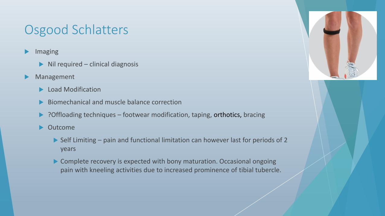
Osgood Schlatters
Imaging
Nil required – clinical diagnosis
Management
Load Modification
Biomechanical and muscle balance correction
?Offloading techniques – footwear modification, taping, orthotics, bracing
Outcome
Self Limiting – pain and functional limitation can however last for periods of 2 years
Complete recovery is expected with bony maturation. Occasional ongoing pain with kneeling activities due to increased prominence of tibial tubercle.

Anterior Cruciate Ligament Injury The number of reported ACL injuries in skeletally
immature athletes is increasing.
Higher participation levels
Greater awareness
Improved imaging techniques
Mechanism of Injury – usually non contact – pivoting on a slightly flexed knee or hyperextension
Examination
Patient History – often describes an audible ‘pop’ and usually unable to Return To Play
Rapid swelling,
Ligamentous tests – Lachman’s / Pivot Shift
Chronic ACL insufficiency – functional instability with change of direction
Trivedi 2017 Stanitski 1993

ACL Injury (Cont)
Imaging
X Rays to exclude tibial eminence fracture (avulsion of attachment) – much more common in younger age groups.
MRI – mid substance tears
Management
Tibial Eminence Fractures – Plaster immobilisation in knee extension or ORIF
Open growth plates – Trial of conservative rehab
Complications
Post traumatic arthropathy – multifactorial
Poor prognosis – increasing age, meniscal injury and lack of full knee extension at disharge post operatively
Growth plate arrest
Shelbourne 2000 Oeistad 2010
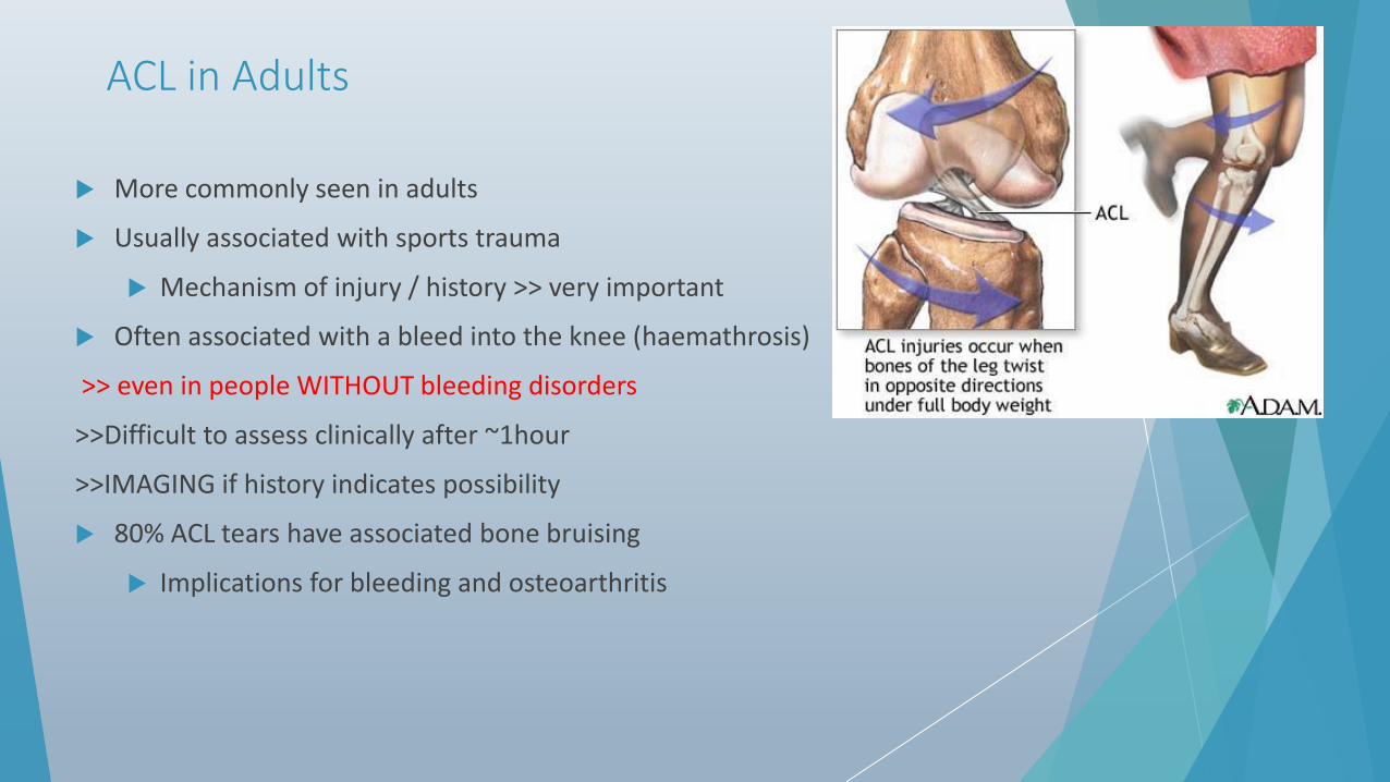
ACL in Adults
More commonly seen in adults
Usually associated with sports trauma
Mechanism of injury / history >> very important
Often associated with a bleed into the knee (haemathrosis)
>> even in people WITHOUT bleeding disorders
>>Difficult to assess clinically after ~1hour
>>IMAGING if history indicates possibility
80% ACL tears have associated bone bruising
Implications for bleeding and osteoarthritis

Normal Effects of Ageing on the Musculoskeletal System
From about age 30 yes, 30!!
The density of bones begins to diminish in men and women.
The cartilage inside a joint becomes thinner, as components of the cartilage decrease.
Connective tissue within ligaments and tendons becomes more rigid and brittle.
Loss of muscle (sarcopenia) the amount of muscle tissue and the number and size of muscle fibres gradually decrease, and the fibre types change, resulting in gradual loss of muscle mass and muscle strength.

Shoulder pain Shoulder impingements, Rotator cuff muscle pathology, Tendinopathy

www.animalclipart.net

Shoulder ‘Impingement’
Shoulder impingements is a clinical sign, not a diagnosis.
Rotator cuff tendons are ‘pinched’ or ‘squeezed’ as they go through the subacromial space
Brukner and Kahn, 2006

Rotator cuff muscle pathology
Can be weakness
Can be a rotator cuff tear (traumatic vs degenerative)
The prevalence of rotator cuff tear increases with the age and the tear should be considered as a physiological condition related to the progressive degeneration of tissues.
Pain and/or functional limitation are not constant in cuff tear
>> So beware imaging
Gumina et al, 2017

Tendinopathy
From age 30 tendinopathy is more common
Tendinopathy arises due to two main issues
1. Overload
2. Overuse
Bursa
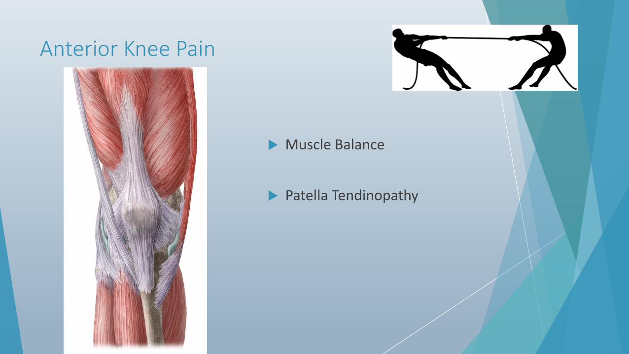
Anterior Knee Pain
Muscle Balance
Patella Tendinopathy

Osteoarthritis
Arthritis may be as a result of bleeding in a joint
Or may be due to genetics/age/weight/adverse dynamics through the chain
Best management of Osteoarthritis
Exercise / Physiotherapy
Weight management
Medication and injection management
Operative

Case Study
39 yo Male, Haem A severe - Treating 1500 units x3/week prophylaxis and on top 1-2/12 for knee bleeds.
He identified mm feels weak but not doing exs given prev.
Description appears not consistent with bleeds>> DW team >> no inhibitor, imaging?

Pt presented with c/o recurrent bleeds in R knee, ongoing for about 2 years.
Yttrium in Early 2011>>no bleeds or pain until x2 in Dec 2011, steroid injection ‘04 Jan 2012. Pt reports does not appear to have made a change as pt reports now bleeds about fortnightly.
(MRI prior to the yttrium shows features of significant internal derangement with osteoarthritis and some mild secondary synovitis).
Pt is on prophylaxis
Description of when gets a bleed:
-swells small amount
-Pain on WB on med side, no P if not WB
-No loss of ROM
-Has episodes about fortnightly on prophylaxis
-occasionally incidences prior but not always able to identify a cause

O/E knee R:
AROM 0-130 deg, PROM 0-130deg
Power: knee E G5, F G5
Lig tests: ACL appears intact/PCL appears intact/MCL appears intact/LCL appears intact
Meniscal testing: Appleys and McMurrays both -ve for med and lat stressing
Squat: able 1/2 ltd by stiff r ankle not knee
Squat with bounce; able, no pain
Lunge; able, no pain
SLS: L 20 sec eo, ec; R eo 15 sec, ec difficult
On palp: no pain elicited over lat jt line, med jt line, tendon insertions, distal pole
VMO active and timely
*Crepitus with F and E*
P/ to attend HTC when have next bleed, pt agreed to this; if pain+++ or unable to manage to treat - but to call HTC first.

Rx DW pt that description is consistent with OA type flare ups rather than bleeds. Advised not to treat next bleed but to come to HTC. DW pt pathology of haemophilic arthritis. DW pt that can get pain and swelling with OA type symptoms. HEP: Additionally given SLS exc x2/day to improve proprioception on R Provided with x2 pcs size F tubigrip with advice

Pt telephoned dept to report that he had swelling in the R knee and ? if bleed. Advised to attend dept, which pt did.
o/e: Knee R has lat and central pocket of swelling.
No redness
No heat
No reduction of range of motion (FROM 0-130 deg, crepitus at PF jt as is usual for pt)
No pain
Clinical diagnosis: Not indicative of a bleed, likely swelling secondary to known degeneration in knee joint.
DW with pt at length. DW pt differences between presentation of a bleed and symptomatic swelling due to degenerative changes.
Medical and Nursing staff observed and agreed.

Rx/ Plan of what to do if this type of swelling occurs: -Do NOT treat with factor replacement unless you have concerns that it is a bleed or swelling WITH pain, reduction in ROM, heat. Can contact dept HTC if unsure. -Tubigrip -Ice -elevation -General rest 24-48hrs; can mobilise if want and no pain -Can use crutches for peace of mind 24-48 hours but not a necessity -Avoid repetitive use of knee during this time and also squatting or twisting (during swelling flare up). -Avoid heavy lifting or mobilising very long distances (during swelling flare up). To call dept if concerned.

Outcome: Pt has had no further reported knee bleeds
Reports knee feels stronger and he has less symptoms and flare ups.
On occasion does have a flare up feels confident in how to manage them.
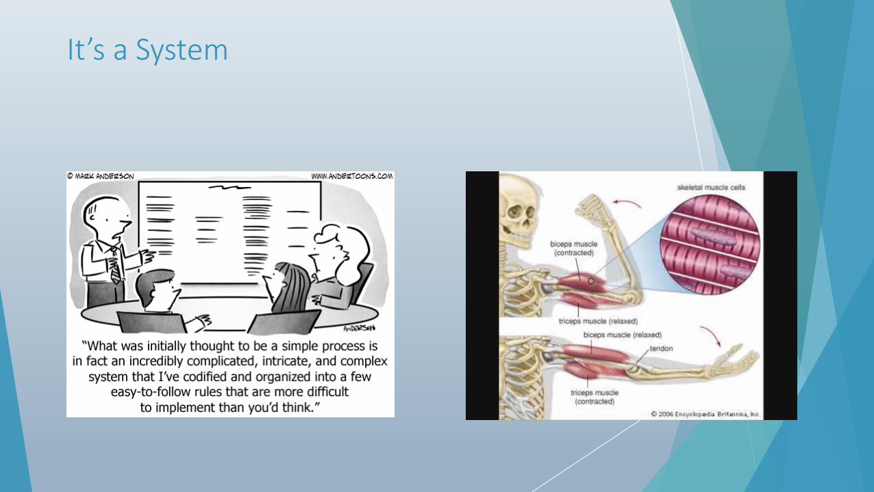
It’s a System

References Bloom OJ, Mackler L & Barbee J. Clinical enquiries. What is the best treatment for osgood-schlatter disease?
Journal of Family Practice. 2004 53(2);153-156
Brukner & Khan. 2006 Clinical Sports Medicine(3rd Ed) McGraw Hlil. Sydney
deLucena GL, dos Santos Gomes G & Guerro RO. 2011 Prevalence and associated factors of osgood-schlatter syndrome in a population based sample of Brazilian adolescents. Am J Sports Med 2011. 3(2);415-420.
Gumina S (Ed) 2017. Rotator Cuff Tears. Spinger
Herngren B, Stenmarker M, Vavruch L et al. Slipped capital femoral epiphysis: a population based study. BMC Musculoskeletal Disorders. 2017 18(1) 304
Kasper JC, Gerhardt MP, Mendelbaum BR. Stress injury leading to a slipped capital femora epiphysis in a competitive adolescent tennis player; a case report. Clinical Journal of Sports Medicine. 2007. 17(1) 72-74
Kose et al. Avulsion fracture of anterior cruciate ligament injury in a nine year old. BMJ Case Reports. 2013
Leeberg 2014
Move(formerly Arthritis Australia) https://www.move.org.au/page/home
Nakase J, Gashima K, Numata H et al. Precise risk factors for osgood-schlatter disease. Arch Orthop Trauma Surg. 2015. 135(9); 1277-81.
Peck D, Voss L & Voss T. SCFE_ Diagnosis and management Rehab post op – protection, pain free ambulation, neuromuscular control stengthening and performance enhancement. American Family Physician. 2017. 95(12) 779-784
Perry DC, Metcalfe D, Costa ML et al. A nationwide cohort study of slipped capital femoral epiphysis. Archives of Disease in Childhood. 2017 Jun (ahead of print)

References (Cont) Pogliacomi F, Calderazzi F, Paterlini et al. Surgical treatment of anterior iliac spine fractures : our experience
Acta Biomed 2014. 24 (85 Suppl 2); 52-58
Rossi F & Dragoni S. Acute avulsion fractures of the pelvis in adolescent competitive athletes; prevalence, location and sports distribution of 203 cases collected. Skeletal Radiology. 2001 30(3) 127-131
Schiller J, DeFroda S & Blood T. Lower extremity avulsion fractures in the pediatric and adolescent athlete. J Am Acad Orthop Surg. 2017 25(4) 251-259
Schoensee S & Nilsson KJ. A novel approach to treatment for chronic avulsion fracture of the ischial tuberosity in three adolescent athletes: a case series. International Journal of Sports Medicine. 2014. 20 9(7) 974-990
Schuett D, Bomar JD & Pennock AT. Pelvic apophyseal fractures; a retrospective review of 228 cases. J Paed Orthopaedics. 2015. 35(6) 617-623
Schuett DJ Bomar JD Pennock AT 2015 Pelvic apophyseal avulsion fractures; a retrospective review of 228 cases J Pediatric Orthopaedics 35(6) 617-623
Stanitski CL. Observations in acute knee hemarthrosis in children and adolescents. Journal of Paediatric Orthopedics 1993. 13(4) 506-510
Trivedi V, Mishra P & Verma D. Pediactric ACL injuries; a review of current concepts. Open Orthopedic Journal. 2017. 11; 378-388
Uvelli K. Treatment for calcaneal apophysitis. American Family Physician 2017. 96(2) 126-127
Weigerinck J, Zwiers R, Sierevelt N et al. Treatment of calcaneal apophysitis; wait and see versus orthotic device versus physical therapy a pragmatic therapeutic randomised trial. Journal of Paediatric Orthopaedics. 2016 36(2) 152-157


ACL Injury Prevention
Neuromuscular training appears to reduce the risk of injury – incorporates plyometrics, strengthening combined with feedback on proper technique
FIFA 11 program
PEP (Prevent Injury and Enhancement Program)