The cannabinoid system and its role in nociception Massimiliano Beltramo, PhD.

Pain and Nociception Unlocking The Gate Control Theory
With Alaa CattaehDiagrams created with Biorender.com
Template created using Canva

Contents
Pain vs Nociception
1
Modulation
5
Phases of Nociception
2
Transduction
3
Transmission
4

What is Pain? Pain vs Nociception
• A psychological state• An unpleasant sensory and emotional experience
associated with actual or potential tissue damage• Can occur without obvious tissue injury
What is Nociception?• Information indicating tissue damage • Process through which potentially damaging stimuli are detected• A noxious stimulus activates a particular set of sensory nerve cells
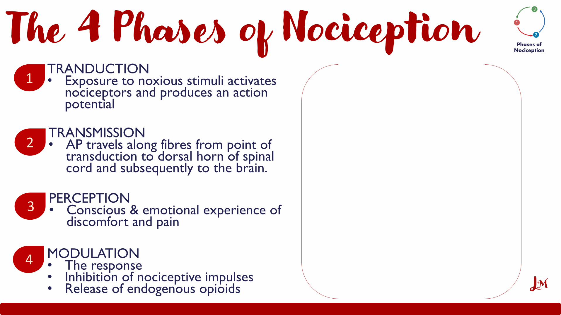
TRANDUCTION• Exposure to noxious stimuli activates
nociceptors and produces an action potential
TRANSMISSION• AP travels along fibres from point of
transduction to dorsal horn of spinal cord and subsequently to the brain.
PERCEPTION• Conscious & emotional experience of
discomfort and pain
1
2
3
The 4 Phases of NociceptionPhases of
Nociception
MODULATION• The response• Inhibition of nociceptive impulses• Release of endogenous opioids
4

SBAAmirah accidently cut her hand with scissors as she was crafting. She felt a sharp scratch around her finger, but the unpleasant sensation only lasted few seconds. Outline the correct order of Amirah’s nociceptive phases during that time.
• Transmission, Transduction, Modulation, Perception• Transmission, Transduction, Perception, Modulation• Transduction, Transmission, Perception, Modulation• Transduction, Transmission, Modulation, Perception

Transduction
Transduction

How Nociceptors are StimulatedWhat is a nociceptor?A ‘pain receptor’ which is a sensory neuron that responds to damaging or potentially damaging stimuli by sending “possible threat” signals to the spinal cord and the brain.
What stimuli activate the nociceptors?Intense mechanical stimuli
Irritant chemical stimuliThermal stimuliProstaglandins
Inflammatory mediators
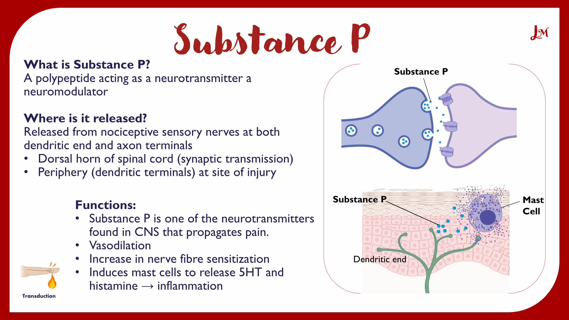
Once nociceptors become activated they produce an action potential.Substance P (a neuropeptide) is released when nociceptive axons carry high frequency trains of action potentials.
Transduction
Nociceptor
Cell Damage
Substance P
Transduction
What is Substance P?A polypeptide acting as a neurotransmitter a neuromodulator
Where is it released?Released from nociceptive sensory nerves at both dendritic end and axon terminals• Dorsal horn of spinal cord (synaptic transmission)• Periphery (dendritic terminals) at site of injury
Functions:• Substance P is one of the neurotransmitters
found in CNS that propagates pain.• Vasodilation• Increase in nerve fibre sensitization• Induces mast cells to release 5HT and
histamine → inflammation
Substance P
Dendritic end
Substance P Mast
Cell

The Nerve Fibers A-delta (Aδ) fibres:• Rapid onset sharp pain• Thin & myelinated axon (AP travels
5-35 m/s)• Small receptive field – well localized
pain• High threshold mechanoreceptors
C fibres:• Slow longer lasting dull throbbing pain• Thin & unmyelinated axon (AP travels
0.5-2 m/s)• Pain is poorly localized• Polymodal
Transduction
A-beta (Aβ) fibres: • Light touch & vibration• Wide & myelinated axon (AP 35-75 m/s)

SBANadia is learning about the different functions of substance P. When it’s released at the site of injury, substance P can lead to inflammation causing the injured area to appear red and swollen.
How can substance P trigger an inflammatory response?
• It activates macrophages• It causes vasoconstriction• It activates mast cells• It releases histamine

SBAMichael was hanging a picture frame up his wall when suddenly one of the nails fell down and he stepped on it.He immediately felt an intense sharp pain in his foot and started to scream.
This sharp onset of pain was caused by which nerve fibre?
• A-beta fibre• A-delta fibre• B fibre• C fibre• D fibre

Pain Transmission
Transmission
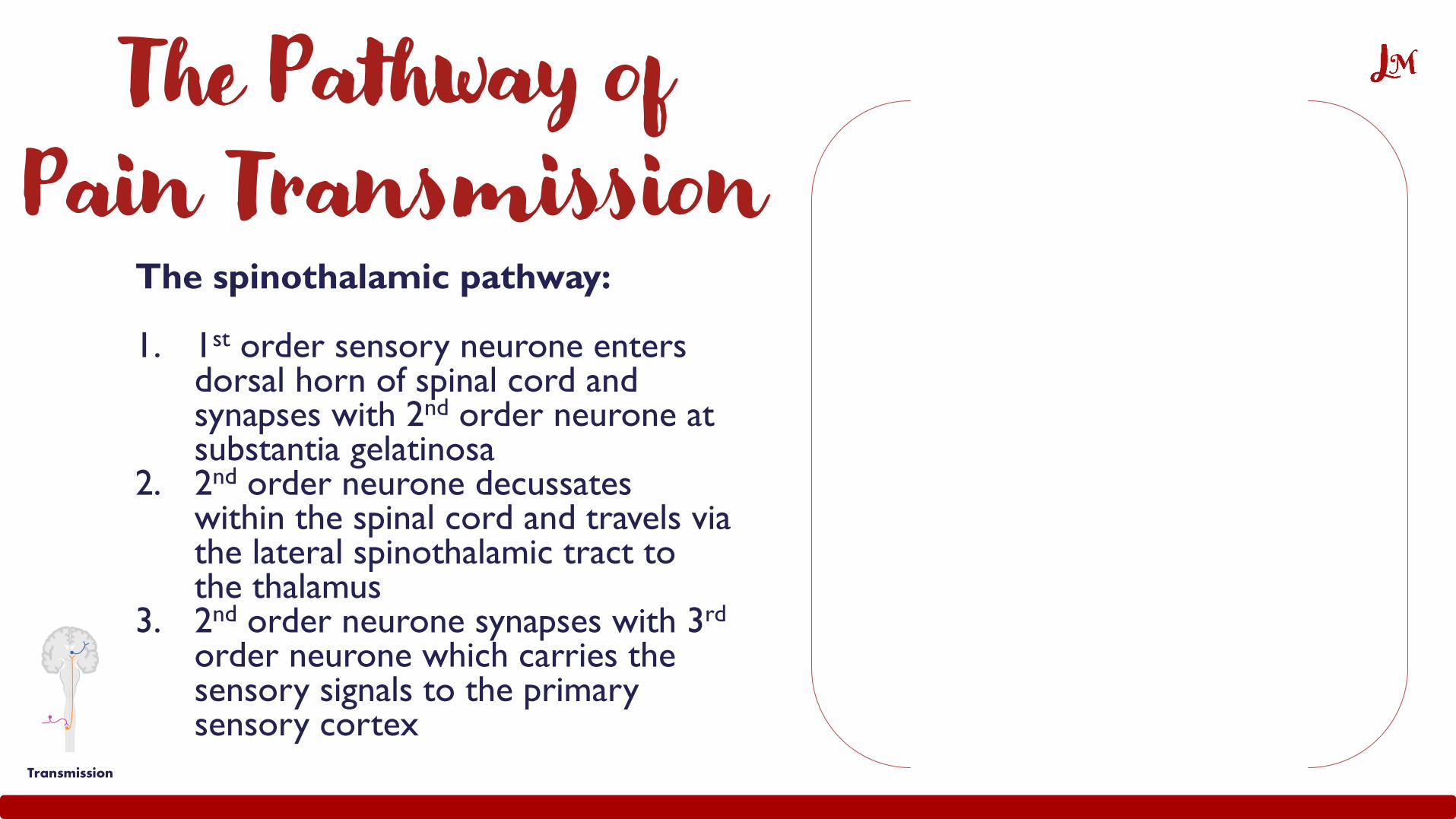
The Pathway of Pain Transmission
The spinothalamic pathway:
1. 1st order sensory neurone enters dorsal horn of spinal cord and synapses with 2nd order neurone at substantia gelatinosa
2. 2nd order neurone decussates within the spinal cord and travels via the lateral spinothalamic tract to the thalamus
3. 2nd order neurone synapses with 3rd
order neurone which carries the sensory signals to the primary sensory cortex
Transmission

Break time!Let's take a 2-3 minute break to recharge...
Any questions? Drop them in the Q&A!

Pain Modulation
Modulation

The Gate Control Theory of Pain (Melzack & Wall 1965)
• Nociceptive signals travel via the spinothalamic pathway to reach the brain where the thalamus first labels the sensation as ‘pain’.
• The theory suggests that the spinal cord contains a neurological "gate" that either blocks pain signals or allows them to continue on to the brain.
• It describes how non-noxious input can override the pain signals and close the gate to prevent them from travelling to the central nervous system.
What is the “gate”?
This is the substantia gelatinosa in the dorsal horn of the spinal
cord which contains inhibitory neurons acting as guards that can
close the gate and block the spinothalamic transmission of pain Modulation

How Non-Noxious Input Suppresses Pain Transmission
Modulation
How Non-Painful Input Closes Gate to Painful Input
Pain signals can be interrupted in the substantia gelatinosa
of the spinal cord, which acts as a gate:
• When you rub the site of pain, A-beta fibres are activated by
touch and produce an AP traveling faster than A-delta and C
nociceptive fibres
• The touch sensory neurone synapses with an inhibitory
interneuron in the substantia gelatinosa which is in contact with
both nociceptive and touch sensory fibres
• The inhibitory interneurone inhibits the nociceptive fibres
carrying the pain signal by preventing the release of substance P

How does the inhibitory interneuron inhibit nociceptive transmission?
Enkephalin
Opioid
receptor
Ca2+
Channel
K+
Channel
C fibre
Inhibitory
interneuron
2nd order
neurone• The inhibitory interneuron
releases enkephalins which bind
to opioid receptors
• This closes Ca2+ channels in the
pre-synaptic nociceptive fiber
meaning that vesicles containing
Substance P neurotransmitter
cannot be released
• This also opens K+ channels in
post-synaptic 2nd order neuron
causing hyperpolarization (makes
it less excitable) and therefore
reduces pain signal transmission
K+
Substance P
Modulation

SBAYoung Chloe was running in the park but accidently fell off and injured her knee. She attempted to lessen the pain by rubbing the bruised area.This stimulated A-beta fibres which in turn activated inhibitory interneurons to release enkephalins causing hyperpolarization of neurons and therefore reduction of pain transmission.
How was hyperpolarization achieved?
• Closure of Ca2+ channels• Opening of Ca2+ channels• Closure of K+ channels• Opening of K+ channels

Descending Pathways that Block Pain Transmission
Modulation
Once the signal from the spinothalamic pathway reaches
the somatosensory cortex, it triggers the descending pain
modulation pathway:
• The main pathway is thought to involve neurons that project from
the Periaqueductal gray (PAG) to serotonin-producing neurons of
the medulla oblongata (raphe nuclei).
• The activated raphe nuclei neurons project down to the dorsal
horn of the spinal cord where they release serotonin which binds
to interneurons in substantia gelatinosa.
• Interneurons release endogenous opioid neurotransmitters that
bind to mu opioid receptors on the incoming nociceptive fibres.
• Activation of opioid receptors produces hyperpolarization of the
neurons, which result in the inhibition of firing and the release of
substance P thereby blocking pain transmission.

SBAUnlike the gate control theory, where closing the gate is stimulated by non-noxious input, descending pain suppression is activated by noxious stimulation because that excites neurons in the nucleus reticularis gigantocellularis which innervate the PAG.
Where is the PAG found?
• Midbrain• Medulla• Dorsal horn• Substantia gelatinosa• Dorsolateral faniculus

Summary
Transduction• Noxious stimuli activate
nociceptors resulting in an AP
• Nociceptors release substance P
• Substance P is a neurotransmitter and a neuromodulator
Transmission• 1st order neurone synapses
with 2nd order neuronewhich travels via lateral spinothalamic tract to synapse with 3rd order neurone at thalamus
• 3rd order neurone carries signal to somatosensory cortex
ModulationGate Control Theory:• A-beta fibers stimulated
by touch activate inhibitory interneurons
• Close the gate
Descending Pain Suppression Theory:• PAG and raphe nucleus
stimulation produces analgesia.
1 2 3
Additional Resources
DCML pathway inhibits
Spinothalamic pain transmission:
https://youtu.be/oQLFfvGM7nI
Mechanisms of pain modulation:
https://nba.uth.tmc.edu/neuroscie
nce/s2/chapter08.html
Pain transduction:
https://youtu.be/lEQyLR6UBW0

Additional Resources
ECG and limb leads: https://button-bubbler-50c.notion.site/ECG-and-Limb-Leads-
c318019e1f9c4e41a20e275fd8e8d671
Regulation of appetite and digestion: https://www.notion.so/Regulation-of-Appetite-and-Digestion-
517d09feb16c4bd8bfb508e4504d8289
Differential diagnosis of dementia: https://button-bubbler-50c.notion.site/Differential-Diagnoses-of-Dementia-
b7ee72bbb67f4760bd29c6bb76e2d20f
Here are links to some of the learning resources I made if you’re interested ☺
Email: [email protected]

Referenceshttps://en.wikipedia.org/wiki/Substance_P
http://faculty.washington.edu/chudler/cv.html
https://teachmeanatomy.info/neuroanatomy/pathways/ascending-tracts-sensory/
https://en.wikipedia.org/wiki/Gate_control_theory
https://nba.uth.tmc.edu/neuroscience/s2/chapter08.html
https://www.youtube.com/watch?v=xkMP4eXp1Oc
Michael-Titus, A. et al. 2010. 5 - PAIN AND ANALGESIA. In: Michael-Titus, A. et al.
eds. The Nervous System (Second Edition). Churchill Livingstone, pp. 79-104.
Diagrams created with Biorender.com

Thank You!