Leeches of the suborder Hirudiniformes (Hirudinea: Haemopidae ...
TERESA MARYANSKA & HALSZKA OSM6LSKA
PACHYCEPHALOSAURlA, A NEW SUBORDER OF ORNITHISCHIANDINOSAURS(plates XXII-XXXI)
Contents
Introduction . . . . . . . . . . . . . . . . . . . . . . .Systematics . . . . . . . . . . . . . . . . . . . . . . .
Systematic position of the Pachycephalosauridae within OrnithischiaDescriptions . . . . . . . . .
Genus Tylocephale n. gen.Tylocephale gilmorei n.sp.
Genus Prenocephale n.gen. .Prenocephale prenes n.sp .
Genus Homalocephale n.gen,Homalocephale calathocercos n.sp,
Anatomy of the Pachycephalosauridae .Osteology of the skull . . . . . . .Some aspects of cranial neurology and circulationOsteology of the postcranial skeletonRemarks on myology
Conclusions . . . . . . . . . . . . .Adaptations and mode of life . . . . . . . . . . . . ..Remarks on the sexual dimorph ism and the supposed phylogeny of the Pachycephalosauria
References . . . . . . . . . . . . . . . . . . . . . . . . . . . . . . . . . . . . . . .
4648485051515353565662627983939797
100101
A bstract. - The present pap er deals with the dino saurs assigned to the family Pachycephalosauridae STERNBERG, 1945.The new mate rial , which was recently recovered in the Upper Cretaceous of Mongolia, is described . It includ es three newgenera and species: Tylocephale gi lmo rei from the Barun Goyot Forma tion, Prenoceph ale prenes and Homalocephale
calathocercos, both from the Nemegt Formation. Th is material as well as that known fro m North America show thatthe Pachycephalosauridae form a separa te group within the Ornithischia , and a new suborder Pachycephalosauria iserected to include thi s family. Pach yceph alosauri a are cha racterized : by the strongly thickened bones of the skull roof;by the complete ossification of the orbit, comparable only to that in some bird s and mammals; by the presence of theepipterygoid, which was never reported in any orni thischian dinosaur. The structure of the pachycephalosaurid pelvic region is unusual for the dinosaurs : the pub is is practically excluded from the acet abular rim; the ischium contacts theilium twice and additionally it contacts also the sacrum by means of the sacral ribs ; the juncture between the sacrumand ilium is weak. The present paper brings the diagnoses of new taxa and the systematic descriptions as well as theosteology of the skull and that of the postcranial skeleton in this group. Some aspect s of the myology, cranial neurology and circulation are discussed, as well as the adaptations, mode of life, sexual dimorphism and the supposed phylogeny of this group. It is believed here that the pach ycephalosaurids were the animals with keen sense of vision andgood olfactory sensitivity which fed on plants and insects ; their cursorial abil ities were limited ; they held hor izontallythe dorsal portion of the vertebral column during the gait , using the fleshy and prot ected by tendons tail as the propduring the rest; the dome might be used as the defensive weapo n; a significance of the dome in pachycep halosaurids asa sexual character seems to he still disputable.
46 TER ESA MARYANSK A & HALSZKA OSM6LSK A
INTRODUCTION
The Pachycephalosauridae are a comparatively rare and scarce group of ornithischiandinosaurs from the Upper Cretaceous. Until recently only two genera were recognized: Stegoceras LAMBE, 1902 and Pachycephalosaurus BROWN & SCHLAIKJER, 1943, both known mainlyfrom North American continent (th e only described Asian species - "S tegoceras" bexelliBOHLIN, 1953 - is represented by a scanty and incomplete material). The new genus Yaverlandia GALTON, 1971 from the Lower Cretaceou s (Wealden) of England was lately added tothe Pachycephalosauridae. During the Polish-Mongolian Paleontological Expeditions to theGobi Desert in 1965, 1970 and 1971 (K IELAN-JAWOROWSKA & DOVCHIN, 1969; KIELAN-JAWOROWSKA & BARSBOLD, 1972) th ree skulls, two of them with the fragmentary postcranialskeleto ns, were reco vered in the Barun Goyot Formation and Nemegt Formation in the localities Khulsan and Nemegt , within Nemegt Basin (GRADZINSKI et al. 1969; GRADZINSKI& JERZYKIEWICZ, 1972). This material , mo stly excellently preserved , is described in the presentpaper under the new generic and spec ific nam es: Ty locephale gilmorei n. gen. et sp.,Prenocephaleprenes n. gen. et sp., Hom alocephale calathocercos n.gen. et sp . The skull of Tylocephalegilniorei with th e mandible attach ed was recovered at Khulsan , laying among loose weatheredblocks on the sayr channel sur face. N o other skeletal elements of thi s species were present.The spec ime n of Prenocephale prenes was found in a weakly cem ented sandstone at the Nemegtlocality, in situ. Th e head was in a nearl y horizontal po sition. Posteri or to it, the rock wa sdamaged and th e anterior portion of th e postcrani al skeleton wa s not preserved. Slightly below,the sacrum with the right side of the pelvis and both femora were found. Several damagedcentra of caudals and numerous free caudal tendons were present in the weathered rock aroundthe caudals. The mo st complete specimen was that of Homalocephale calathocercos. It wasalso found in situ at the Nemegt locality. The skeleton was articulated laying on a shelfabove th e bottom of the sayr, with th e abdomen placed ventrally. The anterior portion of theskull, as well as the anterior portion of th e postcranial skeleton were lacking due to anextensive cleavage of the sandstone block in thi s site . The mode of preservation of theMongolian pachycephalosaurids is exceptional; the y show no traces of transport. The opposite istrue for the American pachycephalosaurids kn own up to dat e, wh ich are mostly restricted tothe sku ll roofs (except for one spec imen of S. validus and that of P. grangeri) and as wa snoticed by STERNBERG (1933) they "show more or less wear, as if they had been rolled alongby water " .
The systematic po sition of th e Pachycephalo sauridae with in the order Ornithischia ha soften caused and still cau ses discussion . They used to be assigned within the suborders : Stegosauria (LAMBE, 1918), Cer atopsia (Nor csx, 1904), Ankylosauria (ROMER, 1927; NOPCSA,1928, 1929), Ornithopoda (GILMORE, 1924; BROWN & SCHLAIKJER, 1943). In 1945 STERNBERGestablished a new familly: Pa chycephalo sauridae, which was assigned by the author to theOrnithopoda, the latter systematic po sition being nowadays generally accepted although nota bsolute ly (i.e. ROZHDESTVENSKY, in 1964, assigned them within Incertae subordinis, and in1972 to the suborder Ankylosauria).
The new material of the pachycephalo saurids which wa s collected in Mongolia convincedus that the establishment of a new suborder Pachycephalosauria within the Ornithischia isfully justified. The representat ives of thi s suborder are bipedaI, sim ilarly as the ornithopodsbut they po ssess a pelvis, in which the pubis is pr actically excluded from the acetabulum. Thisfeature is unique amo ng the dinosau rs. Severa l separa te ossificatio ns are pre sent in the sphe-

PACHYCEPH ALOSAURIA NEW SUBORDER 47
nethmoidal region of the pachycephalosaurids. No similar structure of this region has everbeen reported in any reptile. The new suborder Pachycephalosauria includes for the time beingbut one family Pachycephalosauridae with the following genera: Stenopelix MEYER, 1859,Yaverlandia GALTON, 1971 , Stegoceras LAMBE, 1902, Pachycephalosaurus BROWN & SCHLAIKJER,1943, Tylocephale n. gen., Prenocephale n. gen., Homalocephale n. gen. Stenopelix, which ishere assigned to the Pachycephalosauridae, was thus far placed either within the Hypsilophodontidae (Norcsx, 1928; ROZHDESTVENSKY, 1964) or within the Psittacosauridae (ROMER1956; STEEL, 1969). KOKEN (1887) noticed the unusual structure of the pelvis of Stenopelix.The same structure of pelvis, unique among the dinosaurs, we find in the pachycephalosaurids.This is why we think that Stenopelix cannot be assigned to any ornithopod family. This structureof the pelvis and the pre sence of the strong caudal ribs make Stenopelix close to the Pachycephalosauridae 1.
Because of its excellent pre servation, the new pachycephalosaurid material here describedenables us to provide much additional osteological data on the skull and on the po stcranialskeleton. For thi s reason, the pre sent paper is divided into three parts. The first one is devotedto systematic considerations, the establishing of new taxa and to general descriptions and comparisons. The second part deal s with the detailed comparative osteology of the Pachycephalosauridae based on the new and partly on the old materials, while in the third part the possibleadaptations and the mode of life as well as the pachycephalosaurid phylogeny are discussed .All the comparisons we have made in th is paper, which concern the North American materials,are essentially limited to the complete skulls of Stegoceras validus LAMBE, 1902 (UA No. 2)and Pachy cephalosaurus grangeri BRowN & SCHLAIKJER, 1943 (A.M.N.H.No. 1696). We hadat our disposal the pla ster cast of the skull of S. validus (UA No. 2) as well as the skull roofof the specimens NMC Nos. 138, 8816. We have not revised the species within Stegocerasand Pachycephalosaurus, lacking the adequate materials, and we do not discuss the specificrange of these genera in thi s paper.
We acknowledge with gratitude the help we got in obtaining of the necessary literatureand of the comparative material from the following persons and institutions: Dr. D . A. RUSSELL(National Museum of Natural Sciences, Ottawa) , Dr. P. M. GALTON (University of Bridgeport),Or. E. S. GAFFNEY (American Museum of Natural History, New York), the authorities of theNational Museum of Can ada, Ottawa, and of the American Mu seum of Natural History, NewYork. The special thanks are due to Dr. P. M. GALTON, who read critically the manuscript ,offered many useful suggestions and kindly corrected the English of the present paper. Weprofited also immensely of the discussions with Dr. H. J. OSTROM, Dr. A. K. ROZHDESTVENSKY.Prof. Or. Z. KIELAN-JAWOROWSKA facilitated and encoura ged the investigations. Mrs. K. BuDZYNSKA (Palaeozoological In stitute, Polish Academy of Sciences, Warsaw) made the drawings.Thanks are due to Mrs. M. KLEIBER-MALACHOWSKA (Museum of the Earth , Poli sh Academy of Sciences, Warsaw) and Mr. W. SKARZYNSKI (Palaeozoological Institute, Poli shAcademy of Sciences, Warsaw), who took the photographs.
Abbreviations used :
A.M.N.H. - American Museum of Natural History,G.I. SPS - Geological Institute Section of Palaeontology and Stratigraphy, the Academy of Sciences of the
Mongolian People 's Republic,
1 SCHMIDT (1969) redescribed the holotype of St enopelix valdensis MEYER and stated among others that the pubi stakes part in the formation of the acetabulum to the small extent (I. c., p, 197). Ho wever, the pubis is still insufficientlyillustrated on the drawing given by this author (I . C., Fig. I) to state for sure which is the normal articulation of the pub isin this specimen.

48 TERESA MA RYANSKA & HALSZKA oSM6LSKA
NMC - Na tional Museum of Canada,UA - University of Alberta,Z.Pal. - Palaeozoological Institu te of the Polish Academy of Sciences.
The described material is housed in the Pala eozoological In stitute of the Poli sh Academyof Sciences, Wa rsaw, and in th e Geological In st itut e of the Academy of Sciences of the Mongolian People's Republic, Ulan Bator .
SYSTEMATICS
SYSTEMATIC POSITION OF THE PA CHYCEPHALOSAURIDAEWITHIN THE ORNITHISCHIA
Th e new pach yceph alosaurid mater ial from th e Upper Cretaceous of Mongolia againrai ses the problem of the systema tic position of thi s group within th e Ornithischia. The unusualstructure of th e pach ycephalosaurid pelvis with the pubi s almost excluded from the acetabulum , the sagitta l shortening of the basicran ium and its separa tion from the palatal and suborbital regions, the presence of the accesso ry sphenethmoida l ossification s which close the orbitanteromedially, sepa ra te them from all dinosaurs described to date. Thi s last character isprobably the most exceptional feature of the Pachyceph alosauridae. The complete ossificationof the lamin a orbitonasalis and of the planum suprasepta le ha s never been reported in anyreptile, but it is sometimes pre sent in certain birds (ectethmoid and uncinate bone f ide de BEER, 1971, p. 443). However, the ossification of the orbitonasal region wentmuch furthe r in th e Pach yceph alosauridae, where the bones which can be homologizedwith th ese two menti oned in bird s are accompa nied by three addit iona l orbitonasal ossifications(p . 68). An oth er character which was never repor ted in any ornithischian dinosaur, is the presence of the well developed epipterygoid, which is in the contact with the prootic, similarly asit is in the Squamata. Th e exclusion of th e pubis from the acetabulum is characteristic of thePachycephalosauridae and amo ng th e archosaurs thi s is present only in the crocodiles. It wasalso reported in one other dinosaur - St enopelix MEYER, 1859 (KOKEN, 1877) 2 . Anothercha racter of the Pachyceph alosauridae which is unique for a dinosaur is the art iculation of thesecond and third sacra l ribs with the ischium in the anterior part of the latter. The pubi s isstro ngly reduced in certa in ankylosaurs in which it is p ractically excluded from the acetabulum.However , the ilium in ankylosaurs is quite different and th e acetabulum is closed so that reallythere is no close resemblance between th e pelves of the two groups. Some resemblance exist sbetween the pelvis of the Pachyceph alosauridae and that of the Protoceratopsidae, as wasalready mentioned by BROWN & SCHLAIKJER (1943, p. 145). They deal especially with the shapeof the ischium. H owever, in the Protoceratopsidae the pubi s is strongly reduced and takes partin the formati on of the acetabulum. Th e pre- and postzygapophyses in the dorsal vertebrae ofpachycephalosaurids have the tongue and groove articulation that among dinosaurs was reported only in Protoceratops (fide BROWN & SCHLAIKJER, 1943, p. 145). The tail of pachycephalosaurids is characterized by the strong development of the caudal ribs which are present onsevera l anterior caudals. Similar long cauda l ribs are not reported so far in any other dinosaurs,but it should be ment ioned here th at th ey were present in St enopelix .
The Pachyceph alosauridae were undoubtedly bipedal animals and mo st probably ofornithopod origin (p . 100), bu t they ca nnot be assig ned to the subo rder Ornithopoda as is
• Compare foot-note on p. 47.
PACH YC EPH AL OSA U RI A NE W S U BORD ER 49
usually done. They differ from the ornithopods in numerou s characters such as the presence in thePachycephalosauridae of a heavy skull with a thick roof and a marked tendency for the closureof the supratemporal fene stra. These features, which would be regarded as "normal" in theankylosaurs, are never found in an y ornithopod . Another character, most probably being thecon sequence of the thickening of the skull roof, is the vertical extension of the occipital region ,which is not found in ornithopods. A slightly similar structur e of the occiput is pre sent in theCeratopsia but there the basicranium is placed in quite a different plane to that of the occiput,which is not the case with the Pachyceph alo sauridae. Th e qu adratojugal /qu adrate relat ion inthe Pachycephalosauridae is quite different from th at of ornithopods, where the two bon esonl y contact each other for a short distance, the qu adratojugal never descending very nearto the lower articular su rface of the quadrate as in the pachyceph alo saurids. There are also thedistinctive difference s in the mandibles of both groups. The corono id process is generally betterdeveloped in the Ornithopoda than it is in the Pachyceph alo sauridae. Th e Pachycephalosauridaehave a very strongly pronounced retroarticular process but genera lly thi s is not the case in ornithopods. Among the characters of the skull common to th e pach yceph alo saurids and th eornithopods is the presence of the premaxillary teeth which a re more or less different from themaxillary dentition. The het erodont dentition in ornithopod s is most strongly pronouncedin Lycorhinus HAUGHTON, 1924 (= Heterodontosaurus CROMPTON & CHARIG, 1962, accordingto THULBoRN 1970) in which the premaxillary teeth are of the carnivorous type and there isa diastema between the premaxillary and maxillary teeth to receive the enlarged mandibularcanine. A similar condition is found in some Pach ycephalosauridae (p. 54).
' There are also certain similarities in the po stcranial skeleton of the Pach ycephalo sauridaeand the Ornithopoda, but they are due to bipedalism of both grups. Th e simila rities in the skullas well as in the po stcranial skeleton concern the characters which are primitive in the Pachycephalosauridae and their presence can be expla ined by the fact th at these latter derivedmost probably from a primitive ornithopod group (p. 100). Moreover, it should be mentioned here that Pachycephalosauridae display some similarities to all the suborders of theOrnithischia which was already noticed by man y authors (GILMORE, 1924; BROWN& SCHLAIKJER,1943, and th e pre sent paper. However, the essential characters of the skull and thepostcranial skeleton prevent the assignment of the Pachycephalosauridae to any knownornithischian suborder. For the se reasons we believe th at the esta blishment of a new suborderPachycephalosauria is mo st reasonable and fully justified.
The systematics of the Ornithischia differs significantly from th at accepted in the Saurischiawhere the infraorders are recently recognized within the suborders. To the suborder Theropoda,which includes the bipedal forms, three infraorders are assigned : Coelurosauria, Carnosauri aand Deinonychosauria (COLBERT & RUSSELL, 1969). Were the same practice applied to theOrnithischia, our new suborder Pach ycephalosauria could be regarded as an infraorder withinthe suborder Ornithopoda. Although the unification of systematics of the two dinosaurianorders mentioned seems to be necessary, we are not able to decid e at the moment whether orwhich infraorders should be recognized within the Ornithopoda.
4 - P al aeontologia Po lo nica No . 30
50 l'ERESA MA R YANSKA & IlALSZK A USM6 LSKA
D ESCRIPTIONS
Order ORNITHISCHIASuborder PACHYCEPHALOSAURIA novo
Diagnosis. - As for the family.Family assigned: Pachycephalosauridae STERNBERG, 1945.Stratigraphical and geographical distribution. - Cretaceous of Eurasia and North America.
Family PACHYCEPHALOSAURIDAE STERNBERG, 1945'(= TROODONTlDAE GILMORE, 1924, not TROODONTlDAE sensu RUSSELL, 1948)
Revised diagnosis. - Bipedal , highly specialized ornithischians, small to moderate insize. Pubis does not take part in formation of acetabulum. Skull roof thickened, flat to dome-like.Supratemporal fenestra usually closed in highly domed forms. Antorbital fenestra occasionallypresent. Maxilla penetrated by extensive int ramaxillary sinus. Epipterygoid present and beingin contact with the prootic. Orbits anteriorly and medially closed by accessory sphenethmoidalossifications. Quadrate with tendency to oblique po sition ofits dorsal portion. Jugal excluded fromposterior boundary of infratemporal fenestra and together with quadratojugal strongly expandedventrally to level ofarticular surface ofquadrate. Basicranial region strongly shortened (sag.), nearly completely separated from palatal and suborbital regions by extension of quadrate andpterygoid and by juncture of basisphenoid and pro otic with quadrate wing of the pterygoid.Basal tubera thin, plate-like. Occipital and ba sicranial regions usually placed almost in the samevertical plane. Premaxillary dentition present , heterodonty more or less pronounced. Dentitionweak , teeth arranged in one row, enameled on both sides. External skull bones strongly ornamented. Dorsal vertebrae mostly with tongue and groove articulation of zygapophyses. Caudalribs strongly developed on anterior caudals. Tibi a shorter or equal in length to femur. Pestetradactyl.
Genera assigned : TStenopelix M EYER, 1859, Yaverlandia GALTON, 1971 , Pachycephalosaurus BROWN & SCHLAIKJER, 1943, Stegoceras LAMBE, 1902, Tylo cephale n. geri., Prenocephalen. gen. , Homalocephale n. gen.
Stratigraphical and geographical distribution. - Lower Cretaceous (Wealden) of GreatBritain and Germany, Upper Cretaceous of North America and Asia.
Remarks. - Th e Upper Cretaceous representatives of the Pachycephalosauridae showthe advanced specialization as compared with th eir Wealden predecessors - Stenopelix andYaverlandia. The new element s which sepa rate the frontal from the orbital margin are addedto the skull roof. The postcranial skeleton becomes more lightly built. The sacrum includesthree additional vertebrae, the femur shortens relatively and the shortening of the forelimbis more advanced . It seems that th e po ssibility exists for excluding the two Lower Cretaceousforms from the Pachycephalosauridae and erecting for them a separate family. They are, however , thus far too poorly known and represented by too scanty material. The abdominal ribs ,which are never present in any ornithischian , were often reported in the diagnoses of the familyPachycephalosauridae given by the different authors. The Mongolian material here describedmade us possible to state that the supposed segmented abdominal ribs are , in fact the ossifiedcaudal tendons which form ed a kind of a basket around the po sterior portion of the tail.
I'ACHYC EP HA LOS A URI A NEW S U BO RDE R
Genu s TYLOCEPHALE n.gen .
51
Type species: Tylocephale gilmorei n.sp,Derivation of the name: Gr. tyle = swelling on the skin, cephale = head ; becau se of the thickening of the skull
roof.Genus monot ypi c: diagnosis, stra tigra phic and geographic distribut ion - as for the ' species.
Tylocephale gilmorei n.sp.(pI. XX II , Fig. 3 ; Text-fig. I B)
Type specimen: Onc specimen (Z . Pal. No. MgD-I/105) including : da maged skull with several maxi llary teeth ,lacking braincase, palate and anterior portion of sno ut ; mandible with several mandibular teeth , lacking anterior portionof dentary, as well as articular and prearticul ar.
Type horizon: Upper Cretaceous, Barun Goyot Formation, zo ne of Djadochtatherium catopsaloides KI ELAN-JAWORowSKA, Nemegtbaatar gobiensis KI ELAN-JAWOROWSKA and Chulsanbaatar vulgaris KIELA N-JAWOROWSKA.
Type locality: Khulsan, Nemegt Basin , Gobi D esert , Mongolian Peopl e's Republic (sec G RADZI NSKI & JERZYKIEWICZ, 1972, Text-fig. 4, N o. 10).
Derivation of the name: in hon our of the la te Ch . W. G ILMORE, who first gave the det ailed description of a pachycephalosaurid spec ies.
Diagnosis. - Crani al roof th ickened, highl y elevated, the highest point situated farposteriorly. Po storbital and both supraorbitals incorporated into domed part. Infratemporalfenestra very narrow, long and placed nearl y vertically. Orbit elon gate , oblique, rising upwardspo steriorly. Quadrate nearly vertical. Occipital region narrow, very faintl y depressed. Tooth-bearing edge of maxilla straight nearly to the very po sterio r end. Crowns of maxillary andmandibulary teeth large. Surface of the external bon es stro ngly ornamented, domed part rough .Surface of the jugal at the orbital margin, as well as this of the quadratojugal nearly smooth .
Dimensions - see Table 1.Description. - TIle skull is extremely high and nar row posteriorly. Th e po storbital
portion of the skull is very short. Th e dome is very highly elevated and its highest point is placedvery close to the posterior margin of the skull, so when the skull is viewed posteriorly the domeis visible above the dorsal edge of the occipital region . The squamosal in its dor sal portionforms the thick margin of the skull, wh ich is sha rp and not swollen. Th e ventral portion of thesquamosal contacting the exoccipital is thin . Th e squamosals are separated on th e marginof the skull by a very narr ow wedge of the parietals. The latter widen on the occipital face ofthe skull and become slightly concave where they meet the supraoccipital. All the bones formingthe occipital su rface of the skull are comparatively thin . The quadrate is relatively long so thatits length is nearly equal to the distance between both quadrate s as measured between the man dibular joints. The quadrate is nearly vertical; its lower third is perpendicular to the loweredge of the maxilla and its upper portion is only slightly inclined backwards. The ventral portionsof the quadrates are directed slightly medially. The jugal , from its juncture with the maxillaposteriorly, is directed distinctly laterally. When seen vent rally, the lat eral wall of the splanchnocranium is angularly bent in its po steriormost region along the jugal, quadratojugal and thequadrate, so that the space between the lateral wall and th e pterygoid is relatively very broadtransversally. The infratemporal fenestra is nearly vertical and its po sterior portion is onlyslightly oblique. It is equ ally narrow along its entire length . The orbit is nearly twice as longas broad and its upper boundary is flat. The postorbital bar is narrow and parallel to the quadrate. The po storbital overlap s the jugal anterodorsally along the oblique suture. The anterior
52 TERES A MAR YANSK A & HALSZK A OSM6 LSKA
and the posterior supraorbitals are relatively steeply placed and they were most probablyincorporated into the domed part of the skull roof.
Dentition. The maxilla has 9 posterior teeth preserved. They are arranged in a straightrow but the last tooth is placed slightly outward s from the straight line formed by the otherteeth. The teeth are badly damaged and the denticulation of the crowns is hardly visible. Theteeth are relatively large . The re are seven mandibular teeth preserved. They display high crownsand the ir cutting edges are arched rather than conical in lateral view. The labial face of the toothis concave dorsoventrally and anteroposteriorly. On the inner, lingual face there is a strongvertical ridge with 4 parallel ridges anterior to it and 3 ridges posterior to it. All the ridges arevisible along the entire height of the crown. They end on the cutting edge of the crown and formits denticulation. The similar pattern of the ridge arrangement is present on the labialface of the crown, but it is less distinct there. The lingual face of the maxillary teeth isworn out.
Ornamentation. The external surface of the supraorbitals and the postorbital are quiteregularly ornamented with the tubers of moderate size. The outer surface of the jugal nearthe mandibular joint is covered by large , prominent and irregularly spaced tubers. The pre servedposterior part of the domed roof is rough. Eight node s are present on the squamosal alongthe posterior margin of the skull roof. A single large node is present below the outermost nodeof the squamosal series. A row of the large node s mentioned continues forwards laterally acrossthe squamosal and the postorbital; farther anteriorly the nodes diminish and merge togethercontinuing in the form of a sharp crest along the supraorbitals which constitutes the dorsalmargin of the orbit. Another not so prominent crest is pre sent along the postorbital bar.
Mandible. The coronoid is weakly elevated above the upper contour of the jaw. The adductor fossa is very deep in transverse direction.
Remarks. - The skull of Tylocephale gilmorei was recovered in Khulsan, Barun Goyot Formation. The age of this formation is determined by KIELAN-JAWOROWSKA (1974) as the ?MiddleCampanian, and may be vaguely compared with that of the Belly River Formation, which yieldedthe specimens of Stegoceras validus. The skull of T. gilmorei is comparable in size to the completeskull of S. validus described by GILMORE (1924). It differs from the latter in having the highestpoint of the dome placed very far posteriorly, close to the posterior margin of the skull. The skullof T. gilmorei is the only pachycephalosaurid in which the elevation of the dome is visible whenthe skull is seen from the back . The nearly vertical position of the quadrate and of the infratemporal fenestra also distingui sh T. gilmorei from the other Pachycephalosauridae in whichthe infratemporal fenestra is usually more or less horizontal. The po sterior plane of the skullwhich includes in the Pachycephalosauridae the basicranial and basioccipital regions, is proportionally narrower and higher than it is in S. validus and other Pachycephalosauridae. The poorlypreserved occipital region of T. gilmorei is only slightly concave centrally while it is alway sdistinctly depressed in all the other repre sent atives of the family. Tylocephale gilmorei ha sthe posterior cheek region , bounded by the jugal and quadratojugal, very strongly expandedlaterally to an extent never reported in any other pach ycephalosaurid . The poor state of preservation of the maxillary and mandibulary teeth in the specimen of T. gilmorei makes itimpossible to make detailed comparison s. However , it can be stated that the teeth are relativelylarger than in other Pachyceph alosauridae and not conical as they are in Stegoceras validus.The mandible of T. gilmorei is delicate and devoid of ornamentation, when compared with themandible of S. validus, which ha s so far been the only known mandible in thi s group. Thecharacter of the ornamentation of the skull of T. gilmor ei is similar to that of S. validus but it
PACH YCEPH ALOSAURIA NE W SU BORDER 53
differs in detail s. Taking into account all the significant differences from the other representativesof the family, we are of the opinion that the species above described should be assigned to thenew genus.
Genus PRENOCEPHALE n.gen.
Type species: Prenocephale prenes n. sp.Derivation of the name: G r. prenes = inclined, sloping, cephale = head ; because of the anterior sloping profile
of the head.Genus monotypic: diagnosis, stratigraphic and geographic distribu tion - as for the species.
Prenocephale prenes n.sp.(PI. XXII, Fig. 2, PI. XX III, PI. XXV, Figs 2, 3, PI. XXXI , Fig. 2; Text-figs l e , 2, 3, 6)
Type specimen: One specimen (Z. Pal. No . MgD-I /I04) includ ing perfectly preserved skull with dentition , withoutmandibles , several fragmenta ry dorsal vertebrae, one caudal vertebra, fragmentary dor sal ribs, badly damaged left femur ,right femur lacking greater and lesser trochanters and lateral condyle, numerous free caudal tendons, several of them innatural arrangement.
Type horizon: Upper Cretaceous, Nernegt Formation, zone of Tarbosaurus bataar (MALEYEV) and Saurolophusangust irostris ROZHDESTVENSKY ;
Type locality: Nemegt, Nemegt Basin, Gobi Desert , Mongolian People 's Republi c (see GRADZINSKl & JERZYKIEWICZ, 1972, Text-fig. I , No. 19).
Derivation of the name: see th is for the genus.
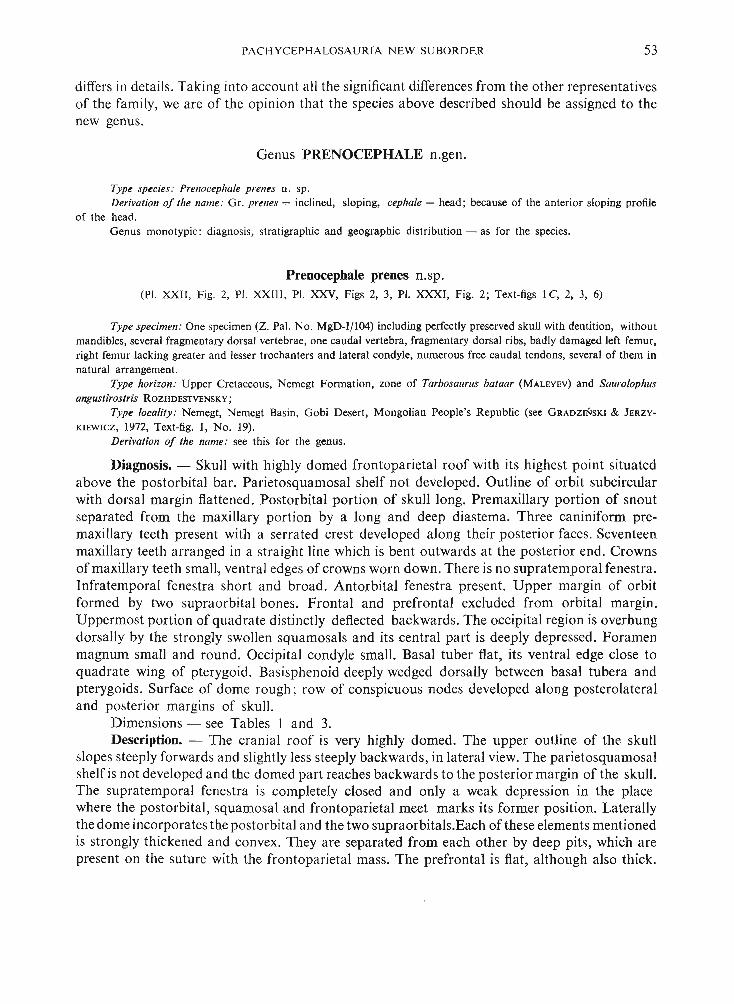
Diagnosis. - Skull with highly domed frontoparietal roof with its highest point situatedabove the postorbital bar. Parietosquamosal shelf not developed. Outline of orbit subcircularwith dor sal margin flattened. Postorbital portion of skull long. Premaxillary portion of snoutseparated from the maxillary portion by a long and deep diastema. Three caniniform premaxillary teeth present with a serrated crest developed along their posterior faces. Seventeenmaxillary teeth arranged in a straight line which is bent outwards at the posterior end. Crownsof maxillary teeth small, ventral edges of crowns worn down . There is no supratemporal fenestra.Infratemporal fenestra short and broad. Antorbital fenestra present. Upper margin of orbitformed by two supraorbital bones. Frontal and prefrontal excluded from orbital margin.Uppermost portion of quadrate distinctly deflected backwards. The occipital region is overhungdorsally by the strongly swollen squamosals and its central part is deeply depressed . Foramenmagnum small and round . Occipital condyle small. Basal tuber flat, its ventral edge close toquadrate wing of pterygoid. Basisphenoid deeply wedged dorsally between basal tubera andpterygoids. Surface of dome rough ; row of conspicuous node s developed along posterolateraland posterior margin s of skull.
Dimensions - see Table s I and 3.Description. - The cranial roof is very highly domed. The upper outline of the skull
slopes steeply forwards and slightly less steeply backwards, in lateral view. The parietosquamosalshelf is not developed and the domed part reaches backwards to the posterior margin of the skull.The supratemporal fenestra is completely closed and only a weak depression in the placewhere the postorbital, squamosal and frontoparietal meet marks its former position. Laterallythe dome incorporates the postorbital and the two supraorbitals.Each of these elements mentionedis strongly thickened and convex. They are separated from each other by deep pits, which arepresent on the suture with the frontoparietal mass. The prefrontal is flat, although also thick.

54 TERESA MA RYAN SKA & HALSZK A OSMOLSKA
The nasal is weakly convex and even somewhat concave close to the suture with its fellow. Thepremaxillary portion of the snout is distinctly differentiated and narrow transversely. The internarial bridge was presumably formed from the premaxillae but it is broken off. The anteriorportion of the premaxilla in front of the external nares is relatively long. The premaxilla reachesfar posteriorly on the lateral wall of the skull. The infratemporal fenestra is comparativelyhigh . The orbit is large subcircular. A rather small antorbital fenestra is present. The quadratealong about two thirds of its length is perpendicular to the lower margin of the maxilla, but itsupper portion is deflected backwards. The foramen magnum and occipital condyle are relativelysmall. The occipital region is extremely expanded dorsally and laterally. It includes the posteriorportion of the squamosals and parietals. The dorsal margin of the occipital region is verystrongly thickened acros s the squamosaIs. The region of the base of the skull is very muchcompressed in connection with the extreme development of the occipital region. The basaltuber is so strongly flattened anteroposteriorl y that it form s a thin plate , which is placed veryclosely to the basisphenoid proce ss. The basicranial plane , anterior to the basal tubera, is locatedin the deep, narrow cleft which is vertically extended. In posterior view, the quadrate is verystrongly developed in the medial direction and it overlaps anteriorly the broad and thin quadraticwing of the pterygoid . Together they form a very broad plate , extended transversely to the longaxis of the skull, onto which is plastered the lateral portion of another flat plate which is builtfrom the prootic and the basisphenoid. The prootic-basisphenoid plate is placed anteriorlyto the flat basal tuber and form s the anterior boundary of the cleft-like basicranial region .As a result of the transverse extension and the union of the bone s mentioned above , the posteriorregion of the skull, including the occiput and the basis cranii , is completely separated frompalate region. The latter is also placed in the plane perpendicular to the posterior region. Thepalate is highly vaulted . The both wings of the pterygoid are well developed. The small postpalatal fenestra are present. The internal nares are large.
Dentition. The three premaxillary teeth are distinctly caniniform, conical and oval in crosssection. Their roots are strong and have a greater diameter than the crowns which are enamelledand bear densely arranged vertical ridges. One of the ridges present on the posterior face ofthe crown is serrated and there are about 8 minute denticle s on I mm. Thi s serrated ridge isvisible on two premaxillary teeth but it was presumably present on all three canines. The crown sof the premaxillary teeth bear on their posteromedial faces the relatively broad, vertical traceof being worn down.Tt is evidence that there were also caniniform teeth along the anteriorportion of the mandible. Between the premaxill ary and the maxillary series of the teeth thereis a long diastema . It is very deep and situated on the suture between the two tooth-bearingbones . The presence of the diastema suggest that there was a strong, caniniform tooth in themandible, which fitted into the deep pit present ventrally on the juncture premaxilla-maxilla.The seventeen maxillary teeth are arranged in a stra ight line, which is slightly bent outwardsonly at the very end. The crowns of the maxillary teeth are low and enamelled on bothsides as is well shown on the replacement teeth . The lingual surface of the crown is convexdorsoventrally and ante ropo steriorly. The labial sur face is concave dorsoventrally and convexanteroposteriorly. The crown is vertically and parallelly ridged. The central ridge on thelingual side is the strongest. There are 4 ridges anterior to it and the anteriormost edge of thecrown is also sharply serrated. All the ridges reach a cingulum-like swelling at the base ofthe crown . Three ridges are pre sent posterior to the medial one but they are thinner thanthe anterior ridges. The posteriormost edge of the crown is serrated. All the ridges end on thecutting edge of the crown to form the denticulation. The median ridge is less pronounced onthe labial face of the crown. Other details of the structure of the labial face of crown are not
PACH YCEPJ-IALOSAURIA NEW SUBORDER 55
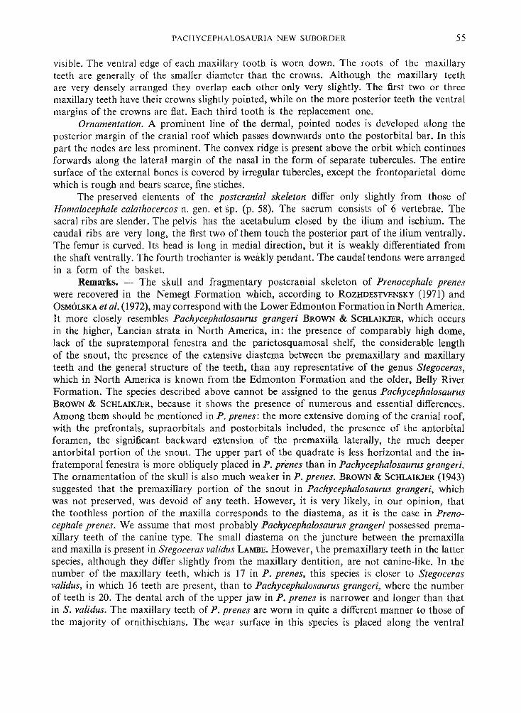
visible. The ventral edge of each maxillary tooth is worn down . The roots of the maxillaryteeth are generally of the smaller diameter than the crowns. Although the maxillary teethare very densely arranged they overlap each other only very slightly. The first two or threemaxillary teeth have their crown s slightly pointed, while on the more posterior teeth the ventralmargin s of the crown s are flat. Each third tooth is the replacement one.
Ornamentation. A prominent line of the dermal, pointed nodes is developed along theposterior margin of the cranial roof which passes downwards onto the postorbital bar. In thi spart the nodes are less prominent. The convex ridge is present above the orbit which continuesforwards along the lateral margin of the nasal in the form of separate tubercules. The entiresurface of the external bones is covered by irregular tubercles, except the frontoparietal domewhich is rough and bear s scarce, fine stiches.
The preserved element s of the postcranial skeleton differ only slightly from those ofHomalocephale calathocercos n. gen. etsp. (p. 58). The sacrum consists of 6 vertebrae. Thesacral ribs are slender. The pelvis has the acetabulum closed by the ilium and ischium. Thecaudal ribs are very long, the first two of them touch the posterior part of the ilium ventrally.The femur is curved. It s head is long in medial direction, but it is weakly differentiated fromthe shaft ventrally. The fourth trochanter is weakly pendant. The caudal tendons were arrangedin a form of the basket.
Remarks. - The skull and fragmentary postcranial skeleton of Prenocephale preneswere recovered in the Nemegt Formation which , according to ROZHDESTVENSKY (l971) andOSMOLSKA et al. (l972), may corre spond with the Lower Edmonton Formation in North America.It more closely resembles Pachycephalosaurus grangeri BROWN & SCHLAIKJER, which occursin the higher , Lancian strata in North America, in: the presence of comparably high dome ,lack of the supratemporal fenestra and the parietosquamosal shelf, the considerable lengthof the snout, the presence of the extensive diastema between the premaxillary and maxillaryteeth and the general structure of the teeth, than any representative of the genus Stegoceras,which in North America is known from the Edmonton Formation and the older, Belly RiverFormation. The species described above cannot be assigned to the genus PachycephalosaurusBROWN & SCHLAIKJER, because it shows the presence of numerous and essential differences.Among them should be mentioned in P. prenes : the more extensive doming of the cranial roof,with the prefrontals, supraorbitals and post orbitals included, the presence of the antorbitalforamen, the significant backward extension of the premaxilla laterally, the much deeperantorbital portion of the snout. The upper part of the quadrate is less horizontal and the infratemporal fenestra is more obliquely placed in P. prenes than in Pachycephalosaurus grangeri.The ornamentation of the skull is also much weaker in P. prenes. BROWN & SCHLAIKJER (l943)suggested that the premaxillary portion of the snout in Pachycepha/osaurus grangeri , whichwas not preserved, was devoid of any teeth. However, it is very likely, in our opinion, thatthe toothless portion of the maxilla corresponds to the diastema, as it is the case in Prenocephale prenes. We assume that most probably Pachycephalosaurus grangeri po ssessed premaxillary teeth of the canine type. The small diastema on the juncture between the premaxillaand maxilla is present in Stego ceras validus LAMBE. Howe ver , the premaxillary teeth in the latterspecies, although they differ slightly from the maxillary dentition, are not canine-like. In thenumber of the maxillary teeth , which is 17 in P. prenes, this species is closer to Stegocerasvalidus, in which 16 teeth are present , than to Pachycephalosaurus grangeri, where the numberof teeth is 20. The dental arch of the upper jaw in P. prenes is narrower and longer than thatin S. validus. The maxillary teeth of P. prenes are worn in quite a different manner to those ofthe maj ority of orn ithischians. The wear surface in th is species is placed along the ventral
56 TE RE SA MAR YAN SKA & HALSZKA OSM6LSKA
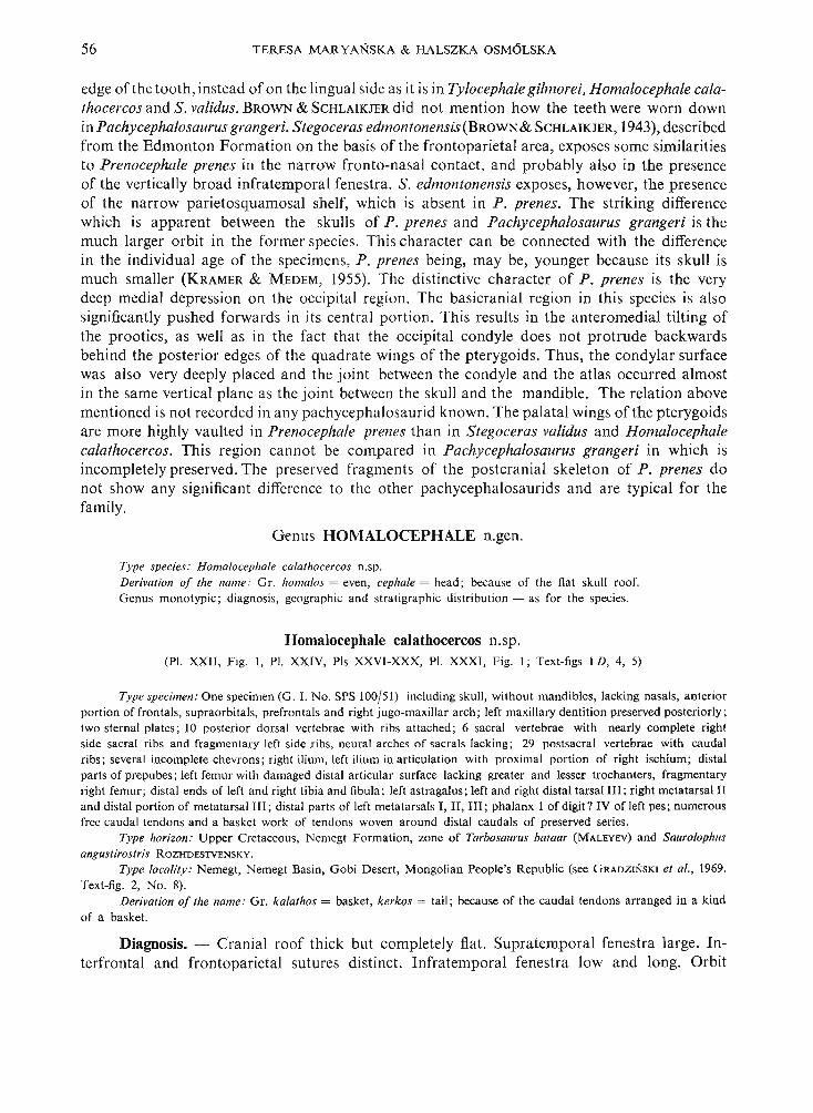
edge of the tooth, instead of on the lingual side as it is in Tylocephale gilmorei, Homalocephale calathocercos and S. validus. BROWN & SCHLAIKJER did not mention how the teeth were worn downin Pachycephalosaurus grangeri. Stegoceras edmontonensis (BROWN& SCHLAIKJER, 1943), describedfrom the Edmonton Formation on the basis of the frontoparietal area, exposes some similaritiesto Prenocephale prenes in the narrow fronto-nasal contact, and probably also in the presenceof the vertically broad infratemporal fenestra. S. edmontonensis exposes, however, the pre senceof the narrow parietosquamosal shelf, which is ab sent in P . prenes. The striking differencewhich is apparent between the skulls of P. prenes and Pachy cephalosaurus grangeri is themuch larger orbit in the former species. This character can be connected with the differencein the individual age of the specimens, P. prenes being, ma y be, younger because its skull ismuch smaller (KRAMER & MEDEM, 1955). The distinctive character of P. prenes is the verydeep medial depression on the occipital region. Th e basicranial region in thi s species is alsosignificantly pushed forwards in its central portion. Thi s results in the anteromedial tilting ofthe prootics, as well as in the fact that the occipital condyle does not protrude backwardsbehind the posterior edges of the quadrate wings of the pterygoids. Thus, the condylar surfacewas also very deepl y placed and the joint between the condyle and the atlas occurred almostin the same vertical plane as the joint between the sku ll and the mandible. The relation abovementioned is not recorded in an y pachycephalosaurid known. The palatal wings of the pterygoidsare more highly vaulted in Preno cephale prenes than in Stegoceras validus and Homalocephalecalathocercos. Thi s region cannot be compared in Pachycephalosaurus grangeri in which isincompletely pre served . The preserved fragments of the po stcranial skeleton of P. prenes donot show any significant difference to the other pachycephalosaurids and are typical for thefamily .
Genus HOMALOCEPHALE n.gen.
Type species: Homalocephale calathocercos n.sp,
Derivation of the name: Gr. homalos = even, cephale = head ; because of the fla t sku ll roof.Genus monotypic ; diagnosis, geographi c and stra tigra phic distribution - as for the species.
Homalocephale calathocercos n.sp.(PI. XXII, Fig. I, PI. XXIV, Pis XXVI-XXX, PI. XXX I, Fig. I ; Text-figs ID, 4, 5)
Type specimen: One specimen (G . I. No. SPS lOO/51) including skull, without mandibles, lacking nasals, anteriorport ion of frontals, supraorbitals, prefrontals and right jugo-maxillar arch ; left maxillary dentition preserved posteriorly ;two sternal plate s ; IO posterior dorsal vertebrae with ribs attached ; 6 sacral vertebrae with nearly complete rightside sacral ribs and fragmentary left side ribs, neural arches of sacrals lacking ; 29 postsacral vertebrae with caudalribs; several incomplete chevron s; right ilium, left ilium in ar ticulation with proximal portion of right ischium; distalpart s of prepubes ; left femur with damaged distal articular surface lacking greater and lesser trochanters, fragmentaryright femur ; distal ends of left and right tibia and fibula ; left astragalus; left and right distal tarsal Ill ; right metatarsal Iland distal portion of metatarsal Ill; distal parts of left metatarsals I, Il, Ill ; phalanx 1 of digit? IV of left pes ; numerousfree caudal tend ons and a basket work of tendons woven around distal caudals of preserved series.
Type horizon: Upper Cretaceous, Nemegt Formation, zone of Tarbosaurus bataar (MALEYEV) and Saurolophusangustirostris RozHDESTVENSKY.
Type locality: Nem egt, Nemegt Basin, Gobi Desert, Mongolian People's Republic (see G RADZINSKI et al., 1969.Text-fig. 2, No . 8).
Derivation of the name: Gr. kalathos = basket , kerkos = tail; becau se of the caudal tend ons arranged in a kindof a basket.
Diagnosis. - Cr ani al roof thick but completely fiat. Supratemporal fenestra large. Interfrontal and frontoparietal sutures distinct. Infratemporal fene stra low and long. Orbit

PACHYCEPHALOSAURI A NEW SUBORD ER 57
large and nearly round. Quadrate deflected backwards along the uppe r half of its length. Occipitalregion moderately concave , deepened centrally. Foramen magnum round and large. Occipitalcondyle large. Basal tubera flat anteroposteriorly, their ventral edges very close to quadraticwings of pterygoids. Ventral maxillary edge arched outwards posteriorl y. Maxilla ry teeth withsmall crowns. Cranial roof roughly ornamented.
Dimensions - see Tables 1-3.
T abl e I
Measurements of the skulls (in mm)
Ty locephale
IPrenocephale
Homalocephalegilmorei prenescalathocercos
Z. Pal. Nos.G. I. No. SPS lOO/51
MgD-I/105 I MgD-I/I04
Length of skull (premaxilla-upper end of quadrate) - 218.0 -Greatest width of skull 99.0 169.0 138.0Greatest height of skull 133.0 170.0 118.0Orbit length 38.0 50.5 53.5Orbit height 16.5 31.5 -Antorbital length of skull - 100.0 -Postorb ital length of skull 35.5 66.5 54.0
Description. - The cranial roof is completely flat , being formed from thick parietals andfrontals, and it slopes forward s. The interfrontal and frontoparietal sutures are very distinctbut the interparietal suture is obliterated. The supratemporal fenestra is large. The infratemporalfenestra is long and low. The orb it is very large and round . The quadrate along its lower halfis perpendicular to the lower edge of the maxilla, in its upper half the quadrate is declinedbackwards. The occipital region is very strongly extended vertically and transversely. It bearsa deep centr al depression. The posterior portions of the squamosals and parietals are includedin the occipital region . The occipital cond yle and the foramen magnum are large. The basicranialregion is compressed and the flat basal tubera nearly touch the quadratic wing of the pterygoidsventrally. Both wings of the pterygoid are strongly developed . The cleft is present anteriorlybetween two palatal wings of the pterygoids. The small foramen is present on the contactof the maxilla , ectopterygoid and palatine. The tooth-bearing edge of the maxilla is archedoutwards posteriorly.
Dentition . The maxillary teeth are arranged in one row and the posterior margin of eachtooth slightly overlaps laterally the anterior margin of the successive tooth. The crowns are low.The labial surface of the crown is conca ve dorsoventrally and the lingual surface is convexanteroposteriorly. On the lingual surface of the crown a thick medial ridge is present. Sixridges are developed anteriorly to it and four ridges posteriorly to it. The posteriormost edgeof the crown is additionally serrated. The preserved maxillary teeth are strongly worn out,the worn out surfaces being placed on the lingual side of the crown. They tend to form a commonplane over the whole maxillary series.
Ornamentation . The sur face of the externa l cran ial bones is very rough , the skull roo fbears centrally a small, smooth field, placed on the parietals close to the frontoparietal suture.The cranial roof is covered on its periphery by deep large pits, towards the medial line the or-
58 TE RESA MA RYANSKA & HALSZK A OS M6LS KA
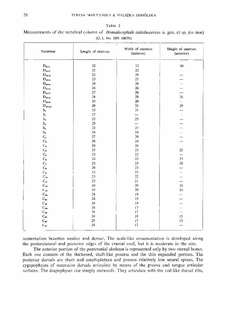
Tabl e 2
Measurements of the vertebral column of Homalocephale calathocercos n. gen. et sp. (in mm)(G. I. No. SPS 100/51)
Vertebrae Length of centrumWidth of centrum Height of centrum
(anterior) (anterior)
D n+l 22 23 20D n+o 22 22 -Dn+s 22 24 -D nH 23 25 -
D n+s 24 26 -Dn+s 26 26 -D n+7 27 26 -Dn+s 28 28 26Dn+s 29 29 -
D n+l. 29 31 29S, 33 31 -S. 27 - -S. 25 25 -S. 25 - -
S. 23 23 -S. 24 24 -C, 27 20 -Cl 26 24 -
Cl 24 24 -
C. 23 23 22C. 23 22 -
C. 23 23 23C7 23 23 20C. 24 23 -C. 23 22 -C,. 23 22 -Cll 23 21 -C12 24 20 16C,. 24 20 16
C•• 24 19 -CIl 24 19 -Cu 24 19 -
C.. 24 17 -
Cs< 24 17 -C•• 24 18 15C•• 25 17 15C.7 24 17 -
namentation becomes weaker and denser. The node-like ornamenta tion is developed alongthe posterolateral and posterior edges of the cranial roof, but it is modera te in the size.
The anterior portion of the postcranial ske leton is represented only by two sternal bone s.Each one consists of the thickened, shaft-like proce ss and the thin expanded portion. Theposterior dorsals are short and amphiplatyan and possess relatively low neural spines. Thezygapophyses of successive dorsals articulate by mean s of the groove and tongue articularsurfaces. The diapoph yses rise steeply outwards. They articulate with the rod-like dorsal ribs,

PACH YCEPHALOSA URI A NEW SUBORDER
T a b le 3
Measurements of the pelvis and hind limbs (in mm)
59
Homalocephale Prenocephalecalathocercos prenes
G. I. No. SPS 100/51 -Z. Pal. No . MgD-I/104
Length of ilium 230-0 225'5eLength of ischium - l85 ·0eLength of prepubis 65·6e -
Femur:Length 218·0 22l -5eProximal transverse width 57'5 62·0eDistal transverse width 46'5e -Least diameter of shaft 25·2 26·0
Tibia :Distal- transverse width 59'5 (dext.) -
57·5 (sin.)Fibula:Distal transverse width 18·8 ~
Pes:Length of metatarsal 11 95·0Length of metatarsal III 99'5eLength of phalan x IV, 32·0
contacting both the capitulum and the tuberculum. The sacrum includes six vertebrae, allwith the rib s pre served. Th e fourth sacral rib is str ongly expanded vertically and flattenedanteroposteriorly. Th e second, third and the ventral part of the fourth rib contact the ischium.Th e caudals are amphiplatyan and bear the long neural spines. The proximal caudals havelong , rod-like caudal ribs. Th e ar ticular surfaces of the prezygapophyses on the anterior caudalsare concave and do rsomedially directed , behind the tenth caudal they are facing medially'. Theacetabulum is completely closed by the ilium and the ischium. Th e prepubis is short, laterallyflattened . The femu r is recurved inward s and its articular head is elongated medially. The ventra ledge of the head is weakly dinstingui shed from the medial edge of the sha ft. The fourth trochanter is weakly pendant. Th e tibia is strongly broadened distally. The pes was probablytetr adactyl , with a weak first digit. Th e posterior portion of the tail is sur rounded by a basketwork of the ossified tendons.
Remarks. - The completely flat skull roof with the distinct sutures distingui shes Homalocephale calathocercos from all the pachycephalo saurids described to date. Even thosePachycephalosauria which are known as being relatively flat-roofed: Yaverlandia bitholusGALToN,1971 and the specimen A.M .N.H. No. 5450 of Stegoceras validus (figured in GALTON,1971 a) expose the slight convexity of the cranial roof, and the sutures in th is region are notvisible in these forms. The flatnes s of the cranial roof and the pre sence of the sutures, characteristic for H. calathocercos, cannot be regarded as being due to the juvenile stage, the skull inquestion being larger than that of any known skull of S. validus. The pitted ornamentation ofthe skull in H. calathocercos is most similar to that of Yaverlandia bitholus from the Wealdenof England. It should be emphasized in th is place that in spite of the differences in the structureof the cranial roof, the other cra nial regions are developed acco rding to the typical pachyceph alo-
El /.~lMJ/~---nta:lr-m:~,......",,-z.>
p-,.,.-' ....
,-,-
"""/
"//
/
"//
//
I/
II
II
II
II
II
{
I -- ,I ~,t.:: --
p
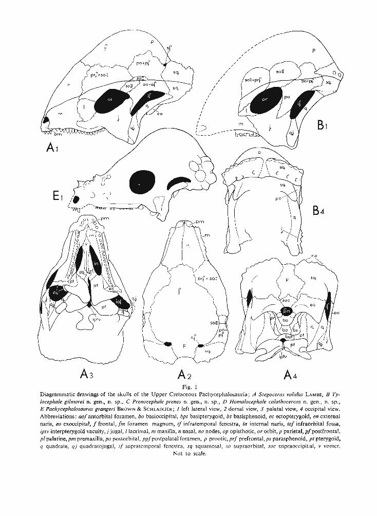
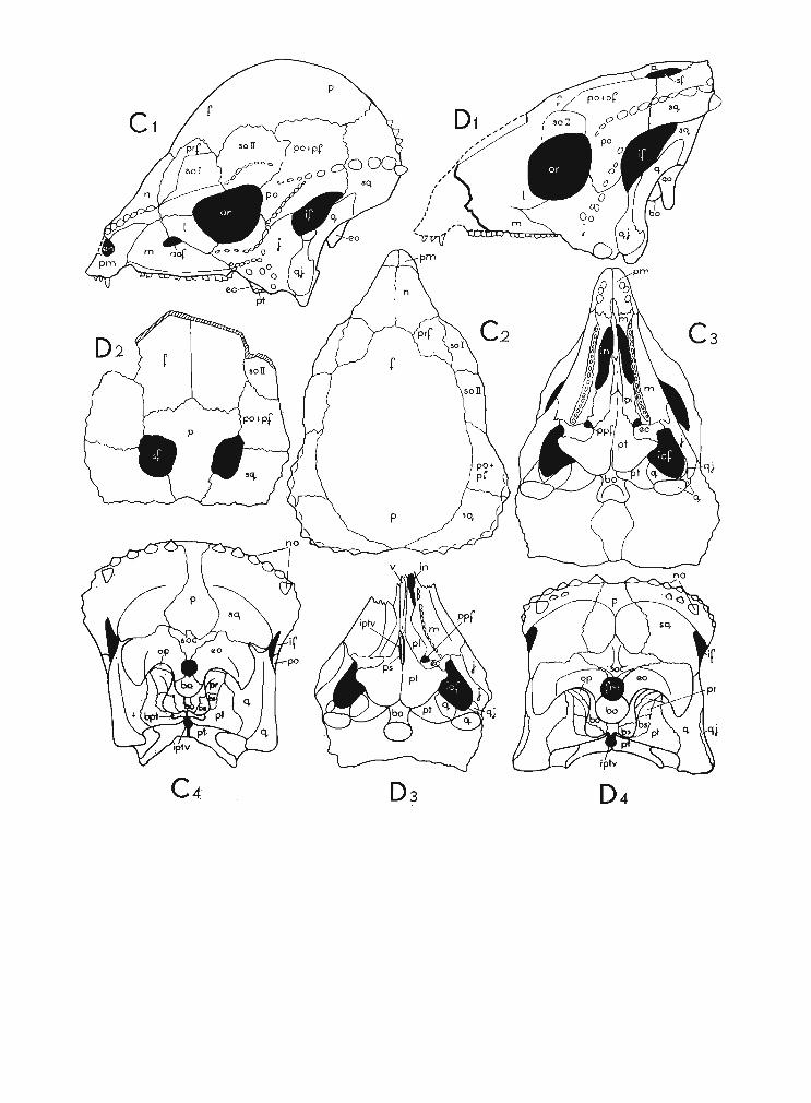
Fig. 1Diagrammatic drawings of the skulls of the Upper Cretaceous Pachycephalosauri a ; A Stegoceras validus LAMBE, B Tylocephale gilmorei n. gen., n. sp., C Prenocephale prenes n. gen., n. sp., D Homalo cephale calathocercos n. gen., n. sp.,E Pachycephalosaurus grangeri BROW N & S CH LAIKJ ER ; 1 left lateral view, 2 dorsal view, 3 palatal view, 4 occipital view.Abbreviations : aof ant orbital foramen , bo basioccipital , bpt basipterygoid, bs basisphenoid, ec ectopterygoid, en externalnaris, eo exoccipital, f frontal , fm foramen magnum, if infrat emporal fenestra , in internal naris , iof infraorbital fossa,ipt v interpterygoid vacuity, j jugal , I lacrimal, m maxilla, n nasal, no nodes, op opisthotic, or orbit, p parietal, pfpostfrontal,pi palatine ,pm premaxilla, po postorbital, ppfpostpalatal foramen , p prootic , prf prefront al, ps parasphenoid, pt pterygoid,q quadrate, qj quadratojugal, sf supratemporal fenestra, sq squamosal, so supraorbital, soc supraoccipit ai, v vomer.
Not to scale.

62 TERESA MA R YANSKA & HALSZK A OSM6LS KA
saurid pattern. H. calathocercos co mes from th e same locality and formation (Nemegt) asPrenocephale prenes. The latter form has th e fro ntopa rieta l dome most strongly developedamong the Pachycephalosauridae kn own to da te . T he skulls of both these species are of aboutthe same size and when th eir occi pit a l reg ions are compared the difference is to be noticedin th e size of th eir foramina magna and of the occi p ita l condyles with both being larger inH. calathocercos. The cranial ro of of H . calathocercos is nar rower in th e posterior view and doesnot overhang the occip ita l region as in P . prenes. H owever, th e ventral portion of the squam osala nd th e exoccip ita l is much br oad er transver sely in H. calathocercos . This is partly causedby th e more steep incl inati on of these bo nes towards th e medi al line in P. prenes. The centraldepression which is presen t in th e occ ipi ta l region of all the Pach ycephalosauridae, in H. ealatho cercos is th e broad est transverse ly but it is much shallower than in P. prenes and aboutas shallow as it is in Stegoceras validus. This depression in H. calathocercos is divided mediallyby a low, vert ica l ridge; th is feat ure is very weakly p ro no u nced in th e two species compared.The basal tubera in H . calathocercos are flatte ned a nte roposte rior ly as it is the usual ca se withall th e pach ycephalosaur ids, but they are st ill th icker tha n those in P. prenes and may be onlyco mpared to th ese in S. validus. H owever, in the latter spec ies the basal tubera are not so closeto th e quad rat ic wing of the pte rygoi d as th ey are in H. calathocercos. The ba sioccipital portionof th e basal tubera in our species is st ro ngly developed , co mparable to th at in Pachycephalosaurus grangeri. In the pa latal view, th e ventra l margin of th e maxilla in Homalocephale ealath ocercos is di stin ctly a rched o utward s in its poster ior sect ion, and it is nearly st ra ight inS tegoceras validus and Ty locephale gilmorei, very slightly arched in Prenocephale prenes andrecurved inwards in Pachycephalosaurus grangeri. The general structure of the maxillary teethis simila r in Pa chyc ephalosaurus grangeri and Prenocephale p renes (altho ugh th e teeth wereused in different ways, jud ging from the different position of th e worn 'out surfaces in th e latterspecies, p . 55). The maxillar y teeth in H. calathocercos have less r idges on the cr owns than th eteeth in P. pre nes. T he st ructu re of th e maxi llary teeth in H. calathocercos an d S. validus isincomparable, because th e crowns are conical in th e latter species.
ANATOM Y OF THE PACH YCEPHALOSA URTDAE
OSTEOLOGY OF THE SKULL(Pis. XXIl-XXIV, PI. XXV, Fig. 2; Text -figs 1-3)
The following detailed descriptio n of the bo nes of the skull is based on the skull of Prenocephale prenes (Z.P aI.No.MgD-I / I04) fro m the Nemegt Formation (U pper Cretaceous) of Nemegtin th e G obi D esert. The state of preservat ion of th is skull is by far th e best known in thi s family,a nd it has allowed us to obtai n some new informat ion s about th e skull st ructure in th eP achycephalosau ridae. The sutures arc for the most part clearl y visible and for this reason wedecid ed 10 ba se th e description of crania l osteology of th e family on th e skull of the specimen.In addition to thi s skull we have two other less complete specime ns - that of Homalocephalecalathoce rcos (G . 1. N o. SPS 100/5 1) from th e same beds an d locality and that of Tylocephalegilmorei (Z .PaJ.No.MgD-l /105) from th e olde r, Barun G oyot Formation of the localityKhulsan in th e G ob i D esert. We had also th e op po rtuni ty to con sult a plaster ca st of the skullof S tegoceras validus LAMBE 1902 (UA. No. 2) described by GILMORE(1924) as well as the cranialroofs of thi s spec ies (N. M.C. Nos. 138, 8816) descr ibed by LAMBE (191 8) from the Belly RiverF o rmatio n in Albert a. Unfo rtu na tely the skull of Pa chycephalosaurus grangeri BROWN &
PACHYC EPHALOSAURI A NEW SUBORDER 63
SCHLAIKJER, 1943 from the Lance Formation in Montana is not accesible to us at the moment. Based on the above material, we are able to state thatthe basic structure ofthe pachycephalosauridskull is very peculiar, although generally a very uniform one. The following cranial osteology iscompleted by some new data dealing with the structu re of: the orbit, the upper jaw and themiddle ear cavity , which were not formerly described. The comparison s of the bones of skullof Preno cephale prenes with those of other pachycephalosaurids from the Upper Cretaceousare given at the end of the description of each particular bone. Th e comparisons of the cranialregions, as well as the description s of the dentition are given in the systematic part of thi s paper(p. 50). The bones of the mandibular segment are not considered here because the mostcomplete lower jaw known so far is that of Stegoceras validus and it was already sufficientlydescribed by GILMORE (1924) . The new Mongolian material at our disposal displays only a fragmentary mandible of Tylocephale gilmorei an d this is described in the systema tic part of thepresent paper.
Neurocranium
Th e bone s forming the brain case are firmly united in the Pachycephalosauridae. Someof them are strongly thickened or domed i. e. th ose of the skull ro of, which gave a very strongpro tection to the bra in. Th e uni on of the neurocranium with the splanch nocranium is verystrong so the skull was aki netic.
Supraoccipital. - The supraoccipita l takes pa rt in the formation of the do rsal marginof the foramen magnum, but it is very narrow th ere. The sup raoccip ital gives lat erally twonarrow wings on the occipita l surface of th e skull, each of them deeply wedged between theexoccip ita l and the squa mo sal. In the midline, the supraoccipit al is elongated in the dorsaldi rection and it gives a tongue which deeply invad es the parietal. Th e supraoccipita l forms th ebottom of the deep central depression and bears medially a vert ica l keel, on the both sidesof which a pair of sho rt deep grooves is present. Th e sup raoccip ita l forms the narrow portionof the roof of the medulla oblonga and anteriorly it expands and bounds the inner ear cavityposteromedially. The supraoccipital tak es part in form at ion of the upper margin of the foramenmagnum in Homalocephale calathocercos and shows st rongly pronounced medial , verticalkeel. The supraoccipita l is similarly developed in Stegoceras valiaus. LAMBE(1918, PI. I , Figs. 1, 2)mistakingly nam ed in S tegoceras validus tue supraoccipita l as the exoccipi ta l. Th is most probablywas cau sed by the presence of th e vertica l c1evage th rough the supraoccipital. BROWN &SCHLAIKJER (1943) stated that the supraoccipital in Pa chycephalosaurus grangeri was excludedfrom the boundary of the fo ramen magnum. Thus, in this respect , the species mentioned wouldbe different from all other known Pachycephalosauridae.
Exoccipital. - Th e exoccipit al forms the major part of the sharp margin of the for amenmagnum and gives the pedicle which participates in the formation of the occipital condyle. Laterally thi s pedicle is perforated by the foram en for the exit for the nerve XII. On the marginof the foramen magnum and within the medulla oblonga it co ntacts the basioccipital ventrallyand the supraoccipital dorsally. It bounds mediolaterally the inner ear cavity. It forms theflat , wing-like paroccipital process which meets the squa mos al dorsolaterally. The suturebetween the squamosal and the exoccipita l is fold ed in its more medial portion and givesupwards the small emb ayment of the exoccip ital within the squamosal. The exoccipital contactsthe opi sthotic under the proximal part of the ventral edge of the paroccipital process. The sutureis distinct, reach es the upper margin of the foram en ovale an d it is very apparent that the entiredistal portion of the paroccip ital process was formed only by the exoccipita l, T he condy lar
64 TER ESA MARYANSKA & HALSZKA OSM6LSKA
pedicle of the exoccipital gives the short process lateroventrally, along the occipital condyle,which forms the upper half of the edge of the basal tuber. This process contacts the basioccipitalmedially in the region of the condylar neck and the basi sphenoid ventrally. The suture betweenthe exoccipital and the basisphenoid extends here upwards on the anterior face of the flatbasal tuber. At the lower margin of the foramen ovale, the exoccipital contacts the horizontalridge of the prootic laterally, and bounds the foramen ovale ventroposteriorly. An extensivedepression is present on the po sterior face of the exoccipital above the foramen magnum whichis transversely elongated. A strongly arched ridge extends above this depression in the lateraldirection and it reaches the ventral edge of the paroccipital process short before its lateroventralextremity. The distal ends of the paroccipital processes are longer and narrower in Homalocephale calathocercos than in Prenocepliale prenes. They are very much shorter in Stegocerasvalidus. In all representatives of the Pachycephalosauridae they are closely attached to thesquamosals and quadrates laterodorsaIly, except P. prenes where their upper distal edges arebroadly sepa rated from the bones mentioned. BROWN & SCHLAIKJER (1943) mentioned thatthe contact of the exoccipital with the prootic is extensi ve in Pachycephalosaurus grangeri,Thi s contact is very limited in P. prenes and Homalocephale calathocercos and it is presentonly at the lower margin of the foramen ovale. Most probably these authors had in minds theventral contact of the exoccipital below the lower edge of paroccipital process. Here however,the exoccipital is ventraII y separated from the prootic by the narrow po sterior inclusion of theopisthotic as it is distinctly visible in both Mongolian species mentioned and also in Stegocerasvalidus.
Basioccipital. - The basioccipital forms nearly the entire occipital condyle. It forms thenarrow portion of the bottom of the medulla oblonga and in the ventroanterior direction itforms the cen tral portion of the basal tubera, marked medially by a sharp keel. The keelis most strongly pronounced in its anterior part. The condylar neck is extremely short. Thearticula r surface of the condylus is developed on its po sterior face , and it continues ventraIly,where it forms a comparatively exten sive triangular area. There is a pair of short grooves presenton both sides of the keel mentioned above. The basioccipital contacts the exoccipitals dorsallyin the region of the occipital condyle, laterally with in the medulla oblonga, as well as laterodorsally outside the condyle. Here on the boundary the foramen for exit of the nerves X and XIis pre sent. The basioccipital contacts the basisphenoid on the po steroventral portions of thebasal tubera. The ventral suture with thi s bone extends transversely close to the bases of theba sipterygoid processes within the deep cleft between the basal tubera posteriorly and thepterygoids anteriorly. The occipital condyles in all the Pachycephalosauridae are simila r inbeing formed mostly by the ba sioccipit al , but they differ in the degree of ventral inclination,which is strongest in Stegoceras validus. The condyle is nearly perpendicular to the line of themaxillary teeth in Homalocephale calathocercos and it has the large articular surface facingventrally. The similar surface seems to be pre sent in Pachycephalosaurus grangeri. It is alsodeveloped in Prenocephale prenes but it is much smaller here. The neck-like portion of thebasioccipital, anterior to the occipital condyle, is very well developed and deep dorsoventrallyin Pachycephalosaurus grangeri, bearing the thick keel in its anteriormost portion. It differsfrom that in Prenocephale prenes, which has only a sharp keel present in this place, and theneck-like portion is nearly ab sent in the latter species. This portion seems to be deepest andnarrowest in Pa chycephalosaurus grangeri.
Opisthotic. - The opi sthotic is a comparatively small, flat bone. It s posterior extensionis wedged between the exoccipital dorsaIly and the prootic ventrally, and its anteromedial edgereaches the boundary of the foramen ovale. Within the infr atemporal fossa, it takes part in
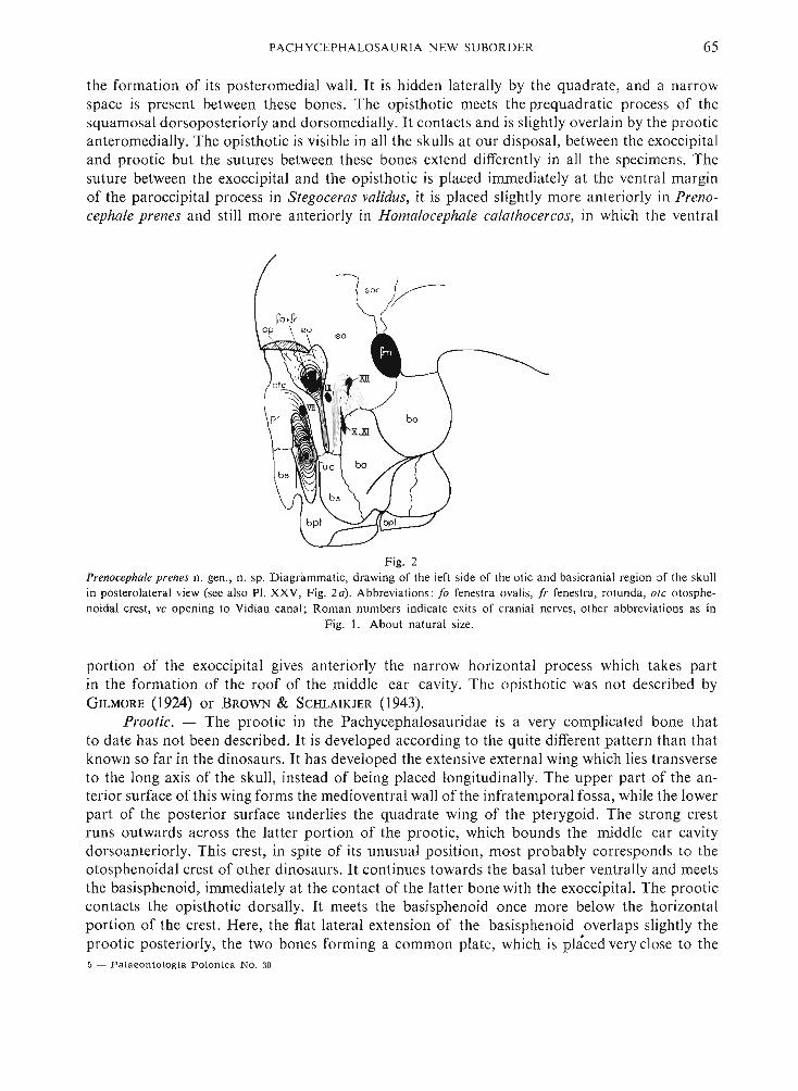
PACHYC EPH ALOSAURI A NEW SUBOR DER 65
the formation of its posteromedial wall. It is hidden laterally by the quadrate, and a narrowspace is present between these bones. The opisthotic meet s the prequadratic process of thesquamosal dorsoposteriorly and dorsomedially. It contacts and is slightly overlain by the prooticanteromedially. The opisthotic is visible in all the skulls at our disposal, between the exoccipitaland prootic but the sutures between these bones extend differently in all the specimens. Thesuture between the exoccipital and the opisthotic is placed immediately at the ventral marginof the paroccipital process in Stegoceras validus, it is placed slightly more anteriorly in Prenocephale prenes and still more anteriorly in Homalo cephale calathocercos, in which the ventral
so c V--
Fig. 2Prenocephale prenes n. gen. , n. sp. D iag rammatic, dr aw ing of the left side of the otic a nd basicranial region of the skullin posterolate ral view (see also PI. XXV, Fig. 2a) . Abbreviati on s : /0 fenestra ova lis, fr fenes tra, ro tunda , ate otosphcnoidal crest , vc opening to Vidian canal ; R om an numbers indi cate exits of crani al nerves, other abbrevia tions as in
Fig. 1. About natural size.
portion of the exoccipital gives anteriorly the narrow hori zontal process which take s partin the formation of the roof of the middle ear cavit y. The opi sthotic was not described byGILMORE (1924) or BROWN & SCHLAIKJER (1943) .
Prootic. - The prootic in the Pachycephalosauridae is a very complicated bone thatto date has not been described. It is developed according to the quite different pattern than thatknown so far in the dinosaurs. It has developed the exten sive external wing which lies transverseto the long axis of the skull, instead of being placed longitudinally. The upper part of the anterior surface of this wing forms the medioventral wall of the infratemporal fossa, while the lowerpart of the posterior surface underlies the quadrate wing of the pterygoid. The strong crestruns outwards across the latter portion of the prootic, which bounds the middle ear cavitydorsoanteriorly. Thi s crest, in spite of its unusual po sition, most probably corresponds to theotosphenoidal crest of other dinosaurs. It continues towards the basal tuber ventrally and meet sthe basisphenoid, immediately at the contact of the latter bone with the exoccipital. The prooticcontacts the opisthotic dorsaIIy. It meets the basisphenoid once more below the horizontalportion of the crest. Here , the flat lateral extension of the basi sphenoid overlaps slightly theprootic po steriorly, the two bone s forming a common plate, which is placed very close to the5 - Pala eo n to logi a Polonica No . 30
66 TER ESA MA R YANSKA & HALSZK A OSMO LSKA
quadrat e wing of the pterygoi d and only a very narrow slit separates the two pla tes. In thi sway , the basicran ial region is nearly completely separated from the suborbital and palatalregions. Th e prootic-basisph enoid plate bounds an terio rly the comparatively broad and verydeep cavity which corresponds to the strongly shortened basicranial region and is limited backwards by th e plate-like ba sal tuber. Within the infratemporal fossa the prootic contacts theopisthot ic po steriorly, the quadrate laterally and dorsally, the pterygoid laterally and ventrally.The contact with the laterosph eno id medially is an extensive one. The co ntact with th e basispheno id anteriorly is only partly visible dor sally and it is nearl y completely hidden by thepterygoid and epipterygo id ventrally. The prooti c meets the epipterygoid anteroventrally andimmediately below th e contact with th e laterosph enoid. The prootic is similarly con structed inHomalocephale calathocercos. Th e nearl y complete separation of the basicranial and the sub orbital region s as well as the presence of the hor izontal crest on the posteri or sur face of thetransverse wing of the prootic in Stegoceras validus, both characteristic of Prenocephale pren esand Homalocephale calathocercos, indicate that the gene ral structur e of th e bone was simila rin thi s species. The crest menti oned seems but slightly more weak ly pronounced in Stegocerasvalidus, but it seems to be quite stro ng in Pa chycephalosaurus grangeri.
Basisphenoid. - Th e basisphenoid is very complicated in th e pachycephalosaurids thanksto the very strong anteroposterior compression of the basicran ial region. Not all its contactswith th e adjoining elements could be observed and intepreted. This is especially true for theanterolateral contact s of the basisph enoid , which can be observed in the medioventral partof the wall of the orbit, where man y additional bony elements are added. In the posteriorview, the basisphenoid forms the lateroventral extension of the flat , plate-like basal tubera, andit contacts here the exoccipita ls dorsally and basioccipit al medially. The ba sisphenoid is invaginated medially on both side of the basicran ium and forms the deep lateral cavities boundedby the basal tubera po steriorly and anteriorly by the plates which are formed by the basisphenoidventrally and the prootic dorsally. Th e bott om of thi s cav ity is pierced in the medioventraldirection by the entra nce to th e Vidian cana l. Th e basisphenoid sends medially and anterovent rally a pai r of the thick basipterygoid processes which atta ch to each other medially, roofingthe po sterior portion of th e interpterygoid cavi ty . In posterior view, th e prootic-basisph enoidplate is underlain along its margin by the quadrate wing of the pt erygoid. It appears withi nthe infraorbital fossa anteriorly, and here the suture between the pro oti c and basisph enoidis not exposed . The suture of th e basisphenoid is here visible dorsally with the presphenoidbackwards and with some additio na l ossifications forwards. The an teriormo st edge of thebasi sphenoid forms the posterior boundary of th e space which joins the two orbits transversely(the interorbital fissure, p . 77). The dor sal rim of thi s fissure is formed by two additional bonyelements and dorsally a pocket is present between them. It is bounded by the palatal wingof the pterygoid anter iorly and ventra lly. The interorbital fissure communicates ventrally withthe interpterygoid vacu ity. The basisph enoid is pierced by the exit for nerve VI and below itby the small foramen which was probably th e exit for the palati ne artery. Th e basisphenoidis generally very similar in all the Pachycephalosauridae where it was described. The pa rtof the basisphenoid which form s the ventra l po rtions of the basal tubera is mo st extendedvent ra lly in Homalocephale calathocercos in which it nearly reach es th e pterygoids ; it is theshortes t vertically in Stegoceras validus in wh ich the comparatively wide space separates thebasisphenoid from the pterygoids. The interorbital fissure mentioned above in Prenocephaleprenes and present in front of th e basisphenoid is nearly closed in Homalocephale calathocercosand Stegoceras validus where thi s bon e seems to be more expa nded forwards. Th e basisph enoidof Pachycephalosaurus grangeri was never described.

I'A CHYC EI'H AL OSA UR IA N EW S U BOR DER 67
Paru sphenoid. - T he parasph en oid in th e Pachycephalosauridae is well developed a nd differentiated from the basisphenoid, alt houg h it overlaps th e latter bon e ventra lly. The par asphenoida l rostrum is developed bu t it seems to be short in. Prenocephale prenes, a nd is visiblewithin the interorbital fissure. The posterior part of th e parasphen oid d ist inctly overlaps ventrallyth e ba sisphenoid in Homalocephale calathoce rcos and it closes nearl y completely th e lumenof the interorbital fissure. The parasphenoidal ros trum is lon g in th is species, being visiblein the palatal view in th e slit between th e two pterygoids. T he parasphe no id seems a lso to closethe lumen of the interorbital fissu re in S tegoceras validus . The pa rasph enoid al rostrum is developed in thi s species simila rly as it is in Homalocephale calathocercos, b ut it exte nds ventra llyin its most anterior part as th e pendant process which was te nta t ively called the presphenoidby GILMORE (1924 , p. 22).
Laterosphen oid. - The lat erosphenoid is coalesced with the adjoining eleme nts excep tthose of the skull roof and most of its su tu res are bu t weakly vis ible. It forms the dorsomedialportion of the crest which marks th e boundary between th e infratemporal fossa and th e or b it.It is expanded mo stly o n th e dorsomedial wall of th e infrat emporal fossa , and only a smallportion is present within th e orbit. The entire posterior bounda ry of th e lat erosphenoid contactsthe parietal. It meets the postorbital posterodorsally and th e fro nta l anterodorsa lly. It contactsthe orbitosphenoid anteriorly, the basisphen oid anteroventra lly and the prootic ventrally. Thenarrow tongue of th e lat erosphen oid mak es th e dorsa l margin of th e exit for cranial nerveVi clo se to the contact of the basisphen oid and pr ootic . T he lat erosphenoid in Homalocephalecalathocercos does not differ from th at in Prenocephale prenes, but its co ntact with the adjoiningbones seems to be looser. T he la tero sphenoid in S tegoceras validus (a lisp heno id of LAMBE
1918 and GILMORE 1924) is less extende d ver t ically because of th e much lower orbit and infratemporal fo ssa. In thi s species it form s the upper anterior bounda ry of th e infrat empor alfossa rather than th e lower anter ior one as was m istakingly sta ted by GILMORE (1924, p. 21).Judging from the description of the bon e give n by B ROWN & SCHLAIKJER (194 3) for Pachy cephalosaurus grangeri it is simila r here to th at bon e of other pachycephalosaurids.
Presphenoid. - It is difficult to indicate the presphenoid in the pachycep ha losa urid sku llbecause of th e many add it iona l ossifications presen t in the orbi ta l reg ion. We thi nk th at it ismo st rea sonable to co nsider as th e presphen oid the small bon e which bounds vent ra lly andslightly anteriorly th e exit of the op tic nerve. Th is bone contacts the proot ic poste rove ntr allyon the lateral wall of th e b ra incase, the bas isphenoid ventra lly and a separa te element of th eadditiona l orbital oss ifica t ion anterovent ra lly. The dor sal contact of th e p resphen oid is exclu sively with the orbitosphenoid. The forarnen is pr esen t in th e poster ior suture between th epresphenoid and orbitosphenoid wh ich rep rese nts th e exit for the third nerve. The exit of sixthnerve is present at the presphenoid /basisphenoid suture. The usual contact between th e presphenoid and the pa ra sphen oid which ca n be observed in many orn ith ischian din osaurs on th elateral wall of the braincase is not present in the pachycepha losau rids. Tt is cau sed by the presenc eof the additional oss ifications which separate the two bones a nd by th e peculi ar com pressionand folding of th e ba sicran ial region in this family. The presphen oid is very similar inHomalocephale calathocercos.
Orbitosphenoid. - The orbitosphen oid is small and fo rms th e dorsoposterior boundaryof the exit for the second nerve. The orbitos phe no id is sutu ra lly connected with the presphen oidon both sides of the optic foramen. It contacts the add itiona l or bita l ossification s anteriorlyand dorsally , and th e laterosphenoid po steriorly. There a re separa te exits for the fourth andthird nerves. That for th e fourth nerve is placed in th e or bitosphenoid, th at for the third nervein the sutu re with th e presphenoid, im med iately posteriorly to the exit for th e second nerve.5"
68 TERE SA MAR YANSK A & HALSZKA OSM6LSKA
The external surface of the orbitosphenoid is very uneven , furrowed irregularly and deeply.The orbitosphenoid is also very small occupying the same position with respect to theadjoining elements in Homalocephale calathocercos and in Stegoceras validus. The surface of thisbone in Homalocephale calathocercos is smoother than in Prenocephale prenes.
Frontoparietal. - The frontal and parietal are not separated by sutures dorsally and theyare extremely strongly domed. The frontoparietal contacts anteriorly the nasal by means of a shortsuture. Laterally it meets the prefrontal along an arched suture. It gives the short wedge whichmeets the supraorbital I laterally. More posteriorly it is bounded laterally by the supraorbital Il,postorbital, and the squamosal. At the juncture of the sutures between the frontoparietalfrom the one side and the prefrontal, supraorbitals from the other side there is a deep pit. Twoother such pits , the anterior deeper and the posterior shallower, are present at the frontoparietalsuture on the juncture with the supraorbital Il-postorbital and the postorbital-sqamosal.Within the orbit the frontal contacts the prefrontal , the accessory orbital ossification 3, theorbitosphenoid, the laterosphenoid, the supraorbitals and the postorbital. The narrow tonguesof the parietal are wedged between the sqamosals on the posterior margin of the skull. Theywiden on the occipital surface of the skull and contact the squamosaIs laterally, and thesupraoccipital ventrally. The parietal ridge is well pronounced along the ventral portion of theparietals and it reaches into the central depression. The parietals take part in formation ofthe central depression. The preserved portion of the frontoparietal shows that the dome inTylocephale gilmorei was placed very much po steriorly in the comparison with other pachycephalosaurids (p. 52). The parietal is nearly vertically placed on the occipital face of the skull ,as a result of the lack of the deep central depression present here in other Pachycephalosauridae.The frontoparietal is very variable in the specimens assigned to Stegoceras validus, which isconnected with the various development of the dome within this species. The frontal portionof the dome in Stegoceras validus is more con vex and better distinguished from the adjacentbones than it is in Prenocephale prenes. Th e parietals are similarly placed in Stegoceras validusas in Prenocephale prenes in connection with the presence of the deep central depression on theoccipital face of the skull. They are , however, relatively broader on the margin of the skulland are devoid of the parietal ridge in Stegoceras validus. One of the specimens of Stegocerasvalidus (NMC No. 2379) exposes dorsally the presence of the distinct interfrontal and frontoparietal sutures (LAMBE 1918, Fig. on p. 25). A similar course of these sutures is exposed inHomalocephale calathocercos, the species which possesses an entirely flat skull roof. A weaktrace of the interparietal suture is preserved in the latter species. The parietals are broader onthe margin of the skull in Homalocephale calathocercos and the occipital portions of these bonesare flatter transversely in connection with the broader central depression than in Prenocephaleprenes. They bear also a thinner but sharper parietal ridge . The parietals bear a large, roundprotuberance in Pachy cephalosaurus grangeri on their occipital portion, which is absent inPrenocephale prenes, and oth er pachycephalosaurids. The ornamentation of the frontoparietalvaries distinctly in the species within this family. It was sometimes regarded as the characterof the sexual dimorphism (p. 100). The most distinct ornamentation of this region was observedin Homalocepha lecalathocercos, where the irregular coarse granulation is present on the frontalsand parietals. The granules are much larger on the posterior portion of the parietals, while theanterior portion is smooth, close to the medial section of the frontoparietal sutur e. The frontoparietal is more or less smooth and more or less den sely pitted in Prenocephale prenes andStegoceros validus. The surface of the dome is smooth in the species of Pachycephalosaurusbut also exposes the perforations.
Accessory orbital ossifications (Text-fig. 3). - The medial and anterior wall of the orbit

PACHYCEPHALOSAUR IA N EW SU BORDE R 69
is completely ossified in the Pachycephalosauridae. The numerous additional ossification s aresurrounded periferally by the bones usually pre sent in this region: the frontal, prefrontal, lacrimalpalatine, basisphenoid, presphenoid , orbitosphenoid, laterosphenoid. These additional elementsform a mosaic of bones which are suturally separated but are often very small. For this reasonthey are rather difficult to describe. However, three of them are more regular, larger and presentin the same place in different specimens. The notations 1-3 are given to them on Text-fig. 3and they are described below. This complete ossification of the anteromedial region of the orbitmeans that the nasal cavity was completely separated from the orbital region and that the nervesand vessels for the snout had to issue through special openings which are present in the anteriorwall of the orbit. Such extensive ossification of the orbital region has not been described inan y other reptile but slightly similar conditions are pre sent in mammals and some bird s. Some
Fig. 3Prenocephale pren es n. gen., n. sp. D iagramma tic dra wing of the right orbit (see also PI. XXV, Fig. 2b). Abbre viation s :d/lacrimal duct, e epipterygoid, fe th foramen ethomoidale, fino intero rbital fissure , f sphp foramen spheno palatinum, Is laterospheno id, os orbito spheno id, pa opening for exit of palatine artery, prs presphen oid, 1, 2, 3, x accessory orb ital
ossificatio ns; other abbreviations as in figs. 1 and 2. About natu ral size.
of these bones most probably represent the separate ossifications within the planum antorbitale(= lamina orbitonasalis of de B EER, 1971), planum supraseptale and comis sura ethmoidalis.Another possible interpretation is that ossification s 2 and 3 aro se within the trabecula communisand consequently they could be interpreted as the sepa rate ossifications of the presphenoid.Others are certainly the membranous bone s formed within the membranes of the orbital region .
Accessory orbital ossification 1. - Thi s bone is the most anterior one and the largest ofthe accessory element s. It form s the major part of the anterior wall of the orbit and contactsanteriorly the prefrontal with its upp er portion, the lacrimal and jugal below, and the palatinemost ventrally. It meets the accessory ossification s 2 and 3 and the narrow upward extensionof the pterygoid posteriorly. Two opening s are present on the suture between the accessoryossifications 1 and 2 and they mo st probably represent the foramina ethmoidale. Anotheropening is present here, at the most ventral exten sion of the accessory ossification 1 betweenthe latter and the palatin e with the jugal. It may be interpreted as the sphenopalatine foramen .It also lead s to the nasal cavity. When seen anteriorly, from the nasal cavity, the accessoryossification 1 forms the extensive posterior wall of the nasal cavity. Thi s wall is convex anteriorlyand contacts the maxilla and prefrontal laterally, the palatine ventrally, the accessory ossification 2 medially and the accessory ossification 3 poste riorly. Th e latter , together with accessory
70 TE RESA MA R YANSKA & HALSZK A OSM 6 LSKA
oss ifica tio ns 2 and 3, tak e pa rt in th e for mation of the floor of the olfactory tr act with acces soryossifica t io n 1 composing its mo st anterior part. The accessory ossifica tion 1 seems to be a homologue of the lateral ethmoid of fishes a nd of the ectethmo id -l-uncina te bone of some birds.D E BEER (1971) di scussing the ossificat ions of the lamina orbito nas alis suggests that the uncinat ebone of birds "apears to be a portion of th e ectethmo id segmented off ..." . The bone describedabove in Prenocephale prenes is well di stingui shabl e also in Homalocephale calathocercos andSt egoceras validus but in the latter species it is distinctly smaller , whi ch is most probably relat edto the much lower orbit of Stegoceras validus. It seems , that th e small fragment of the bonenamed by LAMBE (1918 , PI. I, Figs. I, 2) as parasph enoid corresponds to our access ory orbitaloss ification 1.
Accessory orbital ossification 2. - This ossifica tio n is a small bon e, which bounds theupper foramen ethmoida lis ventra lly an d lower foram en ethmoidalis po steriorly. It meets accesso ry ossification 1 anteriorly, accesso ry ossifica t ion 3 dorsally, one bone of other accessoryelements posteriorly and seve ra l ot hers ventrally. It takes part in the formation of the floorof the olfactory tract. Th is bone is slightly smaller in H omalocephale calathocercos, otherwi sebeing simila r in th e shape. A bone, which most probably represents accessory ossification 2,is pre sent posteriorly to the lower fo ramen ethmo idalis in S tegoceras validus, but it is larger here.
Accessory orbital ossification 3. - I t is a not her large bone of th e accessory orbitaloss ifications. It is elongated in the sagittal di rection and contact s the frontal with a long su turedorsally , the laterosphenoid posteriorl y a nd, with its posterior half, th e orbitosphenoid ventra lly.It meets accessory oss ificat ion 1 a bove th e uppe r fo rame n ethmoida lis and slight ly below th elatter, it is sutura lly connected with accessory ossificat ion 2 anter iorly . One of the small accessory elements is placed anterove ntra lly to accessory ossifica t ion 3. The latter forms th e lat eralwall to the mo st anteri or portion of the cerebrum, th e co ro na l su lcu s a nd the olfactory tract.The acce ssory ossification 3 is a lso well visible in Homalocephale calathocercos and Stegocerasvalidus a nd it is similarly large in both species. In th e specimen NMC N o. 138 of S. validus thisbone wa s det ermined by L AMBE (I. c.) a nd G ALTON (l971 a , Text-fig. 4b) as th e presphenoid .
Splanchnocranium
T he spla nchnocra nium includ es, as here conceived , a ll the skull eleme nts whi ch a re notdirectly involved in th e forma tion of the brain case. They are, however , a ttached directly orindirectly to the neurocranium and all the contacts with the neurocranium seem to be extremelyfirm , excep t, perhap s, th at between th e upper portion of th e quadrat e a nd the skull "roof. Thesquamosa l ha s a rounded , deep art icula r socket for th e head of th e quadrate, which wouldbe for an amortizat ion ra ther th an due to the kinetic ism of the skull.
Premaxilla. - The premaxilla is relati vely large, its lateral wing is wedged far posteriorlybetween th e nasal and ma xilla and sepa rating th ese bones for a very long di stance. This wingis placed in th e vertical plane and is flat. The anterior portion of the prem axilla is long and thickened , slop ing steeply down lat erally. T he alveo la r margin of the premaxilla is comparativelythick and bears three deep alveo lae . The very tip of the premaxill a is devoid of dentition.Ventrally the premaxill a fo rms th e a nterior part of th e slightly concave snout. More po steriorlyit forms the roof of th e deep pit for th e enla rged dentary tooth, and medially its posterior processunderlies the maxilla. The premaxilla bears anteriorly, oppos ite th e first p remaxillary tooth,a small foramen. It presumabl y lead s to th e slit-like op ening placed above and anteriorly toth e bottom of th e na sal cavity . It seems prob abl e that both ope nings were connected with the

I'AC IIYC EI' I-I AL OSA URI A N EW S U BOR DE R 71
Jacobson's organ. The internarial bridge was pro bably formed from the premaxillae, beca useeach of them gives posteriorIy, on the dorsal side, a process which is broken off. The an teriorportion of the nasal cavity is large and deep medially . The side walls of the medial depre ssionare formed by the premaxillae, while its bottom is built from the maxillae. Within the Pachycephalosauridae the premaxillary porti on of the snout is only known in Stegoceras validusand in the here described Pren ocephale prenes. The essential difference between the two speciesin thi s region involves the degree of its development, which is much more extensive in Prenocephale prenes in connection with the development of the relative ly strong cani ne-like premaxillary teeth. In Stegoceras validus the premaxillary portion of the snout is not differenti atedfrom the po sterior portion, and the notch for receiving of the enlarged mandibular tooth islacking, although the short and very slightly deepened diastema is present. The an teriormostportion of the premaxilla in Stegoceras validus is very low and its lateral wing is very short, notseparating the nasal and the maxilla as in Prenocephale pren es. One would expect a similarstructure of the anterior portion of the premaxilla in the other long-sno uted form, Pa chycephalosaurus grangeri. Thi s porti on of the premaxilla is not preserved in this species, and itslateral wing is excluded from the nasal according to BROWN & SCHLAIKJER (1943, p. 134),as in Stegoceras validus. The authors mentioned above suggested that such development ofthe late ral premaxillary wing might be typical of the family , which has appeared to be untrue.
Maxilla . - Th e maxilla is penetrated along nearly its entire length by the extensive sinus,which is here called the intramaxillary sinus 3. It is entirely surrounded by the maxilla in itsant eriormost section. The medial lamin a of the maxilla is here pierced dorsally by a very extensive fenestra, abo ve the maxillary teeth 3-6. This fenestra connects the intra maxillarysinus with the nasal cavity. More posteriorly, the lateral wall of the sinus is perfora ted by theantorbital foramen. The sinus is bounded in this region dorsally by two separated laminae ofthe lacrimal, and by the two laminae of the maxilla. The caudal sect ion of the intramaxillarysinus extends under the lower margin of the orbit. It is bounded here by the jugal dorsallyand by the maxilla vent rally, close to the contact of the latter bone with the ectopterygoid.The sinus opens posteriorly th rough a comparatively small foramen into the pterygopalat inefossa. The lateral wing of the maxilla is very high and placed vertically. It contacts the premaxilla anterodors ally, along the extensive, oblique suture, the lacrimal posterodorsally and thejugal posteroventrally. It s dorsal contact with the nasal is shore The alveolar margin of themaxilla bends weakly backwards, near the place where it meets the jugal and the ectopterygo id.It bears seventeen teeth. A broad narrowing anteriorIy, shelf is present, which is externa l to therow of the teeth. It is pierced by a row of larger and smaller vascular canals. The lingual wingof the maxilla is placed vertically and it form s the medial wall of the intram axillary sinus.It gives the short palatinal process in its posterior part. The row of large fenest ra, which openinto the alveolae, is present immediately below the teeth. The anterior portion of the maxillais thick and medially cont acts its fellow, but ventrally and slightly posteriorIy the vomer iswedged between the two maxillae. The narrow, anteriormost process of the maxilla is overlappeddo rsally and vent rally by the pr ocesses of the premaxilla. The maxi lla is widened in front ofthe first maxillary tooth, and posterior to the contact with the premaxilla. It has ventrally developed an extensive pit , which is bordered laterally by the sharp, toothless margin of the maxilla.Thi s margin rises upward s ant eriorIy, when seen from the side. The alveolar margin of
3 It corresponds, partly at least, to the antorbital recess in Hypsi/ophodon (GALTON, 19700), but it is more exte~sivein the Pachycephalosauridae and seems to be present also in the forms devoid of the antorbital forame n. This is why wedecided to introduce here the term "intrarnaxillary sinus".

72 TERE SA MAR YAN SKA & HAL SZKA OSM6LSKA
the maxilla is straight to the very end in Tylocephale gilmorei. The lateral maxillary shelf isin this species very narrow. The alveolar margin is arched outwards posteriorly in Homalocephalecalathocercos, and the shelf is still more extensive in thi s species than it is in Prenocephaleprenes. The maxilla has a long contact with the na sal laterally in Stegoceras validus, and itborders the external naris posteriorly, which is not the case in Prenocephale prenes. The maxillaryteeth come to the contact between the maxilla and the premaxilla in Stegoceras validus andthere is no pit developed in thi s place. The maxilla bears sixteen teeth in the latter species.The anterior portion of the maxilla is here broad and flat in the ventral view, in which thi sspecies differs from Prenocephale prenes. Otherwise the maxillae of the two species comparedare alike. The maxilla in Pachycephalosaurus grangeri is different from that in Prenocephaleprenes in having a much longer backward process, which contacts the lacrimal ventrally andreaches nearly to the level of the orbit. The difference is also marked in the developmentof the anterior, ventral margin of the maxilla in the two species compared. This in Pachycephalosaurus grangeri does not rise upwards anteriorly, as can be judged from the ilustrationgiven by BROWN & SCHLAIKJER (1943, PI. 38, Fig. 2), but it is similarly sharp as in Prenocephaleprenes. There may be also a difference between the species compared in the contact betweenthe maxilla and the na sal , but it cannot be stated basing on the description and ilustrations givenby BROWN & SCHLAIKJER (I. c.). The lateral wing of the maxilla is con vex outwards inPachycephalosaurus grangeri and Stegoceras validus, while it is flat and vertically placed in Prenocephale prenes.
Lacrimal. - The lacrimal is a relati vely large bone bounded dorsally by the supraorbital Iand anteriorly to the latter, by the small wedge of the na sal. It contacts the maxilla anteriorlyand slightly ventrally along an extensive suture, except for the part, where it bounds posteriorlythe antorbital fene stra. More backwards, it is bounded by th e jugal ventrally, along a shortsuture. In side the orbit, it form s the anterior margin of the latter. The lacrimal duct is locatedimmediately at the margin of the orbit and it lead s into the intramaxillary sinus. The lacrimalcontacts the jugal ventrally, within the orbit , the acce ssory orbital ossification 1 posteriorly,the prefrontal po sterodorsally and the po storbital 1 dorsallv . The lacrimal gives a separatemedial lamina in the region of the antorbital foramen , and thi s lamina bounds dorsomediallythe intramaxillary sinus . The lacrimaJ is su bcircular in Stegoceras validus when seen on thelateral wall of the snout, and forms but the ventra l part of the anterior margin of the orbit.It does not contact the nasal. The lacrimal of Pachycephalosaurus grangeri is more similarto that of Prenocephale prenes, and it occupies the entire anterior margin of the orbit, as inPrenocephale prenes. The lacrimal occupies, however , one third of the orbital margin in Pachycephalosaurus grangeri and less th an one fourth in Prenocephale prenes. The antorbitalforamen seems to be absent in all the species here compared , except Prenoceplzale prenes.
Jugal. - The jugal is a large and long bone, when wiewed from the side, and it forms theentire ventral margin of the orbit. The thin tongue of the jugal underlies medially the postorbital.The jugal forms a short wedge between the lacrimal dorsally and the maxilla ventrally, and itsapex doe s not extend further than the anterior margin of the orbit. The po sterior margin ofthe bone is strongly expanded vertica lly, and it bounds the infratemporal fenestra anteriorly.It contacts the quadratojugal ventrally overlapping the latter bone and contacting the quadrate,above the mandibular joint. In ventral view, the jugal contacts the maxilla anteriorly, and itoverlaps the maxilla laterally along a short sutu re. The short tongue of the jugal is wedgedmedially between the maxilla and the ectopterygoid . The epijugal (= infrajugal of GILMORE
1924) wa s not ob served in Prenocephale prenes. The jugal forms the major portion of the posterior margin of the orbit in Tylocephale gilmorei, when viewed laterally. It is strongly convex
PACHYCEPHALOSAURIA NEW SUBORDER 73
laterally below the ventral margin of the orbit in this species, and in both these features the jugaldiffers from that of Prenocephale prenes. The jugal is similar in Homalocephale calathocercos,but it comes very close to the mandibular joint ventrally. The jugal in Stegoceras validus differsfrom that in Prenocephale prenes in having a very long anterior process, which reaches muchfurther forwards than the anterior suture of the lacrimal. The jugal overlaps laterally in Stegoceras validus the anterior margin of the postorbital along the extensive surface. Pachycephalosaurus grangeri has the jugal very different from that described in Prenocephale prenes. It issmall and has a long anterior process; it overlaps the anterior portion of the postorbital slightlylaterally, and it does not take part in the formation of the anterior margin of the infraorbitalfenestra. The posteroventral margin of the jugal of Pachycephalosaurus grangeri is situatedwell above the mandibular joint of the quadrate, even higher than it is in Prenocephale prenes.The lower, posterior part of the jugal is strongly ornamented in all species of the Pachycephalosauridae known from the Upper Cretaceous.
Quadratojugal. - The quadratojugal consists of two laminae, which overlap the jugalanteriorly and the quadrate posteriorly. The lateral lamina has its quadrate wing stronglydeveloped, while the medial lamina, which is larger than the former, has its jugal wing morestrongly developed . The quadratojugal is constricted centrally between these two laminae. Thebone is pierced by the quadrate foramen. The lateral lamina of the quadratojugal is drop-shaped,being very narrow dorsally and broad ventrally, It reaches the infratemporal fenestra above thecontact of the jugal and quadrate. The medial lamina of the quadratojugal is comparativelynarrow vent rally and broad dorsally. The quadratojugal is extremely strongly ornamented inPrenocephale prenes. The quadratojugal does not have two laminae in Homalocephale ealathocercos. It forms a thin plate of bone, the posterior wing of which overlaps the quadratelaterally and the anterior wing underlies the jugal medially. Its external surface is quite smooth.Otherwise, the quadratojugal in Homalocephale calathocercos resembles that of Prenocephaleprenes. The two laminae of the jugal are less distinctly developed in Stegoceras validus thanthey are in Prenocephale prenes. The bone is not so strongly expanded ventrally on the lateralsurface of the skull as it is in Prenocephale prenes. It is also smoother externally than it is inPrenocephale prenes. The bone is shorter vertically in Pachycephalosaurus grangeri and it isconnected with the weaker vertical expansion of the jugal in its posterior part.
Quadrate. - The quadrate has the strong shaft and the thin, extensive medial wing forthe overlapping contact with the pterygoid. The lower part of the shaft, which meets the quadratojugal anteriorly, is nearly vertical to the line of the teeth and its upper part is inclined backwards and inwards. The upper margin of the quadrate is wedged between the prequadraticand postquadratic processes of the squamosal. A sharp crest is present along the posterior faceof the shaft; it begins above the articular condyle and fades away above half the length of theshaft. The medial wing of the quadrate overlaps the quadrate wing of the pterygoid dorsoanteriorly, and it meets the prootic posteromedially. The medial wing of the quadrate, closeto its posterior contact with the squamosal, lies above the opisthotic but a narrow cleft isleft between the two bones, and it is only posteroventrally, where the two bones contact eachother. The quadrate is incompletely preserved in Tylocephale gilmorei and only its shaft ispresent. It is not deflected backwards in its upper portion, when seen from the side. Its dorsaland ventral portions are directed inwards, in posterior view. In these characters it differs fromthe shafts of the quadrates known in other representatives of the family. The posterior faceof the quadratic shaft is flat in Tytocephale gilmorei and its lateral margin is crest-like. Thequadrate of Homalocephale calathocercos is generally similar to that of Prenocephale prenes,but it has slightly more slender shaft, and its posteriorly declined dorsal portion is longer.

74 TERESA M A R YANS KA & HALSZK A OSM6LSKA
The po steromedial and anterolatera l margins of the sha ft of the quadrate are sharp and cre st-lik ein Stegoceras validus. The po steri orly deflected dorsal portion is very long in thi s species.Ventrally, only the articular sur face for the mandible protrudes below the lower margin of thecheek region. Otherwise the quadrate is genera lly similar in Stegoceras validus and Prenocephaleprenes. The quadrate has a strong shaft in Pachy cephalosaurus grangeri in which it is similarto that of Prenocephale prenes. Th e differences between the two species are distinctly visible inthe po sition of the dorsal portion of the sha ft, which is placed nearly horizontally in Pachycephalosaurus grangeri, as well as in the lack of any cres t on the pos terior face of the shaftin thi s latter species.
Squamosal. - The squamosa l is a very thick bone. Its posterior margin is strongly swollenand overhangs the occipital region of the skull. In do rsal view, th e medial portion of the squamosal is incorporated into the dome , while th e lateral portion is slightly inclined downwardsand outwards and it is placed at an angle to the side of the dome. Th e squa mos al meets th e postorbital anteriorly on the do rsal and lateral surface of the roof and it overla ps thi s bone witha long tongue on the ventral surface. Laterally the squa mosal gives two th in processes in theanteroventral direct ion, which overlap the dorsa l end of the quadrate on its anterior and posterior face. The prequadrati c pr ocess of the squa mosal conta cts the opisthotic and laterosphenoid anteromedially, and the parietal medially. The squa mosa l is stro ngly expa nded onthe po sterior face of the skull. Th e deep and broad groove is developed under the swollen posterior edge of the squamosal. It is arched and extends transversely, invad ing the parieta l inthe medial direction and meeting its fellow within th e central depression of the occipital region.A thick ridge is developed below and parallel to thi s groove. The squamosal contacts the parietalmedially on the po sterior face of th e skull, along the extensive suture. It meets the exoccipitalventrally and gives a small tongue which contacts the supraoccipital and is wedged between thepa rietal and the exoccipital. Th e end of the postquadratic process of the squa mos al is thin andoverla in laterally by th e paroccipital process. A sharp edge is developed ac ross the postquadratic process in the lateral prolongation of the suture between the squamosal and exoccipitaI. Thi s edge is parallel to the upper ma rgin of the paroccipital process but does not contactthe latter. Th e squa mosa l is incompl etely pre served in Tylocephale gilmo rei, but apparent lyits posterior and lateral ma rgins formed a narrow shelf. The occipita l portion of the squamosalis very narr ow in respect to that of Prenocephale prenes. The pos terior margin of this bone isnot overhung and it is not swollen in Ty tocephale gilmo rei, and the ventra l portion of the squa mosal is relatively thin and does not have any ridge or groove . T he squa mosal is very differentin the dorsal view in Homalocephale calathoce rcos, wha t is connected with the entire lack ofthe dome and with the presence of the la rge supratemporal fenestrae. It is sma ll, when vieweddorsally, and anteriorly cont acts th e posto rbital. The large supra tempo ral fenestra separate sthe squamosal from the parietal for most of its length and the two bones contact only posteriorly along a short suture. The posterior margin of the squamosal is not swollen and doesnot overhang the occipital region as in Prenocephale pren es. Similarly as in Ty locephale gilmoreithere are no grooves and ridges on the posterior face of th e squa mosa l. Th e po stquadraticprocess is placed very close to the upp er margin of the paroccipita l process in Homalocephalecalathocercos, in which it differs from Prenocephale prenes. There are no other important differences between the squa mosa ls of the species above men tioned . Th e squa mosal is completelyincorporated into the dome in Pachy cephalosaurus grangeri. Th e pos terior margin of the squamosal is here swollen and overhung similarly as it is in Pr enocephale prenes. The relation betweenthe postquadratic process of the squa mosal and the upper margin of the paroccipital pr ocessis similar in these two species. I n the va rious for ms ass igned within Stegoceras validus the squa mo -

PACHYCEPHALOSAU RIA NEW SUBORDE R 75
sal varies in th e connection with the different development of the shelf. The bone in question iscompletely preserved but in one specimen of Stegoceras validus (UA. N o. 2) and it is not incorporated there int o the dome being instea d placed nearl y in the horizontal plane. The posterior marginis in th is specimen much less swollen than in Prenocephale prenes, bu t there is a similar archedgroove present below the posterior margi n of the bone. The postqua dratic process of the squa mosal is entirely contac ted by the pa roccipi ta l process. I n spec imens of S tegoceras validuswhich are known but from the sku ll roofs (NMC Nos. 138, 8816; LAMBE 1918, PI. 1, Fig. 1)there is a deep "cotylus" visible which receives the dorsal extremity of the qu adrate. It is mostprobably present also in other representatives of the family, because in bot h Prenocephale prenesand Homalocephale calathocercos the juncture between the squamosal and th e qua dra te seemsto be the loosest with in the sku ll. We think that it is developed for the purposes of the amortization rather tha n for the movement s. The posterior and lat eral margins of the squamosalbear a row of dermal spines or nodes which are suturally attached. Those in Pachycepha losaurusgrangeri are the most strongly developed. The dor sal and lateral surfaces of the squa mosalare covered in Stegoceras validus and Ty locephale gilmorei by the irregular tubercles. Th e dorsalsur face of th e squamosal is rough an d covered by the sparse stiches in Prenocephale pre nesand it is deeply pitted in Homalocephale calathocercos.
Postorbital. - The po storbital is large and its dorsal po rtion is quadrangular in sha pe,being incorporated into the dome. It forms the entire upper margin of the infratemporal fenestralaterally, and slightly less tha n a ha lf of the postorbital bar. It contacts the sup raorbita l IIanteriorly on the dorsal and lat era l face of the skull. It meets the frontoparieta l media lly andthe squamosal posteriorly. The sutur e with the jugal extend s oblique ly downwards and forwardson the surface of the postorbital bar. The po storbital is underlain by the jugal along the posteriorma rgin of the orbit. It gives a stout, sharply crested process medially whic h makes th e contac twith the laterosphenoid and dorsally and laterally marks the boundary between the orbitand the infratemporal fossa. The po storbita l is bounded by the fro ntoparieta l pos teromedia llyand it is underlain posteriorly by a long tongue of the squamosal. It ha s a very limited contactwith the fron tal within the orbi t. The postorbi ta l is generally similar in Ty locephale gilmorei,but it is separa ted laterally from the po sterior mar gin of the orbit by the ascending processof the jugal, along a significant distance. The pos torbital reaches med ially the supratemporalfenes tra in H omalocephale calathocercos, but oth erwise is similar to that in Prenocephale prenes.The po storbita l does not form the external margin of the orbit in Stegoceras validus becau sedorsolaterally there is a continuation of the supraorbital and squamosal shelf along the postorbital bar, which is not present in Prenocephale prenes. A similar shelf across the posto rbitalis present in Pachy cephalosaurus grangeri but there it is but a con tinuation of the shelf-likeridge on the supraorbital. Th e postorbital is not incorporated into the dome in Pachy cephalosaurus grangeri. The dorsal surface of the po storbital is very rough and pitted in Prenocephaleprenes and laterally it bears th e continuat ion of the squamosal row of nodes. In this spec iesa row of sma ller tubercles passes obliquely downwards along the pos torbita l bar. The moreor less similar pattern of ornamenta tion of the pos torbital is present in all representa t ives ofthe family. The postorbital was described in Stegoceras validus by G ILMORE (1924) as the complexbo ne including the pos tfrontal. After the examination of the p laster cas t of the specimenUA . No. 2, which was the subject of GILMORE'S description , as well as of the other skulls ofthe Pa chyceph alosau ridae (excluding that of Pachycephalosaurus grangeri) we are able to sta tethat there was no postfronta l present in the repre sentatives of the fami ly. The bone determined by LAMBE (1918) as the postfrontal in Stegoceras validus repre sent s in fact the supraorbital n (p. 76).
76 TE RESA MA RYANS KA & HALSZKA OS M6LS KA
Supraorbitals. - Th ere are two di stinctly sepa rated dermal ossifications forming theheavy upper margin and the roof of th e orbit and th ey represent the supraorbitals I and II.The supraorbital I sends a narrow proce ss anteriorly which contacts the nasal. The contactwith the lacrimal is present on the lateral wall of th e snout and inside the orbit , where the supraorbital I is bounded by the prefrontal a nte rodorsally, and by th e frontal more posteriorly onthe medial wall of the orbit. The posteri or margin of th e supra orbital I makes the contactwith the sup raorbita l II both inside the orbit and on th e dorsal su rface. The supraorb ital I isbounded by the prefrontal medially, on th e dorsal surface of th e skull and by the corner of thefrontal posteriorly. The supraorbital Il forms the posterior part of th e orbital roof, contactingthe supraorbita l I anteriorly a nd the frontal medially. The posterior and ventral margins ofthe supraorbital n make contact with th e postorbital. This bone meets the postorbital posteriorly also inside the orbit, contacting th ere the frontal medially a nd the supra orbita l I anteriorly.Only the posterior portion of the supraorbita l II is preserved in Homalocephale calathocercosin which it contacts the same bones as in Prenocephale prenes, but it is placed in the horizontalplane of the skull roof being only slightly elevated and thicker with respect to the other bonesof the skull roof. Both supraorbitals a re present in Tylocephale gilmo rei and they do not showany significant difference to Prenocephale prenes. LAMBE (1918) indicated in the specimen NMCNo. 138 of Stegoceras validus the su tural facet for th e supraorbita l a nd he di stinguished a smallbone posteriorly to it, which he regarded as the postfrontal (I. C., PI. I , Fig. I, 2). RecentlyGAL10N (1971 a, Text-fig. 4b) ga ve an ilustration of th e same specimen and he determinedth e "postfronta l" of LAMBE (1918) as the prefrontal, in which he partly followed GILMORE(1924) . The latter au thor recognized thi s bone as being in th e union with the supraorbital(l . c. Text-figs. 1, 2) in the specimen UA. N o. 2 of S. validus . The specimens above mentionedwere at our disposal and we were able to state that two supraorbitals, sutu rally separated fromeach other, a re present in Stegoceras validus, the supraorbital I being fused with th e prefrontal as itwas already noticed by GILMORE (1. c.). The su praorbita l II corresponds to th e bone named as the" postfrontal" by LAMBE(1918) and as th e "prefro nta l" by GALTON (1971 a) in th e specimen NMCNo. 138 of Stegoceras validus. The suprao rbita l II is much smaller in th e specimen UA. No. 2 ofStegoceras validus (Text-fig. I A) than it is in Prenocephale pr enes. Both supraorbita ls are notincorporated into the dome in S tegoceras validus, in which th ey differ from Prenocephale pr enes.On e supraorbita l bone was di stinguished in Pa chy cephalosaurus by BROWN & SCHLAIKJER(1943) and it is di stinctly separated from th e prefrontal , simila rly as in Pr enocephale pren es.Two supraorbitals may also be present in Pachy cephalosaurus granger i, but the state of th epreser vation of this region of th e skull in th e species in question does not all ow u s to be su re.The sup raorbitals are rough in Pren ocephale prenes and Pa chy cephalosaurus grangeri anddensely tuberculated in S tegoce ras validus a nd Ty locephale gilmo rei. The tubercles are moreregularly arranged in thi s latter species. The supraorbita l Il is densely pitted and granulatedin Homalocephale calathocercos. A sharp ridge is present along th e supraorbita ls above theorbit in all species of the family, with th e possibl e exception of Pa chy cephalosaurus grangeri.
Prefrontal. - The su tu ra l boundaries of th e prefr ontal are well defined . It is a thick butcomparatively flat bone. On th e dorsal su rface of th e skull it co ntacts th e frontal posteriorlyand posteromedially, th e na sal ante romed ially and th e supraorbita l l laterally. It is also visibleinside the orbit , where it meets th e lac rimal anteriorly, th e sup ra orbital I dorsally, and the accessory ossification 1 (p. 69) posteriorly alo ng a n extensive sutu re. This bone is not preser vedin Homalocephale calath ocercos, a nd it is fused with th e supraorbital I in Stegoceras validus.The prefrontal is extensive an d separated fro m th e adjoin ing bon es in Pa chy cephalosaurusgrangeri.
PACHYCEPH ALOSAURIA NEW SUBORDER 77
Nasal. - The nasal is a thick and ornamented bone displaying in dorsal view a sharplymarked lateral edge. It is roughly triangular, having its longest side medially, along the suturewith its fellow. It makes contact with the premaxilla anteriorly at the internarial bridge. It meetsthe maxilla and lacrimal posteroventrally along a short suture on the lateral face of the snout,and the prefrontal lateroposteriorly along an extensive suture on the dorsal surface of the skull.The nar row, posterior process of the nasal reaches the supra orbital I. The contact of the bonedescribed with the front al is relatively short. Both nasals are slightly concave anteriorly alongthe internasal suture in Prenocephale prenes, being only weakly convex near their comparativelynarrow contact with the frontals. The longitudinal and transverse profiles through the nasalsare here nearly flat on the top. A similar condition is found in Pachycephalosaurus grangeribut the ornamentation of the nasal is more strongly developed, consisting of nodes andspines, while the nasal of Prenocephale prenes is rather moderately and evenly granulated andonly the tubercles along the lateral edge of the bone are slightly larger. The region of thenasals is quite different in Stegoceras validus, their posterior contact with the frontals beingbroader in this species, the crest along the lateral margin of the nasal not pronounced, thenasals taking part with their anterior processes in the formation of the internarial bridge. Thecontact of the nasal with the maxilla is very long in S tegoceras validus. The longitud inal andtransverse profiles across the nasals are distinctly convex in this species.
Pterygoid. - The pterygoid is a complicated and broadly expanded bone in the pachycephalosaurids and it is developed in several different planes. It consists of two broad wings:the palatal wing (roughly horizontal) which is developed mostly in the ventral plane of the skulland the quadrate wing, developed for its most part in the vertical plane. The palatal wing ofthe pterygoid exposes a nearly horizontal, broadly expanded posterior porti on and a taperinganterior portion which is developed in the vertical plane. The latter, vertical portion meetsits fellow medially in the ventral direction. Slightly posteriorIy it is applied along the posteriormargin of the palatine in the dorsal direction being seen within the orbit at its medial wall,slightly anteroventrally. The most dorsal tongue of the bone takes part in the formation of themedial wall of the orbit. It is very narrow here and contact s the accessory orbital ossification 1anteriorIy. The ventral portion of this tongue of the pterygoid forms the anteriorboundary of the slit (interorbital fissure) separating the brain case from the palatal region andcommunicating with the interpterygoid cavity. The latter corresponds to the interpterygoidvacuity, but a s it is laterally closed by the vertical walls of the palatal wings of the pterygoids,which attach each other ventrally, it has the form of a sagitta l cavity. The palatal wing of thepterygoid is overlapped dorsally along its lateral margin by the ectopterygoid and the palatine.The most anterior, medial process of the palatal wing of the pterygoid is overlapped by thevomer ventrally. The close medial contact of the two palatal wings is slightly reduced posteriorly.The posterior margin of the palatal wing is free. The quadrate wing of the pterygoid is developeddorsal to the palatal wing, and the portion of the pterygoid connecting both wings is shortand very stout. The quadrate wing has its small, anterior portion developed in a roughlyhorizontal plane and a much .more extensive posterior portion, which is developed in the verticalplane. The posterior margin of the horizontal portion is free and at the anteromedial edgea short, stout process is developed for close contact with the basipterygoid process. An entranceto interpterygoid cavity mentioned above is present between these two processes and theirfellows of the other side. Posteriorly the quadrate wing of the pterygoid bends upwards at 900
angles. This vertical plate is directed backward s and outwards, where it broadly overlaps posteriorly, along its entire height, the pterygoid wing of the quadrate. The anterior surface ofthe vertical portion of the quadrate wing form s the medioposterior wall of the infraorbital
78 TER ESA M ARYA NSK A & HALSZKA OSM6LSKA
fossa. The quadrate wing of the pterygoid broadly overlaps anteriorly the anterolateral portionof the basisphenoid, above the basipterygoid process and the prootic. The most dorsal portionof the pterygoid is wedged between the two wings of the prootic, the posterior one, mentionedabove, and the anterior one which is visible on the posteromedial wall of the orbit. Ventrallyth e pterygoid bounds the bottom and a part of the po sterior edge of the interorbital fissure .Medially the palatal wing of the pte rygoid rises very steeply upwards in Prenocephaleprenes, but it is not so steep in Stegoceras validus and is only slightly inclined in Homalocephalecalathocercos. It s posterior part is narrowest longitudinally in Stegoceras validus and broadestin Homalocephale calathocercos, where it covers anteriorly a significant portion of the ectopterygo id. The anterior, vertical port ions of the palatal wings of both pterygoids are closelyattached in Prenocephale prenes, while they are sepa rated by the interpterygoid vacuity inHomalocephale calatlzocercos and the cultriformis pr ocess of the parasphenoid is visible above.The po sterior portions of the palatal wings are more distinctly separated in Stegoceras validus.More anteriorly, the pterygoids ar e separated in thi s species by the pendant process of theparasphenoid The horizontal portion of the quadrate wing is very close to the basaltuber in Homalocephale calatlzocercos, wher e it nearly contacts the latter. It is well distant inStegoceras validus and slightly less distant in Prenocephale prenes. Th e horizontal portions ofthe quadrate wings of both pterygoids are sepa rated from each other medially in Prenocephaleprenes and Homalocephale calathocercos by a space leading to th e interpterygoid cavity. Theyare joined medi ally in Stegoceras validus, but it is not clear whether the latter condition was notcaused by the lateral deformation of the skull. The description of the pterygoid in Pachycephalosaurus grangeri ( BROWN & S CHL AIKJER, 1943) is short and the illustration are insufficient forany compa risons.
Epipterygoid. - The bone which is flattened lat erally and directed anteroventrally fromthe prooti c tow ard s the quad rat e wing of the pterygoid is here interpreted as representing theepipterygoid . It meets the prootic po steriorly, immediately below the contact of thi s bonewith the laterosphenoid. The distal end of the epipterygoid is divided into two lips ; the broaderone overlaps laterally the anterior surface of the pterygoid, while the much narrower, mediallip covers the pterygoid dorsally. Th e epipterygoid is more completely preserved in Homaloceplzale calathocercos than in Prenocephale pr enes (in the latter species the distal end of thebone being broken off) but oth erwise it doe s not differ. The epipterygoid ha s not been describedso far in any ornithischian din osaur, and the homology of the bone named as the "epipterygoid"in the present paper with the epip terygoid of saur ischians may be dubious. However, the boneis well developed in both Mongolian species above mentioned and mo st probably also in theskull of the specime n UA. No. 2 of Stegoceras validus; its contacts with the adjacent elementsare the same as of the epipterygoid in some other reptiles; it had presumably played the samerole here as in other reptil es, thus we think it reasonable to describe the bone under the nameof the epipterygoid.
Ectopterygoid, - Th e ectopterygoid is a sma ll, triradiate bone, which contacts the maxillaposter iorly and posterolaterally. It overla ps dorsally the lateral part of the palatal wing of thepterygoid. The anterior margin of the ectopterygoid form s the hind boundary of the posteriorpalatine fenestra. Th e jugal process of the ectopterygoid is stout. The lateral margin of the bonebounds the infraorbital fossa medially. The jugal process is still more stout in Homalocephalecalatlzocercos than in Prenocephale prenes. The ectopterygoid is relatively smaller in Stegocerasvalidus, but it does not differ from that in Prenocephale prenes and the same is true for Pachycephalosaurus grangeri.
Palatine. - Th e palatin e is small, flat and it strongly tapers anterio rly. Medially the bone
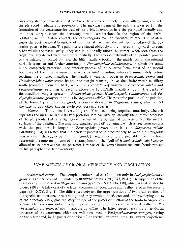
PACHYCEPHALOSAURIA NEW SUBORDER 79
rises very steeply upwards and it contacts the vomer anteriorly. Its maxillary wing contactsthe pterygoid medially and posteriorly. The maxillary wing of the palatine takes part in theformation of the ventroanterior wall of the orbit. It overlaps here the pterygoid laterally andits upper margin meets the accessory orbital ossifications. In the region of the infraorbital fossa the palatine contacts the ectopterygoid over an extensive surface. The palatineforms the posteromedial boundary of the internal naris and the anterior boundary of the posterior palatine fenestra. The palatines are placed obliquely and convergently upwards to eachother within the nasal cavity. They continue dorsally above the vomer, when seen from thefront, but they do not meet each other medially. The anterior extremity of the pointed processof the palatine is located opposite the fifth maxillary tooth, in the mid-length of the internalnaris . It seems to end further anteriorly in Homalocephale calathocercos, in which the snoutis not completely preserved. The anterior process of the palatine forms most of the medialboundary of the internal naris in Stegoceras validus, ending anteriorly immediately beforereaching the cojoined maxillae. The maxillary wing is broader in Prenocephale prenes andHomalocephale calathocercos, its anterior margin reaching above the sixth/seventh maxillarytooth (counting from the back) while it is comparatively narrow in Stegoceras validus andPachycephalosaurus grangeri, reaching above the fourth/fifth maxillary tooth. The depth ofthe maxillary wing is greater in Prenocephale prenes, Homalocephale calathocercos and Pachycephalosaurus grangeri, than it is in Stegoceras validus. The posterior portion of the palatine,at the boundary with the pterygoid, is concave dorsally in Stegoceras validus, which is notthe case in any other known pachycephalosaurid species.
Vomer. - The vomer is very long and Y-shaped, being unpaired anteriorly, where itseparates the maxillae, while its two posterior laminae overlap laterally the anterior processesof the pterygoids. Laterally the dorsal margins of the laminae of the vomer meet the medialsurfaces of the palatines. The anterior, unpaired part of the vomer, which is free from contactwith the palatines, is longer in Prenocephale prenes than it is in Stegoceras validusGILMORE (1924) suggested that the pendant process visible posteriorly between the pterygoidsmay represent the vomer or the presphenoid. It seems to us more probable that this bonerepresents the anterior portion of the parasphenoid. The skull of Homalocephale calathocercosallowed us to observe that the posterior laminae of the vomer bound the cultriformis processof the parasphenoid Iateroventrally.
SOME ASPECTS OF CRANIAL NEUROLOGY AND CIRCULATION
Endocranial cavity. - The complete endocranial cast is known only in Pachycephalosaurusgrangeri as described and illustrated by BROWN& SCHLAIKJER (1943, PI. 41).The upper half of thebrain cavity is preserved in Stegoceras validus (specimen NMC No. 138),which was described byLAMBE (1918). A latex cast of the latter specimen has been made and is illustrated in the presentpaper (PI. XXV, Fig. 1). The differences between the upper portions of two brain cavities ofthe specimens mentioned are striking, and they involve the shorter and the less sloping stalksof the olfactory lobes, plus the steeper slope of the posterior portion of the brain in Stegocerasvalidus. The cerebrum and cerebellum, as well as the optic lobes are separated neither in Pachycephalosaurus grangeri nor in Stegoceras validus. The latter species lacks the anterodorsalprocesses of the cerebrum, which are well developed in Pachycephalosaurus grangeri, havingon the other hand , in the posterior portion of the cerebellum several small backward projections:
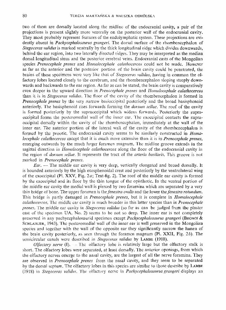
80 TE RESA MA RYANSKA & HALSZKA OSM6LSKA
two of them are dorsally located along the midl ine of the endocranial cavity, a pair of theprojections is present slightly more ventrally on the po sterior wall of the endocranial cavity.They most probably represent features of the endo lympha tic system . These projections are evidently absent in Pachycephalosaurus grangeri. The dorsal sur face of the rhombencephalon ofStegoceras validus is marked ventrally by the th ick longitudinal ridge which divides downwards,behind the ear region, into two laterally directed ridges. They may be interpreted as the mediandorsal longitudinal sinus and the posterio r cere bra l veins . Endo cranial casts of the Mongolianspecies Prenocephale prenes and Homalocephale calathocercos could not be made. Howeveras far as the anterior and the po sterior por tion of the brain cavity could be penetrated, thebrains of these specimens were very like that of S tegoceras validus, having in common the olfactory lobes located closely to the cerebrum , an d the rh ombencephalon sloping steeply downwards and backwards to the ear reg ion. As far as can be stated, the brain cavity is comparativelyeven deeper in the upward direction in Prenocephale prenes and Homalocephale calathocercosthan it is in Stegoceras validus. The floor of the cavi ty of the rh ombencephalon is formed inPrenocephale prenes by the very narrow basioccipita l posteri orly and the broad basisphenoidanteriorly. The basisphenoid rises forwards forming the dorsum sellae. The roof of the cavityis formed po steriorly by the supraoccipital which widens forwards. Posteriorly the supraoccipital forms the posteromedial wall of the inner ear. The exoccipita l contact s the supraoccipi ta l dorsally within the cavity of the rhombencephalon, imme diately at th e wall of theinner ear. The anterior porti on of the lateral wall of the cavity of the rhombencephalon isformed by the prootic. The endocra nial cavity seems to be similarly constructed in Homalocephale calathocercos except th at it is much more extensive than it is in Prenocephale prenes,emerging outwa rds by the much larger foramen magnum. The midline groove extends in thesagittal direction in Homalocephale calathocercos along the floor of the endocranial cavity inth e region of dorsum sellae. It represents the tract of the arteria basilaris. This groove is notmarked in Prenocephale prenes.
Ear. - The middle ear cavity is very deep, vertically elongated and broad dorsally. Itis bounded anteriorly by th e high otosphenoi da l crest and posteriorly by the ventrolateral wingof the exoccipital (Pl. XXV, Fi g. 2a; Text-fig. 2). The ro of of the middle ear cavity is formedby th e exoccipita l an d it s floor by the thin tongu e of the op istho tic. In the ventral portion ofthe middle ear cavity the medial wall is pierced by two foramina which are separa ted by a verythin brid ge of bone. The upper foramen is th e fenestra ovalis and the lower the fenestra rotundum.Thi s bridge is pa rtly damaged in Prenocephale prenes, bu t it is complete in Ho malocephale
, calathocercos. The middle ear cavity is much broader in this latter species than in Prenocephaleprenes. Th e middle ear cavity in Stegoceras validus (so far as can be judged from the plastercast of th e specimen UA. No. 2) seems to be not so deep. The inner ear is not completelypre served in any pachyceph alosaurid spec imen except Pa chycephalosaurus grangeri (BROWN &S CHLAIKJER, 1943). The po steromedial wall of the inner ear is well preserved in the Mongolianspecies and together with the wall of th e opposite ear th ey significantly narrow the lumen ofthe brain cavity po sterio rly, as seen through th e foramen magnum (PI. XXII, Fig. 2b). Thesemicircular canals were described in Stegoceras validus by LAMBE (1918).
Olfa ctory nerve (I). - The olfactory lob e is relatively large but th e olfactory stalk isshort . The olfactory lobes were separa ted, at least dorsally. The anterior openings, from whichthe olfactory nerve s emerge to the nasal cavity, are the largest of all the nerve foramina. Theyare observed in Prenocephale prenes from th e nasal cavity, and they seem to be separatedby the dorsal septum. Th e olfactory lob es in thi s species are similar to th ose describe by LAMBE
(1918) in Stegoceras validus. The olfa ctory nerve in Pachy cephalosaurus grangeri displays an
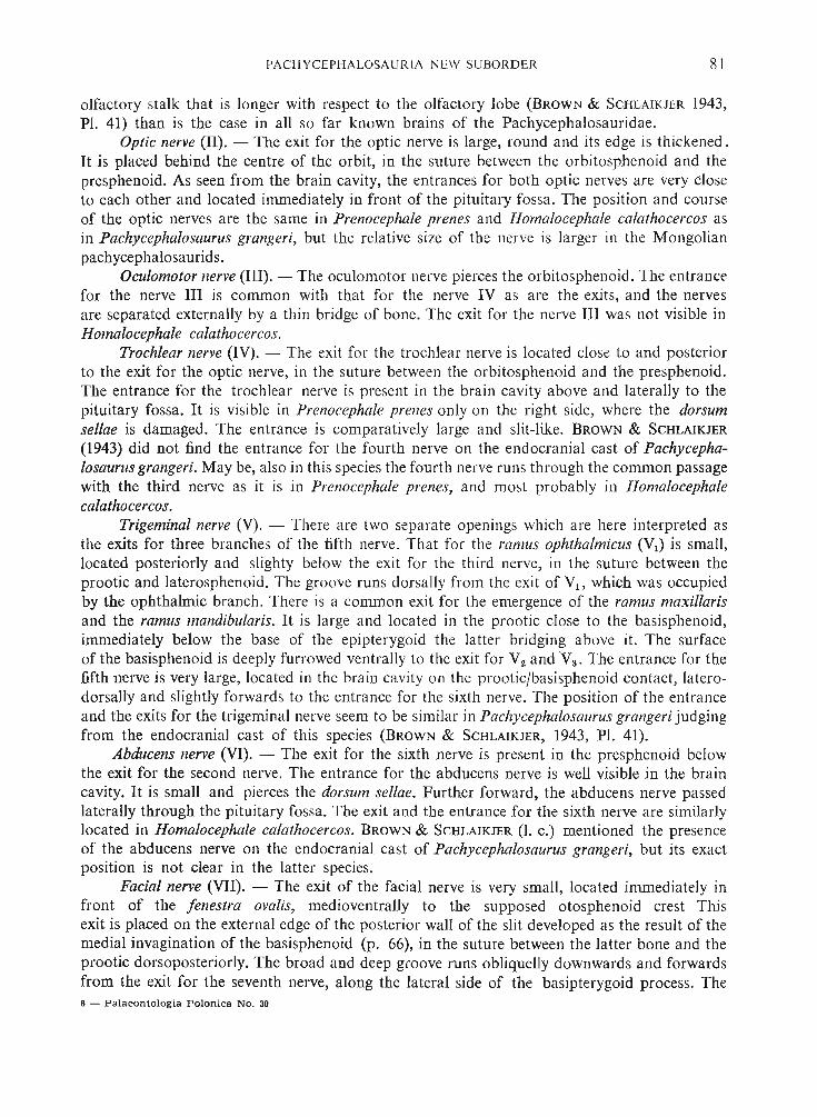
PACl-1YC EPHA LOSAU RIA NE W SUBORD ER 81
olfacto ry stalk that is longer with respect to the olfactory lobe (B ROWN & S CHLAIKJER 1943,Pi. 41) than is the case in all so far known brain s of the Pachyceph alo sauridae.
Optic nerve (Il). - The exit fo r the optic nerve is large, round and its edge is th ickened .It is placed behind the centre of the orbit, in the suture between the orbitosphenoid and thepresphenoid. As seen from the brain cavity, the entrances for bo th optic nerves are very closeto each other and located immediately in front of the pitu itary fossa. Th e po sition and courseof the optic nerves are the same in Prenocephale prenes and Homalocephale calathocercos asin Pachycephalosaurus grangeri, but the relative size of the nerve is larger in the Mongolianpachycephalosaurids.
Oculomotor nerve (Ill). - Th e oculomotor nerve pierces the orbitosphenoid. The entrancefor the nerve HI is common with that fo r th e nerve IV as are th e exits, and the nervesare separated externally by a thi n bridge of bone. Th e exit for th e nerve III was not visible inHomalocephale calathocercos.
Trochlear nerve (IV). - Th e exit for the trochlear nerve is located close to and po steriorto the exit for the optic nerve, in the suture between the or bitosphenoid and the pre sphenoid.The entrance for th e trochlear nerve is present in the brain cavity above and laterally to thepituitary fossa. It is visible in Prenocephale prenes only on the right side, where the dorsumsellae is damaged. The entrance is comparatively large and slit-like. BROWN & SCHLAIKJER
(1943) did not find the entrance for the fourth nerve on the endocranial cast of Pachycephalosaurus grangeri. May be, also in thi s species the fourth nerve run s th rough th e common pa ssagewith the third nerve as it is in Prenocephale prenes, an d most probably in Homalocephalecalathocercos.
Trigeminal nerve (V). - Th ere are two separate openings which ar e here interpreted asthe exits for three branch es of the fifth nerve. That for the ramus ophthalmicus (VI) is small,located po steriorly and slighty below the exit for the th ird nerve, in the 'suture between theprootic and laterosphenoid. Th e gro ove run s dorsally from the exit of VI ' which was occupiedby th e ophthalmic branch . Th ere is a common exit for the emergence of the ramus maxillarisand the ramus mandibularis. It is large and located in the prooti c close to the basispheno id,immediately below the base of the epipterygoid the latt er br idging above it. The sur faceof the basisphenoid is deeply furrowed ventra lly to the exit for V2 an d V 3 • Th e entrance for thefifth nerve is very large, located in the brain cavity on th e prooticJbasisphenoid contact , lat erodorsally and slightly forward s to the ent rance for the sixth nerve. The po sition of the entra nceand the exits for the trigemin al nerve seem to be similar in Pachycephalosaurus grangeri judgingfrom the endocranial cast of thi s species (BROWN & SCHLAIKJER, 1943, PI. 41).
Abducens nerve (VI). - Th e exit for the sixth nerve is present in the presphenoid belowthe exit for the second nerve. The entrance for the abducens nerve is well visible in the braincavity. It is small and pierces the dorsum sellae. Further forward, the abducens nerve pa ssedlaterally through the pituitary fossa. Th e exit and the entra nce for th e sixth nerve are similar lylocated in Homalocephale calathocercos. BROWN & S CHLAIKJER (1. c.) menti oned the presenceof the abducens nerve on th e endocranial cast of Pachycephalosaurus grangeri, but its exactposition is not clear in the latter species.
Facial nerve (VII). - Th e exit of the facia l nerve is very sma ll, located immediat ely infront of the fenestra ovalis, med ioventrally to the supposed otosphenoid crest Thisexit is placed on the external edge of the posterior wall of the slit developed as the result of themedial invagination of the basisphenoid (p. 66), in th e suture between the latter bone and theprootic dorsoposteriorly. Th e broad and deep groove run s obliquelly downwards and forwardsfrom the exit for the seventh nerve, along the lateral side of th e basipterygoid process. The6 - Palae ontologia Polonica NO. 30

82 TE RE SA MA R YANS KA & HALSZK A OSM6 LSK A
groove pre sumably transmitted the palatine ramus of the facial nerve. Further forward s, thepalatine ramus runs ventrally to the basipterygoid process to enter the interpterygoidal cavitytogether with its fellow. The entrance for the facial nerve within the brain cavity, isvery small and located immediately posterior and slightly ventrally to the entrance for thetrigeminal nerve. The exit and the entrance for the seventh nerve are in about the same positionin Prenocephale prenes and in Homalo cephale calathocercos, as is the entrance in Pachycephalosaurus grangeri.
Auditory nerve (VIII). - The entrance for the auditory ner ve cannot be easily observedin any specimen in our disposal, because it is hidden po steriorly by the internal wall of the oticcapsula , which here constricts the cranial cavity.
Glossopharyngeus nerve (IX). - The exit for the glossopharyngeus nerve is placed inthe exoccipital, lateral to the occipital condyle. It is located on the edge of a sharp crest whichis developed parallel to the ba sioccipital in the dorsal prolongation of the basal tuber. Thepassage for the nerve pierces the exoccipital and is separated from the middle ear by a thinbony wall. The entrance for the ninth nerve is located very close to the inner ear cavity, but itsprecise po sition cannot be determined, because of the damage of this portion of the brain cavity.The passage and the exit of the ninth nerve is comparatively large and it is possible that italso contained the jugular vein. The exit for the glossopharyngeus nerve is in the same positionin Homalocephale calathocercos but the po sition of the entrance cannot be determined. B ROW N &SCHLAIKJER (1943) suggested a common exit for nerves IX-Xl in Pachycephalosaurus grangeri.The illustration of the endocranial cast given by the se authors (l . c., PI. 41) does not allow toobserve the position of these nerve s.
Vagus and accessory nerves (X-Xl). - There is a narrow slit present in the suture between the exoccipital and basioccipital placed laterally to the neck-like portion of the occipitalcondyle which locates a common exit for the vagus and accessory nerves. Two grooves leadfrom the exit of the nerves, across the ba sioccipital. One of them, directed ventrally is slightlybroader than the other which is directed anteriorly. Th e common entrance for these two nerve sis located in the exoccipital immediately above the suture with the basioccipital. The exit forthe vagus and accessory nerve s is also common and slit-like in Homalocephale calathocercosand Stegoceras validus. The slit is placed slightly more vertically in the se two species than itis in Prenocephale prenes.
Hypoglossal nerve (XII). - The exit for the hypoglossal nerve is large, funnel-like andit pierces the exoccipital. It is located on the lateral side of the condyle, posterodorsally to theexit for the tenth and eleventh nerve s. Two grooves lead from the exit , one of which is deeper,broader and is directed ventrally, the other-slightly anteriorly. The entrance is well visible withinthe cavity of the medulla oblonga, and is located laterodorsally. The position of this nerve inother known pachycephalosaurid skull s is about the same as it is in Prenocephale prenes.
In addition to the cranial vessels described above and connected with the brain, there areseveral openings on the lateral walls of the brain case and in the orbitonasal region inPrenocephale prenes, which should be interpreted as locating the arteries and veins.
Internal carotid artery. - The entrance for the Vidian canal is located at the very bottomof the invaginated portion of the basisphenoid , dorsal to the basipterygoid processand anteroventral to the exit for the seventh nerve (Text-fig. 2). Within the passage in the basisphenoid posteriorly to the pituitary fossa, the internal carotis gives outwards the palatineartery which emerges through the basisphenoid ventral to the exit for the sixth nerve. Twogrooves extend from the exit for the palatine artery, one of which is directed vent rally and theother po steriorly. They might contain the branches of the palatine artery. The septum between

PACHYC EPHAL OSA URI A NE W SU BO R D ER 83
the orbit and the na sal cavity is pierced by several foramina which resemble the relationsknown in mammals (Text-fig. 3). Two of these foramina , sugge sted here as representing theforamina for the ethmoidal artery, are located between acce ssory orbital ossifications 1 and 2.Dorsally, their exits into the nasal cavity are placed immediately at the midline, and on the sutu rebetween these bones mentioned . Two other foramina are placed more vent rally. One of thempierces the acce ssory ossification 1 at the suture between th e palatine and jugal and emergesinto the nasal cavity laterodorsally, immediately above the palatine. It may represent the sphenopalatine foramen for the entrance of the sphenopalatal artery . The second foramen is locatedslightly above the tongue of the pterygoid which underlies the palatine. It is bounded dorsallyby several small ossifications of the orbital region. Its exit is invi sible because it is located moremedially, and is completely hidden by the palatal wing of the pterygoid. The foramen leadingto the intramaxillary sinus is located in the maxillary portion of th e orbit within the fossa pte rygopalatina and it could conduct forwards th e infraorbital artery as well as the infraorbitalbranch of V2 • In the bottom of the infratemporal fo ssa two foramina are pre sent which areseparated by the broad bridge of the prootic that contacts laterally the quadrate. A distinctgroove leads from the lower of these two foramin a forward s and downwards, under the epip terygoid , and towards the exit of V2 and Va nerves. A broad and shallow recessus is presenton the bottom of the infratemporal fossa which is confluent with the upper of the foraminamentioned above. Both foramina communicate po steriorly with two foramina located belowthe paroccipital process, in the slit between the prootic-opisthotic and quadrate. These structuresmost probably should be also interpreted as the pas sages for the blood vessels. The numerousvascular foramina penetrate the bones of the orbital roof and they are especially aboundanton the frontal.
OSTEOLOG Y OF THE POSTCRANIAL SKELETON(PI. xxv, Fig. 3, PIs XXVI-XXX I; Text-figs 4, 5)
The pachycephalosaurid po stcranial skeleton was until recently kn own only in Stegocerasvalidus LAMBE, 1902, and although being incomplete, it has provided much important information, particularly that dealing with the bipedal po sture of th e animal. The discovery ofanother incomplete postcranial skeleton of Homalo cephale calathocercos n.gen .et sp . (Pi sXXVI-XXX, PI. XXXI, Fig. I ; Text-fig s 4, 5) with mo st of the pre served bones in the articulated conditions as well as some postcranial fragments of Prenocephale prenes n.gen.et sp.(PI. XXV, Fig. 3, PI. XXXI, Fig. 2) allowed us to complete th e data given by GILMORE (1924).It enabled us also to conclude, that the essential characters of the po stcranial skeleton wereprobably very similar in all representatives of the family. The description below, is based onthe postcranial skeleton of Homalo cephale calathocercos, but we ha ve tri ed , as far as it waspossible, to compare the bones of the latter species with the po stcranial elements of Prenocephaleprenes and with those described by GILMORE (I. c.) in Stegoceras validus. The rea son s forbasing the osteological description on the Mongolian species Homalocephale calathocercosis not its completeness, which is generally lesser than in Stegoceras validus, but the fact thatthe preserved portion of the vertebral column is complete, and was reco verd in the naturalarrangment, with the rib s, pelvic girdle and the tail-basket articulated.
Vertebral column. (PIs XXIV-XXVIII; Text-fig. 4). - The preserved part of the vertebral column shows its natural curvature. It rises slightly backwards along the first three presacral dorsals preserved, and then it slo pes towards the last dorsal and th e sacrum, which are6*

84 TERESA MAR YANSKA & l lAL SZKA OSM6LSKA
A
. .~
Fig. 4Homalocephale calathocercos n, gen., n. sp. (G. J. No . SPS l OO/51). Presacral and caudal vertebrae : A dorsal n +4 (seventhcounted from the sacrum) with dorsal ribs att ached, ante rior view, B dorsal n-l-B (third counted from the sacrum) withdorsal ribs attached, anterior view, C dorsal n + 9 (second counted from the sacrum) with dorsal ribs attached, posteriorview, D caudal 4, anterior view, E caudal 5, posterior view, F I caudal 6, anter ior view, F. same caudal, posterior view.
horizontal. Immediately behind the sacrum, the vertebral column slopes relatively steeplydownwards, beginning with the first caudal, and the lateral profile of the vertebral columnbecomes still more steep, along the series between caudal 6 and caudal 13; more posteriorlythe tail is horizontally placed. The dorsal vertebrae are amphiplatyan (Pl, XXVI, Fig. 1;Text-fig. 4A-C). Their centra distinctly increase in size posteriorly. The centra of the first threevertebrae of the preserved series are slightly compressed laterally and have a median keelalong their ventral surfaces. The centra are concave ventrally in lateral view. The successiveposterior dorsal s are devoid of the ventral keel and are more deeply concave in lateral profil e.The sutures between the centra and the neural arches level with the neural canal in thefirst three preserved dorsals, and are slightly lower in the successive dorsals. The neural archis about twice as high as the centrum in the th ree anterior dor sals of the preserved series and itis about one and a half times as high in the posterior dorsals. The diapophyses on these moreanterior dor sals are directed upwards at an angle which is less than 45° to the horizontal levelof the neural canal. Thi s angle increases posteriorly so the diapophyses of the posterior dorsals(except the last one) are very steep. The diapophysis in the last dorsal is nearly horizontal.The diapophyses become shorter and broader toward s the sacrum. They bear two articularsurfaces for the ribs. The part of the diapophysis which contacts the tuberculum becomes short-
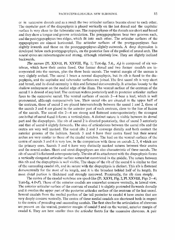
PACHY CEPHALOSAURI A NEW SUBORDER 85
er in successive dorsals and as a result the two articular surfaces become closer to each other.The capitular part of the diapophysis is placed vertically on the last dorsal and the capitularsurface is very close to the tubercular one. The zygapophyses ofthe dorsals are short and broadand they show a tongue and groove articulation. The prezygapophyses bear two grooves each,and the postzygapophyses two ridges, which fit into each other. The articular surfaces of thezygapophyses are almost horizontal. The articular surfaces of the prezygapophyses faceslightly inwards and those on the postzygapophyses slightly outwards. A deep depression isdeveloped below each postzygapophysis, on the posterior face of the pedicel of neural arch. Theneural spines are subrectangular and strong, although relatively low. They are slightly inclinedbackwards.
The sacrum (PI. XXVlI, PI. XXVIII, Fig. 1; Text-fig. 5AI> A 2) is composed of six vertebrae, which have their centra fused. One former dorsal and two former caudals are incorporated into the sacrum, plus the three basic sacrals. The ventral margin of the sacrum isvery slightly arched. The sacral I bears a normal diapophysis, but its rib is fused to the diapophysis, and the capitular and tubercular surfaces are joined. The first sacral rib is very shortand broad, and its distal extremity is thin and flattened dorsoventrally. It attaches loosely to theshallow embayment on the medial edge of the ilium. The ventral surface of the centrum of thesacral 1 is devoid of any keel. The centrum widens posteriorIy and its posterior articular surfacefuses to the successive sacral. The ventral surfaces of sacrals 2-4 bear a keel, which is wellpronounced, although comparatively Iow. Their sacral ribs are situated in the upper half ofthe centrum, those of sacral 2 are placed intervertebrally between the sacral 1 and 2, those ofthe sacrals 3 and 4 are placed in the anterior part of each centrum, close to the line of fusionof the sacrals. The sacral ribs 2-4 are strong and flattened anteroposteriorly; the strongestone is that of sacral 4 and it forms a vertical plate. A distinct suture is visible between its dorsalpart and the diapophysis. The rib of sacral 2 is directed posteriorly, that of sacral 3 anteriorlyand that of sacral 4 slightly forwards. The sites of articulations between the sacral ribs and thecentra are very well marked. The sacral ribs 2 and 3 converge distally and both contact theanterior process of the ischium. Sacrals 5 and 6 have their centra fused but their neuralarches are very similar to those of the caudal vertebra. The keel on the ventral surfaces of thecentra of sacrals 5 and 6 is very Iow, in the comparison with those on sacrals 2, 3,4 which arethe primary ones. Sacrals 5 and 6 have very distinctly marked sutures between their centraand the neural arches. Short and stout diapophyses are also characteristic of these sacrals. Therib of sacral 5 is flattened anteroposteriorly. The site of its attachment with the diapophysi s formsa vertically elongated art icular surface somewhat constricted in the middle. The suture betweenthis rib and the diapophysis is well visible. The shape of the rib of the sacral 6 is similar to thatof the succeeding caudal rib, and its suture with the diapophysis is distinct. This rib is flatteneddorsoventrally for the most of its length, and it is the broadest behind half of its length. Itsmost distal portion is thickened and strongly narrowed. Proximally, the rib rises steeply.
The centra of the caudal vertebrae are spool-like (pI. XXVI, Fig. 2, PI. XXVIII, Figs 2-4;Text-fig. 4 D-F). Those of the anterior caudals are somewhat concave ventrally, in lateral view.The anterior articular surface of the centrum of caudal 1 is slightly protruded forward s dorsallyand it overlies the upper part of the posterior articular surface of the centrum of the last sacral.Several caudals from the medial portion of the tail posterior to caudal 4 have centra that are
, very deeply concave ventrally. The centra of these medial caudals are shortened both in respectto the centra of preceding and succeeding caudals, The first sites for the articulation of chevronsare present on the ventral, posterior margin of caudal 5 and on the ventral, anterior margin ofcaudal 6. They are here smaller than the articular facets for the successive chevrons. A pair

86 TERESA MAR YANSKA & HALSZKA OSM6LSKA
of longitudinal ridges appears on the ventral surface of the each chevron-bearing caudal, andthe ventral face of the centrum, between these ridges, is flat. The posterior articular surfaceof the centrum of caudal 6 is more vertically placed than the others. The chevrons were stillpresent on caudal 29, judging from the presence of the articular facets on the ventral marginsof the centra. The transition point (RUSSELL, 1972) in the caudal series is present on caudal 9.Behind it, each caudal bears a ridge along the side of its centrum. These ridges become longeron the successive centra. The cross-section of the centra changes posteriorly along the entirecaudal series from being round to hexagonal, which is caused by the appearance of the lateraland ventral ridges along the centra. The zygapophyses are relatively short. The caudal prezygapophyses are much longer than the postzygapophyses. The prezygapophyses on the anterior caudals have their articular surfaces concave and directed dorsomedially. They becomegradually flatter and face more medially on the successive caudals, and at about caudal 10 theyface entirely medially. The eight anterior caudaIs bear the caudal ribs, and the distinct suturesare visible where the ribs contact the vertebrae. The first four ribs are placed on the boundarybetween the centrum and the neural arch, more posteriorly their attachment is lower andthe two last caudal ribs are exclusively on the sides of the centra. The ribs on the most anteriorcaudals are very long. The rib of the first caudal differs from the successive ones in shape. Thedistal end of this rib is thickened and it has an articular surface which loosely contacts the uppermargin of the ilium on the very end of the latter. The shaft of the rib is flattened, being thebroadest close to the distal end. This flattening diminishes on the three successive caudal ribsand the last ones are more rod-like. The arching of the caudal ribs changes along the caudalseries. The first four ribs are more strongly arched and they rise upwards along their proximalhalves; they become nearly straight and directed downwards, beginning with the caudal 5;the last, eight caudal rib is the shortest and is developed as a short process; there is only a tubercle developed, instead of the rib, on the caudal 9. The neural spines in the caudal vertebraeare very long. The first four spines are broader than the others. They are very slightly inclinedbackwards in the proximal vertebrae and the inclination becomes stronger in the more posteriorcaudals.
The portion of the vertebral column preserved in Homalocephale calathocercos is thus farthe most complete in the family Pachycephalosauridae. GILMORE (1924) had at his disposalbut the two dorsals and the eight caudals from the different parts of the tail, in the specimen ofStegoceras validus. He suggested that the vertebra illustrated by him (l. c. PI. 12, Figs 8, 9)represents the medial dorsal, because of the strong upward direction of its diapophyses. This,however, should not be necessary true, because the preserved portion of the presacral vertebralcolumn shows in Homalocephale calathocercos that the diapophyses on the several penultimatedorsals display the most upwards direction. The diapophyses are similarly directed as thesein Homalocephale calathocercos, in Thescelosaurus edmontonensis STERNBERG 1940, although,it seems that the relation is reversed in Thescelosaurus neglectus where the more posterior dorsaldiapophyses are less steep than those anterior ones. Two sacral bones are present in the materialrecovered in Mongolia and that of the above described species, Homalocephale calathocercos,is more complete. The sacrum and the sacral ribs of another Mongolian species, Prenocephaleprenes, do not show any important differences, except for being, perhaps, slightly more slender.The caudal vertebra illustrated by GILMORE (I. c., PI. 12, Fig. 2) in Stegoceras validus, wasconsidered by him as the third one. It exposes the presence of the articular facet for the contactwith chevron, on the lower posterior margin of the centrum. The first articular facet for thechevron in Homalocephale calathocercos is present on the caudal 5. Thus, the caudal in question,

PACH YCEPH ALOSAURI A NEW SUBORDE R 87
illustrated by GILMORE (I. c.) more probably repre sents the caudal 5, becau se also the centraof the two vertebra here compared have the similar shape.
Sternum (PI. XXX, Fig. I). - Both sternal plates are not fused medially. The sternumis 74.5 mm long and has its posterior, shaft-like process directed outwards and thickened distally.It broadens and thins medially. The lateral margin of the expanded portion is thickened, especially at the proximal end , where it exposes a rough surface. The medial margin of the expanded portion sends the hook-like process, close to the shaft. The process is directed posteriorly.The sternaI plates of Homalocephale calathocercos described above were for the first time recovered within the Pachycephalosauridae. They resemble very closely in the shape the sternalplate s of Anatosaurus annectens (MARSH, 1892) and are one more evidence for the bipedalityof the pachycephalosaurids.
Ribs. - Nine posterior dorsal ribs are preserved in Homalocephale calathocercos 4.
They rise slightly outwards in the proximal portion , and are gently arched more distally, except the last dorsal rib , which is more strongly vaulted than the others. The ribs become broadermore posteriorly. The fifth and the tenth rib of the preserved presacral series are complete. Theyare correspondingly 83 mm and 73 mm long across the curve and 142 mm and 117 mm longalong the curve when measured from the capitulum. Both the capitulum and the tuberculumarticulate with the diapophysis, but they are distinctly separated. They are closer to each otherin the more posterior ribs. The penultimate dorsal rib has the capitulum and the tuberculumstill well separated, but the last dorsal rib has the articular facet s separated only when the ribis seen ventroposteriorly. The tuberculum is placed outwards and posteriorly with respect to thecapitulum on all the ribs, the last one excluding. Here, the two facet s are nearly equidistantfrom the median line of the body, and the tuberculum is placed abo ve the capitulum. The dorsalribs are directed slightly po sterolaterally but the last rib has its long axis about normal to theline of the vertebral column. The more anterior ribs of the pre served series are narrower thanthe posterior ones. The last rib, which contacts the ilium freely by its medial portion, is herethe flatte st and the broadest, while its distal extremity is rounded in cross-section . The distalends of the more anterior ribs are oval in cross-section. Only five fragmentary dorsal ribs arepreserved in Prenocephale prenes, and as far as can be stated, they do not differ from tho se inHomalocephale calathocercos. The same seems be true for Stegocercas validus.
Pelvic girdle (PI. XXV. Fig. 3, PI. XXIX, PI. XXX, Fig. 2 ; Text-fig. 5A , B). - The pelvicgirdle is broad and widens posteriorly. The ilium has its anterior portion dorsoventrally flattened .The posterior portion is developed in the form of a vert ical blade and it bears dorsally a flat ,subtriangular inward extension , immediately behind the acetabulum. There is no antitrochanterdeveloped. A shallow embayment for the reception of the distal end of the first sacral rib ispresent on the medial edge of the anterior portion of ilium, close to its anterior extremity. Thepubic peduncle is exceptionally wide and flattened dorsoventrally. It is directed forwards anddownwards and bears the narrow, transversely elongated articular surface for the prepubis.The medial corner of the pubic peduncle contacts the ischium. The ischiac peduncle is very heavy,thick and expanded tran sverselly. It is long, extending below the ventral margin of the posteriorportion of the ilium. Laterally it exhibits a strong, rough protuberance above the articular facetfor the ischium. In posterior view, the ischiac peduncle is a strong, transverse plate. It continuesupwards and merges dorsally with the anteromedial border of the inward extension of ilium.The dorsal acetabular rim is very broad transversely. A vertically elongated depression is present
• For the description of sacral and caudal ribs see p. 85.
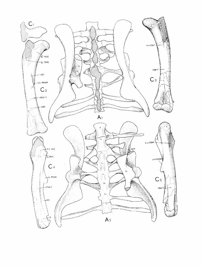
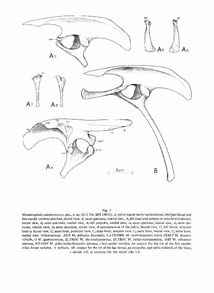
As "
pp
Fig. 5Homalocephale calathocercosn. gen., n. sp. (G. 1. No. SPS lOO/51).Al pelvicregion partlyreconstructed.with'last'dorsal andfirst caudal vetebrae attached, dorsal view, As same specimen, ventral view, A. left ilium and ischium in natural articulation,lateral view, A, same specimen, medial view, As left prepubis, medial view, As same specimen, lateral view, A7 same specimen, ventral view, As same specimen, dorsal view, B reconstruction of the pelvis, lateral view, Cl left femur, articularhead in dorsal view, C. same bone, posterior view, C3 same bone, anterior view, C, same bone, lateral view, C. same bone,medial view. Abbreviations: ADD M. adductor femorali s, CA-FEMBR M. caudi-femoralis brevis FEM-T M. femoro-tibialis, G M. gastrocnemius, IL-TROC M. iIio-trochantericus, IS-TROC M. ischio-trochantericus, osr M. obturaturinternus, P-I-FINT M. pubo-ischio-femoralis internus , c first caudal vertebra, I er contact for the rib of the first cauda l,d last dorsal vertebra, is ischium, Idr contact for the rib of the last dorsal, pp postpubi s, ppd pubic peduncle of the ilium,
s sacrals 1-6, sr contact s for the sacral ribs 1-6.
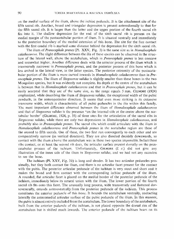
90 TE RESA MA R YANSKA & HALSZK A OSM 6LSKA
on the medial surface of the ilium , above the ischiac peduncle. It is the attachment site of thefifth sacral rib. Another, broad and triangular depression is pre sent anterodorsally to that forthe fifth sacral rib. It is larger than the latter and the upper portion of the fourth sacral ribfits into it. The shallow depression for the end of the sixth sacral rib is pre sent on themedial margin of the postacetabular po rti on of ilium. It is situated ventrally and immediatelyto the po sterior boundary of the medial extension of thi s bone . The site for the free contactwith the first caudal rib is marked some distanc e behind the depression for the sixth sacral rib .
The ilium of Prenocephale prenes (PI. XXV, Fig. 3) is the same size as in Homalocephalecalathocercos. The slight difference between the ilia of these species can be observed in the structure of the lateral wall, above the acetabulum, which in Prenocephale prenes is less concaveand somewhat higher. Another difference deals with the anterior process of the ilium which istransversely narrower in Prenocephale prenes, and the po sterior process of thi s bone which isless arched in the lateral view, in the latter species. The po sterior extremit y of the po stacetabular portion of the ilium is mo re curve d inwards in Homalocephale calathocercos than in Prenocephale prenes. The ilium of Stegoceras validus is slightly smaller than these bones in the twoMongolian species, but it was evidently not complete. It s depth at the centre of the acetabulumis between that in Homalo cephale calathocercos and that in Prenocephale prenes, but it can beeas ily accepted that they are of the same size, as the ra nge equals 5 mm . GILMORE (1924)emphasized, while describing the ilium of Stegoceras validus, the except ional width of the pubi cpenducle, in the anteroposterior direction . It seems that even more striking is its extensivetransverse width, which is characteristic of all pubi c peduncles in the ilia within thi s family.The most important difference observed between the ilium of Homalocephale calathocercosand that of Stegoceras validus is the presence "o n the intern al side at the center above the acetabular border" (GILMORE, 1924, p. 35) of three sites for the articulation of the sacral ribs inStegoceras validus, while there are only two depressions in Hom alocephale calathocercos, andprobably also in Prenocephale prenes. Th e sacral ribs which could ar t iculate with the ilium inHomalocephale calathocercos and Prenocephale prenes in the acetabular region are the se ofthe second to fifth sacrals. Out of these, the two first run convergently to each other and arecomparatively narrow (in vertica l direction). They are also directed decidely downwards, socontact with the ilium a bove the aceta bulum was in these two species impossible. In fact theseribs contact, or at least the second rib does, the ar ticular surface present dorsally on the preacetabular process of the ischium. Unfortunately, G ILMORE (I. c.) did not give anyillustration of the inner side of the ilium in Stegoceras validus, and we had not any occas ionto see the bone.
The ischium (PI. XXV, Fig. 3b) is long and slender. It has two articular peduncles proximally, but they both contact the ilium, and there is no articular facet present for the contactwith the pubis. Th e posterior articular peduncle of the ischium is very stout and sho rt and itmak es the broad and firm contact with the correspo nding ischiac peduncle of the ilium .A rounded, flat articular facet is placed on the medial bo rder of the posterior peduncle of theischium, immediately below its su tural union with the ilium. Th e lower portion of the fourthsacral rib fits onto thi s facet. Th e unusually long process, wide transversely and flattened dorsoventrally, extends anterovent rally fro m the posterior peduncle of the ischium. This processconstitutes the anter ior peduncle of thi s bone. It bounds the acetabulum ventrally, contactinglaterally the anteromedial articular surface of the pubic peduncle of the ilium. In thi s manner,the pubis is almost entirely excluded from the acetabulum. Th e lower boundary of the acetabulum,built from the anterior peduncle of the ischium, is not placed opposite the dorsal rim of theaceta bulum but is shifted much inwards. The anter ior pedu ncle of the ischium bear s on its

PACHYCEPHALOSAURIA NE W SUBORDER 91
anterior extremity, a large rounded articular facet , which face s slightly medially. It makes thecontact with the distal end of the second, and most probably also with the third sacral ribs.The lateroventral sur face of the anterior peduncle of the ischium is flat and its lateral margin,below the anterior portion of the acetabulum, is sharp. Th e shaft of the ischium is very slenderand it broadens slightly distall y. It s lateroventral face is flat. Th e shaft is slightly bent inwardtowards its fellow.
GILMORE (1924), while describing the ischium of Stegoceras validus noticed that the anteriorpeduncle is long and that its extremity is opposite the pubic peduncle of the ilium. He did notob serve the se two bones in the articulated po sit ion , as we did , and moreover , he did not havethe pubis in the material at his dispo sal. Our mater ial allowed us to state that the unusual lengthof the anterior peduncle of isch ium is connected with the closure of the acetabulum by meansof the ischium and the ilium exclusively. It may serve as the evidence that the same structureof the pelvic girdle was th e case in S tegoceras validus. Wh en the ischia of Stegoceras vatidus,Homalocephale calathocercos and Prenocephale pr enes are compared, the slight difference isobserved in the curvature of their sha fts . The portion of the ischium preserved in Homalocephalecalathocercos shows th at the shaft was less curve d inwards in thi s species.
The prepubis (PI. XXIX, Fig. 2; Text-fig. 5A 5 _ S) is flattened dorsoventrally inits proximal portion, and laterally in the distal one. It s a rt icular sur face, which contacted thepubic peduncle of the ilium, is damaged, as well as the pubis (or po stpubis). The dorsal marginof the prepubis is slightly rised up distally. The very distal extremity is thickened and rough.The base of a thin, broken off process, which corresponded to the pubis, is proximally presentat the medial margin of the prepubis. This is the only pubi s thus far recovered in thi s family.
H ind limb (PI. XXX, Figs. 3- 6, Pl. XXXI; Text-fig. 5e). - The femur isrecurved inwards. Th e articular head is slightly compressed anteroposteriorl y and its articularsurface faces slightly forwards. It is separated from the anterior face of the shaft by a sharpridge, and from the greater trochanter dorsally by a deep groove. The fourth trochanter is moderately developed and faintly pendant. It lies enti rely in the proximal half of the femur. Thedistal end ha s two condyles separated by a sha llow intercondylar groove. Th e lateral condyleis stronger than the medi al one. Along the po sterolateral face of the femur extends the relativelysharp ridge, which disappears towards the proximal end, on the level of the fourth trochanter.The smooth, flat surface is pre sent on the posterior face of the sha ft. It extends downwardsand laterally to the fourth trochanter , and is sepa rated from the anterior face of the shaft bythe sha rp ridge. Th e furrow extends upwards, on the lateral sur face of the proximal end, abovethe fourth trochanter. The femur of Prenocephale pr enes (PI. XXXI, Fig. 2) is slightly morerecurved inwards than thi s in Homalocephale calathocercos (PI. XXXI, Fig. 1). It s articularhead is still less distinctly distinguished from the medial and anterior faces of the shaft. Thearticular head is slightly longer and more slender in Prenocephale pr enes, when viewed from thetop. Th e greater and the lesser trochanters are lacking in both Mongolian species, but theirbase s are pre served. Th ese latter are much thicker in Prenocephale prenes than in Homalocephalecalathocercos, and con sequently the trochanters should be stronger in the species first mentioned .The femur of Stegoceras validus was damaged and its description and illustr ation (GILMORE,
1924) are insufficient for the detailed comparison s.The pre served distal half of the tibia in Homalocephale calathocercos (PI. XXX, Figs. 3, 4)
shows th at its shaft was slender and the distal pa rt strongly broadened lateromedially. Theinternal margin of the distal end is sharp and the po steromedial face of thi s bone is flat behindthi s margin. The lateral condyle is the more prominent of the two . The flattened surface forco ntact with the distal end of the fibula is present on the an terior face of the tibi a , immediately
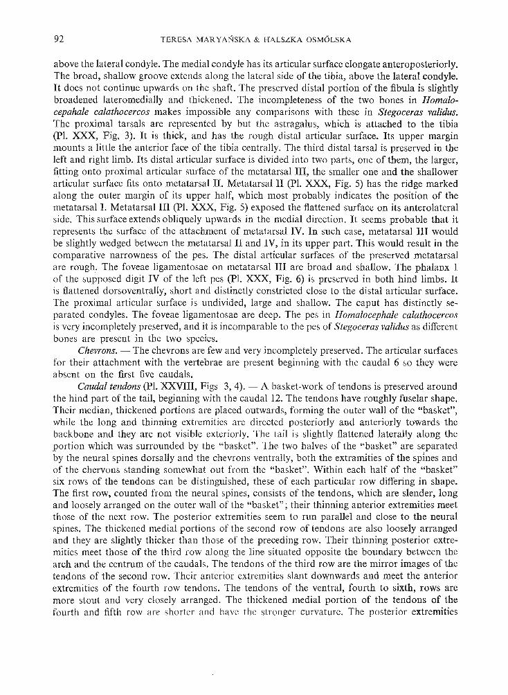
92 TERESA MARYANSKA & HALSZKA OSM6LSKA
above the lateral condyle. The medial condyle has its articular surface elongate anteroposteriorly,The broad, sha llow gro ove extends along the lateral side of the tibia, above the lateral condyle.It doe s not continue upwards on the shaft. The preserved distal portion of the fibula is slightlybroadened lat eromedially and thickened . The incompleteness of the two bones in Homalocepahale calathocercos makes impossible any comparisons with these in Stegoceras validus,Th e proximal tarsals are represented by but the astragalus, which is attached to the tibi a(pI. XXX, F ig, 3). It is th ick, an d has the rough distal articular surface. Its upper marginmounts a little th e anterior face of the tibia centrally. The third distal tarsal is preserved in th eleft and right limb. Its distal articular surface is divided into two parts, one of them, the larger,fitting onto pr oximal articular surface of the metatarsal Ill, the smaller one and th e shallowerarticular sur face fits on to metatarsal n. Metatarsal 11 (PI. XXX, Fig. 5) has the ridge markedalong th e outer margin of its upper ha lf, which mo st probably indicates the position of th emetatarsal I. Metatarsal 111 (PI. XXX, Fig. 5) exposed the flattened surface on its anter ola teralside. This surface extends obliquely upwards in the medial direction . It seems probable th at itrep resents the surface of th e attachment of metatarsal IV. In such case, metatarsal III wouldbe slightly wedged between the metatarsal II and IV, in its upper part. This would res ult in thecomparative narrowness of the pes. The distal articular surfaces of the preserved metatarsalare rough. The foveae ligamentosae on metatarsal III are broad and shallow. The phalanx Iof the supposed digit IV of the left pes (PI. XXX, Fig. 6) is preserved in both hind limbs. Itis flattened dorsoventrally, short and distinctly constricted close to the dista l articular surface.The proximal art icular surface is undivided, large and shallow. The caput has distinctly separated condyles. The foveae ligamentosae are deep. Th e pes in Homalocephale calathocercosis very incompletely preserved, and it is incomparable to th e pes of Stegoceras validus as differentbones are present in the two species.
Chevrons. - The chevrons are few and very incompletely preserved . The articular surfacesfor their attachment with the vertebrae are present beginning with the caudal 6 so they wereabsent on the first five caudals.
Caudal tendons (PI. XXVIII, Figs 3, 4). - A basket-work of ten dons is preserved aroundthe hind part of the tail, beginning with the caudal 12. The tendons have roughly fuselar shape.Their median, thickened portions are placed outwards, forming th e outer wall of the "basket",while the long and thinning extremities are directed posteriorly and anteriorly towards thebackbone an d they are not visible exteriorly. Th e tail is slightly flattened la terally alo ng th eportion which was surrounded by the "bas ket" . The two halves of the "basket" are separatedby the neural spines dorsally and the chevrons ventra lly, both the extramities of the spines andof the chervons standing somewha t out from th e "basket" . Within each half of the "basket"six rows of the tendons can be distinguished, th ese of each particular row differing in shape.The first row, counted from the neural spines, consists of the tendons, which are slender, longand loo sely arra nged on the outer wall of the "basket"; their thinning anterior extremities meetthose of the next row. The po sterior extremities seem to run parallel and close to the neuralspines. The thickened med ial portions of the second row of tendons are also loosely arrangedand they are slightly thicker than those of the preceding row. Their thinning posterior extremities meet those of the third row along th e line situated opposite the boundary between thearch and th e centru m of the caudals. The tendons of the third row are the mirror images of th etendons of the second row. Their anterior extremities slant downwards and meet the anteriorextremities of the fourth row tendons. The tendons of the ventral, fourth to sixth, rows aremore stout and very closely arranged. Th e thickened medial portion of th e tendons of th efourth and fifth row are shorter and have the stronger curvature. Th e posterior extremities

PACHYCEPHALOSAURIA NEW SUBORDER 93
of the fourth row of tendons approach the tendons of the more ventral row, opposite the medialline of the thickened portions of the latter, and they are directed more inwards towards the back.The tendons of the fifth row are the most strongly curved inwards. Their anterior, thinningextremities meet those of the next row while the posterior extremities rise slightly and approachthe median portions of the tendons of the more dorsal row. The last, sixth row of tendons isplaced entirely ventrally and it flanks the chevrons. Its tendons are very stout, their thickened, medial portions being comparatively long and closely attached to each other. The posteriorextremities of the sixth row of tendons are directed inwards and run parallel to the sides ofthe chevrons. It seems, judging from the preserved fragments, that the tendons within eachrow become more slender towards the end of the tail. One can noticed on cross-section of thetail, that the thin extremities of all tendons , in all the rows, are arranged parallel to the backbone. Sometimes the extremities of the tendons , especially these anterior ones of the first and second row, are fused together. Similar, ossified were recovered together with the postcranialskeleton of Prenocephale prenes, although mostly they were not in their original arrangement.As the specimens of Homalocephale calathocercos and Prenocephale prenes here describedhave proved, the elements described by GILMORE (1924, p. 31) in Stegoceras validus as theabdominal ribs, represent in fact the caudal tendons. On the other hand, the ossifications described by this author (1. c.) as the tendons are but the thinned extremities of the tendons.The caudal tendons in the three species mentioned, where they were recovered, do not differ,and, most probably, the presence of the "basket" around the major part of the tail is characteristic of the family Pachycephalosauridae.
REMARKS ON MYOLOGY
Muscle scars are visible on most of the bones and especially on the skull. We do notprovide the present paper with any reconstruction of the musculature, but interpretations ofthe attachment sites are here presented. In the interpretation of the muscle attachments onthe head we mainly followed OELRICH (1956) and OSTROM (1961), while our interpretationsof the muscle scars of the pelvic region and the hind limbs are based on ROMER (1927) andGALTON (1969).
Mandibular musculature. - Several scars corresponding to the origins of the mandibularadductors can be distinguished on the skull of the pachycephalosaurids. Their position doesnot deviate from that found in other ornithischian dinosaurs i.e, in the hadrosaurs (OSTROM,1961) except perhap s, that they are generally placed more posteriorly in relation to their insertions on the mandible than in the latter group, in which they slightly resemble the conditionsin Protoceratops (RASS, 1955; OSTROM, 1964, 1966). Internal adductors. The attachment of theM. pterygoideus dorsalis is visible on the dorsal surface of the ectopterygoid and partly on theventral wing of the pterygoid. The attachment of the M. pterygoideus ventralis is visible on thedorsal surface of the ventral wing of the pterygoid along its posterior margin. The attachmentof the M. pseudotemporalis is present within the infratemporal fossa, on the ventral surface ofthe postorbital and partly on the lateral surface of the parietal. The posterior boundary of theattachments is indicated by the comparatively sharp crest present on the squamosal , along itscontact with the parietal. The origin of the M. pseudotemporalis is present in Homalocephalecalathocercos just in front of the anterior boundary of the supratemporal fenestra. Externaladductors. The origin of the M. adductor ex ternus superficialis is visible on the laterodorsalsurface of the prequadratic process of the squamosal. The origin site of the M. adductor externus
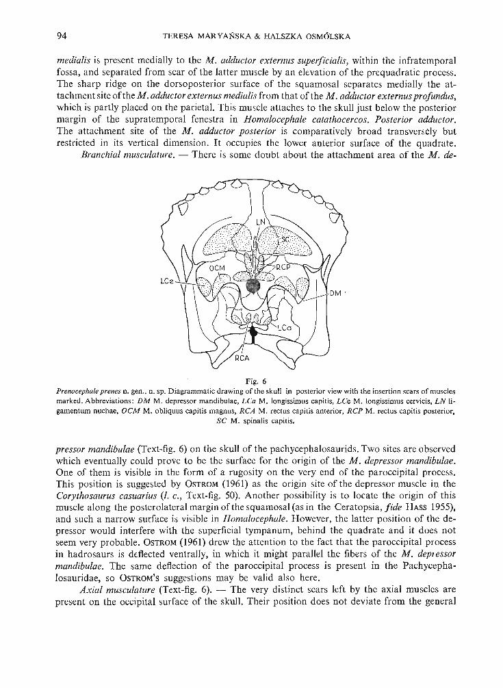
94 TERESA MAR YANSKA & HALSZKA OSM6LSKA
medialis is present medially to the M. adductor ex ternus superficialis, within the infratemporalfossa, and separated from scar of the latter muscle by an elevation of the prequadratic process .The sharp ridge on the dorsoposterior surface of the squamosal separates medially the attachment site ofthe M .adductor externus medialis from that of the M. adductor externusprofundus,which is partly placed on the parietal. This muscle attaches to the skull just below the posteriormargin of the supratemporal fenestra in Homalocephale catathocercos. Posterior adductor.The attachment site of the M. adductor posterior is comparatively broad transversely butrestricted in its vertical dimension. It occupies the lower anterior surface of the quadrate.
Branchial musculature. - There is some doubt about the attachment area of the M. de-
Fig. 6Prenocepha/e prenes n. gen., n. sp, Diagrammatic drawing of the skull in posterior view with the insertion scars of musclesmarked. Abbreviations : DM M. depressor mandibulae, LCa M. longissimus capitis, LCe M. longissimus cervicis, L N ligamentum nuchae, GCM M. obliquus capitis magnus, RCA M. rectus capitis anterior, RCP M. rectus capitis posterior.
SC M. spinalis capitis.
pressor mandibulae (Text-fig. 6) on the skull of the pachycephalosaurids. Two sites are observedwhich eventually could prove to be the surface for the origin of the M . depressor mandibulae.One of them is visible in the form of a rugosity on the very end of the paroccipital process.This position is suggested by OSTROM (I961) as the origin site of the depresso r muscle in theCorythosaurus casuarius (I. c., Text-fig. 50). Another possibility is to locate the origin of thismuscle along the posterolateral margin of the squamosal (as in the Ceratopsia, f ide HASS 1955),and such a narrow surface is visible in Homa locephale. However, the latter position of the depressor would interfere with the superficial tympanum, behind the quadrate and it does notseem very probable. OSTROM(1961) drew the attention to the fact that the paroccipital processin hadrosaurs is deflected ventrally, in which it might parallel the fibers of the M. depressormandibulae. The same deflection of the paroccipital process is present in the Pachycephalosauridae, so OSTROM'S suggestions may be valid also here.
Axial musculature (Text-fig. 6). - The very distinct scars left by the axial muscles arepresent on the occipital surface of the skull. Their position does not deviate from the genera l
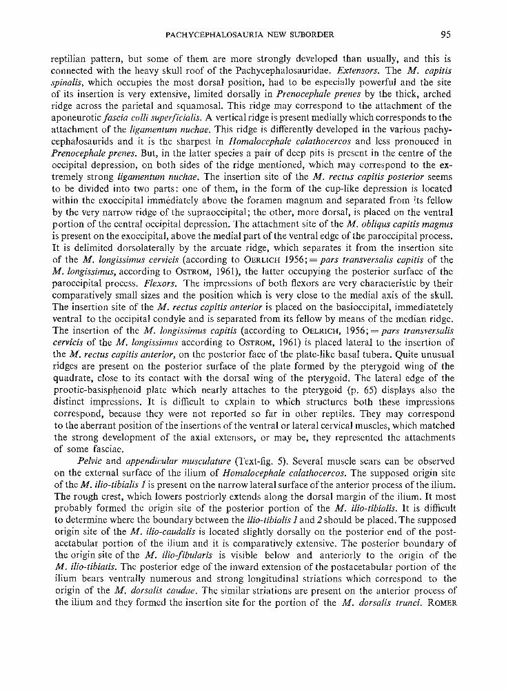
PACHYCEPHALOSAURIA NEW SUBORDER 95
reptilian pattern, but some of them are more strongly developed than usually, and this isconnected with the heavy skull roof of the Pachycephalosauridae. Extensors. The M. capitisspinalis, which occupies the most dorsal position, had to be especially powerful and the siteof its insertion is very extensive, limited dorsally in Prenocephale prenes by the thick, archedridge across the parietal and squamosal. This ridge may correspond to the attachment of theaponeurotic fascia colli superficialis. A vertical ridge is present medially which corresponds to theattachment of the ligamentum nuchae. This ridge is differently developed in the various pachycephalosaurids and it is the sharpest in Homalocephale calathocercos and less pronouced inPrenocephale prenes. But, in the latter species a pair of deep pits is present in the centre of theoccipital depression, on both sides of the ridge mentioned, which may correspond to the extremely strong ligamentum nuchae. The insertion site of the M. rectus capitis posterior seemsto be divided into two parts: one of them, in the form of the cup-like depression is locatedwithin the exoccipital immediately above the foramen magnum and separated from its fellowby the very narrow ridge of the supraoccipital; the other, more dorsal, is placed on the ventralportion of the central occipital depression. The attachment site of the M. obliqus capitis magnusis present on the exoccipital, above the medial part of the ventral edge of the paroccipital process.It is delimited dorsolaterally by the arcuate ridge, which separates it from the insertion siteof the M. longissimus cervicis (according to OERLlCH 1956; = pars transversalis capitis of theM. longissimus, according to OSTROM, 1961), the latter occupying the posterior surface of theparoccipital process. Flexors. The impressions of both flexors are very characteristic by theircomparatively small sizes and the position which is very close to the medial axis of the skull.The insertion site of the M. rectus capitis anterior is placed on the basioccipital, immediatetelyventral to the occipital condyle and is separated from its fellow by means of the median ridge.The insertion of the M. longissimus capitis (according to OELRICH, 1956; = pars transversaliscervicis of the M. longissimus according to OSTROM, 1961) is placed lateral to the insertion ofthe M. rectus capitis anterior, on the posterior face of the plate-like basal tubera. Quite unusualridges are present on the posterior surface of the plate formed by the pterygoid wing of thequadrate, close to its contact with the dorsal wing of the pterygoid. The lateral edge of theprootic-basisphenoid plate which nearly attaches to the pterygoid (p. 65) displays also thedistinct impressions. It is difficult to explain to which · structures both these impressionscorrespond, because they were not reported so far in other reptiles. They may correspondto the aberrant position of the insertions of the ventral or lateral cervical muscles, which matchedthe strong development of the axial extensors, or may be, they represented the attachmentsof some fasciae.
Pelvic and appendicular musculature (Text-fig. 5). Several muscle scars can be observedon the external surface of the ilium of Homalocepha/e calathocercos. The supposed origin siteof the M. ilio-tibialis 1 is present on the narrow lateral surface of the anterior process of the ilium.The rough crest, which lowers postriorly extends along the dorsal margin of the ilium. It mostprobably formed the origin site of the posterior portion of the M. ilio-tibialis. It is difficultto determine where the boundary between the ilio-tibialis 1 and 2 should be placed. The supposedorigin site of the M. ilio-caudalis is located slightly dorsally on the posterior end of the postacetabular portion of the ilium and it is comparatively extensive. The posterior boundary ofthe origin site of the M. ilio-fibularis is visible below and anteriorly to the origin of theM. ilio-tibiatis, The posterior edge of the inward extension of the postacetabular portion of theilium bears ventrally numerous and strong longitudinal striations which correspond to theorigin of the M. dorsalis caudae. The similar striations are present on the anterior process ofthe ilium and they formed the insertion site for the portion of the M. dorsalis trunci. ROMER
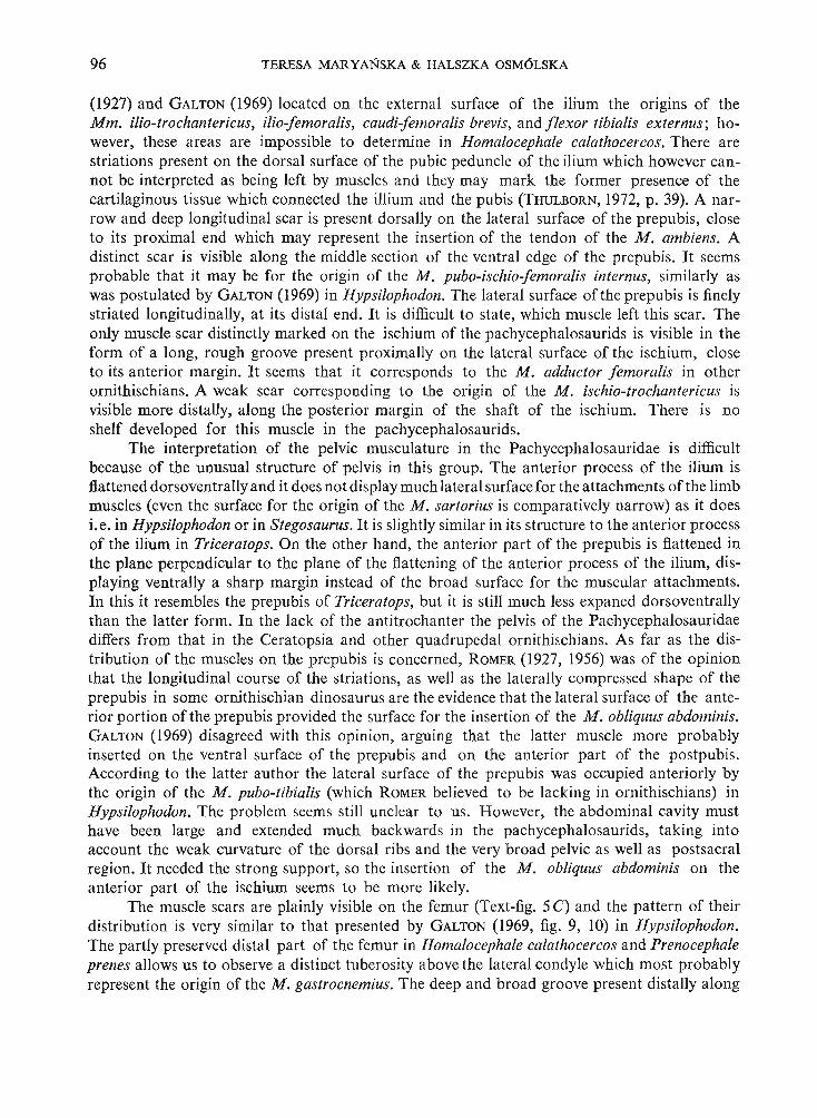
96 TERESA MAR YANSKA & HALSZKA OSM6LSKA
(1927) and GALTON (1969) located on the external surface of the ilium the origins of theMm. ilio-trochantericus, ilio-femoralis, caudi-femoralis brevis, and flexor tibialis externus; however, these areas are impossible to determine in Homalocephale calathocercos. There arestriations present on the dorsal surface of the pubic peduncle of the ilium which however cannot be interpreted as being left by muscles and they may mark the former presence of thecartilaginous tissue which connected the illium and the pubis (THULBORN, 1972, p. 39). A narrow and deep longitudinal scar is present dorsally on the lateral surface of the prepubis, closeto its proximal end which may represent the insertion of the tendon of the M. ambiens. Adistinct scar is visible along the middle section of the ventral edge of the prepubis. It seemsprobable that it may be for the origin of the M. pubo-ischio-femoralis internus, similarly aswas postulated by GALTON (1969) in Hypsilophodon. The lateral surface of the prepubis is finelystriated longitudinally, at its distal end. It is difficult to state, which muscle left this scar. Theonly muscle scar distinctly marked on the ischium of the pachycephalosaurids is visible in theform of a long, rough groove present proximally on the lateral surface of the ischium, closeto its anterior margin. It seems that it corresponds to the M. adductor femoralis in otherornithischians. A weak scar corresponding to the origin of the M. ischio-trochantericus isvisible more distally, along the posterior margin of the shaft of the ischium. There is noshelf developed for this muscle in the pachycephalosaurids.
The interpretation of the pelvic musculature in the Pachycephalosauridae is difficultbecause of the unusual structure of pelvis in this group. The anterior process of the ilium isflattened dorsoventrally and it does not display much lateral surface for the attachments of the limbmuscles (even the surface for the origin of the M. sartorius is comparatively narrow) as it doesi.e. in Hypsilophodon or in Stegosaurus. It is slightly similar in its structure to the anterior processof the ilium in Triceratops. On the other hand, the anterior part of the prepubis is flattened inthe plane perpendicular to the plane of the flattening of the anterior process of the ilium, displaying ventrally a sharp margin instead of the broad surface for the muscular attachments.In this it resembles the prepubis of Triceratops, but it is still much less expaned dorsoventrallythan the latter form. In the lack of the antitrochanter the pelvis of the Pachycephalosauridaediffers from that in the Ceratopsia and other quadrupedal ornithischians. As far as the distribution of the muscles on the prepubis is concerned, ROMER (1927, 1956) was of the opinionthat the longitudinal course of the striations, as well as the laterally compressed shape of theprepubis in some ornithischian dinosaurus are the evidence that the lateral surface of the anterior portion of the prepubis provided the surface for the insertion of the M. obliquus abdominis.GALTON (1969) disagreed with this opinion, arguing that the latter muscle more probablyinserted on the ventral surface of the prepubis and on the anterior part of the postpubis.According to the latter author the lateral surface of the prepubis was occupied anteriorly bythe origin of the M. pubo-tibialis (which ROMER believed to be lacking in ornithischians) inHypsilophodon. The problem seems still unclear to us. However, the abdominal cavity musthave been large and extended much backwards in the pachycephalosaurids, taking intoaccount the weak curvature of the dorsal ribs and the very broad pelvic as well as postsacralregion. It needed the strong support, so the insertion of the M. obliquus abdominis on theanterior part of the ischium seems to be more likely.
The muscle scars are plainly visible on the femur (Text-fig. 5C) and the pattern of theirdistribution is very similar to that presented by GALTON (1969, fig. 9, 10) in Hypsilophodon.The partly preserved distal part of the femur in Homalocephale calathocercos and Prenocephaleprenes allows us to observe a distinct tuberosity above the lateral condyle which most probablyrepresent the origin of the M. gastrocnemius. The deep and broad groove present distally along
PACHYCEPHALOSAURIA NEW SUBORDER 97
the lateral margin of the tibia posteriorly to its attachment with the fibula, may represent theorigin of the muscles moving the halux.
Tail musculature. - Very little is known about the musculature of the tail in dinosaursand any comparison with that of the pachycephalosaurids is difficult. The rather strange skeletalstructure of the tail in the latter group with extremely long caudal ribs in the anterior part ofthe tail and the basket-like arrangement of the tendons in the posterior part indicate that thecaudal musculature was extremely strongly developed and played an important role. Distinct,vertically arranged striations are present on the lateral surfaces of the high spinal processesof the proximal caudal vertebrae. They cover the entire lateral surface of the spine on its toppart, and are grouped at the anterior margin more ventrally. The striations are also presenton the caudal ribs close to the centra and along the external surface of the shafts of the anteriorcaudal ribs. It indicates that the epaxial musculature of the tail was well developed. Thesestriations may correspond to the attachments of the muscles of the: transversospinalis system(Mm. interspinales, M. spinalis, M. dorsomedialis), longissimus system (M. sacro-coccygeusdorsolateralis) and iliocostalis system (M. sacrococcygeus lateralis) of the mammals. Thestriations corresponding to the ventral musculature of the tail are generally distinct. They arelocated along the centra and along the internal surfaces of the flattened shafts of the caudal ribsclose to their anterior margin. These striations seem to correspond to the M. caudi-femoralisand the M. ilio-ischio-caudalis.
CONCLUSIONS
ADAPTATIONS AND MODE OF LIFE
The suggestions made below dealing with the possible habits of the Pachycephalosauridaeare of a speculative nature although in our opinion they seem to be generally consistent withwhat is known of their morphology. The habits of the Pachycephalosauridae were previouslythe subject of discussions by several authors, among others GILMORE (1924) and STERNBERG(1933). Recently GALTON (1970b, 1971 a) has discussed the possible function of the pachycephalosaurid dome.
Feeding habits. - Attention should be drawn to the fact that in spite of the generalstructure of the teeth which is similar in all the pachycephalosaurids, the pattern of wear ofthe teeth varies very much within this group. This implies that the mode of using the teeth hadto vary, which is reflected by the different position of the wearing surfaces in the different generaI.e. Homalocephale exposes the extensive, worn out lingual surfaces of the maxillary teeth, andthese surfaces over the whole maxillary series tend to form a common plane. Consequently,the cutting edges of all the maxillary teeth are horizontal and they form here almost continuousline over the entire series. A similar pattern is present in Tylocephale, although the teeth arehere much larger with respect to the size of the skull. Each maxillary tooth in Stegocerasbears the surface of wearing antero-medially, and the cutting edges over the full maxillary seriesproduce the "megaserration", the teeth being conical (GILMORE, 1924, p. 27). There are nosharp cutting edges on the maxillary teeth in Prenocephale, because the narrow wearing surfaceof each tooth is present along its ventral margin. This, together with the canine-like premaxillaryteeth presumably of prehensile function make the dentition of this genus quite unique amongthe late ornithischians, except perhaps Pachycephalosaurus. Thi s wide variation in thedentition of the Pachycephalosauridae speaks for different kinds of food, which might includethe plants (leaves, fruit s and seeds) and the insects as well. In spite of the very variable dentition,7 - Palaeon tolog ia Polonica No . 30

98 TERESA MARYANSKA & HALSZKA OSM6LSKA
the construction of the suspensorium is very alike in all the Pachycephalosauridae (p. 62.)The functional analysis of the mandibular joint in this group is limited by the fact, that thecomplete mandible is known only in one specimen (Stegoceras validus). Thus we are only ableto offer some vague suggestions. The striking character of the pachycephalosaurid suspensoriumis the complete external covering of the insertion area of the main adductors on the mandible(M. adductor externus and M. pseudotemporalis, p. 93). It is achieved by the high position ofthe precoronoidal portion of the surangular and especially by the strong downward developmentof the jugal and quadratojugaI. As a result the space for upward passage of the main adductorswas very narrow, and was the broadest in Tylocephale, where the outward protrusion of thejugal and quadratojugal region is the strongest (p. 51). The narrow passage for these adductorsindicates that the bulk of the muscles was not very large. The relatively high position of the insertion of M. adductor externus would have produced a shortening of this muscle, were not itsorigins shifted slightly backwards (p. 93). However, the length of the adductor externus can byno means be compared to that in the advanced Ceratopsia, but to some extent it resembledthe length of the superficial portion of the M. adductor externus in Protoceratops. The mandibularjoint in the Pachycephalosauridae did not allow for any protraction and retraction, as can bededuced from the small glenoid facet in Stegoceras validus. Additional evidence for the lackof such a motion .is found in the mutual relation between the dorsal, posterior portion of themandible and the suborbital fossa of the skull which excluded any aft and fore motion in thisspecies. The presence of premaxillary teeth and the toothed anterior portion of the dentary(at least in Stegoceras and Prenocephale) as well as the character of the wear on these teeth,also speak against protraction of the mandible. Judging from the mutual relation of the originsite of the M. adductor posterior and its insertion (around the Meckelian fossa), the courseof the fibers of this muscle was nearly parallel to that of the main adductors, so it generated anadductive force in the same direction, rather than retraction of the mandible. The premaxillahas a thick, rounded ventral margin which is in our opinion the evidence against the presence ofa beak and suggests the development of the soft and comparatively extensive lip around thetip of the snout. This lip might have been larger in Prenocephale than in Stegoceras.
Vision. - Vision presumably played an important role in the life of the Pachycephalosauridae. The orbits are very large and deep and their bony walls gave perfect protectionto the eye. The orbits are facing laterally, but there is a strongly marked tendency to extendthe field of vision more anteriorly. It is realized during phylogeny by the gradual narrowingforwards of the snout in its preorbital portion and by the enlargment (especially in the verticaldirection) of the orbit. This tendency reaches its climax in Prenocephale, where additionallythe orbit is also facing slightly anteriorly. The optic nerve is one of the largest, which speaksfor the high degree of the visua perception.
Olfaction. - The endccranilal casts of the pachycephalosaurid skulls are characterizedby the enormous sizeof the first nerve, which speaks for good olfactory abilities. There is a welldeveloped external nasal cavity in the pachycephalosaurids, which displays the extensive floor.It might have been covered by mucous membrane for the increasing of olfactory sensitivity.
Posture. - The cervical vertebrae are known in no pachycephalosaurid specimen, whichmakes impossible the reconstruction of the neck region and of the exact position of the headin these animals. However, the very strong development of the extensors of the headwas necessary for keeping the heavy skull in the horizontal position while the animal was walking.Such a position of the skull during the normal gait seems to us the most reasonable one, becausethe development of the laterally extending shelves above the eyes would limit (at least in P renocephale and Stegoceras) the forwards vision if the head is kept vertically. We think that

PACH YCEPHALOSAURIA NEW SUBORDER 99
the position with the lowered head and the ant eriorly directed dome was attained when the head(and the dome) was used as the defensive weapon. Th e batt ering role for the dome in the pachycephalosaurid s was suggested by G ALTON (I970b, 1971 a). Thi s assumption seems to us wellfounded and logical. Int erpretation of the dome as being connected with sexual dimorphismseems to us still disputable. The need for the development of a defensive weapon seemsto be very essential in animals which, like the pachycephalosaurid s, were comparatively slow(judging from the tibial femur ratio, p. 101)and unprotected by armour. The flexors of the headare weak in the Pach ycephalosauridae in contrast to the strong development of the extensors.Moreover, they insert on the very weak, plate-like basal tub ers. Thi s indicates that the actiongenerated by these muscles was less powerful than that of the extensors. The location of themain flexors immediately on the medial axis of the skull might result in stronger developmentof the lateral or ventra l cervical musculature to mat ch the transverse development of the extensors. We suppose that the neck in the Pachycephalosauridae was fleshy, broad andshort. The ant erior dorsal region is also unk nown in the Pachycephalosauridae. The weakcurvature of the posterior dorsal ribs, the broad pelvis, and the presence of unusually long ribson the anterior caud als mean that the animal was very broad in top view. The groove and tonguearticulation ofthe zygapophyses on the (at least) posterior dorsal vertebrae completely eliminatedlateral flexions of the trunk. A similar lack of lateral flexion is found in the posterior portionof the tail, where it is prevented by the vertical position of the articular surfaces of the zygapophyses. The only laterally flexible part of the tail was its anterior portion, where the zygapophyseal articular surfaces are horizontal. Here however, the lateral flexion was limited bythe long caudal ribs. A slightly stronger lateral flexions of the tail was possible in the point placedimmediately behind the rib-bear ing portion of the tail , between the eight and ninth vertebra,where the zygapophyses have small, but completely horizontal articular surfaces, which are notonly more horizontal than the zagypophyses of the succeeding caudal, but also than those ofthe preceding ones. Vertical movements were possible along the tail , but they were limited bythe very high spinal processes.The basket-work oftendons gave perfect protection to the tail, beingat the same time relatively light and elastic in thevertical direction. The arrangement ofthe thickesttendons along the ventral surface of the ta il, as well as the numerous traces of damages presenton these very tendons pro ve that it was the "working surface" of the tail. As was already mentioned above the structure of the pachycephalosaurid pelvis is most unusual. It is notonly very broad (for a bipedal animal), but the acetabulum practically lacks its lower rim, asa result of the inward shifting of the anterior articular process of the ischium towards the sacrum.Moreover, the ischium contacts the sacrum at two points by means of the sacral ribs, one infront and the other behind the acetabulum. The contact between the ilium and the sacrumis weakened in the anterior portion of the pelvis, and it is restricted to a loose attachment between the anterior process of the ilium and the first sacral rib. The only firm union betweenthe sacrum and ilium is that behind the acetabulum, by means of the upper portion of thefourth sacral rib and the fifth sacral rib. Thi s latt er contact seems to be addi tionally strenghtenedby the vertical extension of the ilium in the medial direction which closely attaches to theposterior surface of the fifth sacral rib. Mo st pr obably this aberrant structure of the widenningbackwards pelvic region was caused by the development of the large (also posteriorly) abdominalcavity. It may suggest that the Pachycephalosaurid ae were viviparous. The generallyloose contact between the pelvis and sacrum might be important for the amortization of theabd omen when the animal was fighting and blows were conducted backwards along therigid vertebral column. The rigid structure of the vertebral column may indicate that it washeld nearly horizontal while the animal walked (GALTON, 1970b), but the deeper acetabulum7·
100 TERESA MARYANSKA & HALSZKA OSM6LSKA
and the stronger pubic peduncle in the Pachycephalosauridae, than they were i.e. in the hadrosaurs, indicate that the more inclined upwards posture of the vertebral column and the iliumcould be also achieved during the rest, with the tail acting as the prop and with the centre ofgravity placed behind the acetabulum.
REMARKS ON THE SEXUAL DIMORPHISM AND THE SUPPOSED PHYLOGENYOF THE PACHYCEPHALOSAURIA
The first authors who discussed the problem of the sexual dimorphism in the Pachycephalosauridae were BROWN & SCHLAIKJER (1943). In their opinion, the degree of development ofthe parietosquamosal shelf and the surface of the domed part of the skull roof are the charactersconnected with the sexual dimorphism; the forms with the smooth frontoparietal dome, whichnearly lacked the shelf, were regarded by these authors as females, while the forms with therough dome and the extensive shelf represented males. Recently GALTON (1971a) reconsideringthis problem, drew attention to the fact that the development of the shelf is connected withthe development of the dome. He also reversed BROWN & SCHLAIKJER'S sex designationsuggesting that forms with the relatively larger domes could be males. Although the majorityof the Pachycephalosauridae have their skull roofs domed, among them the two forms areknown which have them flat. They are: Homalocephale calathocercos from Mongolia and theskull No. A. M. N. H. 5450 of S. validus from Canada (GALTON, 1971a, Text-figs 5, 6). Werethe latter Canadian specimen the female, the only other unquestionable female should be thespecimen of H. calathocercos. If the hypothesis about the sexual dimorphism within the pachycephalosaurids were applied to our Mongolian material, Prenocephale prenes and H. ealathocercos have to be considered as male and female (or vice versa) of the same species becausethey were contemporary, being recovered in the beds of the same age (Nemegt Formation).But, the differences between the "flatly roofed" H. calathocercos and domed P. prenes are notrestricted to the structure of the skull roof. They also deal with many other characters of theskull and the postcranial skeleton as well. Certainly, some of these differences are connectedwith the change in the thickness of the skull roof, or caused by their different mechanic promotedby the dissimilar position of the weight centres. There exists, however, still a number of differences between the two skulls compared, which seem to be independent: e.g. the differencesin the size and shape of the foramen magnum and the medulla oblonga, in the shape of the archof upper teeth and in their being worn out in the different manner, in the shape and the mutualrelation between the pterygoids, etc. They speak, in our opinion, for the taxonomical separationof the two specimens in question rather than in favour of their sexual dimorphism, Nevertheless,the problem of the sexual dimorphism of the Pachycephalosauridae can be definitively solvedonly on the basis of a large sample, such as that from Belly River Formation of Alberta, whichis far the richest in this familly, as suggested by GALTON (1971a). We fully agree here withGALTON (I. c.) that the characters regarded by BROWN & SCHLAIKJER (1943) as of sexual valueare far from being convincing, and a revision of the North American species, especially of"Stegoceras validus" is badly needed.
The earliest Pachycephalosauria known, represented by the Stenopelix and Yaverlandia,were reported from the Lower Cretaceous of Europe (MEYER, 1859; GALTON, 1971a). ThePachycephalosauria most probably should be derived from the Jurassic Hypsilophodontidae,as they have the premaxillary dentition still preserved, and they expose the similar general structure of the skull (GALTON, 1971a). However, the earliest hypsilophodontid with the known

PACHYCEPHALOSAURIA NEW SUBORDER 101
postcranial skeleton - Fabrosaurus australis GINSBURG, 1964 from the African Upper T riassic(Thulborn, 1972) - has alrea dy th e tibia distinctly longer than the fem ur, which rela tion isthe constant character within the main hyp silophodontid lineage. The relat ion between th efemur and the tibia is more primitive in the Pachycephalosauria. In the Wealden StenopelixMEYER, and in the Upper Cretaceous Pachycephalosauridae as well, th e femur is never shorte rthan the tibia. This fact , and the exten sive differences in th e pelvic structure, ind icat e th at thePachycephalosauria might hav e ari sen very ea rly (T riassic?) from th e Hypsiloph od ontidae,before their advanced, cursorial trend wa s achieved. H owever , as already not iced a bove (p. 48),there are sim ila rities in the st ructure of the pelvis, as well as so me in th e sku ll, between thePachycephalosauria and th e Ceratopsia. These two suborders had appeared in th e orn ithisc h ianhi story very late (Cretaceous). In our opinion , both groups ca n be der ived from th e common ,side line of the Hypsilophodontidae . It co uld be eas ily presumed th at th eir separa tion tookplace during the Jurassic. Pachycephalosauria occu r from th e Belly River F orm at ion to th eLance Formation in North America inclusively (Upper Campa nian to Upper Maast richtian,according to Russell ; personal communication). In A sia , th ei r ea rlies t representative "S tegoceras" bexelli BOHLIN, 1953 (BOHLIN, 1953) is known from th e T sondolein -Khuduk local ityin Inner Mongolia, and these beds a re, perhaps, an equ ivalent of the Djad okhta F orm ationaccording to ROZHDESTVENSKY (1971 , Table 2). The age of the Dj adokh ta F orm ati on isConiacian or Santonian according to K IELAN-JAWOROWSKA (1970). Recently, th e P olish-Mongolian Expeditions recovered pachycephalosaurid representati ves in Barun G oyot F ormation (?Middle Campanian, according to KIELAN-JAWOROWSKA, 1974), and in th e N emegtFormation (Maastrichtian , according to ROZHDESTVENSKY, 1971, a nd Up per Campa nia n orMaastrichtian, according to OSM6LSKA et al., 1972). If the bed s in T sondolein-Khuduk mayreally be correlated with th e Djadokhta Formation, th e poorly kn own species "Stegoceras"bexelli, which occurs in these beds, is the oldest known representative of th is famil y inAsia.
Museum of the Earthof the Polish Academy of S ciences
00-488 Warsza ll'a Al. Na Skarpie 20/26,Septemb er 1972
REFERENCES
Pa/aeozoological Instituteof the Polish Academy of Sciences
02·089 Warszall'a Al. Zwirki i Wigury 93,September 1972
BOHLlN, B. 1953. Fossil repti les from Mongolia and Kansu. - The Sino-Swedish Exped. Publ. 37, Stockholm.BROWN, B. & SCHLAIKJER E. M. 1943. A study of the Tr oodon dinosaurs with the description of a new genus and four
species. - 8ull. Amer. Mu s. Na t, His t., 82, 121-149, New York.BEER, DE G. 1971. The development of the vertebrate skull. 1-554, Londo n.COLBERT, E. H. & R USSELL, D. A. 1969. The small Cretaceo us dinosaur Dromaeosau rus. - All/er. M us. Novit ., 2380,
1-49, New Yor k.GALTON, P. M. 1969. The pelvic musculature of the dinosaur Hypsiloph odon (Reptilia: Ornithischia). - Postilla, 131,
1-64, New Haven.1970a. Orn ithischian dinosaurs and the origin of birds. - Evolution, 24, 448-462, Lancaster.1970b. Pachycephal osaurids - dinosaurian battering ra ms. - Discovery , 6, 23·32, New Haven .1971a. A primit ive dome-headed dinosaur (Ornithischia : Pachycephalosauridae) from the Lower Cretaceousof England and the function of the dome of pachycephalosau rids. - J. Palaeont ., 45, I , 40-47, Tulsa.1971b. The posture of had rosaurid dinosaurs. - J. Pa/aeont ., 44, 3, 465-473, Tulsa.
GILMORE, C. W. 1924. On Troodon validus an orthopodous dino sau r from the Belly River Cretaceous of Alberta, Canada . - Bull . Alberta Univ., 1, 1-43, Edmonton.
GRADZINSKI, R., KAZMIERCZAK J. & LEFELD, J. 1969. Geograpfical and geological data from the Polish-Mongolian Palaeontological Expeditions. Results Polish-Mongol. Palaeon t. Exped, I. - Palaeont. Pol., 19, 33-82. Warszawa.
102 TERESA MA R YANSKA & HALSZKA OSM6LSKA
GRADZINSKI, R. & JERZYKIEWICZ, T . 1972. Addit ional geographical and geological data from the Polish-MongolianPalaeontological Expeditions. - Resul ts ..., IV. - Ibidem, 27, 17-32.
Hxss, G . 1955. The jaw musculature in Protocera tops and in other ceratopsians. - Am er, Mus. Novit., 1729, 1-24,New York.
KIELAN-JAWOROWSKA, Z. 1970. New Upper Cretaceous multituberculate genera fro m Bayn Dzak, Gobi Desert.Results ..., 11. - Palaeont . Pol. 21, 35-49, Wa rszawa.1974. Mu ltit uberc ulate succession in Late Cretaceo us of the Go bi Deser t, Mongolia. Resul t. .., V. - Ibidem, 30,
KIELAN-JAWOROWSKA, Z.& BARsBoLD, R. 1972. Narrat ive of the Poli sh-Mongolian Palaeontological Expeditions 1967-1971.Result s ... , IV. - Ibidem, 27, 5-13. .
KIELAN-JAWOROWSKA, Z. & DOVCHIN, N. 1969. Narrative of the Pol ish-M ong olian Palaeontological Expeditions 1963-65.Result s ..., 1. - Ibidem, 19, 7-30.
KOKEN, E. 1887. Die Dinosaurier, Crocodil ien und Sauro pterig ier des norddeutschen Wealden. - Palaeont. Abh., j,5, 1-111, Berlin.
KRAMER, G. & MED EM, F. 1955/56. Ueber wachstumsbedingte proport ions-anderungen bei K rokodilien. - Zool-. Jb.,III Abt, allg. Zoo l. Physiol. Tiere. 66, 1, 67-74, Jena.
LAMBE, L. M. 1902. New genera and species from the Belly River series (Mid-Cretaceous). - Contr. Can. Palaeont,Geol. Surv ., 3, 2, 25-81.1918. The Cretaceous genus Stegoceras typifyi ng a new family referre d provisionally to the Stcgosauria. - Trans.Roy, Soc. Canada, ser. lI I, 12, 23-36, Ott awa.
MARSH, O. C. 1896. The dinosau rs of North America. 143-244, Washington. ,MEYER, H . 1859. Stenopelix Valdensis, ein Rep til aus der Wealden-Form ation Deutschlands. - Palaeont ogr ., 7, 1, 25-34,
Cassel.NOPCSA, F . 1904. Dinosaurierreste aus Siebenbilrgen. III - Denksch. Kais. Akad. wi«; 74, 229-263.
1928. Di nosaurierreste aus SiebenbUrgen . IV. - Palaeontol. Hungar., 1, 273-302. Budapest.1929. Dinosaurierreste aus Siebenburgen, V. - Geol. Hung. ser. palaeont., 4, 1676. Budapest.
OELRICH, T. M. 1956. The anato my of the head of Cte nosaura pectinata (Iguanidae). - Mise. Publ , Univ. MichiganM us. Zoo l., 94, 1-122, Ann Ar bor.
OSMOLSKA, H. , RONIEWICZ, E. & BARsBoLD, R . 1972. New dinosau r, Gallimimus bullatus n. gen., n. sp, (Ornithornimidae)from the Upper Cretaceous of Mongol ia . Results ... IV. - Palaeollt. Pol., 27, 103-143, Warszawa.
OSTROM, J . H. 1961. Cranial morpho logy of the had rosau rian dinosaurs of North America. - Bull. Amer. Mus. Nat,Hist ., 122, 39-186, New Yo rk.1964. A funct ional ana lysis of jaw mecha nics in the din osau r Triceratops. - Postil/a , 88, 1-35, New Haven.1966. Functional morpho logy and evolution of the cera to psia n dinosaurs. - Evolution, 22, 3, 290-308, Lancaster.
ROMER, A. S. 1927. T he pelvic musculature of orn ithisc hian dinosau rs. - Acta Zool., 8, 225-275, Stockholm.1956. Os teo logy of the Reptili a. 1-772, Chicago .
ROZHDESTVENSKY. A. K . - see Pom~EcTBEHCHIIH , A. K.RUSSELL, D . A. 1972. Os trich dinosaurs from the Lat e Cre taceous of Western Canada. - Can. J. Nat. Sci., 9, 4, 375-402,
Ottawa .R ussELL, L. S. 1948. T he dentary of Troodon a genu s of Thero po d dinosaurs. - J. Palaeont. , 28, 5, 625-629. Tulsa.SCHMIDT, H . 1969. Stenopelix valden sis H. v. Meyer, der kleine D inosaurier des norddeutschen Wealden. - Paliiont ~ Z :.
43, 3/4, 194-198, Stut tgart.STEEL, R. 1969. Ornithischia, in O. K uhn, Handbuch der Palaoherpetologie. 1-81, Stuttgart.STERNBERG, C. M. 1933. Relat ionships and hab ita t of Troo don and the nodo saurs , - Ann. Mag. Nat. Hist. , (10), 11,
231-235, London .1945. Pachycephalosau ridae proposed for dom e-headed dinosau rs Stegoceras lambei, n. sp., described. - J.
Palaeont., 19, 5, 534-538, T ulsa.THULDORN, R. A. 1972. T he post-cran ial skeleton of the Triassic orn ithischian dinosaur Fabrosaurus au stralis. - Pa
laeontology, 15, 1, 29-60, London .PomgEcTBEHCKHH, A. K. 1964. CeMeHCTBo Pachyceph alosauridae. In OPJIOB, 10. A. (ed .) OCIIOBbI I1aJIeOIITOJIO
rHH, 588-58 9. 11ocHBa.1971. Hayxenne ~IIII03aBpOB 110IIr OJIHll 11 HX POJIb B paCtIJIeHeHllII I{OIITlllIeHTaJIbHOrO Me3030fl. <1laylIa MC3030H 11 HallH030H 3anagHoH 110IIro JIllll , 21-32 . 110CI<Ba .1971. Pa3BHTHe 11 BbIMHpaIllle na pytience MeJIa II naneorena, In. Paasnrne H caena oprana uecicoro MIIpa Hapyfiexce Me3030H 11 I<aHlIo30H. Te3HCbI ~OHJI . H MeTo~. MaTep . AHa~. Hayx CCCP, 51-56 110CI< Ba.

Palaeontologia Polonica, No. 30, 1974
T. MAR YANSKA & H. OSM6LSKA: PACHYCEPHALOSAURIA, NEW SUBORDER
PLATE XXII
Homalocephale calathocercos n. gen., n. sp.(see also Plates XXIV, XXVI-XXX and XXXI, Fig. I)
Upper Cretaceous, Nemegt Formation, Nemegt, Gobi Desert, Mongolia
Fig. 1. Stereo-photograph of the damaged skull in right lateral view. Type specimen (G. 1. No. SPS 100/51); about1/3 of natural size.
Prenocephale prenes n. gen., n. sp.(see also Plates XXIII, XXV, Figs. 2, 3 and XXXI, Fig. 2)
Upper Cretaceous, Nemegt Formation, Nernegt, Gobi Desert, Mongolia
Fig. 2. Stereo-photographs of the skull in: a - palatal view, b - occipital view. Type specimen (Z. Pal.No. MgD-I/I04); about 1/3 of natural size.
Tylocephale gilmorei n. gen., n. sp.
Upper Cretaceous, Barun Goyot Formation, Khulsan, Gobi Desert, Mongolia
Fig. 3. Skull in: a - dorsal view, b - right lateral view (with the mandible attached), c - palatal view, d occipital view. Type specimen (Z. Pal. No. MgD-I /I05) ; about 1/3 of natural size.
Photo : M. Kleib er, W. Skariynski
Page
56
53
51

Palaeontologia Polonica, No. 30, 1974
1
T. MARYANSKA & H. O SM6 LSKA: P ACH YCEPHALOSAURl A NEW SUBORDER
3d
PI. XXII

Palaeontologia Polonica, No. 30, 1974
T. MARYANSKA & H. OSM6LSKA: PACHYCEPHALOSAURI A, NEW SUBORDER
PLATE XXIII
Prenocephale prenes n. gen., n. sp. . . . .(see also Plates XXII, Fig. 2; XXV, Figs. 2, 3; and XXXI, Fig. 2)
Upper Cretaceous, Nernegt Formation, Nemegt, Gobi Desert, Mongolia
Fig. 1. Stereo-photographs of the skull in: a - dorsal view, b - right lateral view, d - anterior view, c left lateral view. Type specimen (Z. Pal. No. MgD-I/I04); about 1/3, of natural size.
Photo : W. Skariysisk!
Page
53

Palaeontologia Polonica, No. 30, 1974 PI. X XIlI
T. MARYANSKA & H. OSM6LSKA : PACHYCEPHALOSAURlA NEW SUBORDER
Palaeontologia Polonica, No. 30, 1974
T. MARYANSKA & H. OSM6LSKA : PACHYCEPHALOSAURIA, NEW SUBORDER
PLATE XXIV
Homalocephale calathocercos n. gen., n. sp,(see also Plates XXII, Fig. 1; XXVI-XXX and XXXI, Fig. 1)
Upper Cretaceous, Nemegt Formation, Nernegt, Gobi Desert, Mongolia
Fig. 1. Stereo-photographs of the damaged skuIl in : a - dorsal view, b - left lateral view, c - occipital view,d - palatal view. Type specimen (G. I. No. SPS 100/51); about 1/3 of natural size.
Photo: W. Skar iyriski
Page
56

Palaeontologia Polonica, No. 30, 1974 Pi. XXIV
T. MARYANSKA & H. O SM6LSKA : PACHYCEPHALOSAURlA NEW SUBORDER

Palaeontologia Polonica, No. 30, 1974
T. MARYANSKA & H. o SM6LSKA : PACHYCEPHALOSAURIA, NEW SUBORDER
PLATE xxv
Stegoceras validus LAMBE, 1902
Upper Cretaceous, Belly R iver Fo rmat ion , Red D eer River, Alberta, Canada
Fig . I. Latex cast of the endocranial cavity of specimen NMC No. 138: a - stereo-photograph in dorsal view,b - posterior view (medulla oblonga), c - left lateral view. Specimen illustrated also by LAMBE, 1918,PI. I , Fig. I ; x I.
Prenocephale prenes n.gen., n.sp.(see also Plates XXII, Fig. 2, XXIII and XXXI, Fig. 2)
Upper Cretaceous, Nemegt Formation, Nemegt, Gobi Desert , Mongolia
Fig. 2a. Left middle ear and basicranial region in oblique, posterolateral view (see also Text-fig. 2). Typespecimen (Z. Pal. No . MgD- I/ I04); x r.
Fig . 2b. Media l wall of the right orbit with accessory ossifications visible (see also Text-fig. 3). Same specimen ;x l.
Fig . 3a. Do rsal view of the sacral and caudal ribs with ilium in natural articulation . Same specimen ; about 1/3of nat ural size.
Fig. 3b. Ventral view of the sacral region, right ilium and ischium, the latter shifted slightly in horizontal direct ion from the natural position. Same specimen; about 1/3 of natural size.
Photo: W. Skari yriski
Pag e
79
53
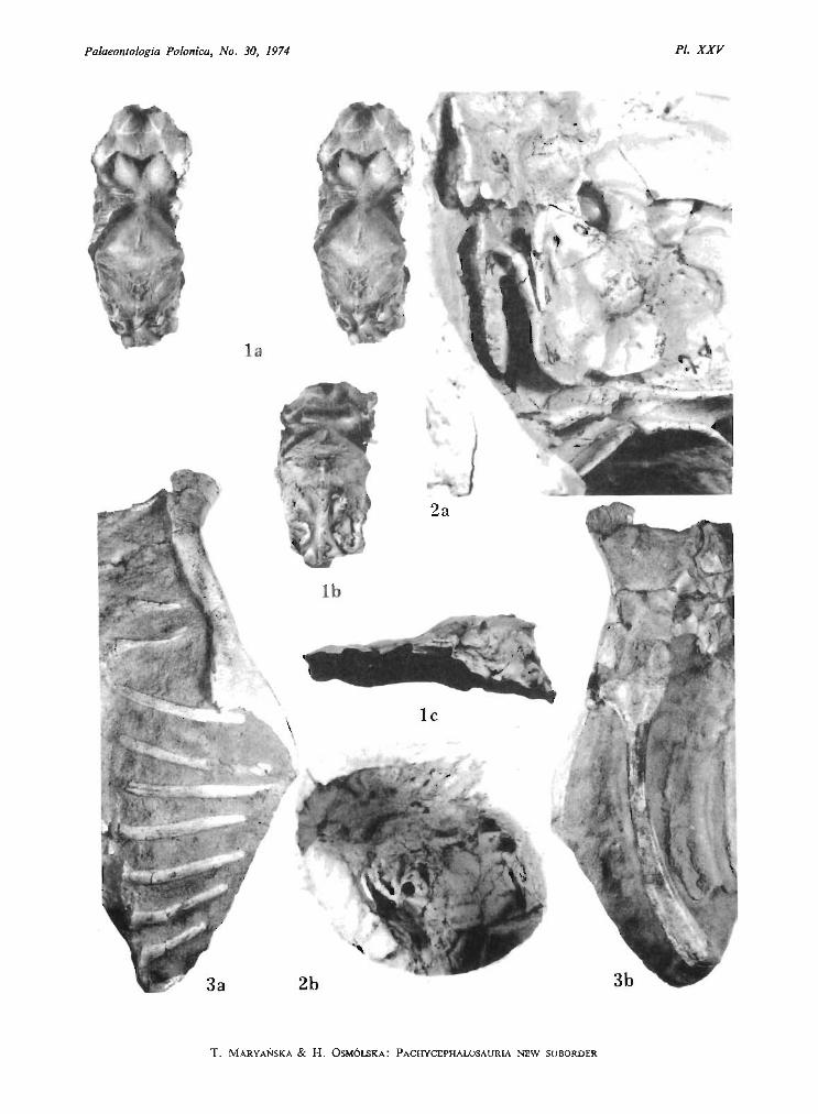
Pa/aeonfologia Polonica, No. 30, 1974 PI. XXV
....t ~ .
:. ,..t _ .
la
T. MARYANSKA & H. OSM6LSKA : PACHYCEPHALOSAURlA NEW SUBORDER
Palaeontologia Polonica, No. 30, 1974
T. MARYAN SKA & H. OSM6LSKA: PACHYCEPHALOSAURIA, NEW SUBORDER
PLATE XXVI
Homalocephale calathocercos n.gen., n.sp.(see also Plates XXII , Fig. 1; XXIV, XXVII-XXX and XXXI, Fig. 1)
Upper Cretaceous, Nemegt Formation, Nemegt, Gobi Desert, Mongolia
Fig. 1. Posterior dorsal portion of the vertebral colum, the penultimate dorsal lacking, in : a - right lateralview, b - dorsal view. Type specimen (G. I. No. SPS 100/51); about 1/2 of natural size.
Fig. 2. Postsacral portion of the vertebral column in dorsal view. Same specimen; about 1/2 of natural size.
Photo: M . Kleiber
Page
56
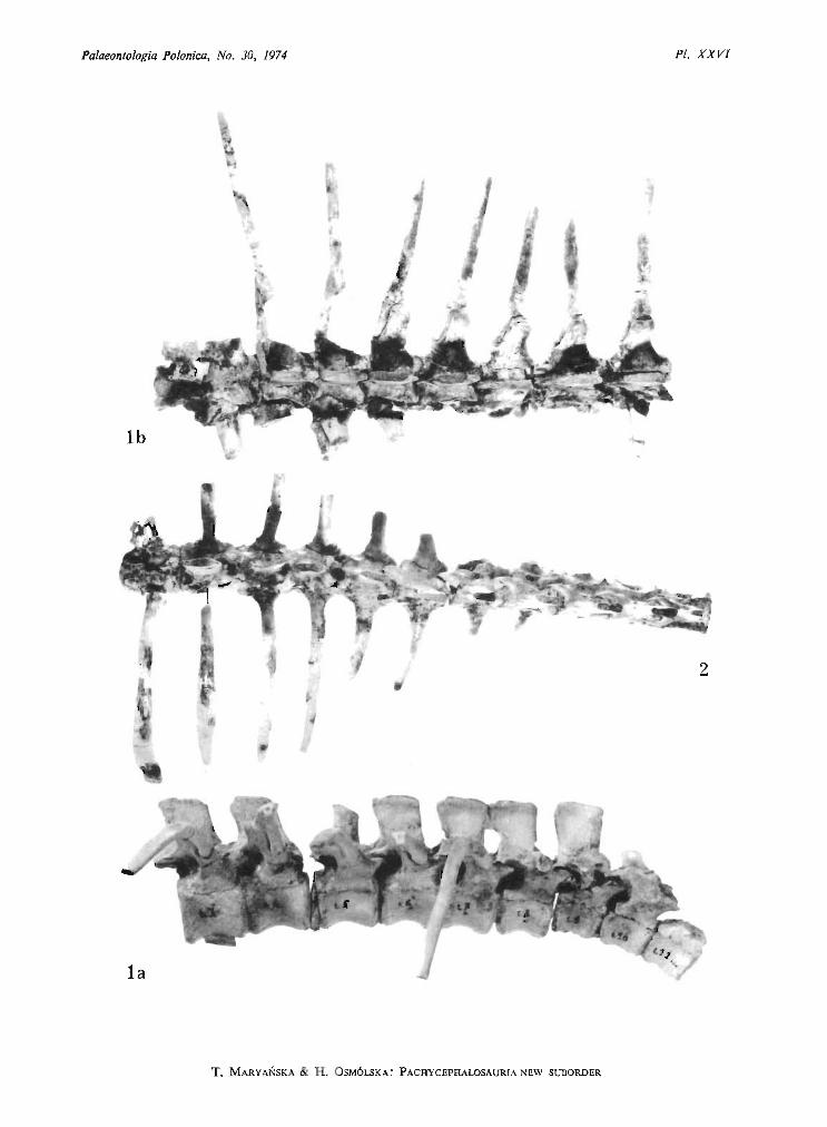
Palaeontologia Polonica, No. 30, 1974 PI. X XVI
Ib
T. MARYANSKA & H. OSM6LSKA : PACHYCE PHALOSAURlA NEW SUBORDER
Palaeontologia Polonica, No . 30, 1974
T. MARYANSKA & H. OSM6LSKA: PACHYCEPHALOSAURIA, NEW SUBORDER
PLATE XXVII
Homalocephale calathocercos n.gen., n.sp.(see also Plates XXII , Fig. 1, XXIV, XXVI, XXVIlI-XXX and XXXI, Fig. 1)
Upper Cretaceous, Nemegt Formation, Nemegt, Gobi Desert, Mongolia
Fig. 1. Stereo-photograph of the damaged sacrum, with the last dorsal attached, in: a - dorsal view, b ventral view. Type specimen (G. I. No. SPS 100/51.); about 1/2 of natural size.
Photo: W . Skar i yrisk l
Page
56

Palaeontologia Polonica, No. 3D, 1974PI. XXVII
Ib
T. MARYANSKA & H. OSM6LSKA: P ACHYCEPHALOSAURIA NEW SUBORD ER

Palaeontologia Polonica, No. 30, 1974
T. MARYANSKA & H. oSM6LSKA: PACHYC EPHALOSAURIA, NEW SUBORDER
PLATE XXVIII
Page
Homa locephale calathocercos n.gen., n.sp. 56(see also Plates XXII, Fig. 1, XXIV, XXVI, XXVII, XXIX, XXX and XXXI, Fig. 1)
Upper Cretaceous, Nemegt Formation , Nemegt, Gobi Desert, Mongolia
Fig. 1. Sacrum in right lateral view. Type specimen (G. I. No. SPS l OO/51); about 1/2 of natural size.Fig. 2. Postsacral portion of the vertebral column in left lateral view. Same specimen; about 1/2 of natural size.Fig. 3. Posterior port ion of the tail, with the basket-work of tendons visible, in left lateral view. Same specimen;
about 1/2 of natura l size.Fig. 4. Posteriormost preserved portion of the tail, partly with tendons, in left lateral view. Same specimen;
about 1/2 of natural size.Photo : M . Kleiber
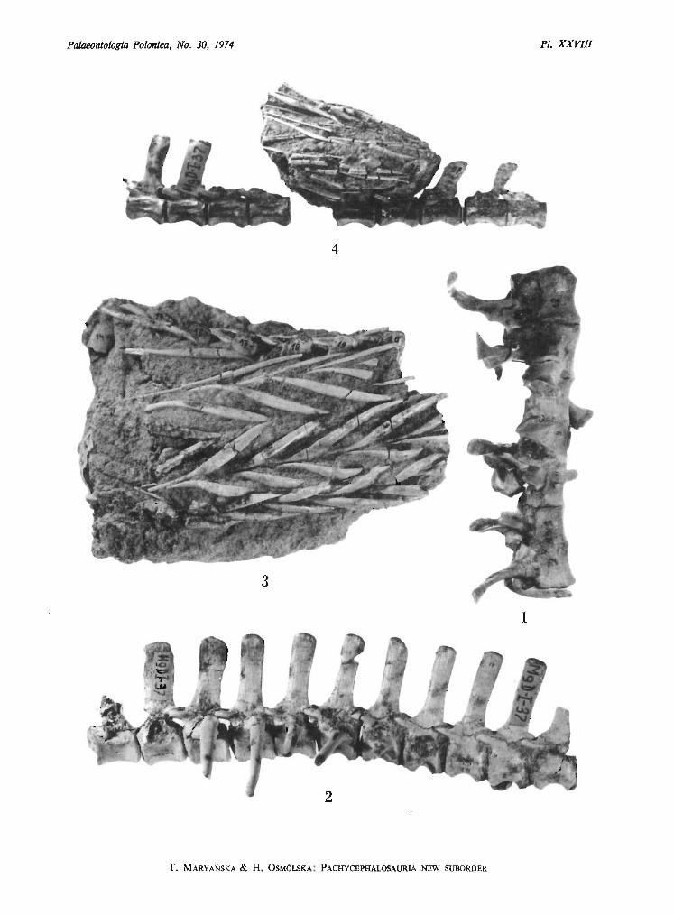
Palaeontologia Polonica, No. 3D, 1974 PI. XXVIll
4
3
1
T. MARYANSKA & H. O SM6LS KA: PACHYCEPHALOSAURlA NEW SUBORDER

Palaeontologia Polonica, No. 30. 1974
T. MARYANSKA & H. oSM6LSKA: PACHYCEPHALOSAURIA, NEW SUBORDER
PLATE XXIX
Homatocephale calathocercos n.gen., n.sp.(see also Plates XXII , Fig. 1, XXV, XXVI-XXVIII, XXX and XXXI, Fig. 1)
Upper Cretaceous, Nemegt Formation, Nemegt, Gobi Desert, Mongolia
Fig. 1. Left ilium with proximal portion of the ischium in natural articulation : a - stereo-photograph in lateralview, b - stereo-photograph in medial view, c - dorsal view. Type specimen (G. I. No. SPS 100/51);about 1/2 nat ural size.
Fig. 2. Left prepubis in: a - lateral view, b - medial view. Same specimen; about 1/2 of natural size.
Phot o: M. Kleiber
Page
56
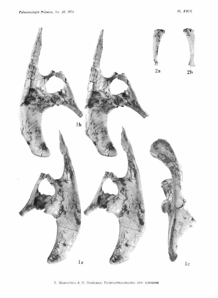
Palaeontologia Polonica, No. 30, 1974
2a
T. M ARYANSKA & H. OSM6LSKA: P ACHYCEPH ALOSAUR IA NEW SUBORD ER
PI. XXiX
JJ
2b
le

Palaeontologia Polonica, No. 30, 1974
T. MARYANSKA & H. OSM6LSKA: PACHYCEPHALOSAURIA, NEW SUBORDER
PLATE XXX
Homalocephale calathocercos n.gen., n.sp.(see also Plates XXII, Fig. I, XXIV, XXVI-XXIX and XXXI, Fig . 1)
Upper Cretaceous, Nemegt Formation, Nemegt, Gobi Desert, Mongolia
Fig. 1. Sternal bones in ventral view. Type specimen (G. I. No. SPS 100/51); about 1/2 of natural size.Fig. 2. Stereo-photograph of the left ilium with fragment of the ischium, in natural articulation, ventral view.
Same specimen; about 1/2 of natural size.Fig. 3. Distal portion of the right tibia and fibula, with astragalus attache d, in : a - posterior view, b - anterior
view. Same specimen; about 1/2 of natural size.Fig. 4. Distal portion of the left tibia, in: a - posterior view, b - anterior view. Same specimen; 1/2 of natural
size.Fig. 5. Left metatarsal 11 and damaged metatarsal III in : a - anterior view, b - posterior view. Same speci
men; about 1/2 of natural size.Fig. 6. Phalanx 1 of the left digit IV (1) in : a - dorsal view, b - ventral view. Same specimen; about 1/2 of
natural size.Photo: M. Kletber, W. Skarzyliskl
Page
56
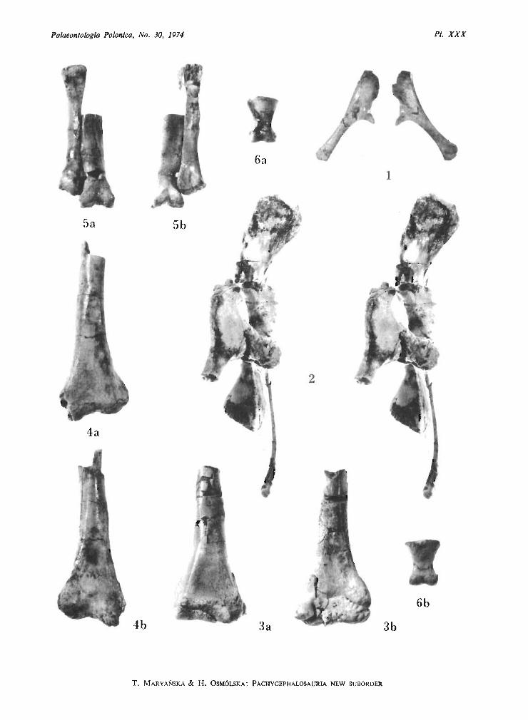
Palaeontologia Polonica, No. 30, 1974 PI. XXX
6b
1
3b
2
3a
6a
5b
4b
4a
5a
T. M ARYANSKA & H. OSM6LSKA: PACHYCEPHALOSAURIA NEW SUBORDER
Palaeontologia Polonica, No. 30, 1974
T. MARYANSKA & H. OSM6LSKA: PACHYCEPHALOSAURIA, NEW SUBORDER
PLATE XXXI
Homalocephale calathocercos n.gen., n.sp.(see also Plates XXII, Fig. 1, XXIV and XXVI-XXX)
Upper Cretaceous, Nemegt Formation, Nemegt, Gobi Desert, Mongolia
Fig. 1. Stereo-photograph of the left femur in: a - anterior view, b - posterior view. Type specimen (G. 1.No. SPS 100/51); about 1/2 of natural size.
Prenocephale prenes n.gen., n.sp.(see also Plates XXII , Fig. 2, XXIII and XXV, Figs. 2, 3)
Upper Cretaceous, Nemegt Formation, Nemegt, Gobi Desert, Mongolia
Fig. 2. Right femur in: a - anterior view, b - posterior view, c - medial view. Type specimen (Z. Pal.No. MgD-I/I04); about 1/2 of natural size.
Phot o: M. Klelber , W. Skorfyriskl
Page
56
53
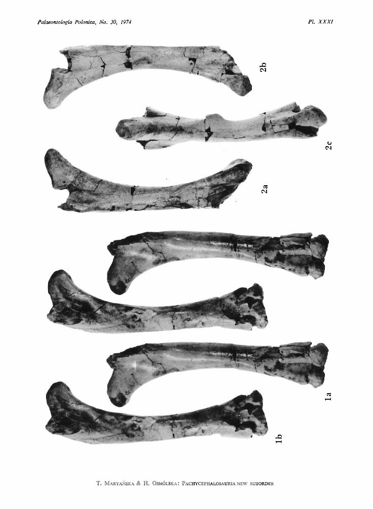
Palaeontologia Polonica. No. 30. 1974 PI. XXXI
T. MARYANSKA & H. OSM6 LSKA: PACHYCEPHALOSAURlA NEW SUBORDER