P450-Glo™ Assays - Promega/media/Files/Resources/Protocols/Technical...P450-Glo™ Assays...
54
Revised 9/16 TB325 TECHNICAL BULLETIN P450-Glo™ Assays Instrucons for Use of Products V8321, V8322, V8421, V8422, V8751, V8752, V8761, V8762, V8771, V8772, V8781, V8782, V8791, V8792, V8801, V8802, V8811, V8812, V8881, V8882, V8891, V8892, V8901, V8902, V8911, V8912, V9001 and V9002
Transcript of P450-Glo™ Assays - Promega/media/Files/Resources/Protocols/Technical...P450-Glo™ Assays...

Revised 9/16 TB325
T E C H N I C A L B U L L E T I N
P450-Glo™ AssaysInstructions for Use of Products V8321, V8322, V8421, V8422, V8751, V8752, V8761, V8762, V8771, V8772, V8781, V8782, V8791, V8792, V8801, V8802, V8811, V8812, V8881, V8882, V8891, V8892, V8901, V8902, V8911, V8912, V9001 and V9002
Promega Corporation · 2800 Woods Hollow Road · Madison, WI 53711-5399 USA · Toll Free in USA 800-356-9526 · 608-274-4330 · Fax 608-277-2516 1www.promega.com TB325 · Revised 9/16
All technical literature is available at: www.promega.com/protocols/ Visit the web site to verify that you are using the most current version of this Technical Bulletin.
E-mail Promega Technical Services if you have questions on use of this system: [email protected]
P450-Glo™ Assays
1. Description .........................................................................................................................................2
2. Product Components and Storage Conditions ........................................................................................7
3. Overview of Biochemical CYP Inhibition Assay Protocol ....................................................................... 14
Biochemical Assays 4. Preparation of Buffers and Solutions .................................................................................................. 16
4.A. Preparing the Reconstituted Luciferin Detection Reagent ............................................................ 174.B. Preparing the P450-Glo™ Luminogenic Substrates ..................................................................... 184.C. Preparing the 2X NADPH Regeneration System .......................................................................... 194.D. Preparing the 4X CYP Reaction Mixture ..................................................................................... 204.E. Suggested Plate Layout with Controls ......................................................................................... 22
5. Protocol for Performing Biochemical Assays ....................................................................................... 23
6. Results ............................................................................................................................................. 24
7. Quantifying P450-Glo™ Signals with d-Luciferin Standard Curves ........................................................ 247.A. Generating a d-Luciferin Standard Curve.................................................................................... 267.B. Data Analysis ........................................................................................................................... 27
8. Km Measurements ............................................................................................................................. 28
9. General Considerations ..................................................................................................................... 289.A. Substrate Specificity ................................................................................................................. 289.B. Cytochrome P450 Concentration ............................................................................................... 289.C. Assay Time and Temperature ..................................................................................................... 28
Cell-Based Assays 10. Protocol for Performing Cell-Based Assays .......................................................................................... 33
10.A. Measuring CYP Activity in Cultured Cells ................................................................................... 3410.B. Nonlytic P450-Glo™ Assays Using Cultured Cells in Monolayers ................................................. 3510.C. Lytic P450-Glo™ Assays Using Cultured Cells in Monolayers....................................................... 37
11. General Considerations for Cell-Based Assays ..................................................................................... 3811.A. Examples of CYP Gene Inducers ................................................................................................ 3811.B. Fresh Hepatocyte Cell Cultures ................................................................................................. 3911.C. Cryopreserved Hepatocytes ....................................................................................................... 4011.D. Normalizing to Viable Cell Number ........................................................................................... 41
12. Troubleshooting................................................................................................................................ 42

2 Promega Corporation · 2800 Woods Hollow Road · Madison, WI 53711-5399 USA · Toll Free in USA 800-356-9526 · 608-274-4330 · Fax 608-277-2516TB325 · Revised 9/16 www.promega.com
13. Appendix .......................................................................................................................................... 4713.A. Composition of Buffers and Solutions ........................................................................................ 4713.B. Reagent Suppliers .................................................................................................................... 4713.C. References .............................................................................................................................. 4913.D. Related Products...................................................................................................................... 50
14. Summary of Changes ......................................................................................................................... 52
1. Description
P450-Glo™ Assays(a–e) provide a luminescent method to measure cytochrome P450 (CYP) activity (1–4). The assays measure the activities of CYP enzymes from recombinant and native sources and test the effects of drugs and new chemical entities on CYP activities. All of these assays can be used for cell-free CYP inhibition studies. Many of these assays also can be used for cell-based CYP induction assays. The P450-Glo™ Substrates are CYP enzyme substrates that are proluciferins, derivatives of beetle luciferin [(4S)-4,5-dihydro-2-(6´-hydroxy-2´-benzothiazolyl)-4-thiazolecarboxylic acid]. The derivatives are converted by CYP enzymes to luciferin products. d-luciferin is formed and detected in a second reaction with the Luciferin Detection Reagent (Figure 1 and Table 1). The amount of light produced in the second reaction is proportional to CYP activity.
The P450-Glo™ Assays provide a luminogenic CYP substrate, a lyophilized Luciferin Detection Reagent and a reconstitution buffer. The user supplies a CYP preparation with the requisite buffer and NADPH, which is supplied by the NADPH Regeneration System (Cat.# V9510). In cell-based assays, NADPH in the cell is sufficient to support CYP activity. Protocols are configured for multiwell plate formats but can be easily adapted for single-tube applications. An overview of the protocol is provided in Figure 2.
5276
MC
Light
S
N
HO
R
LuciferinDetectionReagent
B.
A.
CYP Enzyme
P450-Glo™ Substrate(proluciferin)
Figure 1. Conversion of P450-Glo™ substrate by cytochrome P450. CYP enzymes act on a luminogenic P450-Glo™ substrate (Reaction A) to produce a luciferin product that generates light with the Luciferin Detection Reagent (Reaction B), which is added after the CYP reaction has been completed. Cytochrome P450 substrate selectivity depends on the specific structure of the proluciferin substrate (Table 1). R varies as shown in Table 1.

Promega Corporation · 2800 Woods Hollow Road · Madison, WI 53711-5399 USA · Toll Free in USA 800-356-9526 · 608-274-4330 · Fax 608-277-2516 3www.promega.com TB325 · Revised 9/16
Figure 2. Flow diagram showing preparation and use of the reconstituted Luciferin Detection Reagent. Use the Reconstitution Buffer provided to reconstitute the lyophilized Luciferin Detection Reagent. Reconstitution Buffer for CYP2C19, CYP2D6 and CYP3A4/Luciferin-IPA assays contains esterase, and the Reconstitution Buffer for all other CYP assays does not contain esterase. Label the blank space on the Luciferin Detection Reagent label with the appropriate CYP name to ensure the correct Luciferin Detection Reagent is used.
The P450-Glo™ Assays are ideal for measuring:• CYP Inhibition: Screen drugs and new chemical entities for inhibition of CYP activities in native or
recombinant fractions.• Recombinant CYP Activity: Measure recombinant CYP activities in membrane fractions from heterologous
expression systems, such as insect cells and E. coli.• Native CYP Activity: Measure native CYP activities in microsomal fractions from tissues (e.g., liver).• CYP Induction: Identify and characterize inducers of CYP gene expression by measuring CYP activity within
intact cells (e.g., hepatocytes).
4154
MA
05_3
A
Luciferin Detection
Reagent
reconstituted Luciferin
Detection Reagent
luminogeniccytochrome P450 assay
in a white plate
luminometer
Reconstitution Buffer or Reconstitution Buffer with esterase

4 Promega Corporation · 2800 Woods Hollow Road · Madison, WI 53711-5399 USA · Toll Free in USA 800-356-9526 · 608-274-4330 · Fax 608-277-2516TB325 · Revised 9/16 www.promega.com
1. Description (continued)
Table 1. Cytochrome P450 Enzymes, Recommended Substrates and Assay Formats.
Substrate1P450 Enzyme Assays2 (Biochemical Assays)
Cell-Based Assays
7695
MB
O
N
S S
N OH
O
Luciferin-ME
CYP1A2, CYP2C8, CYP2C9, CYP2J2,
CYP4A11, CYP4F3B, CYP19
Not recommended
9589
MA
O
N
SN
Luciferin-1A23
CYP1A2 CYP1A2 induction
7694
MB
O
N
S S
N OH
O
Cl
Luciferin-CEE
CYP1A1, CYP1B1, CYP3A7
CYP1A induction
OO
O N
SN
1239
6MA
Luciferin-2B63
CYP2B6 CYP2B6 induction
7693
MBN
S S
N OH
O
Luciferin-H
CYP2C9 CYP2C9 induction
7696
MB
O
N
S S
N OOH
O
Luciferin-ME EGE
CYP2D6, CYP1A1, CYP1A2, CYP2B64
Not recommended
1The arrow indicates the site of modification by CYP.2The indicated substrate is provided with kits for the CYP shown in bold. These kits also can be used to assay the other CYP enzymes listed for a given substrate.3The product of the CYP1A2/Luciferin-1A2 and CYP2B6/Luciferin-2B6 reactions is a luciferin precursor. d-luciferin is formed and detected by the Luciferin Detection Reagent supplemented with d-Cysteine.4CYP2B6 activity was diminished when reactions were supplemented with cytochrome b5.
(continued, next page)

Promega Corporation · 2800 Woods Hollow Road · Madison, WI 53711-5399 USA · Toll Free in USA 800-356-9526 · 608-274-4330 · Fax 608-277-2516 5www.promega.com TB325 · Revised 9/16
Table 1. Cytochrome P450 Enzymes, Recommended Substrates and Assay Formats (continued).
Substrate1P450 Enzyme Assays2 (Biochemical Assays)
Cell-Based Assays
7697
MBN
S S
N OOH
O
Luciferin-H EGE
CYP2C19, CYP1A1, CYP1A2
Not recommended
7692
MB
O
N
S S
N OH
O
Ph
Luciferin-BE
CYP3A4, CYP3A5, CYP3A7, CYP4F12
Not recommended
7875
MB
O
FF
F
F
F
N
S S
N OH
O
Luciferin-PFBE
CYP3A4, CYP3A5, CYP3A7
CYP3A induction
7691
MBO
N
S S
N OH
O
NN
Ph
Luciferin-PPXE
CYP3A4, CYP3A5, CYP3A7
Not recommended
7907
MBN
S S
N
HO
O
O
Luciferin-IPA
CYP3A4 CYP3A induction
1The arrow indicates the site of modification by CYP.2The indicated substrate is provided with kits for the CYP shown in bold. These kits also can be used to assay the other CYP enzymes listed for a given substrate.
Notes for Table 1
Four distinct substrates are available for the CYP3A enzymes.
Luciferin-IPA is the most sensitive and selective substrate for all CYP3A4 applications (2), including cell-free or cell-based inhibition assays and cell-based induction assays. Luciferin-IPA shows minimal cross-reactivity with CYP3A5 and 3A7 (Figure 6). The CYP3A4 reaction with Luciferin-IPA is only modestly inhibited by DMSO.

6 Promega Corporation · 2800 Woods Hollow Road · Madison, WI 53711-5399 USA · Toll Free in USA 800-356-9526 · 608-274-4330 · Fax 608-277-2516TB325 · Revised 9/16 www.promega.com
1. Description (continued)
Notes for Table 1 (continued)
Luciferin-PPXE cross-reacts with CYP3A4, 3A5 and 3A7 (Figure 6). The CYP3A4 reaction with Luciferin-PPXE is highly insensitive to DMSO, with little or no inhibition at or below 0.25% DMSO.
Luciferin-PFBE is useful for cell-based CYP3A assays. For cell-free enzyme assays Luciferin-PFBE differs from Luciferin-BE in that Luciferin-PFBE is nonreactive with CYP4F12, background luminescence is typically lower than that with Luciferin-BE and its reaction with CYP3A4 is less sensitive to inhibition by DMSO than Luciferin-BE.
Luciferin-BE is the original luminogenic CYP3A substrate that cross-reacts with CYP3A5, 3A7 and 4F12. CYP3A4 reactions with Luciferin-BE are inhibited substantially by DMSO, so DMSO should be eliminated from reactions or kept at or below 0.1%.
Additional luminogenic CYP substrates: Additional substrates are available as standalone products. These include substrates for CYP3A7, CYP2J2, CYP4F2/3, CYP4A, CYP4F12 and a non-selective substrate referred to as Luciferin-MultiCYP. See Section 13.D for Promega catalog numbers for these substrates.
Advantages of the P450-Glo™ Assays include:
Speed: The luminescence format eliminates the need for time-consuming analyses such as liquid chromatograph/mass spectrophotometry or thin-layer chromatography.
Simplified Method: Simple protocols make the assays amenable to high-throughput screening in multiwell plates.
Greater Sensitivity: Less CYP is required than in conventional methods because of enhanced sensitivity. This provides a cost-saving benefit and allows more accurate kinetic analysis.
No Fluorescence Interference: By using luminescence to monitor enzyme activity, the P450-Glo™ Assays obviate problems associated with fluorescent assays. In luminescent assays, there is no concern about the possible overlap between the fluorescent excitation and emission wavelengths of analytes, NADPH and CYP substrates. Such overlaps in fluorescent assays confound analysis and present misleading or irrelevant data.
Low False-Positive Rate: Use of a proprietary stabilized firefly luciferase (Ultra-Glo™ Luciferase) and a proprietary luciferase assay formulation minimizes the incidence of false positives due to luciferase inhibition.
Signal Stability: Glow-type luminescence provides a stable signal with a half-life of greater than 2 hours.
Cell Permeability: The substrates and reaction products of cell-based P450-Glo™ Assays are cell- permeant and amenable to a nonlytic format. This allows multiplexing with a cell viability assay so that cytochrome P450 activity can be normalized to the number of viable cells.
DMSO Tolerance: The P450-Glo™ reactions, except Luciferin-BE and Luciferin-PFBE with CYP3A4, are not inhibited substantially by DMSO at concentrations typically encountered (e.g., ≤0.25%).

Promega Corporation · 2800 Woods Hollow Road · Madison, WI 53711-5399 USA · Toll Free in USA 800-356-9526 · 608-274-4330 · Fax 608-277-2516 7www.promega.com TB325 · Revised 9/16
2. Product Components and Storage Conditions
P R O D U C T S I Z E C AT. #
P450-Glo™ CYP1A1 Assay 10ml V8751
Each system contains sufficient reagents for 200 biochemical assays at 50µl per assay in 96-well plates. Includes:
• 1 × 70µl Luciferin-CEE, 5mM• 1 vial Luciferin Detection Reagent (lyophilized)• 1 × 10ml Reconstitution Buffer
P R O D U C T S I Z E C AT. #
P450-Glo™ CYP1A1 Assay 50ml V8752
Each system contains sufficient reagents for 1,000 biochemical assays at 50µl per assay in 96-well plates. Includes:
• 1 × 350µl Luciferin-CEE, 5mM• 1 vial Luciferin Detection Reagent (lyophilized)• 1 × 50ml Reconstitution Buffer
P R O D U C T S I Z E C AT. #
P450-Glo™ CYP1A2 Assay 10ml V8771
Each system contains sufficient reagents for 200 biochemical assays at 50µl per assay in 96-well plates. Includes:
• 1 × 200µl Luciferin-ME, 5mM• 1 vial Luciferin Detection Reagent (lyophilized)• 1 × 10ml Reconstitution Buffer
P R O D U C T S I Z E C AT. #
P450-Glo™ CYP1A2 Assay 50ml V8772
Each system contains sufficient reagents for 1,000 biochemical assays at 50µl per assay in 96-well plates. Includes:
• 1 × 1ml Luciferin-ME, 5mM• 1 vial Luciferin Detection Reagent (lyophilized)• 1 × 50ml Reconstitution Buffer

8 Promega Corporation · 2800 Woods Hollow Road · Madison, WI 53711-5399 USA · Toll Free in USA 800-356-9526 · 608-274-4330 · Fax 608-277-2516TB325 · Revised 9/16 www.promega.com
2. Product Components and Storage Conditions (continued)
P R O D U C T S I Z E C AT. #
P450-Glo™ CYP1A2 Induction/Inhibition Assay 10ml V8421
Each system contains sufficient reagents for 200 biochemical assays at 50µl per assay in 96-well plates. Includes:
• 1 × 30µl Luciferin-1A2, 6mM• 1 vial Luciferin Detection Reagent (lyophilized)• 1 × 10ml Reconstitution Buffer• 1 × 100µl d-Cysteine, 500X
P R O D U C T S I Z E C AT. #
P450-Glo™ CYP1A2 Induction/Inhibition Assay 50ml V8422
Each system contains sufficient reagents for 1,000 biochemical assays at 50µl per assay in 96-well plates. Includes:
• 2 × 30µl Luciferin-1A2, 6mM • 1 vial Luciferin Detection Reagent (lyophilized) • 1 × 50ml Reconstitution Buffer • 1 × 100µl d-Cysteine, 500X
P R O D U C T S I Z E C AT. #
P450-Glo™ CYP1B1 Assay 10ml V8761
Each system contains sufficient reagents for 200 biochemical assays at 50µl per assay in 96-well plates. Includes:
• 1 × 70µl Luciferin-CEE, 5mM• 1 vial Luciferin Detection Reagent (lyophilized)• 1 × 10ml Reconstitution Buffer
P R O D U C T S I Z E C AT. #
P450-Glo™ CYP1B1 Assay 50ml V8762
Each system contains sufficient reagents for 1,000 biochemical assays at 50µl per assay in 96-well plates. Includes:
• 1 × 350µl Luciferin-CEE, 5mM• 1 vial Luciferin Detection Reagent (lyophilized)• 1 × 50ml Reconstitution Buffer

Promega Corporation · 2800 Woods Hollow Road · Madison, WI 53711-5399 USA · Toll Free in USA 800-356-9526 · 608-274-4330 · Fax 608-277-2516 9www.promega.com TB325 · Revised 9/16
P R O D U C T S I Z E C AT. #
P450-Glo™ CYP2B6 Assay 10ml V8321
Each system contains sufficient reagents for 200 biochemical assays at 50µl per assay in 96-well plates. Includes:
• 1 × 15µl Luciferin-2B6, 3mM• 1 vial Luciferin Detection Reagent (lyophilized)• 1 × 10ml Reconstitution Buffer• 1 × 100μl d-Cysteine Solution, 2M
P R O D U C T S I Z E C AT. #
P450-Glo™ CYP2B6 Assay 50ml V8322
Each system contains sufficient reagents for 1,000 biochemical assays at 50µl per assay in 96-well plates. Includes:
• 1 × 60µl Luciferin-2B6, 3mM• 1 vial Luciferin Detection Reagent (lyophilized)• 1 × 50ml Reconstitution Buffer• 1 × 100μl d-Cysteine Solution, 2M
P R O D U C T S I Z E C AT. #
P450-Glo™ CYP2C8 Assay 10ml V8781
Each system contains sufficient reagents for 200 biochemical assays at 50µl per assay in 96-well plates. Includes:
• 1 × 300µl Luciferin-ME, 5mM• 1 vial Luciferin Detection Reagent (lyophilized)• 1 × 10ml Reconstitution Buffer
P R O D U C T S I Z E C AT. #
P450-Glo™ CYP2C8 Assay 50ml V8782
Each system contains sufficient reagents for 1,000 biochemical assays at 50µl per assay in 96-well plates. Includes:
• 2 × 750µl Luciferin-ME, 5mM• 1 vial Luciferin Detection Reagent (lyophilized)• 1 × 50ml Reconstitution Buffer

10 Promega Corporation · 2800 Woods Hollow Road · Madison, WI 53711-5399 USA · Toll Free in USA 800-356-9526 · 608-274-4330 · Fax 608-277-2516TB325 · Revised 9/16 www.promega.com
2. Product Components and Storage Conditions (continued)
P R O D U C T S I Z E C AT. #
P450-Glo™ CYP2C9 Assay 10ml V8791
Each system contains sufficient reagents for 200 biochemical assays at 50µl per assay in 96-well plates. Includes:
• 1 × 200µl Luciferin-H, 5mM• 1 vial Luciferin Detection Reagent (lyophilized)• 1 × 10ml Reconstitution Buffer
P R O D U C T S I Z E C AT. #
P450-Glo™ CYP2C9 Assay 50ml V8792
Each system contains sufficient reagents for 1,000 biochemical assays at 50µl per assay in 96-well plates. Includes:
• 1 × 1ml Luciferin-H, 5mM• 1 vial Luciferin Detection Reagent (lyophilized)• 1 × 50ml Reconstitution Buffer
P R O D U C T S I Z E C AT. #
P450-Glo™ CYP2C19 Assay 10ml V8881
Each system contains sufficient reagents for 200 biochemical assays at 50µl per assay in 96-well plates. Includes:
• 1 × 123µg Luciferin-H EGE• 1 vial Luciferin Detection Reagent (lyophilized)• 1 × 10ml Reconstitution Buffer with esterase
P R O D U C T S I Z E C AT. #
P450-Glo™ CYP2C19 Assay 50ml V8882
Each system contains sufficient reagents for 1,000 biochemical assays at 50µl per assay in 96-well plates. Includes:
• 2 × 123µg Luciferin-H EGE• 1 vial Luciferin Detection Reagent (lyophilized)• 1 × 50ml Reconstitution Buffer with esterase

Promega Corporation · 2800 Woods Hollow Road · Madison, WI 53711-5399 USA · Toll Free in USA 800-356-9526 · 608-274-4330 · Fax 608-277-2516 11www.promega.com TB325 · Revised 9/16
P R O D U C T S I Z E C AT. #
P450-Glo™ CYP2D6 Assay 10ml V8891
Each system contains sufficient reagents for 200 biochemical assays at 50µl per assay in 96-well plates. Includes:
• 1 × 240µg Luciferin-ME EGE• 1 vial Luciferin Detection Reagent (lyophilized)• 1 × 10ml Reconstitution Buffer with esterase
P R O D U C T S I Z E C AT. #
P450-Glo™ CYP2D6 Assay 50ml V8892
Each system contains sufficient reagents for 1,000 biochemical assays at 50µl per assay in 96-well plates. Includes:
• 1 × 900µg Luciferin-ME EGE• 1 vial Luciferin Detection Reagent (lyophilized)• 1 × 50ml Reconstitution Buffer with esterase
P R O D U C T S I Z E C AT. #
P450-Glo™ CYP3A4 Assay (Luciferin-IPA) 10ml V9001
Each system contains sufficient reagents for 200 biochemical assays at 50µl per assay in 96-well plates. Includes:
• 1 × 15µl Luciferin-IPA, 3mM• 1 vial Luciferin Detection Reagent (lyophilized)• 1 × 10ml Reconstitution Buffer with esterase
P R O D U C T S I Z E C AT. #
P450-Glo™ CYP3A4 Assay (Luciferin-IPA) 50ml V9002
Each system contains sufficient reagents for 1,000 biochemical assays at 50µl per assay in 96-well plates. Includes:
• 1 × 60µl Luciferin-IPA, 3mM• 1 vial Luciferin Detection Reagent (lyophilized)• 1 × 50ml Reconstitution Buffer with esterase

12 Promega Corporation · 2800 Woods Hollow Road · Madison, WI 53711-5399 USA · Toll Free in USA 800-356-9526 · 608-274-4330 · Fax 608-277-2516TB325 · Revised 9/16 www.promega.com
2. Product Components and Storage Conditions (continued)
P R O D U C T S I Z E C AT. #
P450-Glo™ CYP3A4 Assay 10ml V8801
Each system contains sufficient reagents for 200 biochemical assays at 50µl per assay in 96-well plates. Includes:
• 1 × 100µl Luciferin-BE, 5mM• 1 vial Luciferin Detection Reagent (lyophilized)• 1 × 10ml Reconstitution Buffer
P R O D U C T S I Z E C AT. #
P450-Glo™ CYP3A4 Assay 50ml V8802
Each system contains sufficient reagents for 1,000 biochemical assays at 50µl per assay in 96-well plates. Includes:
• 1 × 500µl Luciferin-BE, 5mM• 1 vial Luciferin Detection Reagent (lyophilized)• 1 × 50ml Reconstitution Buffer
P R O D U C T S I Z E C AT. #
P450-Glo™ CYP3A4 Assay (Luciferin-PFBE) Cell-Based/Biochemical Assay 10ml V8901
Each system contains sufficient reagents for 200 biochemical assays at 50µl per assay in 96-well plates. Includes:
• 1 × 500µl Luciferin-PFBE, 2mM• 1 vial Luciferin Detection Reagent (lyophilized)• 1 × 10ml Reconstitution Buffer
P R O D U C T S I Z E C AT. #
P450-Glo™ CYP3A4 Assay (Luciferin-PFBE) Cell-Based/Biochemical Assay 50ml V8902
Each system contains sufficient reagents for 1,000 biochemical assays at 50µl per assay in 96-well plates. Includes:
• 3 × 500µl Luciferin-PFBE, 2mM• 1 vial Luciferin Detection Reagent (lyophilized)• 1 × 50ml Reconstitution Buffer
Promega Corporation · 2800 Woods Hollow Road · Madison, WI 53711-5399 USA · Toll Free in USA 800-356-9526 · 608-274-4330 · Fax 608-277-2516 13www.promega.com TB325 · Revised 9/16
P R O D U C T S I Z E C AT. #
P450-Glo™ CYP3A4 Assay (Luciferin-PPXE) DMSO Tolerant Assay 10ml V8911
Each system contains sufficient reagents for 200 biochemical assays at 50µl per assay in 96-well plates. Includes:
• 1 × 15µl Luciferin-PPXE, 50mM• 1 vial Luciferin Detection Reagent (lyophilized)• 1 × 10ml Reconstitution Buffer
P R O D U C T S I Z E C AT. #
P450-Glo™ CYP3A4 Assay (Luciferin-PPXE) DMSO Tolerant Assay 50ml V8912
Each system contains sufficient reagents for 1,000 biochemical assays at 50µl per assay in 96-well plates. Includes:
• 2 × 15µl Luciferin-PPXE, 50mM• 1 vial Luciferin Detection Reagent (lyophilized)• 1 × 50ml Reconstitution Buffer
P R O D U C T S I Z E C AT. #
P450-Glo™ CYP3A7 Assay 10ml V8811
Each system contains sufficient reagents for 200 biochemical assays at 50µl per assay in 96-well plates. Includes:
• 1 × 300µl Luciferin-BE, 5mM• 1 vial Luciferin Detection Reagent (lyophilized)• 1 × 10ml Reconstitution Buffer
P R O D U C T S I Z E C AT. #
P450-Glo™ CYP3A7 Assay 50ml V8812
Each system contains sufficient reagents for 1,000 biochemical assays at 50µl per assay in 96-well plates. Includes:
• 2 × 750µl Luciferin-BE, 5mM• 1 vial Luciferin Detection Reagent (lyophilized)• 1 × 50ml Reconstitution Buffer
Storage Conditions: Store components at –20°C, except Luciferin-PPXE, which must be stored at –70°C. Store components protected from light. Store CYP enzyme preparations purchased separately at –70°C.
Luciferin-H EGE and Luciferin-ME EGE that are reconstituted in acetonitrile and the reconstituted Luciferin Detection Reagent can be stored at –20°C for up to 3 months. For convenience, the reconstituted Luciferin Detection Reagent can be stored at room temperature (approximately 23°C) without loss of activity for 24 hours or at 4°C for 1 week. Avoid multiple freeze-thaw cycles of components.

14 Promega Corporation · 2800 Woods Hollow Road · Madison, WI 53711-5399 USA · Toll Free in USA 800-356-9526 · 608-274-4330 · Fax 608-277-2516TB325 · Revised 9/16 www.promega.com
3. Overview of Biochemical CYP Inhibition Assay Protocol
P450-Glo™Assays are performed in two steps (Figure 1).
Step 1. The CYP Reaction: A CYP enzyme and a P450-Glo™ substrate are combined in potassium phosphate (KPO4) buffer with or without a test compound of interest, and the reaction is initiated by adding an NADPH regenerating system. Table 2 indicates the reaction components, final reagent concentrations and incubation times for Step 1. A convenient approach is to prepare a 4X concentrated mixture of CYP enzyme, substrate and KPO4 reaction buffer. A volume of this mixture (e.g., 12.5µl in a 96-well plate) is combined with an equal volume of test compound solution to give one-half of the final reaction volume (e.g., 12.5µl added to bring the volume to 25µl in a 96-well plate). The reaction is initiated by adding two volumes of 2X concentrated NADPH Regeneration System (Cat.# V9510) (e.g., 25µl added for a final volume of 50µl in a 96-well plate).
Note: “2X” and “4X” refers to a reagent that is prepared at two or four times the final reagent concentration, respectively.
Step 2. The Luciferin Detection Reaction: In this step, the luciferin product produced in Step 1 of the P450-Glo™ Assays is detected as a luminescent signal from a luciferase reaction. Step 2 is initiated by adding an equal volume of Luciferin Detection Reagent (e.g., 50µl added to a 50µl CYP reaction in a 96-well plate). This reagent simultaneously stops the CYP reaction and initiates a luminescent signal that is proportional to the amount of CYP activity in Step 1. Signals then are allowed to stabilize for 20 minutes at room temperature before reading luminescence on a luminometer.
Note: Do not use a fluorometer, which uses excitation light that will interfere with the luminescent readout.

Promega Corporation · 2800 Woods Hollow Road · Madison, WI 53711-5399 USA · Toll Free in USA 800-356-9526 · 608-274-4330 · Fax 608-277-2516 15www.promega.com TB325 · Revised 9/16
Table 2. Reaction Components in the P450-Glo™ Assay.
Human CYP Preparation
CYP per Reaction
(96-Well Plate)KPO4
Concentration Substrate Concentration (Km)
Incubation Time
(37°C/RT)1
CYP1A1 0.5pmol 100mM 30µM Luciferin-CEE 10/30 minutes
CYP1A2 0.5pmol 100mM 100µM Luciferin-ME 10/30 minutes
CYP1A2 0.5pmol 100mM 6µM Luciferin-1A2 10/10 minutes
CYP1B1 1.0pmol 100mM 20µM Luciferin-CEE 20/30 minutes
CYP2B6 0.1pmol 100mM 3µM Luciferin-2B6 10/10 minutes
CYP2C8 1.0pmol 50mM 150µM Luciferin-ME 30/45 minutes
CYP2C9 0.5pmol 25mM 100µM Luciferin-H 30/30 minutes
CYP2C19 0.25pmol 50mM 10µM Luciferin-H EGE 20/30 minutes
CYP2D6 0.25pmol 100mM 30µM Luciferin-ME EGE 30/45 minutes
CYP3A4 0.1pmol 100mM 3µM Luciferin-IPA2 10/10 minutes
CYP3A4 1.0pmol 100mM3 50µM Luciferin-BE 30/30 minutes
CYP3A4 1.0pmol 100mM3 50µM Luciferin-PFBE 10/30 minutes
CYP3A4 0.5pmol 100mM3 25µM Luciferin-PPXE 15/30 minutes
CYP3A7 1.0pmol 100mM 150µM Luciferin-BE 30/30 minutes
Liver microsomes4 20µg 100mM 20–150µM substrate of choice 30/30 minutes
Liver microsomes with Luciferin-IPA
1µg 100mM 8µM 10/10 minutes
Liver microsomes with Luciferin-1A2
1µg 100mM 3µM 10/10 minutes
Liver microsomes with Luciferin-2B6
1µg 100mM 3µM 10/10 minutes
1RT = room temperature, which is defined as 20–25°C.2Luciferin-IPA is the preferred substrate for cell-based CYP3A4 assays.3CYP3A4 activity with these substrates is somewhat enhanced in 200mM KPO4 compared to that in 100mM KPO4.4Liver microsomes are not recommended for CYP1A2/Luciferin-ME, 2C8, 2C19 or 2D6 assays because of substrate cross-reactivity with other CYPs (Figure 6).

16 Promega Corporation · 2800 Woods Hollow Road · Madison, WI 53711-5399 USA · Toll Free in USA 800-356-9526 · 608-274-4330 · Fax 608-277-2516TB325 · Revised 9/16 www.promega.com
Biochemical Assays
4. Preparation of Buffers and Solutions
Materials to Be Supplied by the User
(Solution compositions are provided in Section 13.A.)• 1M KPO4 buffer (pH 7.4)• distilled or deionized water• active CYP preparation that includes CYP450 reductase (see Section 13.B for supplier information)• preparation that lacks CYP activity for the minus-P450 control reactions• acetonitrile (for CYP2D6 or CYP2C19 assays)• NADPH Regeneration System (Cat.# V9510)• 100mM Tris-HCl (pH 7.5) to dilute Luciferin-PPXE for CYP3A4 assays
• white opaque polystyrene nontreated flat-bottom multiwell plates (e.g., 96-well Costar® plates, Corning Cat.# 3912, or white 96 MicroWell™ plates, Nunc Cat.# 236108) Do not use treated plates, black plates or clear plates.
• luminometer or charge-coupled device (CCD) capable of reading multiwell plates (If using a multifunctional reader be sure it is operating in the luminescence, not fluorescence, mode.)
• Optional: multichannel pipette or automated pipetting station
For best results when performing assays with CYP2B6, 2C8, 2C9, 3A4, 3A5 and 3A7, use a CYP preparation that is supplemented with cytochrome b5 in addition to CYP450 reductase.!

Promega Corporation · 2800 Woods Hollow Road · Madison, WI 53711-5399 USA · Toll Free in USA 800-356-9526 · 608-274-4330 · Fax 608-277-2516 17www.promega.com TB325 · Revised 9/16
4.A. Preparing the Reconstituted Luciferin Detection Reagent
1. Equilibrate the lyophilized Luciferin Detection Reagent with the appropriate Reconstitution Buffer to room temperature. Reconstitute the Luciferin Detection Reagent with the appropriate Reconstitution Buffer as indicated in the following table for each assay (see note about d-cysteine supplementation in Step 2 below).
CYP Assay Reconstitution Buffer Supplement
CYP1A1/Luciferin-CEE Reconstitution Buffer —
CYP1A2/Luciferin-ME Reconstitution Buffer —
CYP1A2/Luciferin-1A2 Reconstitution Buffer d-Cysteine
CYP1B1/Luciferin-CEE Reconstitution Buffer —
CYP2B6/Luciferin-2B6 Reconstitution Buffer d-Cysteine
CYP2C8/Luciferin-ME Reconstitution Buffer —
CYP2C9/Luciferin-H Reconstitution Buffer —
CYP2C19/Luciferin-H EGE Reconstitution Buffer with esterase —
CYP2D6/Luciferin-ME EGE Reconstitution Buffer with esterase —
CYP3A4/Luciferin-BE Reconstitution Buffer —
CYP3A4/Luciferin-PFBE Reconstitution Buffer —
CYP3A4/Luciferin-PPXE Reconstitution Buffer —
CYP3A4/Luciferin-IPA Reconstitution Buffer with esterase —
CYP3A7/Luciferin-BE Reconstitution Buffer —
The Reconstitution Buffer and Reconstitution Buffer with esterase are not interchange able. Use the appropriate buffer for each assay. Label the bottle of reconstituted Luciferin Detection Reagent with the appropriate CYP name.
2. Transfer the contents of one bottle of reconstitution buffer to the amber bottle containing the lyophilized Lucif-erin Detection Reagent. For the CYP1A2/Luciferin-1A2 and CYP2B6/Luciferin-2B6 assays, add 20μl and 100μl of the supplied 2M d-Cysteine Solution to 10ml (Part# V859A and V865A) and 50ml (Part# V859B and V865B) in reconstituted Luciferin Detection Reagent, respectively. Mix by swirling or inverting several times to obtain a homogeneous solution. Store at room temperature until ready to use.
Note: The reconstituted Luciferin Detection Reagent can be stored at room temperature for 24 hours or at 4°C for 1 week without loss of activity. For long-term storage, store at –20°C for up to 3 months. Be sure to mix the thawed Luciferin Detection reagent well before use.
!

18 Promega Corporation · 2800 Woods Hollow Road · Madison, WI 53711-5399 USA · Toll Free in USA 800-356-9526 · 608-274-4330 · Fax 608-277-2516TB325 · Revised 9/16 www.promega.com
4.B. Preparing the P450-Glo™ Luminogenic Substrates
There are many substrates available with the P450-Glo™ Assays for biochemical and cell-based assays. The indicated substrate is provided with kits for the CYP shown in bold in Table 1 (Section 1). Table 3, below, provides specific instructions for preparing each of the luminogenic substrates.
Table 3. Preparation of Luminogenic Substrates.
Substrate Substrate Stocks Processing Storage Notes
Luciferin-ME Luciferin-CEE Luciferin-H Luciferin-BE Luciferin-PFBE
5mM (in 5mM NaCitrate [pH 5.5]) 5mM (in 5mM Tris-HCl [pH 7.5]) 5mM (in 5mM NaCitrate [pH 5.5]) 5mM (in 100mM KPO4 [pH 7.4]) 2mM (in 25mM Tris-HCl [pH 8.0])
• Thaw.• Keep on ice.
• Store at or below –20°C.
• Protect from light.
If Luciferin-ME precipitation or Luciferin-BE viscosity are observed, heat to 37°C and vortex.
Luciferin-1A2 Luciferin-2B6 Luciferin-IPA
6mM (in DMSO) 3mM (in DMSO) 3mM (in DMSO)
• Thaw.• Keep at room
temperature.
• Store at or below –20°C.
• Protect from light.
—
Luciferin-PPXE 50mM (in DMSO plus 0.05N HCl) • Thaw.• Keep at room
temperature.
• Store at or below –70°C.
• Protect from light.
—
Luciferin-H EGE Luciferin-ME EGE
Dried pellets • Suspend to 10mM in acetonitrile.*
• Store at or below –20°C.
• Protect from light.
Suspend pellets in acetonitrile by vigorous vortex mixing.*
*To 123µg of Luciferin-H EGE, add 40µl of acetonitrile. To 240µg of Luciferin-ME EGE, add 70µl of acetonitrile. To 900µg of Luciferin-ME EGE, add 265µl of acetonitrile.

Promega Corporation · 2800 Woods Hollow Road · Madison, WI 53711-5399 USA · Toll Free in USA 800-356-9526 · 608-274-4330 · Fax 608-277-2516 19www.promega.com TB325 · Revised 9/16
4.C. Preparing the 2X NADPH Regeneration System
Note: The NADPH Regeneration System (Cat.# V9510) is not supplied with the P450-Glo™ Assays and must be purchased separately. See Section 13.B for more information. The NADPH Regeneration System is not required for cell-based assays. In cell-based assays, NADPH in the cell is sufficient to support CYP activity.
Prepare the 2X NADPH regeneration system as directed in Tables 4 and 5. For CYP3A4 reactions with Luciferin-BE, Luciferin-PPXE or Luciferin-PFBE, prepare the 2X NADPH regeneration system with KPO4 buffer as directed in Table 5. Use Tables 4 and 5 to calculate the volume of each component.
For 96-well plates, prepare 25µl of the appropriate 2X NADPH regeneration system for each well. Prepare the 2X NADPH regeneration system on the day of use, and store at room temperature until ready to use.
Table 4. Preparation of the 2X NADPH Regeneration System1.
ComponentVolume Per
Reaction ×Number of Reactions = Total Volume
Luciferin-Free Water 22.0µl
Solution A1 2.5µl
Solution B1 0.5µl
Final volume 25.0µl1For use in all assays containing 100mM or less final KPO4 buffer concentration.
Table 5. Preparation of the 2X NADPH Regeneration System with KPO4 Buffer1.
ComponentVolume Per
Reaction ×Number of Reactions = Total Volume
Luciferin-Free Water 12.0µl
1M KPO4 buffer 10.0µl
Solution A2 2.5µl
Solution B2 0.5µl
Final volume 25.0µl1For use in assays with Luciferin-BE, Luciferin-PPXE and Luciferin-PFBE when using 200mM KPO4 buffer to ehnance activity (see Table 2, footnote 3).2If you prefer to use purified NADPH in place of the 2X NADPH regeneration system, the 2X concentration of NADPH is 200µM.
!

20 Promega Corporation · 2800 Woods Hollow Road · Madison, WI 53711-5399 USA · Toll Free in USA 800-356-9526 · 608-274-4330 · Fax 608-277-2516TB325 · Revised 9/16 www.promega.com
4.D. Preparing the 4X CYP Reaction Mixture
The volumes recommended here are for 50µl CYP reactions in 96-well plates. For smaller well formats, scale volumes as necessary. Thaw CYP preparations immediately before use to minimize enzyme instability.
Note: We suggest dispensing the membranes into smaller volume aliquots and storing at –70°C to minimize freeze-thaw cycles.
1. Thaw the CYP preparation rapidly at 37°C, then place on ice.
2. Prepare 12.5µl of 4X CYP reaction mixture for each CYP reaction. Reaction mixtures include the CYP enzyme, its luminogenic substrate and KPO4 buffer.
Note: CYP3A4 assays can be performed in 100mM final KPO4 buffer (400mM 4X KPO4) according to Table 2. CYP3A4 activity with Luciferin-BE, Luciferin-PFBE and Luciferin-PPXE is slightly enhanced in 200mM final KPO4, but substrates precipitate in the 4X mixtures with 800mM KPO4. To avoid precipitation, eliminate the KPO4 from the 4X CYP mixture and include it in the NADPH mixture as shown in Table 5.
Use the concentration of each component listed in Table 6. When preparing 4X CYP reaction mixtures with substrates other than Luciferin-PPXE, use water to bring the reaction mixture to the final volume. When preparing 4X CYP reaction mixtures with Luciferin-PPXE, use 100mM Tris-HCl (pH 7.5) to enhance Luciferin-PPXE solubility. Mix well after each component is added, and add the CYP enzyme last. The membranes in the CYP preparation may settle to the bottom of the tube, so it may be necessary to mix before dispensing. Store the 4X CYP reaction mixture on ice until ready to use.
3. For minus-P450 control reactions to measure background, prepare a 4X negative control reaction mixture using an equivalent amount of membrane protein from a membrane preparation that lacks CYP activity. Prepare 12.5µl of 4X control reaction mixture for each minus-P450 control reaction in a 96-well plate. The membranes in the control preparation may settle to the bottom of the tube, so it may be necessary to mix before dispensing. Store mixture on ice until ready to use.

Promega Corporation · 2800 Woods Hollow Road · Madison, WI 53711-5399 USA · Toll Free in USA 800-356-9526 · 608-274-4330 · Fax 608-277-2516 21www.promega.com TB325 · Revised 9/16
Table 6. Components of the 4X CYP Reaction Mixtures.
CYP IsoformAmount of
CYP/12.5µl1
KPO4 Concentration Substrate Concentration
CYP1A1 0.5pmol 400mM 120µM Luciferin-CEE
CYP1A2 0.5pmol 400mM 400µM Luciferin-ME
CYP1A2 0.5pmol 400mM 24µM Luciferin-1A2
CYP1B1 1.0pmol 400mM 80µM Luciferin-CEE
CYP2B6 0.1pmol 400mM 12µM Luciferin-2B6
CYP2C8 1.0pmol 200mM 600µM Luciferin-ME
CYP2C9 0.5pmol 100mM 400µM Luciferin-H
CYP2C19 0.25pmol 200mM 40µM Luciferin-H EGE
CYP2D6 0.25pmol 400mM 120µM Luciferin-ME EGE
CYP3A4 0.1pmol 400mM 12µM Luciferin-IPA
CYP3A4 1.0pmol 400mM2 200µM Luciferin-BE
CYP3A4 1.0pmol 400mM2 200µM Luciferin-PFBE
CYP3A4 0.5pmol 400mM2 100µM Luciferin-PPXE
CYP3A7 1.0pmol 400mM 600µM Luciferin-BE3
Liver microsomes4 20µg 400mM 80–600µM substrate of choice
Liver microsomes with Luciferin-IPA
1µg 400mM 32µM Luciferin-IPA
Liver microsomes with Luciferin-1A2
1µg 400mM 12µM Luciferin-1A2
Liver microsomes with Luciferin-2B6
1µg 400mM 12µM Luciferin-2B6
1Add the recommended amount of CYP to a solution containing 4X concentrated substrate and KPO4 buffer (i.e., 4X CYP reaction mixture) for a final volume of 12.5µl. This 4X CYP reaction mixture becomes 1X when included in a 50μl final reaction volume. For smaller reaction volumes, scale the amount as necessary.2For enhanced activity, use 200mM final KPO4. In this case, substrate precipitation is avoided when the KPO4 buffer is withheld from the 4X CYP reaction mixture and is added at a 2X concentration (400mM) as a component of the 2X NADPH regeneration system (Section 4.C).3To avoid substrate precipitation when preparing the 4X CYP3A7 reaction mixture, add the Luciferin-BE substrate last.4For CYP1A2/Luciferin-ME, 2C8, 2C19 and 2D6 assays, the best results are obtained with a recombinant or purified CYP preparation; we do not recommend use of liver microsomes with the substrates for selective detection of these CYPs because of cross-reactivity with other CYP enzymes present (Figure 6).)
22 Promega Corporation · 2800 Woods Hollow Road · Madison, WI 53711-5399 USA · Toll Free in USA 800-356-9526 · 608-274-4330 · Fax 608-277-2516TB325 · Revised 9/16 www.promega.com
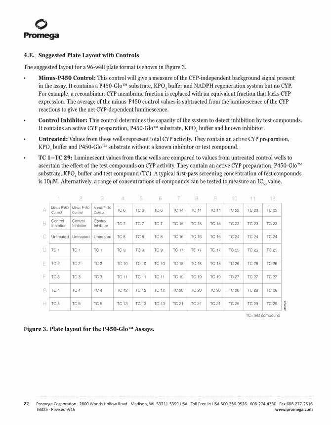
4.E. Suggested Plate Layout with Controls
The suggested layout for a 96-well plate format is shown in Figure 3.
• Minus-P450 Control: This control will give a measure of the CYP-independent background signal present in the assay. It contains a P450-Glo™ substrate, KPO4 buffer and NADPH regeneration system but no CYP. For example, a recombinant CYP membrane fraction is replaced with an equivalent fraction that lacks CYP expression. The average of the minus-P450 control values is subtracted from the luminescence of the CYP reactions to give the net CYP-dependent luminescence.
• Control Inhibitor: This control determines the capacity of the system to detect inhibition by test compounds. It contains an active CYP preparation, P450-Glo™ substrate, KPO4 buffer and known inhibitor.
• Untreated: Values from these wells represent total CYP activity. They contain an active CYP preparation, KPO4 buffer and P450-Glo™ substrate without a known inhibitor or test compound.
• TC 1–TC 29: Luminescent values from these wells are compared to values from untreated control wells to ascertain the effect of the test compounds on CYP activity. They contain an active CYP preparation, P450-Glo™ substrate, KPO4 buffer and test compound (TC). A typical first-pass screening concentration of test compounds is 10µM. Alternatively, a range of concentrations of compounds can be tested to measure an IC50 value.
4857MA
Minus P450 Minus P450 Minus P450 Control Control Control TC 6 TC 6 TC 6 TC 14 TC 14 TC 14 TC 22 TC 22 TC 22
Control Control ControlInhibitor Inhibitor Inhibitor TC 7 TC 7 TC 7 TC 15 TC 15 TC 15 TC 23 TC 23 TC 23
Untreated Untreated Untreated TC 8 TC 8 TC 8 TC 16 TC 16 TC 16 TC 24 TC 24 TC 24
TC 1 TC 1 TC 1 TC 9 TC 9 TC 9 TC 17 TC 17 TC 17 TC 25 TC 25 TC 25
TC 2 TC 2 TC 2 TC 10 TC 10 TC 10 TC 18 TC 18 TC 18 TC 26 TC 26 TC 26
TC 3 TC 3 TC 3 TC 11 TC 11 TC 11 TC 19 TC 19 TC 19 TC 27 TC 27 TC 27
TC 4 TC 4 TC 4 TC 12 TC 12 TC 12 TC 20 TC 20 TC 20 TC 28 TC 28 TC 28
TC 5 TC 5 TC 5 TC 13 TC 13 TC 13 TC 21 TC 21 TC 21 TC 29 TC 29 TC 29
1 2 3 4 5 6 7 8 9 10 11 12
A
B
C
D
E
F
G
H
TC=test compound
Figure 3. Plate layout for the P450-Glo™ Assays.
Promega Corporation · 2800 Woods Hollow Road · Madison, WI 53711-5399 USA · Toll Free in USA 800-356-9526 · 608-274-4330 · Fax 608-277-2516 23www.promega.com TB325 · Revised 9/16
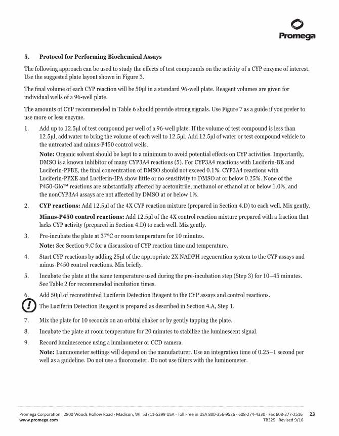
5. Protocol for Performing Biochemical Assays
The following approach can be used to study the effects of test compounds on the activity of a CYP enzyme of interest. Use the suggested plate layout shown in Figure 3.
The final volume of each CYP reaction will be 50µl in a standard 96-well plate. Reagent volumes are given for individual wells of a 96-well plate.
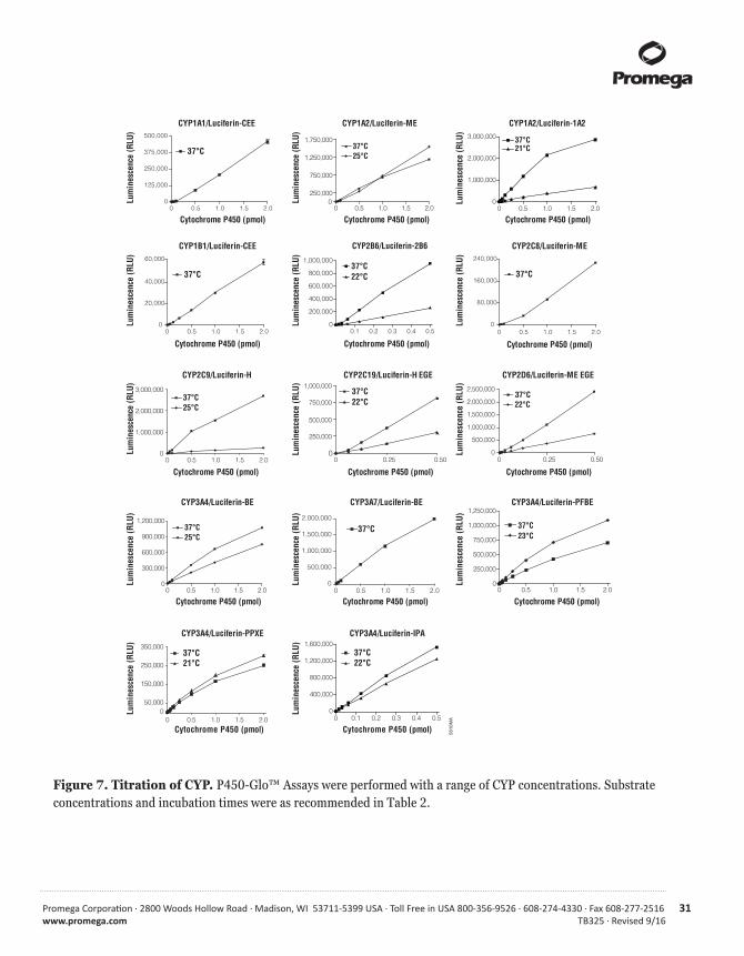
The amounts of CYP recommended in Table 6 should provide strong signals. Use Figure 7 as a guide if you prefer to use more or less enzyme.
1. Add up to 12.5µl of test compound per well of a 96-well plate. If the volume of test compound is less than 12.5µl, add water to bring the volume of each well to 12.5µl. Add 12.5µl of water or test compound vehicle to the untreated and minus-P450 control wells.
Note: Organic solvent should be kept to a minimum to avoid potential effects on CYP activities. Importantly, DMSO is a known inhibitor of many CYP3A4 reactions (5). For CYP3A4 reactions with Luciferin-BE and Luciferin-PFBE, the final concentration of DMSO should not exceed 0.1%. CYP3A4 reactions with Luciferin-PPXE and Luciferin-IPA show little or no sensitivity to DMSO at or below 0.25%. None of the P450-Glo™ reactions are substantially affected by acetonitrile, methanol or ethanol at or below 1.0%, and the nonCYP3A4 assays are not affected by DMSO at or below 1%.
2. CYP reactions: Add 12.5µl of the 4X CYP reaction mixture (prepared in Section 4.D) to each well. Mix gently.
Minus-P450 control reactions: Add 12.5µl of the 4X control reaction mixture prepared with a fraction that lacks CYP activity (prepared in Section 4.D) to each well. Mix gently.
3. Pre-incubate the plate at 37°C or room temperature for 10 minutes.
Note: See Section 9.C for a discussion of CYP reaction time and temperature.
4. Start CYP reactions by adding 25µl of the appropriate 2X NADPH regeneration system to the CYP assays and minus-P450 control reactions. Mix briefly.
5. Incubate the plate at the same temperature used during the pre-incubation step (Step 3) for 10–45 minutes. See Table 2 for recommended incubation times.
6. Add 50µl of reconstituted Luciferin Detection Reagent to the CYP assays and control reactions.
The Luciferin Detection Reagent is prepared as described in Section 4.A, Step 1.
7. Mix the plate for 10 seconds on an orbital shaker or by gently tapping the plate.
8. Incubate the plate at room temperature for 20 minutes to stabilize the luminescent signal.
9. Record luminescence using a luminometer or CCD camera.
Note: Luminometer settings will depend on the manufacturer. Use an integration time of 0.25–1 second per well as a guideline. Do not use a fluorometer. Do not use filters with the luminometer.
!

24 Promega Corporation · 2800 Woods Hollow Road · Madison, WI 53711-5399 USA · Toll Free in USA 800-356-9526 · 608-274-4330 · Fax 608-277-2516TB325 · Revised 9/16 www.promega.com
6. Results
Calculate the net luminescence of each CYP assay by subtracting the luminescence of the average of the minus-P450 control wells [untreated – minus-P450 control = net total CYP activity; treated – minus-P450 control = net treated CYP activity]. Treatment effects are typically seen as decreases due to CYP inhibition. However, some test compounds increase signal because they exhibit positive cooperativity with the P450-Glo™ substrate. This phenomenon has been reported for CYP2C9 and 3A4 (5–7).
7. Quantifying P450-Glo™ Signals with d-Luciferin Standard Curves
The concentration of d-luciferin generated by CYP in P450-Glo™ Assays can be determined by comparing luminescence from CYP reactions to luminescence from a d-luciferin standard curve. The range of d-luciferin concentrations generated in P450-Glo™ Assays is in the linear portion of the standard curve for d-luciferin as illustrated in Figure 4. Standard curve measurements should be performed at the same time and in the same plate as samples. Use the plate layout shown in Figure 5. By comparing signals from CYP reactions to those from d-luciferin standards, the quantity of d-luciferin generated by CYP can be determined.
See Section 7.B for an explanation of why interpolated values for Luciferin-PPXE reactions are multiplied by two.

Promega Corporation · 2800 Woods Hollow Road · Madison, WI 53711-5399 USA · Toll Free in USA 800-356-9526 · 608-274-4330 · Fax 608-277-2516 25www.promega.com TB325 · Revised 9/16
4856
MA
A.
B.
C.
Inhibition of CYP2C9
0
100,000
200,000
300,000
0.001 0.01 0.1 1 10
Sulfaphenazole Concentration (µM)
D-luciferin Concentration (µM)
Sulfaphenazole Concentration (µM)
Lum
ines
cenc
e (R
LU)
Lum
ines
cenc
e (R
LU)
D-luciferin Standard Curve
0.0 0.5 1.0 1.5 2.00
500,000
1,000,000
1,500,000
0.25
0.75
1.25
0.001 0.01 0.1 1 10
Inhibition of CYP2C9
CYP2
C9 A
ctiv
ity(p
mol
D-l
ucife
rin/
pmol
CYP
2C9/
min
ute)
0
Figure 4. Representative P450-Glo™ Assay data. CYP2C9 reactions were performed in the presence or absence of the CYP2C9 inhibitor, sulfaphenazole, as described in Section 5. Panel A. Inhibition of CYP2C9 by sulfaphenazole is expressed in terms of relative light units (RLU). Panel B. A d-luciferin standard curve was performed in parallel with CYP2C9 reactions as described in Section 7.A and analyzed by linear regression (r2 = 0.99). Panel C. Luminescent signals from CYP2C9 reactions were compared to those from the d-luciferin standard curve to interpolate the d-luciferin concentrations. d-luciferin concentrations then were used to calculate CYP2C9 reaction rates (pmol d-luciferin/pmol CYP2C9/minute). The IC50 value derived from Panel A or C is 0.2µM. Luminescence was measured using a POLARstar luminometer (BMG Labtech).
26 Promega Corporation · 2800 Woods Hollow Road · Madison, WI 53711-5399 USA · Toll Free in USA 800-356-9526 · 608-274-4330 · Fax 608-277-2516TB325 · Revised 9/16 www.promega.com
7. Quantifying P450-Glo™ Signals with d-Luciferin Standard Curves (continued)
4858
MA
2.0µM 2.0µM 2.0µM TC 1 TC 1 TC 1 TC 9 TC 9 TC 9 TC 17 TC 17 TC 17
0.4µM 0.4µM 0.4µM TC 2 TC 2 TC 2 TC 10 TC 10 TC 10 TC 18 TC 18 TC 18
0.08µM 0.08µM 0.08µM TC 3 TC 3 TC 3 TC 11 TC 11 TC 11 TC 19 TC 19 TC 19
0.016µM 0.016µM 0.016µM TC 4 TC 4 TC 4 TC 12 TC 12 TC 12 TC 20 TC 20 TC 20
0.0µM 0.0µM 0.0µM TC 5 TC 5 TC 5 TC 13 TC 13 TC 13 TC 21 TC 21 TC 21
Control Control ControlInhibitor Inhibitor Inhibitor TC 6 TC 6 TC 6 TC 14 TC 14 TC 14 TC 22 TC 22 TC 22
TC 7 TC 7 TC 7 TC 15 TC 15 TC 15 TC 23 TC 23 TC 23
Untreated Untreated Untreated TC 8 TC 8 TC 8 TC 16 TC 16 TC 16 TC 24 TC 24 TC 24
1 2 3 4 5 6 7 8 9 10 11 12
A
B
C
D
E
F
G
H
TC=test compound
D-Luciferin Standards
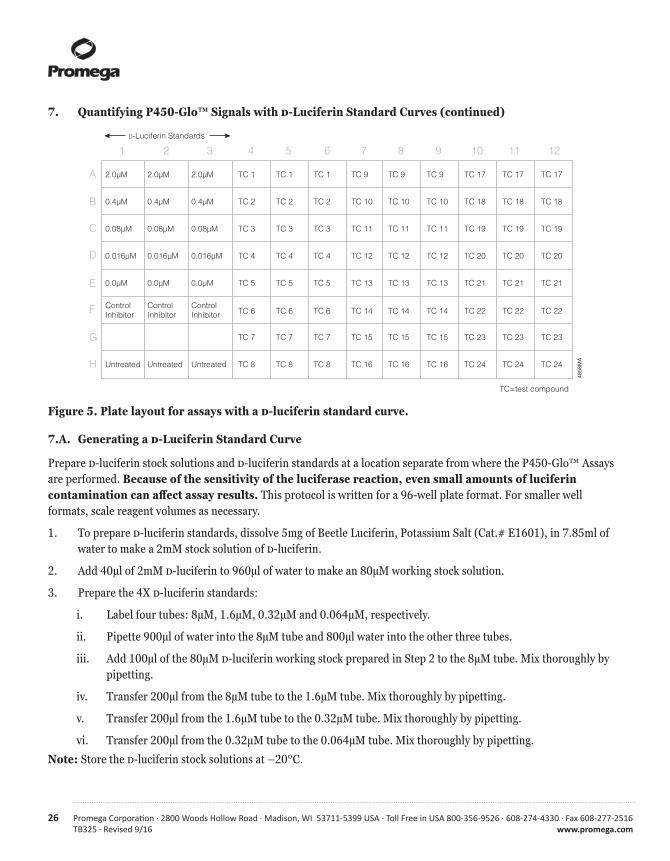
Figure 5. Plate layout for assays with a d-luciferin standard curve.
7.A. Generating a d-Luciferin Standard Curve
Prepare d-luciferin stock solutions and d-luciferin standards at a location separate from where the P450-Glo™ Assays are performed. Because of the sensitivity of the luciferase reaction, even small amounts of luciferin contamination can affect assay results. This protocol is written for a 96-well plate format. For smaller well formats, scale reagent volumes as necessary.
1. To prepare d-luciferin standards, dissolve 5mg of Beetle Luciferin, Potassium Salt (Cat.# E1601), in 7.85ml of water to make a 2mM stock solution of d-luciferin.
2. Add 40µl of 2mM d-luciferin to 960µl of water to make an 80µM working stock solution.
3. Prepare the 4X d-luciferin standards:
i. Label four tubes: 8µM, 1.6µM, 0.32µM and 0.064µM, respectively.
ii. Pipette 900µl of water into the 8µM tube and 800µl water into the other three tubes.
iii. Add 100µl of the 80µM d-luciferin working stock prepared in Step 2 to the 8µM tube. Mix thoroughly by pipetting.
iv. Transfer 200µl from the 8µM tube to the 1.6µM tube. Mix thoroughly by pipetting.
v. Transfer 200µl from the 1.6µM tube to the 0.32µM tube. Mix thoroughly by pipetting.
vi. Transfer 200µl from the 0.32µM tube to the 0.064µM tube. Mix thoroughly by pipetting.
Note: Store the d-luciferin stock solutions at –20°C.

Promega Corporation · 2800 Woods Hollow Road · Madison, WI 53711-5399 USA · Toll Free in USA 800-356-9526 · 608-274-4330 · Fax 608-277-2516 27www.promega.com TB325 · Revised 9/16
4. Prepare the 4X CYP reaction mixture, 4X control reaction mixture and 2X NADPH regeneration system as described in Section 4. Prepare enough 4X control reaction mixture for all standards. Also, be sure to add the appropriate P450-Glo™ substrate to the 4X control reaction mixture.
5. Add 12.5µl of 4X d-luciferin standards to the appropriate wells (8µM standards to wells labeled 2µM, 1.6µM standards to wells labeled 0.4µM, 0.32µM standards to wells labeled 0.08µM, and 0.064µM to wells labeled 0.016µM). Add 12.5µl of water to 0µM d-luciferin wells. Take care to avoid cross-contaminating the wells with d-luciferin.
Note: The luminescence from the sample labeled as 0µM is equivalent to the minus-P450 control in Figure 3.
6. Add 12.5µl of the 4X control reaction mixture to the 2µM, 0.4µM, 0.08µM, 0.016µM and 0µM standard wells.
7. Set up wells with control and test compounds, and proceed with the assay as described in Section 5.
7.B. Data Analysis
1. Subtract the average luminescence of the 0µM d-luciferin standard wells from all luminescence values (including 0µM d-luciferin).
2. Perform linear regression analysis of luminescence from standards to generate a standard curve, where X represents the d-luciferin concentration and Y represents the luminescence (in relative light units, RLU).
3. Interpolate CYP-generated d-luciferin concentrations in test samples by comparing their RLU values to those of the standard curve.
4 To convert d-luciferin concentrations to a CYP reaction rate, consider the interpolated d-luciferin concentration, reaction volume, incubation time and amount of CYP assayed. For example, a 30-minute reaction with 1pmol of CYP generates 1µM d-luciferin. In a 50µl reaction volume, 1µM d-luciferin is 50pmol. The activity is 50pmol d-luciferin/pmol CYP/30 minutes or 1.67pmol d-luciferin/pmol CYP/minute.
Note: The luminescence from all samples should not be higher than that of the 2µM standard. If any values exceed that of the highest standard, the range of the standard curve can be extended by including standards at higher concentrations (e.g., 10µM and 50µM d-luciferin).
Luciferin concentrations interpolated for Luciferin-PPXE reactions are half of their true values because Luciferin-PPXE is provided as a 50:50 mixture of d- and l-forms and the Luciferin Detection Reagent only detects d-luciferin. The rate of metabolism by CYP enzymes of d-luciferin-PPXE and l-luciferin-PPXE are equal, so the reaction product is a 50:50 mixture of d-luciferin and l-luciferin. To calculate the true values, multiply interpolated values by two.
!

28 Promega Corporation · 2800 Woods Hollow Road · Madison, WI 53711-5399 USA · Toll Free in USA 800-356-9526 · 608-274-4330 · Fax 608-277-2516TB325 · Revised 9/16 www.promega.com
8. Km Measurements
The Km value for a given CYP may vary somewhat between enzyme preparations (8). The concentrations of P450-Glo™ substrates recommended here are representative Km concentrations for recombinant CYP enzyme preparations. When measuring Km values, Luciferin-H, Luciferin-ME, Luciferin-CEE, Luciferin-H EGE and Luciferin-ME EGE cause a partial inhibition of luciferase at the upper end of the concentration ranges tested and thus diminish the brightness of the detection step. Such luciferase inhibition is not observed with Luciferin-BE, Luciferin-PFBE, Luciferin-PPXE, Luciferin-IPA, Luciferin-1A2 or Luciferin-2B6. Without compensating for luciferase inhibition by the former sub-strates, the system is less sensitive to detect luciferin at the high end of the substrate concentration range than at the low end, resulting in an underestimate of the Km value. For the Km values of reactions with Luciferin-ME, Luciferin-CEE, Luciferin-H, Luciferin-H EGE and Luciferin-ME EGE reported in Table 2, compensation for luciferase inhibition was made by performing CYP reactions at a range of substrate concentrations, stopping the reaction by adding the reconstituted Luciferin Detection Reagent, then adjusting the substrate concentration in all reactions to the highest concentration in the range. In this way the sensitivity of luciferase to detect CYP-generated luciferin was equalized across the range of substrate concentrations. No compensation was made for the substrate consumed during the CYP reaction because less than 1% of the total substrate was consumed. Km values measured using this method were in good agreement with values determined by integration of a luciferin peak using HPLC (data not shown).
9. General Considerations
9.A. Substrate Specificity
Some P450-Glo™ substrates have enhanced CYP enzyme selectivity over many conventional substrates. However, different CYP enzymes can react with more than one P450-Glo™ substrate (Figure 6). For best results, we recommend the enzyme and substrate combinations shown in Tables 1 and 2.
9.B. Cytochrome P450 Concentration
Although it is necessary to use enough CYP enzyme to generate a detectable amount of luciferin, large amounts of protein or phospholipid from microsome preparations can bind nonspecifically to a drug or inhibitor, leading to a reduction in the effective concentration and overestimation of Km and Ki values (8). General recommendations for the amount of CYP are made in Table 2. CYP concentrations can be increased for brighter signals or reduced further as a cost-saving measure or to reduce nonspecific binding. The enzyme titration curves shown in Figure 7 can be used as a guide if you prefer to use more or less enzyme.
9.C. Assay Time and Temperature
CYP reactions are generally performed at 37°C, but they also may be performed at room temperature (20–25°C) as shown in Figure 8. The suggested incubation times (Table 2) give strong signals within the linear range of the assays (Figure 8). If you prefer a different incubation time, refer to Figure 8 to determine if a shorter time gives adequate signal or if a longer time is within the linear range.
Promega Corporation · 2800 Woods Hollow Road · Madison, WI 53711-5399 USA · Toll Free in USA 800-356-9526 · 608-274-4330 · Fax 608-277-2516 29www.promega.com TB325 · Revised 9/16
5563
MA
Luciferin-CEE
cont
rol
CYP
1A1
CYP
1A2
CYP
1B1
CYP
2A6
CYP
2B6
CYP
2C8
CYP
2C9
CYP
2C18
CYP
2C19
CYP
2D6
CYP
2E1
CYP
2J2
CYP
3A4
CYP
3A5
CYP
3A7
CYP
4A11
CYP
4F2
CYP
4F3A
CYP
4F3B
CYP
4F12
CYP
19
0
25,000
50,000
75,000
100,000
125,000
150,000
175,000Lu
min
esce
nce/
pmol
CYP
450
Lum
ines
cenc
e/pm
ol C
YP45
0 Luciferin-H
cont
rol
CYP
1A1
CYP
1A2
CYP
1B1
CYP
2A6
CYP
2B6
CYP
2C8
CYP
2C9
CYP
2C18
CYP
2C19
CYP
2D6
CYP
2E1
CYP
2J2
CYP
3A4
CYP
3A5
CYP
3A7
CYP
4A1 1
CYP
4F2
CYP
4F3A
CYP
4F3B
CYP
4F12
CYP
19
0
100,000
200,000
300,000
400,000
Lum
ines
cenc
e/pm
ol C
YP45
0 Luciferin-H EGE
cont
rol
CYP
1A1
CYP
1A2
CYP
1B1
CYP
2A6
CYP
2B6
CYP
2C8
CYP
2C9
CYP
2C18
CYP
2C19
CYP
2D6
CYP
2E1
CYP
2J2
CYP
3A4
CYP
3A5
CYP
3A7
CYP
4A1 1
CYP
4F2
CYP
4F3A
CYP
4F3B
CYP
4F12
CYP
19
50,000
150,000
250,000
350,000
450,000
550,000
650,000
A. B.
C. D.
E. F.
Lum
ines
cenc
e/pm
ol C
YP45
0
Luciferin-ME
cont
rol
CYP
1A1
CYP
1A2
CYP
1B1
CYP
2A6
CYP
2B6
CYP
2C8
CYP
2C9
CYP
2C18
CYP
2C19
CYP
2D6
CYP
2E1
CYP
2J2
CYP
3A4
CYP
3A5
CYP
3A7
CYP
4A11
CYP
4F2
CYP
4F3A
CYP
4F3B
CYP
4F12
CYP
19
0
25,000
50,000
75,000
100,000
125,000
225,000
Luciferin-1A2
cont
rol
CYP
1A1
CYP
1A2
CYP
1B1
CYP
2A6
CYP
2B6
CYP
2C8
CYP
2C9
CYP
2C18
CYP
2C19
CYP
2D6
CYP
2E1
CYP
2J2
CYP
3A4
CYP
3A5
CYP
3A7
CY
P4A
11C
YP4F
2C
YP
4F3A
CY
P4F
3BC
YP
4F12
CYP
19
0
250,000
500,000
750,000
1,000,000
1,250,000
Lum
ines
cenc
e/0.
5pm
ol C
YP45
0
cont
rol
CYP
1A1
CYP
1A2
CYP
1B1
CYP
2A6
CYP
2B6
CYP
2C8
CYP
2C9
CYP
2C18
CYP
2C19
CYP
2D6
CYP
2E1
CYP
2J2
CYP
3A4
CYP
3A5
CYP
3A7
CY
P4A
11C
YP4F
2C
YP
4F3A
CY
P4F
3BC
YP
4F12
CYP
19Lum
ines
cenc
e/0.
1pm
ol C
YP45
0
Luciferin-2B6
0
300,000
200,000
100,000
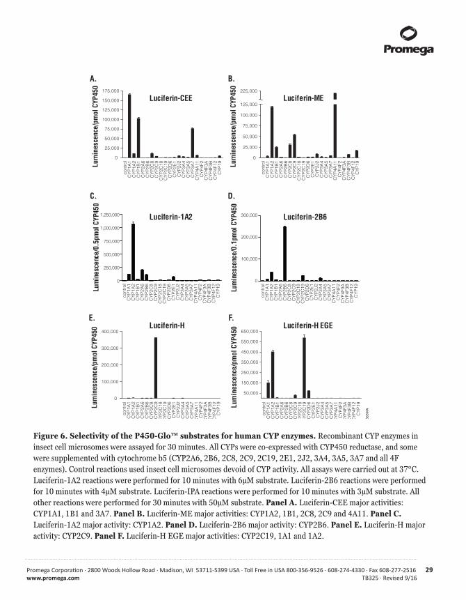
Figure 6. Selectivity of the P450-Glo™ substrates for human CYP enzymes. Recombinant CYP enzymes in insect cell microsomes were assayed for 30 minutes. All CYPs were co-expressed with CYP450 reductase, and some were supplemented with cytochrome b5 (CYP2A6, 2B6, 2C8, 2C9, 2C19, 2E1, 2J2, 3A4, 3A5, 3A7 and all 4F enzymes). Control reactions used insect cell microsomes devoid of CYP activity. All assays were carried out at 37°C. Luciferin-1A2 reactions were performed for 10 minutes with 6µM substrate. Luciferin-2B6 reactions were performed for 10 minutes with 4µM substrate. Luciferin-IPA reactions were performed for 10 minutes with 3µM substrate. All other reactions were performed for 30 minutes with 50µM substrate. Panel A. Luciferin-CEE major activities: CYP1A1, 1B1 and 3A7. Panel B. Luciferin-ME major activities: CYP1A2, 1B1, 2C8, 2C9 and 4A11. Panel C. Luciferin-1A2 major activity: CYP1A2. Panel D. Luciferin-2B6 major activity: CYP2B6. Panel E. Luciferin-H major activity: CYP2C9. Panel F. Luciferin-H EGE major activities: CYP2C19, 1A1 and 1A2.

30 Promega Corporation · 2800 Woods Hollow Road · Madison, WI 53711-5399 USA · Toll Free in USA 800-356-9526 · 608-274-4330 · Fax 608-277-2516TB325 · Revised 9/16 www.promega.com
5563
MD
H.
I. J.
K.
Lum
ines
cenc
e/pm
ol C
YP45
0 Luciferin-BE
50,000
150,000
100,000
200,000
1,300,000
cont
rol
CYP
1A1
CYP
1A2
CYP
1B1
CYP
2A6
CYP
2B6
CYP
2C8
CYP
2C9
CYP
2C18
CYP
2C19
CYP
2D6
CYP
2E1
CYP
3A4
CYP
3A5
CYP
3A7
CYP
2J2
CYP
4A11
CYP
4F2
CYP
4F3A
CYP
4F3B
CYP
4F12
CYP
19
0
Luciferin-PPXE
cont
rol
CYP
1A1
CYP
1A2
CYP
1B1
CYP
2A6
CYP
2B6
CYP
2C8
CYP
2C9
CYP
2C18
CYP
2C19
CYP
2D6
CYP
2E1
CYP
2J2
CYP
3A4
CYP
3A5
CYP
3A7
CYP
4A11
CYP
4F2
CYP
4F3 A
CYP
4F3B
CYP
4F12
CYP
19
0
100,000
200,000
300,000
400,000
Lum
ines
cenc
e/pm
ol C
YP45
0Luciferin-IPA
cont
rol
CYP
1A1
CYP
1A2
CYP
1B1
CYP
2A6
CYP
2B6
CYP
2C8
CYP
2C9
CYP
2C18
CYP
2C19
CYP
2D6
CYP
2E1
CYP
2J2
CYP
3A4
CYP
3A5
CYP
3A7
CYP
4A11
CYP
4F2
CYP
4F3A
CYP
4F3B
CYP
4F12
CYP
19
0
100,000
200,000
300,000
400,000
500,000
Lum
ines
cenc
e/0.
1pm
ol C
YP45
0Lu
min
esce
nce/
pmol
CYP
450
cont
rol
CYP
1A1
CYP
1A2
CYP
1B1
CYP
2A6
CYP
2B6
CYP
2C8
CYP
2C9
CYP
2C18
CYP
2C19
CYP
2D6
CYP
2E1
CYP
2J2
CYP
3A4
CYP
3A5
CYP
3A7
CYP
4A11
CYP
4F2
CYP
4F3A
CYP
4F3B
CYP
4F12
CYP
19
0
25,000
50,000
75,000
100,000
Luciferin-PFBE450,000
G.Lu
min
esce
nce/
pmol
CYP
450
Luciferin-ME EGE
cont
rol
CYP
1A1
CYP
1A2
CYP
1B1
CYP
2A6
CYP
2B6
CYP
2C8
CYP
2C9
CYP
2C18
CYP
2C19
CYP
2D6
CYP
2E1
CYP
2J2
CYP
3A4
CYP
3A5
CYP
3A7
CYP
4A11
CYP
4F2
CYP
4F3A
CYP
4F3B
CYP
4F12
CYP
19
0
1,500,000
125,000
250,000
375,000
500,000
Figure 6. Selectivity of the P450-Glo™ substrates for human CYP enzymes (continued). Panel G. Luciferin-ME EGE major activities: CYP2D6, 1A1 and 1A2. Panel H. Luciferin-IPA major activity: CYP3A4. Panel I. Luciferin-BE major activities: CYP3A4, 3A5, 3A7 and 4F12. Panel J. Luciferin-PFBE major activities: CYP3A4, 3A5 and 3A7. Panel K. Luciferin-PPXE major activities: CYP3A4, 3A5 and 3A7.
Promega Corporation · 2800 Woods Hollow Road · Madison, WI 53711-5399 USA · Toll Free in USA 800-356-9526 · 608-274-4330 · Fax 608-277-2516 31www.promega.com TB325 · Revised 9/16
CYP1A1/Luciferin-CEE
0 0.5 1.0 1.5 2.00
125,000
250,000
375,000
500,000
37°C
Cytochrome P450 (pmol)
Lum
ines
cenc
e (R
LU)
CYP1B1/Luciferin-CEE
0 0.5 1.0 1.5 2.00
20,000
40,000
60,000
37°C
Cytochrome P450 (pmol)
Lum
ines
cenc
e (R
LU)
CYP2C8/Luciferin-ME
0 0.5 1.0 1.5 2.00
80,000
160,000
240,000
37°C
Cytochrome P450 (pmol)
Lum
ines
cenc
e (R
LU)
5610
MA
CYP3A7/Luciferin-BE
0 0.5 1.0 1.5 2.00
500,000
1,000,000
1,500,000
2,000,000
37°C
Cytochrome P450 (pmol)
Lum
ines
cenc
e (R
LU)
CYP1A2/Luciferin-ME
Cytochrome P450 (pmol)
Lum
ines
cenc
e (R
LU)
0 0.5 1.0 1.5 2.0
37°C25°C
250,000
750,000
1,250,000
1,750,000
0
CYP2C9/Luciferin-H
Cytochrome P450 (pmol)
Lum
ines
cenc
e (R
LU)
0 0.5 1.0 1.5 2.00
1,000,000
2,000,000
3,000,00037°C25°C
Cytochrome P450 (pmol)
CYP2C19/Luciferin-H EGE
Lum
ines
cenc
e (R
LU)
37°C22°C
0 0.25 0.500
250,000
500,000
750,000
1,000,000CYP2D6/Luciferin-ME EGE
Cytochrome P450 (pmol)
Lum
ines
cenc
e (R
LU)
0 0.25 0.500
500,000
1,000,000
1,500,000
2,000,000
2,500,000
22°C37°C
CYP3A4/Luciferin-BE
Cytochrome P450 (pmol)
Lum
ines
cenc
e (R
LU)
0 0.5 1.0 1.5 2.00
300,000
600,000
900,000
1,200,00037°C25°C
Cytochrome P450 (pmol)
Lum
ines
cenc
e (R
LU)
CYP3A4/Luciferin-PFBE
0.5 1.0 1.5 2.00
0
250,000
500,000
750,000
1,000,000
1,250,000
23°C37°C
CYP3A4/Luciferin-PPXE
0 0.5 1.0 1.5 2.00
21°C37°C
Cytochrome P450 (pmol)
Lum
ines
cenc
e (R
LU)
50,000
150,000
250,000
350,000
Lum
ines
cenc
e (R
LU)
22°C37°C
Cytochrome P450 (pmol)
CYP3A4/Luciferin-IPA
0 0.1 0.2 0.3 0.4 0.50
400,000
800,000
1,200,000
1,600,000
37°C21°C
CYP1A2/Luciferin-1A2
0 0.5 1.0 1.5 2.00
1,000,000
2,000,000
3,000,000
Cytochrome P450 (pmol)
Lum
ines
cenc
e (R
LU)
Cytochrome P450 (pmol)
Lum
ines
cenc
e (R
LU)
CYP2B6/Luciferin-2B6
0.1 0.2 0.3 0.4 0.50
200,000
400,000
600,000
800,000
1,000,00037°C22°C
Figure 7. Titration of CYP. P450-Glo™ Assays were performed with a range of CYP concentrations. Substrate concentrations and incubation times were as recommended in Table 2.

32 Promega Corporation · 2800 Woods Hollow Road · Madison, WI 53711-5399 USA · Toll Free in USA 800-356-9526 · 608-274-4330 · Fax 608-277-2516TB325 · Revised 9/16 www.promega.com
5574
MA
CYP1B1/Luciferin-CEE
0 15 30 45 60
20,000
50,000
80,000
110,000
140,000
25°C37°C
Time (minutes)
Lum
ines
cenc
e (R
LU)
CYP1A2/Luciferin-ME
0 15 30 45 60 75 90
250,000
500,000
750,000
0
37°C25°C
Time (minutes)
Lum
ines
cenc
e (R
LU)
CYP2C8/Luciferin-ME
0 15 30 45 60 75 900
60,000
120,000
180,000
25°C37°C
Time (minutes)
Lum
ines
cenc
e (R
LU)
CYP2C19/Luciferin-H EGE
150,000
350,000
550,000
0 15 30 45 60 70
25°C37°C
Time (minutes)
Lum
ines
cenc
e (R
LU)
CYP2C9/Luciferin-H
0 15 30 45 60 75 90
100,000
300,000
500,000
700,000
25°C37°C
Time (minutes)
Lum
ines
cenc
e (R
LU)
CYP2D6/Luciferin-ME EGE
0 15 30 45 60 75 90
1,000,000
2,000,000
3,000,000
4,000,000
5,000,000
0
25°C37°C
Time (minutes)
Lum
ines
cenc
e (R
LU)
CYP3A7/Luciferin-BE
0 15 30 45 60 75 900
300,000
600,000
900,000
1,200,00037°C25°C
Lum
ines
cenc
e (R
LU)
Time (minutes)0 10 20 30 40 50 60 70 80 90
37°C25°C
CYP3A4/Luciferin-BE
0
250,000
500,000
750,000
1,000,000
Time (minutes)
Lum
ines
cenc
e (R
LU)
37°C25°C
Time (minutes)
Lum
ines
cenc
e (R
LU)
CYP3A4/Luciferin-PFBE
10 20 30 40 50 600
125,000
250,000
375,000
500,000
CYP1A1/Luciferin-CEE
0 15 30 45 60 75 90
125,000
250,000
0
25°C37°C
Time (minutes)
Lum
ines
cenc
e (R
LU)
187,500
62,500
CYP3A4/Luciferin-PPXE
0 10 20 30 400
100,000
200,000
150,000
50,000
37°C25°C
Time (minutes)
Lum
ines
cenc
e (R
LU)
Lum
ines
cenc
e (R
LU)
37°C22°C
Time (minutes)
CYP3A4/Luciferin-IPA
0 5 10 15 20
50,000
150,000
250,000
350,000
450,000
37°C21°C
CYP1A2/Luciferin-1A2
0 5 10 15 20 25 300
500,000
1,000,000
1,500,000
2,000,000
Time (minutes)
Lum
ines
cenc
e (R
LU)
Time (minutes)
Lum
ines
cenc
e (R
LU)
CYP2B6/Luciferin-2B6
0 5 10 15 200
100,000
200,000
300,000
400,000
500,00037°C22°C
Figure 8. Incubation time and temperature. P450-Glo™ reactions (50µl) were performed with the enzyme and substrate concentrations indicated in Table 2. Reactions were incubated at room temperature (20–25°C) or 37°C for up to 90 minutes before adding reconstituted Luciferin Detection Reagent.

Promega Corporation · 2800 Woods Hollow Road · Madison, WI 53711-5399 USA · Toll Free in USA 800-356-9526 · 608-274-4330 · Fax 608-277-2516 33www.promega.com TB325 · Revised 9/16
Cell-Based Assays
10. Protocol for Performing Cell-Based Assays
The P450-Glo™ substrates and reaction products are cell-permeable. This allows development of cell-based assays (1–4). In these assays, a luminogenic substrate is incubated with cultured cells for an appropriate period of time. Intracellular CYP enzymes convert the substrate to luciferin product, which passes out of cells and then can be detected with the Luciferin Detection Reagent (Figure 9). The luciferin produced is measured using either a nonlytic assay (perform the P450-Glo™ Assay with an aliquot of intact-cell supernatant) or a lytic assay (perform the P450-Glo™ Assay in a well containing cells). In either type of assay, the light output of the luciferase reaction is proportional to CYP activity. In a nonlytic assay, after an aliquot of the reaction medium is removed, the remaining cells can be used for additional cell-based testing; for example, a cell viability assay may be run to normalize CYP activities to viable cell number (e.g., CellTiter-Glo® Luminescent Cell Viability Assay; Section 13.D).
Proluciferin(P450-Glo™ Substrate)
Proluciferin
LuciferinProduct
1240
2MA
LightLiigggggghhtLight
+ CYP450 Luciferin DetectionReagent
Figure 9. Cell-based P450-Glo™ Assay principle. A P450-Glo™ substrate (proluciferin) enters cells from the culture medium. Inside the cell, a P450 enzyme converts the proluciferin to a luciferin product, and luciferin is formed and detected with Luciferin Detection Reagent (LDR).
Cell-based applications of P450-Glo™ Assays include measurements of basal CYP activities, induction of these activities by test compounds and inhibition of both basal and induced CYP activities by test compounds (9). CYP inductions also can be observed as marker events to detect nuclear receptor and aryl hydrocarbon receptor ligands as exemplified by the following:
• CYP3A/Luciferin-IPA and CYP3A/Luciferin-PFBE activities are induced by ligands for the pregnane X receptor (PXR), constitutive androstane receptor (CAR) and glucocorticoid receptor (GR).
• CYP1A2/Luciferin-1A2 activity is induced by ligands for the aryl hydrocarbon receptor (AHR).
• CYP2B6/Luciferin-2B6 activity is induced by ligands for the constitutive androstane receptor (CAR) and pregnane X receptor (PXR).
• CYP1A/Luciferin-CEE activity is induced by ligands for the aryl hydrocarbon receptor (AHR).

34 Promega Corporation · 2800 Woods Hollow Road · Madison, WI 53711-5399 USA · Toll Free in USA 800-356-9526 · 608-274-4330 · Fax 608-277-2516TB325 · Revised 9/16 www.promega.com
10. Protocol for Performing Cell-Based Assays (continued)
Chemically mediated CYP gene inductions have been observed in numerous cultured cell types. Transcriptional inducers of CYP genes cause increased CYP enzyme levels, which can be measured using the P450-Glo™ Assay. The following protocols relate specifically to assays of monolayer hepatocyte cultures; however the general methods are applicable to other cell types that express CYPs (e.g., HepaRG, HepG2, MCF7, FA2N-4, DPX-2). Cell-based CYP induction assays are typically performed with cells grown in monolayers. With hepatocytes, it is essential to follow a well defined protocol to maintain CYP expression and an inducible phenotype. The P450-Glo™ Assay is used to measure gene induction in hepatocytes cultured according to the methods described in Sections 11.B and 11.C, and we expect that other established culture/induction schemes can be followed (10).
10.A. Measuring CYP Activity in Cultured Cells
The P450-Glo™ Assays recommended for cell-based applications are:
• P450-Glo™ CYP3A4 Assay with Luciferin-IPA (Cat.# V9001, V9002) and P450-Glo™ CYP3A4 Assay with Luciferin-PFBE (Cat.# V8901, V8902)
These assays can be used to selectively measure CYP3A activity in hepatocytes. Significant activities of other human CYPs have not been detected with Luciferin-IPA or Luciferin-PFBE.
• P450-Glo™ CYP1A2 Assay with Luciferin-1A2 (Cat.# V8421, V8422)
This assay can be used to measure CYP1A2 activity in hepatocytes. Luciferin-1A2 is highly selective for CYP1A2.
• P450-Glo™ CYP2B6 Assay with Luciferin-2B6 (Cat.# V8321, V8322)
This assay can be used to measure CYP2B6 activity in hepatocytes. Luciferin-2B6 is highly selective for CYP2B6.
• P450-Glo™ CYP2C9 Assay with Luciferin-H (Cat.# V8791, V8792)
This assay can be used to measure CYP2C9 activity in human hepatocytes and other human cell types. Luciferin-H is highly selective for CYP2C9.
• P450-Glo™ CYP1A1 Assay (Cat.# V8751, V8752) and P450-Glo™ CYP1B1 Assay (Cat.# V8761, V8762) with Luciferin-CEE.
The P450-Glo™ CYP1A1 Assay can be used to selectively measure CYP1A1 activity in hepatocytes where cross-reacting CYP1B1 and CYP3A7 are absent. However, CYP1A1 and CYP1B1 assays detect a mixture of CYP1A1 and CYP1B1 activities in cell types that express both enzymes.
Consider including an inhibition control for induced P450 activity with a selective P450 inhibitor, such as 5μM a-naphthoflavone for CYP1A2/Luciferin-1A2, 1µM clopidogrel for CYP2B6/Luciferin-2B6, 2μM sulfaphenazole for CYP2C9/Luciferin-H, and 1μM ketoconazole for CYP3A4/Luciferin-IPA. The inhibition control will confirm that the induced activity is from the desired CYP enzyme. Please refer to Figure 6 for the P450-Glo™ substrate selectivities.
Note: The NADPH Regeneration System is not required for cell-based assays. In cell-based assays, NADPH in the cell is sufficient to support CYP activity.!
Promega Corporation · 2800 Woods Hollow Road · Madison, WI 53711-5399 USA · Toll Free in USA 800-356-9526 · 608-274-4330 · Fax 608-277-2516 35www.promega.com TB325 · Revised 9/16
10.B. Nonlytic P450-Glo™ Assays Using Cultured Cells in Monolayers
Reserve some empty wells for background measurements. Recommendations for cell culture conditions are given in Sections 11.B and 11.C.
Optional: If you intend to combine the P450-Glo™ Assay with a luminescent cell viability assay, consider culturing cells on white-walled, collagen-coated plates with clear bottoms (e.g., BioCoat® 96-well plates, Corning Cat.# 354650/ 356650).
1. Treat cells with test compounds. For CYP gene induction studies, cells are typically treated with inducers for 24–72 hours. Optimal treatment time should be determined empirically; however, 48 hours is a common starting point. See Section 11.A for examples of CYP gene inducers that can be used as positive controls. Change medium with test compounds, etc., once daily for the duration of the treatment time.
2. For CYP3A4/Luciferin-IPA, CYP3A4/Luciferin-PFBE, CYP2C9/Luciferin-H, CYP1A1/Luciferin-CEE, and CYP1B1/Luciferin-CEE assays, replace culture medium with fresh medium containing a luminogenic CYP substrate after the experimental treatment as indicated in Table 7. See Note 1.
Optional: Wash cells with medium or phosphate buffered saline before adding medium with substrate. Some compounds that induce CYP gene expression also inhibit the CYP enzyme activity that has been induced. To observe induction, you may need to remove the inducer by including a wash step prior to adding a luminogenic substrate.
The CYP1A2/Luciferin-1A2 and CYP2B6/Luciferin-2B6 assays must be performed in Krebs-Henseleit Buffer (e.g., BioreclamationIVT Cat.# Z99074, Sigma Cat.# K3753) or in phosphate buffered saline (PBS) after the treatment of cells. Remove the culture medium completely after treatment and wash cells twice with two volumes of Krebs-Henseleit buffer or PBS. Add diluted Luciferin-1A2 or Luciferin-2B6 in Krebs-Henseleit Buffer or phosphate buffered saline (PBS) containing 3mM salicylamide as indicated in Table 7 (see Note 3). These two P450-Glo™ Assays cannot be performed in most cell culture media because the media contain l-cysteine, which interferes with the assays.
Notes:
1. See Section 4.B for substrate preparation instructions.
2. If inhibitors of basal or induced CYP enzyme activity are being tested, add them at this point with the luminogenic substrate.
3. Salicylamide (e.g., Sigma Cat.# 860417) is added to inhibit phase II conjugation of the CYP product from CYP1A2/Luciferin-1A2 and CYP2B6/Luciferin-2B6. Prepare a fresh 3M salicylamide stock (1000X) in DMSO from the powder. When diluting salicylamide in Krebs-Henseleit Buffer or PBS, a white precipitate of salicylamide may occur. With mixing, all of the salicylamide will dissolve in the Krebs-Henseleit Buffer or PBS.
3. To determine background luminescence, add luminogenic substrate in medium to a set of empty wells (no cells). For CYP1A2/Luciferin-1A2 and CYP2B6/Luciferin-2B6 assays, add Luciferin-1A2 and Luciferin-2B6, respectively, in the Krebs-Henseleit Buffer or PBS containing 3mM salicylamide to a set of empty wells (no cells).
4. Incubate plates at 37°C for the appropriate time as indicated in Table 7.

36 Promega Corporation · 2800 Woods Hollow Road · Madison, WI 53711-5399 USA · Toll Free in USA 800-356-9526 · 608-274-4330 · Fax 608-277-2516TB325 · Revised 9/16 www.promega.com
10.B. Nonlytic P450-Glo™ Assays Using Cultured Cells in Monolayers (continued)
5. During the incubation prepare the Luciferin Detection Reagent by transferring the entire contents of the bottle of the appropriate reconstitution buffer to the amber bottle containing Luciferin Detection Reagent, as explained in Section 4.A, Step 1.
6. Transfer 25µl of culture medium or buffer from each well to a 96-well opaque white luminometer plate at room temperature, and add 25µl of Luciferin Detection Reagent to initiate a luminescent reaction (for single-tube luminometers use appropriate luminometer tubes or cuvettes).
7. Incubate the plate at room temperature for 20 minutes, then read luminescence using a luminometer or CCD camera.
Note: Luminometer settings will depend on the manufacturer. Use an integration time of 0.25–1 second per well as a guideline. Do not use a fluorometer. Do not use filters with the luminometer.
8. Calculate net signals by subtracting background luminescence values (no-cell control) from test compound-treated and untreated (vehicle control) values.
9. Calculate percent change by dividing net treated values by net untreated values and multiplying by 100.
10. Optional: Perform the CellTiter-Glo® Luminescent Cell Viability Assay (Cat.# G7570, G7571, G7572 and G7573) to normalize P450-Glo™ Assay values to cell number; see Section 11.D.
Table 7. P450-Glo™ Assay Setup.
Assay SubstrateFinal Substrate Concentration
Medium Volume Required for Various Plate Sizes
Incubation Time
96- Well Plate
48- Well Plate
24- Well Plate
6- Well Plate
CYP3A4 Luciferin-IPA 3µM (1:1,000 dilution*)
50µl 150µl 300µl 1.0ml 30–60 minutes
CYP3A4 Luciferin-PFBE 50µM (1:40 dilution*)
50µl 150µl 300µl 1.0ml 3–4 hours
CYP2C9 Luciferin-H 100µM (1:50 dilution*)
50µl 150µl 300µl 1.0ml 3–4 hours
CYP1A1 or CYP1B1
Luciferin-CEE 100µM (1:50 dilution*)
50µl 150µl 300µl 1.0ml 3 hours
CYP1A2 Luciferin-1A2 6µM (1:1,000 dilution**)
50µl 150µl 300µl 1.0ml 30–60 minutes
CYP2B6 Luciferin-2B6 3µM (1:1,000 dilution**)
50µl 150µl 300µl 1.0ml 60–120 minutes
*Dilute the provided substrate stock solution in culture medium.
**Dilute Luciferin-1A2 and Luciferin-2B6 stock solutions in the Krebs-Henseleit Buffer or PBS containing 3mM salicylamide.

Promega Corporation · 2800 Woods Hollow Road · Madison, WI 53711-5399 USA · Toll Free in USA 800-356-9526 · 608-274-4330 · Fax 608-277-2516 37www.promega.com TB325 · Revised 9/16
10.C. Lytic P450-Glo™ Assays Using Cultured Cells in Monolayers
Note: If you intend to read luminescence directly from culture wells, cells should be cultured on white-walled, collagen-coated culture plates with clear bottoms (e.g., BioCoat® 96-well plates, Corning Cat.# 354650/356650).
1. Treat cells with test compounds. For CYP gene induction studies, cells are typically treated with inducers for 24–72 hours. Optimal treatment time should be determined empirically; however, 48 hours is a common starting point. See Table 8 for examples of CYP gene inducers that can be used as positive controls. Change the medium with test compounds, etc., once daily for the duration of the treatment time.
2. For CYP3A4/Luciferin-IPA, CYP3A4/Luciferin-PFBE, CYP2C9/Luciferin-H, CYP1A1/Luciferin-CEE, and CYP1B1/Luciferin-CEE assays, replace culture medium with fresh medium containing a luminogenic CYP substrate after the experimental treatment as indicated in Table 7. See Section 4.B for substrate preparation instructions.
Optional: Wash cells with medium or phosphate buffered saline before adding medium with substrate. Some compounds that induce CYP gene expression also inhibit the CYP enzyme activity that has been induced. To observe induction, you may need to remove the inducer by including a wash step prior to adding a luminogenic substrate.
The CYP1A2/Luciferin-1A2 and CYP2B6/Luciferin-2B6 assays must be performed in Krebs-Henseleit Buffer (e.g., BioreclamationIVT Cat.# Z99074, Sigma Cat.# K3753) or in phosphate buffered saline (PBS) after the treatment of cells. Remove the culture medium completely after treatment and wash cells twice with two volumes of Krebs-Henseleit buffer or PBS. Add diluted Luciferin-1A2 and Luciferin-2B6, respectively, in Krebs-Henseleit Buffer or phosphate buffered saline (PBS) containing 3mM salicylamide as indicated in Table 7 (see Note 3). These two P450-Glo™ Assays cannot be performed in most cell culture media because the media contain l-cysteine, which interferes with the assays.
Notes:
1. See Section 4.B for substrate preparation instructions.
2. If inhibitors of basal or induced CYP enzyme activity are being tested, add them at this point with the luminogenic substrate.
3. Salicylamide (e.g., Sigma Cat.# 860417) is added to inhibit phase II conjugation of the CYP product from CYP1A2/Luciferin/1A2 and CYP2B6/Luciferin-2B6. Prepare a fresh 3M salicylamide stock in DMSO (1000X) from the powder. When diluting salicylamide in Krebs-Henseleit Buffer, a white precipitate of salicylamide may occur. With mixing, all of the salicylamide will dissolve in the Krebs-Henseleit Buffer or PBS.
3. To determine background luminescence, add luminogenic substrate in medium to a set of empty wells (no cells). For CYP1A2/Luciferin-1A2 and CYP2B6/Luciferin-2B6 assays, add Luciferin-1A2 and Luciferin-2B6, respectively, in the Krebs-Heneseleit Buffer of PBS containing 3mM salicylamide to a set of empty wells (no cells).
4. Incubate plates at 37°C, 5% CO2 for the appropriate time as indicated in Table 7.
5. During the incubation, prepare the Luciferin Detection Reagent as described in Section 4.A, Step 1.
6. Add an equal volume of Luciferin Detection Reagent to each well, and mix briefly on a multiwell plate shaker or by gently tapping or swirling the plate to form a lysate.

38 Promega Corporation · 2800 Woods Hollow Road · Madison, WI 53711-5399 USA · Toll Free in USA 800-356-9526 · 608-274-4330 · Fax 608-277-2516TB325 · Revised 9/16 www.promega.com
10.C. Lytic P450-Glo™ Assays Using Cultured Cells in Monolayers (continued)
7. Option 1 (preferred): Read luminescence directly from cell culture plate. In this case, to avoid luminescent cross talk between wells, cells must be grown in a white-walled culture plate with clear-bottom wells.
Option 2: Transfer 50µl of lysate from each well to a 96-well opaque white luminometer plate at room temperature (for single-tube luminometers use appropriate luminometer tubes or cuvettes).
Note: Transferring the lysate containing detergent may impact accuracy of transferred volume. If transferring reaction to a 96-well white plate is preferred, it can be carried out before the addition of Luciferin Detection Reagent. Please refer to Section 10.B for details.
8. Equilibrate the plate at room temperature for 20 minutes.
Note: Luminescence from lysates of some, but not all, cell types may decay rapidly, so it might be necessary to record luminescence within 20 minutes after adding Luciferin Detection Reagent.
9. Read luminescence using a luminometer or CCD camera.
Note: Luminometer settings will depend on the manufacturer. Use an integration time of 0.25–1 second per well as a guideline. Do not use a fluorometer. Do not use filters with the luminometer.
10. Calculate net signals by subtracting background luminescence values (no-cell control) from test compound-treated and untreated (vehicle control) values.
11. Calculate percent change by dividing net treated values by net untreated values and multiplying by 100.
11. General Considerations for Cell-Based Assays
11.A. Examples of CYP Gene Inducers
Upregulation of CYP genes occurs when a test compound binds one of several nuclear receptors and then activates transcription of a CYP gene. The pregnane X receptor (PXR) and constitutive androstane receptor (CAR) regulate the CYP2C8, CYP2C9, CYP2C19 and CYP3A4 genes (11–14); the aryl hydrocarbon receptor (AHR) regulates CYP1A and CYP1B1 (15) genes; the constitutive androstane receptor (CAR) and pregnane X receptor (PXR) regulate CYP2B6 genes.

Promega Corporation · 2800 Woods Hollow Road · Madison, WI 53711-5399 USA · Toll Free in USA 800-356-9526 · 608-274-4330 · Fax 608-277-2516 39www.promega.com TB325 · Revised 9/16
The compounds listed in Table 8 are well known CYP gene inducers. These can be included as positive controls for induction when screening test compounds with unknown effects on CYP gene expression.
Table 8. Common CYP Gene Inducers.
CYP Gene Inducers Pathway
CYP3A4
25µM rifampicin PXR
500µM phenobarbitol CAR
50µM dexamethasone GR
CYP2C925µM rifampicin
PXR/CAR500µM phenobarbitol
CYP1A1/1A2/1B1
10nM 2,3,7,8-tetrachloro-dibenzo-p-dioxin (TCDD)
AHR1µM 3-methylcholanthrene
100µM omeprazole
CYP2B6 1mM phenobarbitol CAR
11.B. Fresh Hepatocyte Cell Cultures
Fresh human hepatocytes adherent on collagen-coated plates or in suspension can be obtained for overnight delivery from various vendors (e.g., BioreclamationIVT and In Vitro ADMET Laboratories). The following considerations for adherent hepatocyte culture conditions have been used successfully with the P450-Glo™ Assay.
Note: The hepatocyte phenotype is best maintained when cells are near confluence.
1. On day 1, viable hepatocytes from a vendor arrive in a multiwell plate at ambient temperature. Remove adhesive seal from the plate, and replace with a loose-fitting multiwell plate lid. Incubate the plate for 2 hours in a 5% CO2, 37°C incubator in the shipping medium.
2. After the 2-hour incubation, remove the shipping medium, and replace with 37°C culture medium formulated for hepatocytes (e.g., In Vitro GRO Hepatocyte Incubation Medium, BioreclamationIVT).
3. Return the plates to the incubator for 1 hour if you are using the sandwich culture method (Step 4). Otherwise, incubate overnight in the incubator.
4. Sandwich culture method (optional): After the 1-hour incubation, remove culture medium, add cold medium (approximately 4°C) containing 0.125mg/ml Matrigel® (Corning) to each well. Return the plates to the incubator overnight.
5. On day two, remove the culture medium, and replace with fresh serum-free medium (without Matrigel®) that contains a control inducer, test compound or vehicle alone.
6. Change the medium with test compounds or controls once daily for the duration of the experiment. For CYP gene induction studies, cells are typically exposed for 24–72 hours. Optimal length of treatment time should be determined empirically, though maximal inductions are typically reached after 48 hours of exposure to an inducer.
7. Perform the P450-Glo™ Assay as described in Section 10.B or 10.C.

40 Promega Corporation · 2800 Woods Hollow Road · Madison, WI 53711-5399 USA · Toll Free in USA 800-356-9526 · 608-274-4330 · Fax 608-277-2516TB325 · Revised 9/16 www.promega.com
11.C. Cryopreserved Hepatocytes
Cryopreserved primary human and animal hepatocytes are available from several suppliers (e.g., BioreclamationIVT and In Vitro ADMET Laboratories). Store the cells in liquid nitrogen. Note that some cryopreserved hepatocytes may not adhere well to 96-well plates and may require wells with a larger surface area. For optimal results, use cells designated as platable (i.e., cells adhere and form monolayers in culture) by the supplier.
1. On day 1, thaw the cells as recommended by the supplier. This typically involves a specialized isolation procedure or thawing medium (e.g., InVitroGro Hepatocyte Thawing Medium, BioreclamationIVT).
2. Estimate the percentage of live cells (e.g., by trypan blue exclusion).
3. Seed approximately 1.5 × 10⁵ cells per cm2 on collagen-coated tissue culture plates in a hepatocytes culture medium (e.g., In Vitro GRO Hepatocyte Plating Medium, BioreclamationIVT).
4. Incubate cells for 6 hours if you are using the sandwich culture method (Step 5). Otherwise, incubate overnight.
5. Sandwich culture method (optional): After the 6-hour incubation, remove culture medium, add cold medium (approximately 4°C) containing 0.125mg/ml Matrigel® (Corning) to each well. Return the plates to the incubator overnight.
6. On day two, remove the culture medium, and replace with fresh serum-free medium without Matrigel® (e.g., In Vitro GRO Hepatocyte Incubation Medium, BioreclamationIVT) that contains a control inducer, test compound or vehicle alone.
7. Change the medium with test compounds or controls once daily for the duration of the experiment. For CYP gene induction studies, cells are typically exposed for 24–72 hours days. Though maximal inductions are typically reached after 48 hours of exposure to an inducer, empirically determine optimal length of treatment time.
8. Perform the P450-Glo™ Assay as described in Section 10.B or 10.C.
Promega Corporation · 2800 Woods Hollow Road · Madison, WI 53711-5399 USA · Toll Free in USA 800-356-9526 · 608-274-4330 · Fax 608-277-2516 41www.promega.com TB325 · Revised 9/16
11.D. Normalizing to Viable Cell Number
After sampling reaction for the nonlytic P450-Glo™ Assay (Section 10.B), the cells remain intact. We recommend performing a CellTiter-Glo® Luminescent Cell Viability Assay (Cat.# G7570, G7571, G7572, G7573) for each culture well to estimate cell number. Results then can be normalized to cell number by dividing the P450-Glo™ Assay values by the CellTiter-Glo® Assay values. This compensates for variability in cell number due to inconsistent plating efficiency, toxicity or proliferative effects of certain test compounds.
For information about the CellTiter-Glo® Assay, refer to CellTiter-Glo® Luminescent Cell Viability Assay Technical Bulletin #TB288 while considering the following examples:
• When using the CellTiter-Glo® Luminescent Cell Viability Assay, avoid cross-contamination of the P450-Glo™ reactions. The CellTiter-Glo® Reagent contains luciferin, which will interfere with the P450-Glo™ Assay. Prepare the CellTiter-Glo® Reagent away from the area where the P450-Glo™ reaction will be performed. If contamination does occur, wash the area well with clean water. Use aerosol-resistant tips.
• After Step 6 in Section 10.B (Nonlytic P450-Glo™ Assays Using Cultured Cells in Monolayers), there is 25µl of medium remaining in the P450-Glo™ Assay wells. Add an equal volume (25µl) of the CellTiter-Glo® Reagent to those wells. If cells are in a plate with white walls and clear bottoms, read luminescence directly in the plate after shaking for a few minutes. If cells are in a clear plate, remove an aliquot of lysate (e.g., 40μl) to an opaque white luminometer plate to measure luminescence.
• In order to perform the additional CellTiter-Glo® Luminescent Cell Viability Assay, it is preferred to grow cells in a plate with white walls and a clear bottom. Luminescence from the CellTiter-Glo® Assay can be recorded directly from the plate. Transferring the reaction containing detergent to another plate may impact the accuracy of the transferred volume.
42 Promega Corporation · 2800 Woods Hollow Road · Madison, WI 53711-5399 USA · Toll Free in USA 800-356-9526 · 608-274-4330 · Fax 608-277-2516TB325 · Revised 9/16 www.promega.com
12. Troubleshooting
For questions not addressed here, please contact your local Promega Branch Office or Distributor. Contact information available at: www.promega.com. E-mail: [email protected]
Symptoms Causes and CommentsHigh background luminescence Luciferin contamination in one or more of the reaction
components.
• Avoid workspaces and pipettes that are used with luciferin-containing solutions, including luminescence-based cell viability, apoptosis or gene reporter assays.
• Decontaminate work surfaces by wiping with clean water. Rinse pipettes and other labware with distilled water multiple times. For automated dispensing systems, replace any components that have been used to dispense luciferin-containing solutions.
Contamination of minus-P450 control reactions with a CYP isoform that reacts with the luminogenic substrate of interest.
• Choose a control preparation known to be free of CYP activity.
• Avoid contact between the inactive control and active preparations of CYP.
Substrate was stored improperly. Store Luciferin-PPXE at or below –70°C, protected from light; store all other P450-Glo™ substrates at –20°C, protected from light.
Low luminescent signal Use only white opaque luminometer plates. Do not use black plates or clear plates. Best results are obtained with nontreated, white, polystyrene plates (e.g., Costar® 96-well plates, Cat.# 3912, or white 96 MicroWell™ plates, Nunc Cat.# 236108). CYP activity may be inhibited nonspecifically by binding of CYP membranes or substratesto a surface that has been treated to enhance hydrophobicity. Reactions with Luciferin-IPA are sensitive to this effect.

Promega Corporation · 2800 Woods Hollow Road · Madison, WI 53711-5399 USA · Toll Free in USA 800-356-9526 · 608-274-4330 · Fax 608-277-2516 43www.promega.com TB325 · Revised 9/16
Symptoms Causes and CommentsLow luminescent signal (continued) Low CYP activity in enzyme preparation.
• Store the CYP preparations at –70°C. Storing CYP enzymes at –20°C leads to a loss of activity. Dispense the membranes into single-use aliquots to avoid multiple freeze-thaw cycles.
• Thaw the CYP preparation immediately before use. Extended incubations on ice or at room temperature may lead to enzyme inactivation.
• For recombinant CYP2B6, 2C8, 2C9, 2C19, 3A4, 3A5 and 3A7, use enzyme preparations that are supplemented with CYP450 reductase and cytochrome b5.
Use Figure 8 to determine whether brighter signals are obtained at room temperature or 37°C for a given reaction time.
Luciferin Detection Reagent was reconstituted with the wrong buffer. For CYP1A1, 1A2, 1B1, 2B6/Luciferin-2B6, 2C8, 2C9, 3A4/Luciferin-BE, 3A4/Luciferin-PFBE, 3A4/Luciferin-PPXE and 3A7 assays, use the supplied Reconstitution Buffer to reconstitute the lyophilized Luciferin Detection Reagent. For the CYP2C19, 2D6 and 3A4/Luciferin-IPA assays, use the supplied Reconstitution Buffer with esterase to reconstitute the lyophilized Luciferin Detection Reagent. These buffers are not interchangeable.
The P450-Glo™ substrate formed a precipitate upon thawing or dilution in aqueous mixtures.
• Briefly warm thawed solution to 37°C, then vortex to dissolve the substrate.
• KPO4 buffer was added directly to the 4X CYP3A4 reaction mixture. The 4X KPO4 buffer concentration for CYP3A4 reactions with Luciferin-BE, Luciferin-PFBE and Luciferin-PPXE is 800mM, and this may cause the substrate to precipitate. By introducing KPO4 buffer to the reaction as part of the NADPH regeneration mixture, the substrate is not exposed to the high KPO4 buffer concentration, and precipitation is avoided.

44 Promega Corporation · 2800 Woods Hollow Road · Madison, WI 53711-5399 USA · Toll Free in USA 800-356-9526 · 608-274-4330 · Fax 608-277-2516TB325 · Revised 9/16 www.promega.com
12. Troubleshooting (continued)
Symptoms Causes and CommentsLow luminescent signal from The Luciferin Detection Reagent was not supplemented with CYP1A2/Luciferin-1A2 and d-Cysteine. d-Cysteine is required to convert the product of CYP2B6/Luciferin-2B6 assays CYP1A2/Luciferin-1A2 and CYP2B6/Luciferin-2B6 reactions
into d-luciferin.
Unexpected inhibition of P450-Glo™ Assay Luciferase or esterase inhibition. Screen compounds using by test compound multiple CYP enzymes. Inhibition of only a subset of enzymes
indicates that the test compound is not a luciferase inhibitor.
Luciferase or esterase inhibition. Luciferase is used to generate luminescence in the P450-Glo™ Assays. A mixture of porcine esterases is used in the CYP2C19, 2D6 and 3A4/Luciferin-IPA Luciferin Detection Reagent to process the product of their respective reactions. The potential for inhibition of luciferase or esterase has been minimized by using an engineered luciferase, maintaining high enzyme concentrations and using reaction chemistries that reduce the effects of potential inhibitors. For example, 10µM resveratrol inhibits wildtype firefly luciferase (16) but does not inhibit the luciferase used in the P450-Glo™ Assay. Also, 10µM of the esterase competitive inhibitors ethyl butyrate, ethyl acetate and 4-nitrophenyl acetate had little or no effect on assay signal (98.5% ± 2.1%, 98.8% ± 1.1% and 98.4% ± 1.6% of control reactions, respectively).
To test for luciferase inhibition, assemble two reactions, one with equal volumes of reconstituted Luciferin Detection Reagent and 400nM Beetle Luciferin, Potassium Salt (Cat.# E1601), and a second reaction with equal volumes of reconstituted Luciferin Detection Reagent and 400nM beetle luciferin plus the test compound. Incubate reactions for 10 minutes at room temperature, then measure the luminescence. A decrease in luminescence in the presence of the test compound indicates luciferase inhibition.

Promega Corporation · 2800 Woods Hollow Road · Madison, WI 53711-5399 USA · Toll Free in USA 800-356-9526 · 608-274-4330 · Fax 608-277-2516 45www.promega.com TB325 · Revised 9/16
Symptoms Causes and CommentsUnexpected inhibition of P450-Glo™ Assay If luciferase inhibition has been ruled out as a possible cause, by test compound (continued) perform the following test for esterase inhibition (CYP2C19,
2D6 and 3A4/Luciferin-IPA assays only). Perform CYP2C19, 2D6 or 3A4/Luciferin-IPA reactions without test compound. Add Luciferin Detection Reagent to one reaction and Luciferin Detection Reagent plus the test compound to a second reaction. Diminished signal in the second reaction indicates esterase inhibition.
Inhibition of the NADPH regeneration system. Concerns that test compounds may inhibit the NADPH regeneration system and cause apparent inhibition of CYP activity are unwarranted. The system generates an excess of NADPH, which remains at a nonlimiting concentration over the course of a reaction even in the absence of continual synthesis.
Inhibition of CYP by an organic solvent. Minimize solvent concentration, or use a different solvent for the test compound. DMSO is a known CYP3A4 inhibitor (5).
Luciferin-PPXE substrate formed a precipitate in Use 100mM Tris-HCl (pH 7.5), not water, to prepare the 4X the 4X Luciferin-PPXE/CYP3A4 reaction mixture Luciferin-PPXE/CYP3A4 reaction mixture. Luciferin-PPXE has
greater solubility in Tris-HCl.
When preparing the 4X Luciferin-PPXE/CYP3A4 4X reaction mixture, mix the Luciferin-PPXE and Tris-HCl (pH 7.5) immediately upon combining.

46 Promega Corporation · 2800 Woods Hollow Road · Madison, WI 53711-5399 USA · Toll Free in USA 800-356-9526 · 608-274-4330 · Fax 608-277-2516TB325 · Revised 9/16 www.promega.com
12. Troubleshooting (continued)
Symptoms Causes and CommentsP450 induction expected but not Cell culture conditions did not support maintenance of the observed in a cell-based assay inducible phenotype. See Sections 11.B and 11.C.
The CYP gene inducer is also an inhibitor of the target CYP enzyme. Induction may have occurred at the transcriptional level, resulting in an increase in the amount of CYP present, but an increase in activity is not observed because the gene inducer inhibits the enzyme activity. For reversible inhibitors, relieve enzyme inhibition by performing a wash step to remove the inducer before adding the P450-Glo™ substrate.
The amount and extent of CYP enzyme induction in human hepatocytes is donor-dependent. Some individuals may exhibit poor P450 induction. Other individuals may show unexpected P450 induction.
Low luminescent signal from cell-based The Luciferin Detection Reagent was not supplemented with assays with Luciferin-1A2 and Luciferin-2B6 d-Cysteine. d-Cysteine is required to convert the product of
CYP1A2/Luciferin-1A2 and CYP2B6/Luciferin-2B6 reactions into d-luciferin.
The P450-Glo™ Assay was performed in cell culture medium, not in Krebs-Henseleit Buffer. The l-cysteine in the cell culture medium interferes with the Luciferin-1A2 and Luciferin-2B6 substrates in the P450-Glo™ Assay.
Salicylamide was not added to Luciferin-1A2 and Luciferin-2B6 substrate solutions in Krebs-Henseleit Buffer. Salicylamide inhibits phase II conjugation reaction of the P450 product.
High luminescent signal in Possible luciferin contamination. Avoid luminometers that are random wells of the plate used with luciferin-containing solutions, including luminescence-
based cell viability, apoptosis or gene reporter assays.

Promega Corporation · 2800 Woods Hollow Road · Madison, WI 53711-5399 USA · Toll Free in USA 800-356-9526 · 608-274-4330 · Fax 608-277-2516 47www.promega.com TB325 · Revised 9/16
13. Appendix
13.A. Composition of Buffers and Solutions
1M potassium phosphate buffer (pH 7.4) 13.94g potassium phosphate dibasic, anhydrous 2.72g potassium phosphate monobasic, anhydrousBring the volume to approximately 90ml with deionized water. Adjust to pH 7.4 with KOH or H3PO4. Add deionized water to a final volume of 100ml.
Solution A, NADPH Regeneration System (20X concentration) 26mM NADP+ 66mM glucose-6-phosphate 66mM MgCl2
Solution B, NADPH Regeneration System (100X concentration) 40U/ml glucose-6-phosphate dehydrogenase in 5mM sodium citrate (pH 5.5)
13.B. Reagent Suppliers
Promega offers the P450-Glo™ Screening Systems, which include recombinant CYP enzymes and control membranes devoid of CYP activity. P450-Glo™ Screening Systems are available for CYP1A2, 2B6, 2C9, 2C19, 2D6 and 3A4 (Section 13.D).
The NADPH Regeneration System (Cat.# V9510) is not supplied with the P450-Glo™ Assays and must be purchased separately. See below for more information. The NADPH Regeneration System is not required for cell-based assays. In cell-based assays, NADPH in the cell is sufficient to support CYP activity.
Active Cytochrome P450 Preparations
Active CYP preparations include recombinant CYP1A1, 1A2, 1B1, 2B6, 2C8, 2C9, 2C19, 2D6, 3A4, 3A7 and human liver microsomes. The CYP preparations should contain enough CYP450 reductase to support CYP activity. Some preparations also may contain cytochrome b5 for enhanced activity. Most recombinant CYP preparations are not limited for CYP450 reductase because they are prepared from a heterologous expression system with co-expression of CYP and CYP450 reductase. Control membrane preparations are typically made from cells of the heterologous expression system but without recombinant CYP expression. Insect cells or E. coli expression systems are most commonly used. Native levels of CYP450 reductase present in liver microsomes are sufficient to support cytochrome P450 activity. Suppliers of active CYP preparations are listed below.
Corning, Inc. Sigma-Aldrich Co.
Life Technologies Xenotech, LLC
Cypex New England Biolabs
Oxford Biomedical Research BioreclamationIVT
Moltox, Inc.

48 Promega Corporation · 2800 Woods Hollow Road · Madison, WI 53711-5399 USA · Toll Free in USA 800-356-9526 · 608-274-4330 · Fax 608-277-2516TB325 · Revised 9/16 www.promega.com
13.B. Reagent Suppliers (continued)
NADPH Regeneration System
The NADPH regeneration system reduces NADP+ to NADPH. The NADPH Regeneration System available from Promega (Cat.# V9510) consists of two reagents, Solution A and Solution B. Solution A contains the substrates NADP+ and glucose-6-phosphate and is supplied at a 20X concentration. Solution B contains the enzyme glucose-6-phosphate dehydrogenase at a 100X concentration. The two solutions are combined before use at a 2X concentration, and the NADPH generated serves as the source of electrons for the CYP oxidative reactions. When Solution A and Solution B are combined, reduction of NADP+ to NADPH occurs rapidly. Within 5–10 minutes at room temperature, the regeneration system is fully charged. The P450-Glo™ Assays are initiated by adding the 2X NADPH regeneration system to the CYP assays.
The 2X NADPH regeneration system for use with all P450-Glo™ Assays except CYP3A4 with Luciferin-BE, Luciferin-PFBE or Luciferin-PPXE contains 2.6mM NADP+, 6.6mM glucose-6-phosphate, 6.6mM MgCl2 and 0.8U/ml glucose-6-phosphate dehydrogenase. The 2X NADPH regeneration system for CYP3A4 with Luciferin-BE, Luciferin-PFBE or Luciferin-PPXE contains 2.6mM NADP+, 6.6mM glucose-6-phosphate, 6.6mM MgCl2, 0.8U/ml glucose-6-phosphate dehydrogenase and 400mM KPO4 buffer. A typical 50µl CYP assay will contain 2.5µl of Solution A and 0.5µl of Solution B.
Purified NADPH can be substituted for the 2X NADPH regeneration system in P450-Glo™ Assays. Final concentration in the CYP assay should be 100µM. NADPH can be purchased from Sigma-Aldrich and other chemical suppliers.
The NADPH Regeneration System is not required for cell-based assays. In cell-based assays, NADPH in the cell is sufficient to support CYP activity.
Stability
Solution A and B are stable for up to five freeze-thaw cycles. Both solutions may be held at room temperature for up to 2 hours without significant loss in the ability to generate NADPH. When combined, the resulting NADPH regeneration system remains charged at room temperature for up to 2 hours and is stable for repeated freeze-thaw cycles.

Promega Corporation · 2800 Woods Hollow Road · Madison, WI 53711-5399 USA · Toll Free in USA 800-356-9526 · 608-274-4330 · Fax 608-277-2516 49www.promega.com TB325 · Revised 9/16
13.C. References
1. Cali, J.J. et al. (2012) Bioluminescent assays for ADME evaluation: dialing in CYP selectivity with luminogenic substrates. Expert Opin. Drug Metab. Toxicol. 8, 1115–30.
2. Meisenheimer, P.L. (2011) Proluciferin acetals as bioluminogenic substrates for cytochrome p450 activity and probes for CYP3A inhibition. Drug Metab. Dispos. 39, 2403–10.
3. Cali, J.J. et al. (2008) Bioluminescent assays for ADMET. Expert Opin. Drug Metab. Toxicol. 4, 103–20.
4. Cali, J.C. et al. (2006) Luminogenic cytochrome P450 assays. Expert. Opin. Drug. Metab. Toxicol. 2, 629–45.
5. Busby, W.F., Jr., Ackermann, J.M. and Crespi, C.L. (1999) Effect of methanol, ethanol, dimethyl sulfoxide, and acetonitrile on in vitro activities of cDNA-expressed human cytochrome P-450. Drug Metab. Dispos. 27, 246–9.
6. Ueng, Y.F. et al. (1997) Cooperativity in oxidations catalyzed by cytochrome P450 3A4. Biochemistry 36, 370–81.
7. Hutzler, J.M., Hauer, M.J. and Tracy, T.S. (2001) Dapsone activation of CYP2C9-mediated metabolism: Evidence for activation of multiple substrates and a two-site model. Drug Metab. Dispos. 29, 1029–34.
8. Obach, R.S. (1997) Nonspecific binding to microsomes: Impact on scale-up of in vitro intrinsic clearance to hepatic clearance as assessed through examination of warfarin, imipramine, and propranolol. Drug Metab. Dispos. 25, 1359–69.
9. Weinkers, L.C. and Heath, T.G. (2005) Predicting in vivo drug interactions from in vitro drug discovery data. Nat. Rev. Drug Discov. 4, 825–33.
10. Madan, A. et al. (2003) Effects of prototypical enzyme inducers on cytochrome P450 expression in cultured human hepatocytes. Drug Metab. Dispos. 31, 421–31.
11. Stevens, J.C. (2006) New perspectives on the impact of cytochrome P450 3A expression for pediatric pharma-cology. Drug Discov. Today 11, 440–5.
12. Willson, T.M. and Kliewer, S.A. (2002) PXR, CAR and drug metabolism. Nature Rev. Drug Disc. 1, 259–66.
13. Ferguson, S.S. et al. (2005) Human CYP2C8 is transcriptionally regulated by the nuclear receptors constitutive androstane receptor, pregnane X receptor, glucocorticoid receptor, and hepatic nuclear factor 4alpha. Mol. Pharmacol. 68, 747–57.
14. Chen, Y. et al. (2003) Identification of constitutive androstane receptor and glucocorticoid receptor binding sites in the CYP2C19 promoter. Mol. Pharmacol. 64, 316–24.
15. Nebert, D.W. et al. (2000) Role of the aromatic hydrocarbon receptor and [Ah] gene battery in the oxidative stress response, cell cycle control, and apoptosis. Biochem. Pharmacol. 59, 65–85.
16. Bakhtiarova, A. et al. (2006) Resveratrol inhibits firefly luciferase. Biochem Biophys Res. Commun. 351, 481–4.
50 Promega Corporation · 2800 Woods Hollow Road · Madison, WI 53711-5399 USA · Toll Free in USA 800-356-9526 · 608-274-4330 · Fax 608-277-2516TB325 · Revised 9/16 www.promega.com
13.D. Related Products
Product Size Cat.#NADPH Regeneration System 1,000 assays V9510
Beetle Luciferin, Potassium Salt 5mg E1601
50mg E1602
250mg E1603
1g E1605
P450-Glo™ CYP1A2 Screening System 1,000 assays V9770
P450-Glo™ CYP2B6 Screening System 1,000 assays V9781
P450-Glo™ CYP2C9 Screening System 1,000 assays V9790
P450-Glo™ CYP2C19 Screening System 1,000 assays V9880
P450-Glo™ CYP2D6 Screening System 1,000 assays V9890
P450-Glo™ CYP3A4 Screening System 1,000 assays V9800
P450-Glo™ CYP3A4 Screening System (Luciferin-PPXE) DMSO Tolerant Assay 1,000 assays V9910
P450-Glo™ CYP3A4 Screening System with Luciferin-IPA 1,000 assays V9920
Luminogenic Enzyme Substrates
Product Size Cat.#Luciferin-NAT2 3mg P1721
Luciferin-3A7 3mg P1741
Luciferin-4A 3mg P1621
Luciferin-4F2/3 3mg P1651
Luciferin-4F12 3mg P1661
Luciferin-2J2/4F12 (ester) 3mg P1671
Luciferin-MultiCYP (ester) 3mg P1731
Luciferin Detection Reagents
Product Size Cat.#Luciferin Detection Reagent 50ml V8921
Luciferase Detection Reagent with esterase 50ml V8931
Additional sizes available. Note: Use Cat.# V8921 with Cat.# P1621, P1651, P1661, P1721, and P1741. Use Cat.# V8931 with Cat.# P1671 and P1731.

Promega Corporation · 2800 Woods Hollow Road · Madison, WI 53711-5399 USA · Toll Free in USA 800-356-9526 · 608-274-4330 · Fax 608-277-2516 51www.promega.com TB325 · Revised 9/16
Monoamine Oxidase Assay
Product Size Cat.#MAO-Glo™ Assay 200 assays V1401
1,000 assays V1402
UGT Activity Assays
Product Size Cat.#UGT-Glo™ Assay 200 assays V2081
1,000 assays V2082
UGT-Glo™ UGT1A1 Screening System 200 assays V2120
1,000 assays V2121
UGT-Glo™ UGT2B7 Screening System 200 assays V2130
1,000 assays V2131
P-glycoprotein Assays
Product Size Cat.#Pgp-Glo™ Assay System 10ml V3591
Pgp-Glo™ Assay System with P-glycoprotein 10ml V3601
Glutathione-S-Transferase Assay
Product Size Cat.#GSH-Glo™ Glutathione Assay 10ml V6911
50ml V6912
Cell Viability Assays
Product Size Cat.#CellTiter-Glo® Luminescent Cell Viability Assay (ATP) 10 × 10ml G7571
CellTiter 96® AQueous One Solution Cell Proliferation Assay (MTS) 5,000 assays G3581
CellTiter-Blue® Cell Viability Assay (Resazurin) 10 × 100ml G8082
MultiTox-Fluor Multiplex Cytotoxicity Assay 2 × 50ml G9202
CellTiter-Fluor™ Cell Viability Assay 2 × 50ml G6082
Many of these products are available in additional sizes.

52 Promega Corporation · 2800 Woods Hollow Road · Madison, WI 53711-5399 USA · Toll Free in USA 800-356-9526 · 608-274-4330 · Fax 608-277-2516TB325 · Revised 9/16 www.promega.com
13.D. Related Products (continued)
Apoptosis Assays
Product Size Cat.#Apo-ONE® Homogeneous Caspase-3/7 Assay 100ml G7791
Caspase-Glo® 3/7 Assay 100ml G8092
Caspase-Glo® 8 Assay 100ml G8202
Caspase-Glo® 9 Assay 100ml G8212
Caspase-Glo® 6 Assay 50ml G0971
Caspase-Glo® 2 Assay 50ml G0941
All of these products are available in additional sizes.
Luminometers
Product Size Cat.#GloMax®-Multi Base Instrument* 1 each E7031
GloMax®-Multi Luminescence Module 1 each E7041
GloMax®-Multi Fluorescence Module 1 each E7051
GloMax®-Multi Absorbance Module 1 each E7061
GloMax® 96 Microplate Luminometer 1 each E6501
GloMax® 20/20 Luminometer 1 each E5311
*Cat.# E7031 must be purchased with at least one detection module (Cat.# E7041, E7051, E7061).
14. Summary of Change
The following change was made to the 9/16 version of this document:
Added vendor ordering information for salicylamide in Sections 10.B and 10.C.
Promega Corporation · 2800 Woods Hollow Road · Madison, WI 53711-5399 USA · Toll Free in USA 800-356-9526 · 608-274-4330 · Fax 608-277-2516 53www.promega.com TB325 · Revised 9/16
(a)U.S. Pat. Nos. 6,602,677, 7,241,584, 8,030,017 and 8,822,170, European Pat. No. 1131441, Japanese Pat. Nos. 4537573 and 4520084 and other patents pending. (b)U.S. Pat. Nos. 7,692,022 and 8,106,052 and other patents pending. (c)U.S. Pat. No. 8,288,559 and other patents pending. (d)U.S. Pat. No. 8,551,721 and other patents pending. (e)U.S. Pat. No. 8,592,172 and other patents pending.
© 2003, 2005, 2007–2009, 2011–2016 Promega Corporation. All Rights Reserved.
Apo-ONE, Caspase-Glo, CellTiter 96, CellTiter-Blue, CellTiter-Glo and GloMax are registered trademarks of Promega Corporation. CellTiter-Fluor, GSH-Glo, MAO-Glo, P450-Glo, Pgp-Glo, UGT-Glo and Ultra-Glo are trademarks of Promega Corporation.
BioCoat, Costar and Matrigel are registered trademarks of Corning, Inc. MicroWell is a trademark of Thermo Fisher Scientific.
Products may be covered by pending or issued patents or may have certain limitations. Please visit our Web site for more information.
All prices and specifications are subject to change without prior notice.
Product claims are subject to change. Please contact Promega Technical Services or access the Promega online catalog for the most up-to-date information on Promega products.