OxyBlot™ Protein Oxidation Detection Kit...
24
OxyBlot™ Protein Oxidation Detection Kit S7150 FOR RESEARCH USE ONLY Not for use in diagnostic procedures USA & Canada Phone: +1(800) 437-7500 • Fax: +1 (909) 676-9209 • Europe +44 (0) 23 8026 2233 Australia +61 3 9839 2000 • Germany +49-6192-207300 • ISO Registered Worldwide www.chemicon.com • [email protected] • [email protected]
Transcript of OxyBlot™ Protein Oxidation Detection Kit...
OxyBlot™ Protein Oxidation
Detection Kit
S7150
FOR RESEARCH USE ONLY Not for use in diagnostic procedures
USA & Canada Phone: +1(800) 437-7500 • Fax: +1 (909) 676-9209 • Europe +44 (0) 23 8026 2233 Australia +61 3 9839 2000 • Germany +49-6192-207300 • ISO Registered Worldwide www.chemicon.com • [email protected] • [email protected]
(C) 2003 COSMO BIO CO.,LTD.
Added by COSMO BIO CO.,LTD.

This page left blank intentionally.
(C) 2003 COSMO BIO CO.,LTD.
Added by COSMO BIO CO.,LTD.

i
________________________________________________________________
TABLE OF CONTENTS
I. INTRODUCTION ..............................................................................1 Using this Manual ..................................................................................... 1 Background ............................................................................................... 1 Principles of the Procedure ....................................................................... 2
II. KIT COMPONENTS .........................................................................3 Materials Required But Not Supplied ....................................................... 3 Precautions ................................................................................................ 4
III. PROTOCOL .....................................................................................5 Fig. 1: Flow Diagram Of OxyBlot™ Protocol..................................... 5 Preparation of Protein Lysate.................................................................... 5 Derivatization of Protein Mixture ............................................................. 6 Reagents ............................................................................................... 6 Protocol ................................................................................................ 6 SDS-PAGE, Western Transfer and Immunodetection .............................. 7 Reagents ............................................................................................... 7 Protocol ................................................................................................ 8
IV. DATA ANALYSIS ........................................................................... 10 Molecular Weight Protein Standards ...................................................... 10 Fig. 2: Molecular Weight Protein Standard....................................... 10
V. TROUBLESHOOTING ....................................................................11 VI. APPENDIX ..................................................................................... 13
Preparation of Oxidized Proteins for Positive Controls .......................... 13 Reagent Preparation ................................................................................ 13 Sensitivity of DNP Detection.................................................................. 15 Fig. 3: Sensitivity of DNP Detection Using the OxyBlot™ Kit .......... 15
VII. REFERENCES ................................................................................16 References Cited in the Manual .............................................................. 16
Disclaimers ............................................................................................. 18 Warranty.................................................................................................. 19
(C) 2003 COSMO BIO CO.,LTD.
Added by COSMO BIO CO.,LTD.

1
I. INTRODUCTION
Using This Manual
OxyBlotTM Protein Oxidation Detection Kit (S7150) provides the chemical and immunological reagents necessary to perform the immunoblot detection of carbonyl groups introduced into proteins by oxidative reactions with ozone or oxides of nitrogen or by metal catalyzed oxidation. Please read the entire manual before beginning the procedure. Should additional questions arise, assistance is available from Chemicon Technical Service at (800) 437-7500 or at [email protected]
Background Oxygen-derived free radicals have been implicated in important roles in aging, apoptosis, cancer, neurodegenerative diseases, chronic inflammatory diseases, pulmonary diseases, and cardiovascular diseases (for reviews, see ref. 1-4).
Oxygen free radicals are generated by environmental factors such as ionizing radiation and chemical substances. They are also produced during normal cellular metabolism by mitochondrial electron transport and the cellular redox system, and by immune responses (5-7). Although cells have developed various antioxidant defenses to protect against free radicals (7-9), free radicals that escape the defenses attack and modify subcellular components – nucleic acids, lipids and proteins (7, 10-12). In some cases, cells respond to the oxidative stimuli and allow the organism to adapt to the oxidative stress (13-15).
Proteins are one of the major targets of oxygen free radicals and other reactive species. Oxidative modification of proteins modifies the side chains of methionine, histidine, and tyrosine and forms cysteine disulfide bonds (16-19). Metal catalyzed oxidation of proteins introduces carbonyl groups (aldehydes and ketones) at lysine, arginine, proline or threonine residues in a site-specific manner (16, 20-22).
The oxidative modification of proteins can modulate biochemical characteristics of proteins such as enzymatic activity (21-23), DNA binding activities of transcription factors (26-28), and the susceptibility to proteolytic degradation (12, 27-30). While a relationship between protein oxidation and aging has been suggested (31-33), little is known about the importance of oxidative modification of individual proteins in the pathophysiology of free radical mediated processes.
(C) 2003 COSMO BIO CO.,LTD.
Added by COSMO BIO CO.,LTD.

2
OxyBlotTM Protein Oxidation Detection Kit provides a sensitive and simple methodology for detection and quantification of proteins modified by oxygen free radicals and other reactive species.
Principles of the Procedure
Oxidative modification of proteins by oxygen free radicals and other reactive species such as hydroxynonenal occurs in physiologic and pathologic processes. As a consequence of the modification, carbonyl groups are introduced into protein side chains by a site-specific mechanism. The OxyBlotTM Kit provides reagents for simple and sensitive immunodetection of these carbonyl groups, which is a hallmark of the oxidation status of proteins.
The carbonyl groups in the protein side chains are derivatized to 2,4-dinitrophenylhydrazone (DNP-hydrazone) by reaction with 2,4-dinitrophenylhydrazine (DNPH). The DNP-derivatized protein samples are separated by polyacrylamide gel electrophoresis followed by Western blotting. Alternatively, the derivatized protein samples can be directly dot blotted onto a membrane filter. After either method of preparation, the filters are incubated with primary antibody, specific to the DNP moiety of the proteins. This step is followed by incubation with a horseradish peroxidase-antibody conjugate directed against the primary antibody (secondary antibody: goat anti-rabbit IgG). The filters are then treated with chemiluminescent reagents (luminol and enhancer). The luminol is converted to a light-emitting form at wavelength 428 nm by the antigen/primary antibody/secondary antibody/peroxidase complex (bound to the membrane) in an H2O2 catalyzed oxidation reaction. The light is detected by short exposure to blue-light sensitive films.
Under the conditions recommended in the kit, as little as 5 femtomoles of carbonyl residue can be detected. This sensitivity is at least 100 times greater than those obtained by other procedures such as radioisotope methodology utilizing 3H-labeled NaBH4.
The detection of oxidatively modified proteins by immunoblotting has additional advantages. Individual oxidized proteins are separated and identified from a complex mixture by SDS-PAGE. The oxidative status of each protein can be analyzed quantitatively by comparison of the signal intensity of the same protein in different lanes on the same or different gels.
(C) 2003 COSMO BIO CO.,LTD.
Added by COSMO BIO CO.,LTD.
3
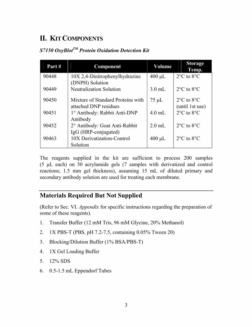
II. KIT COMPONENTS S7150 OxyBlotTM Protein Oxidation Detection Kit
Part # Component Volume Storage Temp.
90448 10X 2,4-Dinitrophenylhydrazine (DNPH) Solution
400 µL 2°C to 8°C
90449 Neutralization Solution 3.0 mL 2°C to 8°C
90450 Mixture of Standard Proteins with attached DNP residues
75 µL 2°C to 8°C (until 1st use)
90451 1° Antibody: Rabbit Anti-DNP Antibody
4.0 mL 2°C to 8°C
90452 2° Antibody: Goat Anti-Rabbit IgG (HRP-conjugated)
2.0 mL 2°C to 8°C
90463 10X Derivatization-Control Solution
400 µL 2°C to 8°C
The reagents supplied in the kit are sufficient to process 200 samples (5 µL each) on 30 acrylamide gels (7 samples with derivatized and control reactions; 1.5 mm gel thickness), assuming 15 mL of diluted primary and secondary antibody solution are used for treating each membrane.
Materials Required But Not Supplied
(Refer to Sec. VI. Appendix for specific instructions regarding the preparation of some of these reagents).
1. Transfer Buffer (12 mM Tris, 96 mM Glycine, 20% Methanol)
2. 1X PBS-T (PBS, pH 7.2-7.5, containing 0.05% Tween 20)
3. Blocking/Dilution Buffer (1% BSA/PBS-T)
4. 1X Gel Loading Buffer
5. 12% SDS
6. 0.5-1.5 mL Eppendorf Tubes
(C) 2003 COSMO BIO CO.,LTD.
Added by COSMO BIO CO.,LTD.

4
7. Reagents necessary to perform SDS-PAGE
8. Transfer Apparatus
9. Chemiluminescent Reagent
10. Nitrocellulose or PVDF Membrane
11. Distilled water (dH2O)
Precautions
1. Components 90448 and 90463 contain trifluoroacetic acid. Component 90448 also contains 2-4-dinitrophenyl hydrazine (DNPH). Harmful if swallowed or inhaled; avoid contact with skin and eyes (wear gloves and eye protection); wash areas of contact immediately.
2. Component 90450 can be stored at 2°C to 8°C until first use. After using, the component should be stored at -15°C to -25°C.
(C) 2003 COSMO BIO CO.,LTD.
Added by COSMO BIO CO.,LTD.

5
III. PROTOCOL Figure 1: Flow Chart of OxyBlot™ Procedure.
Prepare Protein Solution ↓
Derivatize the Carbonyl Groups ↓
Separate the Protein Sample by SDS-PAGE ↓
Transfer to a Membrane ↓
Block Non-Specific Sites ↓
Incubate in Primary Antibody ↓
Incubate in HRP-Conjugated Secondary Antibody ↓
Add Chemiluminescent Reagent ↓
Expose to film
Preparation of Protein Lysate
The protein lysate may be prepared using a variety of protocols, such as those detailed in ref. 38 and 39. It is recommended that a reducing agent be added to the lysis buffer to prevent the oxidation of proteins that may occur after cell lysis. Lysis buffer containing either 1-2% 2-mercaptoethanol or 50 mM DTT sufficiently inhibits this oxidation, but has no adverse effect on the derivatization reaction in the OxyBlotTM protocol. Purified protein samples are also suitable for analysis with the OxyBlotTM Kit.
(C) 2003 COSMO BIO CO.,LTD.
Added by COSMO BIO CO.,LTD.

6
Derivatization of Protein Mixture
Reagents a. 1X DNPH Solution: Dilute 1 volume of the 10X DNPH stock solution with
9 volumes of dH2O. If some precipitation of DNPH is observed, spin down the reagent prior to adding to the protein samples. This does not alter the performance of the reaction. The dilution can be prepared in advance and stored for 6 months at 2°C to 8°C.
b. Derivatization-Control Solution: Dilute 1 volume of the 10X stock solution with 9 volumes of dH2O. The dilution can be prepared in advance and stored for 6 months at 2°C to 8°C.
c. Neutralization Solution
d. 12% SDS
Protocol Two aliquots of each specimen to be analyzed will be treated. One aliquot will be subjected to the derivatization reaction. The other aliquot will serve as a negative control, by substituting the 1X Derivatization-Control Solution for the 1X DNPH Solution.
1. Transfer 5 µL of a protein sample (crude or purified) into each of 2 eppendorf tubes (0.5-1.5 mL). The volume of protein samples can vary depending on the well size of the gel and the concentration of the sample. When a sample volume other than 5 µL is used, adjust the volume of other reagents accordingly.
Note: Using the protocol and reagents of this kit, 10 femtomoles of DNP residues in a protein will be readily detectable by a 30 second exposure (see Appendix). This is about equal to those in 2.5 ng of protein with M.W. 50,000 containing 0.2 mole carbonyl group/mole of protein.
(C) 2003 COSMO BIO CO.,LTD.
Added by COSMO BIO CO.,LTD.
7
Note: Generally, 15-20 µg of protein is recommended per derivatization reaction. Users may need to try several protein concentrations to obtain desirable results.
Note: Avoid high protein concentration (>10 µg/µL) to assure solubility during the derivatization reaction.
2. Denature each 5 µL aliquot of protein by adding 5 µL of 12% SDS for a
final concentration of 6% SDS.
3. Derivatize the sample by adding 10 µL of 1X DNPH Solution to one of the tubes. To the aliquot designated as the negative control, add 10 µL of 1X Derivatization-Control Solution instead of the DNPH solution.
4. Incubate both tubes at room temperature for 15 minutes. Do not allow the reaction to proceed more than 30 minutes, as side reactions other than hydrazone linkage may occur.
5. Add 7.5 µL of Neutralization Solution to both tubes. If reduction of the protein sample is desired and a reducing reagent was not present during lysis, add 2-mercaptoethanol to the sample mixture to achieve a final concentration of 0.74 M solution (1-1.5µL; 5%v/v).
6. Both the treated sample and the negative control are ready to load into a polyacrylamide gel.
Note: At this point, the samples can be stored at 4°C for up to one week. Thaw at room temperature.
SDS-PAGE, Western Transfer and Immunodetection Reagents a. Transfer Buffer
b. Blocking/Dilution Buffer: 1% BSA/PBS-T
c. PBS-T: PBS (Phosphate buffered saline, pH 7.2-7.5) containing 0.05% Tween-20.
d. DNP-lated molecular weight standard protein (90450).
e. 1° Antibody: dilute 1° Antibody stock (90451) to 1:150 with Blocking/ Dilution Buffer
f. 2° Antibody: dilute 2° Antibody stock (90452) to 1:300 with Blocking/ Dilution Buffer
(C) 2003 COSMO BIO CO.,LTD.
Added by COSMO BIO CO.,LTD.
8
g. Chemiluminescent reagent: Refer to the manufacturer's instructions for preparation. Prepare the reagent just prior to use. About 0.125 mL of reagent per cm2 of membrane is sufficient.
Protocol
1. Upon first use, warm the standard protein mixture to room temperature. Transfer the amount necessary for the first experiment into one tube and divide the remaining mixture among several tubes. Store the aliquots at -15˚C to -25˚C. Since the mixture contains SDS which might be precipitated during storage, it may be necessary to heat the stock solution to 50˚C to redissolve the SDS. Aliquoting the mixture protects the protein standard from excessive freeze-thaw and heating cycles. Combine 2.5 µL of the molecular weight standard with attached DNP residues and 20 µL of 1X Gel Loading Buffer prior to loading onto the gel. (See Sec. IV. Data Analysis, Molecular Weight Protein Standards.) Perform SDS-PAGE according to standard procedures (34).
Note: It is not necessary to add 1X Gel Loading Buffer to the samples prior to loading the gel, since the addition of the Neutralization Solution makes the sample dense enough to sink to the bottom of the well. However, the addition of Loading Buffer will not adversely affect the electrophoresis of the sample.
Note: Do not heat samples prior to loading into the gel.
Note: The DNP-BSA in 2.5 µL solution contains 100 femtomoles of DNP residues (determined spectrophotometrically).
Note: A portion or all of the derivatized and neutralized sample can be applied directly on SDS-PAGE or dot blotted onto a filter. The free DNPH in the derivatization mixture does not hamper the following detection reaction.
2. Electroblot proteins to a membrane (35).
Note: Refer to the transfer apparatus manual for specific transfer instructions and recommendations.
Note: If using PVDF membrane, pre-wet with 95% ethanol, then rinse with dH2O.
Note: Optional: If non-reduced protein samples will be analyzed, soak the gel in 1% 2-mercaptoethanol in 1X Gel Running Buffer prior to Western transfer. This pretreatment will enhance the detection sensitivity (36).
3. Incubate the membrane in Blocking/Dilution Buffer for 1 hour with gentle shaking. Use 0.3 mL/cm2 of membrane.
(C) 2003 COSMO BIO CO.,LTD.
Added by COSMO BIO CO.,LTD.
9
4. Dilute the 1° Antibody stock 1:150 in Blocking/Dilution Buffer just before use. Use enough solution so that the membrane is completely immersed in the solution (15 mL for a 10 x 10 cm2 membrane). An alternative method would be to place the membrane in a heat-sealable plastic bag (in this case, the amount of solution required will be less than that used for a membrane treated in a open container). Incubate the membrane in the 1° Antibody solution for ~1 hour at 18°C to 25°C with gentle shaking.
5. Rinse the membrane 2 times with 1X PBS-T. Wash the membrane with 1X PBS-T once for 15 minutes, then twice for 5 minutes each at 18°C to 25°C.
6. Dilute the 2° Antibody stock 1:300 in Blocking/Dilution Buffer. Incubate the membrane in the 2° Antibody solution (15 mL for a 10 x 10 cm2 membrane) for ~1 hour at 18°C to 25°C with gentle shaking.
7. Wash the membrane as in step 5.
8. Drain the excess buffer from the membrane, place it on a plastic sheet with the protein side up, and cover the membrane. Prepare the chemiluminescent reagents according to manufacturer's directions just before use. Make just enough to cover the membrane with chemiluminescent reagent. The final reagent volume required is 0.125 mL/cm2. Incubate for 1 minute at 18˚C to 25°C.
9. Drain off excess reagent by holding the membrane vertically and touching the edge against filter paper. As an alternative, the membrane may be dried by placing it between sheets of filter paper. Place the membrane inside a plastic page protector. Remove air pockets.
10. Place the membrane, protein side up, in a film cassette. Make sure that there is no free reagent in the cassette.
11. In a darkroom, carefully place an autoradiography film on top of the membrane.
12. Expose the film for 30 seconds, then develop. When all the procedures are correctly performed, the bands of molecular weight standard proteins should be readily visible after a 30 second exposure.
13. Place a second film on top of the membrane. Expose the film for the appropriate amount of time. Because each protein will differ in efficiency of transfer to a membrane and in the immunodetection of the attached DNP moieties, the extent of the carbonyl content in each protein is best investigated by comparison of the signal intensity of the same protein. The signal intensity of the molecular weight protein standards provides a convenient control for the experimental variations between separate experiments.
(C) 2003 COSMO BIO CO.,LTD.
Added by COSMO BIO CO.,LTD.

10
IV. DATA ANALYSIS Proteins which have undergone oxidative modification will be identified by appearing as a band only in the lane containing the derivatized sample, but not in the lane containing the negative control. If proteins bands of the same molecular weight appear in both lanes, this is probably due to non-specific binding of either of the antibodies and does not indicate a modified protein.
Molecular Weight Protein Standards
Protein Molecular Weight Phosphorylase B 97,400 Bovine serum albumin 68,000 Ovalbumin 43,000 Carbonic anhydrase 29,000 Trypsin inhibitor 21,000
Figure 2: Molecular Weight Protein Standard. 2.5 µL of DNP-protein mixture was mixed with 20 µL of 1X SDS-Sample Buffer, separated on a 12% SDS polyacrylamide gel and electroblotted onto nitrocellulose membrane. The membrane was processed according to the procedure outlined in the OxyBlot™ Kit protocol, using reagents in the kit. The protein bands on the filter were detected using a chemiluminescent detection kit. The chemiluminescent signal was captured by a 15 second exposure on film.
The molecular weight protein standard mixture is composed of five proteins containing 1-3 DNP residues per molecule. The attached DNP residues permit the visualization of the proteins by the detection system of the kit. Since the proteins already have DNP residues, the protein standard mixture should NOT be subjected to the derivatization reaction. However, it is necessary to add 1X Gel Loading Buffer to the protein standards prior to gel loading (2.5 µL of protein standard plus 20 µL of 1X Gel Loading Buffer). In order to maintain consistency, gel loading buffer which contains a reducing agent must be added to the protein standard if the sample also contains a reducing agent. This standard serves as an internal control for the electrophoresis, western transfer and immunodetection steps of the OxyBlot™ procedure.
(C) 2003 COSMO BIO CO.,LTD.
Added by COSMO BIO CO.,LTD.

11
V. TROUBLESHOOTING
No signal or poor signal is obtained. 1. Potential Problem: No transfer during Western Blot Procedure.
Recommendations: a. Check the power supply and electroblot apparatus. b. Check the orientation of the membrane and gel with respect to the
electrodes of the transfer apparatus.
2. Potential Problem: Poor transfer during Western Blot Procedure.
Recommendations: a. Re-evaluate the blotting procedure. (Check the transfer efficiency by
staining procedure the blotting membrane (refer to ref. 37) or by transfer of pre-stained marker). Check the transfer time, current, transfer buffer, buffer temperature, gel concentration, thickness, etc.
b. Check that the membrane was hydrated according to manufacturer's instructions.
3. Potential Problem: Detection System.
Recommendations: a. Check that antibodies and other reagents are being stored correctly and
properly used as recommended in the protocol. b. Make sure that chemiluminescent reagents are properly used and film
developer and fixer are fresh.
?
(C) 2003 COSMO BIO CO.,LTD.
Added by COSMO BIO CO.,LTD.

12
Diffused and/or distorted bands are observed. 1. Potential Problem: Electrophoresis.
Recommendations: a. Gel was overloaded with protein – apply less electrophoresis sample to the
gel. b. Make sure no air bubbles are trapped during electrophoresis. c. Check the electrophoresis conditions: make sure the buffer was made
correctly. 2. Potential Problem: Blotting.
Recommendations: d. Inspect the transfer cassette. e. Make sure that firm contact was made between the gel and the membrane. f. Make sure that any air bubbles that may have been trapped between the gel
and the membrane are carefully removed prior to transfer.
High background is observed 1. Potential Problem: Inadequate Blocking of Membrane
Recommendations: g. Check that the Blocking/Dilution Buffer was properly prepared. h. Increase the incubation time of the blocking step.
?
?
(C) 2003 COSMO BIO CO.,LTD.
Added by COSMO BIO CO.,LTD.
13
VI. APPENDIX
Preparation of Oxidized Proteins for Positive Controls
1. Prepare a 5-10 mg/mL protein sample in the following buffer:
25 mM HEPES buffer, pH 7.2 25 mM ascorbate 100 µM FeCl3 2. Incubate at 37˚C for 5 hours.
3. Dialyze the sample vs. 50 mM HEPES, 1 mM EDTA.
Reagent Preparation
1. Electroblot Transfer Buffer To make 1 L:
Tris Base 1.45 g Glycine 7.20 g dH2O 800 mL Adjust pH to 8.3 using HCl. Methanol 200 mL
2. 10X Phosphate Buffered Saline (PBS) To make 1 L:
H2PO4 • H2O 26 g Na2HPO4 115 g NaCl 85 g Adjust the pH to 7.2-7.5 using NaOH or HCl.
(C) 2003 COSMO BIO CO.,LTD.
Added by COSMO BIO CO.,LTD.

14
3. 1X PBS-T To make 1 L:
10X PBS 100 mL Tween 20 0.5 mL dH2O 899.5 mL
4. Blocking/Dilution Buffer To make 1 L:
1X PBS-T 1000 mL BSA 10 g Adjust the pH to 7.2-7.4.
5. 1X Gel Loading Buffer To make 8 mL:
0.5 M Tris-HCl, pH 6.8 1.0 mL Glycerol 0.8 mL 14.3 M 2-Mercaptoethanol 0.1 mL (Reagent Grade) 10% SDS 1.6 mL 0.05% Bromophenol Blue 0.32 mL dH2O 4.18 mL
Divide into 1 mL aliquots and store at -15°C to -25˚C.
(C) 2003 COSMO BIO CO.,LTD.
Added by COSMO BIO CO.,LTD.
15
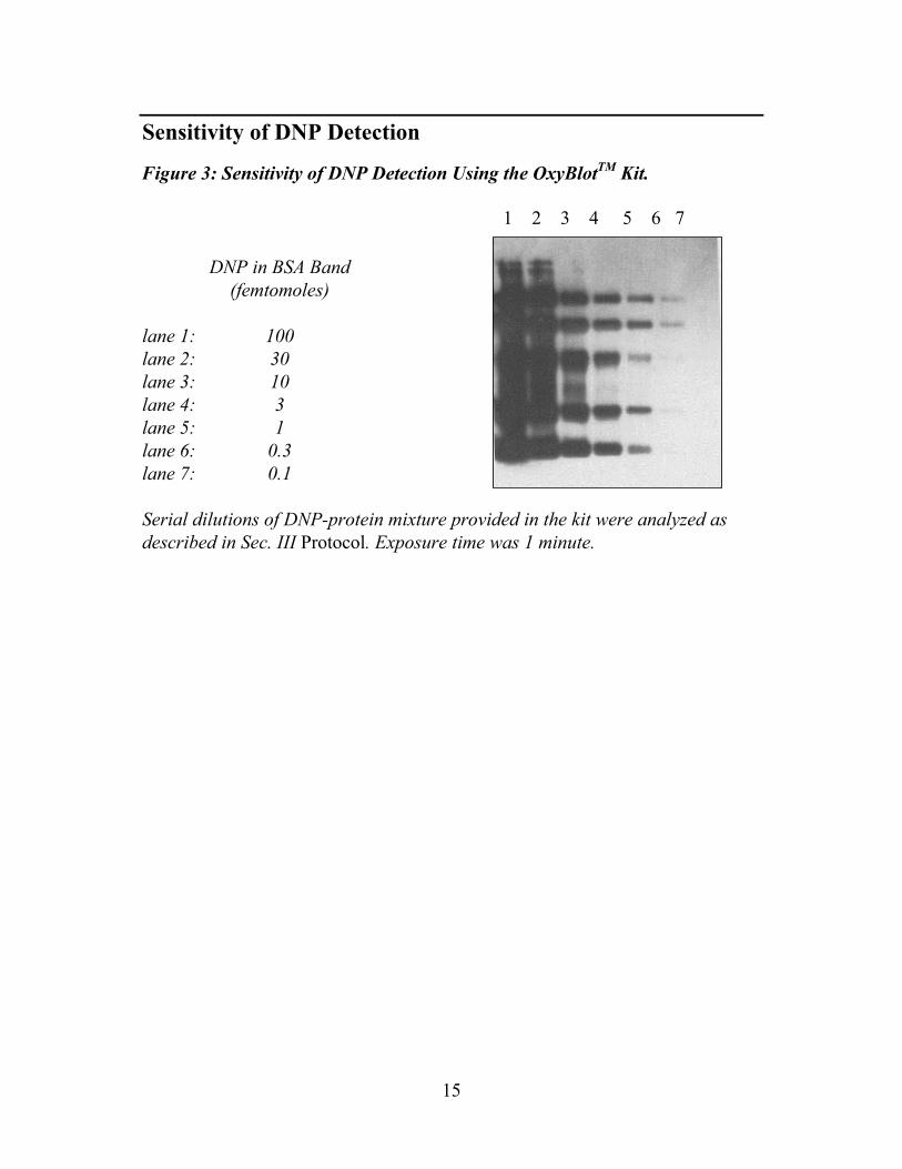
Sensitivity of DNP Detection
Figure 3: Sensitivity of DNP Detection Using the OxyBlotTM Kit.
1 2 3 4 5 6 7 DNP in BSA Band (femtomoles)
lane 1: 100 lane 2: 30 lane 3: 10 lane 4: 3 lane 5: 1 lane 6: 0.3 lane 7: 0.1
Serial dilutions of DNP-protein mixture provided in the kit were analyzed as described in Sec. III Protocol. Exposure time was 1 minute.
(C) 2003 COSMO BIO CO.,LTD.
Added by COSMO BIO CO.,LTD.

16
VII. REFERENCES
References Cited in this Manual
1. Halliwell, B. and Gutteridge, J.M.C. (1990) Role of free radicals and catalytic metal ions in human diseases: an overview. Methods in Enzymol. 186:1.
2. Halliwell, B. and Gutteridge, J.M.C. (1989) Free Radicals. Biology and Medicine, 2nd Ed. (Oxford, Clarendon Press).
3. Stadtman, E.R. (1992) Protein oxidation and aging. Science 257:1220.
4. Ching Kuang Chow, ed. (1988) Cellular Antioxidant Defense Mechanisms (CRC Press, FL).
5. Weizman, S.A. and Gordon, L.L. (1990) Inflammation and cancer: Role of phagocyte-generated oxidants in carcinogenesis. Blood 76:655.
6. Babior, B.M. and Woodman, R.C. (1990) Chronic granulomatous disease. Semin. Hematol. 27:247.
7. Cance, B., et al. (1979) Hydroperoxide metabolism in mammalian organs. Physiol. Rev. 59:527.
8. Halliwell, B. (1994) Free radicals, antioxidants, and human disease: curiosity, cause, or consequence? Lancet 344:721.
9. Miquel, J., et al., eds. (1989) Handbook of free radicals and antioxidant in biomedicine (CRC Press, FL).
10. Dizdaroglu, M. Chemistry of free radical damage to DNA and nucleoprotein in B. Halliwell and I. Aruomao, eds., (1993) DNA and Free Radicals (Chichester, Ellis Harwood), p19.
11. Ames, B.N., et al. (1991) Oxidative Damage and Repair: Chemical, Biological and Medical Aspects, K.J.A. Davies, ed. (Pergamon, Elmsford, NY), p181.
12. Davies, K.J.A. (1987) Protein damage and degradation by oxygen radicals. J. Biol. Chem. 262:9895.
13. Schreck, R. et al. (1991) Reactive oxygen intermediates as apparently widely used messengers in the activation of the NF-kB transcription factor and HIV-1. EMBO J. 10:2247.
14. Demple, B. and Halbrook, J.H. (1983) Nature 304:466.
(C) 2003 COSMO BIO CO.,LTD.
Added by COSMO BIO CO.,LTD.

17
15. Christman, M. F., et al. (1985) Cell 41:753.
16. Stadtman, E. R. (1993) Oxidation of free amino acids and amino acid residues in proteins by radiolysis and by metal-catalyzed reactions. Ann. Rev. Biochem. 62:797.
17. Davies, et al. (1987) Protein damage and degradation by oxygen radicals II. Modification of amino acids. J. Biol. Chem. 262:9902.
18. Uchida, K. and Kawakishi, S. (1990) Site-specific oxidation of angiotensin I by copper(II) and L-ascorbate: conversion of histidine residues to 2-imidazolones. Arch. Biochem. Biophys. 283:20.
19. Heinecke, J.W., et al. (1993) Dityrosine, a specific marker of oxidation, is synthesized by the myeloperoxidase-hydrogen peroxidase system of human neutrophil and macrophages. J. Biol. Chem. 268:4069.
20. Farber, J.M. and Levine, R.L. (1986) Sequence of a peptide susceptible to mixed function oxidation. Probable cation binding site in glutamine synthetase. J. Biol. Chem. 261:4574.
21. Climent, I., et al. (1989) Derivatization of r-glutamyl semialdehyde residues in oxidized proteins by fluoresceninamine. Anal. Biochem. 182:226.
22. Levine, R.L. (1983) Oxidative modification of glutamine synthetase I. Interaction is due to loss of one histidine residue. J. Biol. Chem. 258:11823.
23. Oliver, C.N. (1987) Inactivation of enzymes and oxidative modification of proteins by stimulated neutrophils. Arch. Biochem. Biophys. 253:62.
24. Abate, C., et al. (1990) Expression and purification of the leucine zipper and DNA-binding domaines of FOS and Jun: Both Fos and jun contact DNA directly. Proc. Natl. Acad. Sci. USA. 87:1032.
25. Hainaut, P. and Milner, J. (1993) Redox modulation of p53 conformation and sequence-specific DNA binding in vitro. Cancer. Res. 53:4469.
26. Pognonec, P., et al. (1992) The helix-loop/leucine repeat transcription factor USF can be functionally regulated in a redox-dependent manner. J. Biol. Chem. 267: 24563.
27. Rivett, A.J. (1986) Regulation of intracellular protein turnover: covalent modification as a mechanism of marking proteins for degradation. Curr. Top. Cell Regul. 28:291.
28. Wolf, S.P. and Dean, R.T. (1986) Fragmentation of proteins by free radicals and its effect on their susceptibility to enzymic hydrolysis. Biochem J. 234:399.
(C) 2003 COSMO BIO CO.,LTD.
Added by COSMO BIO CO.,LTD.

18
29. Davies, et al. (1987) Protein damage and degradation by oxygen radicals IV. Degradation of denatured protein. J. Biol. Chem. 262:9914.
30. Levine, R.L., et al. (1981) Turnover of bacterial glutamine synthetase: Oxidative inactivation proceeds proteolysis. Proc. Natl. Acad. Sci. USA 78:2120.
31. Oliver, C.N., et al. (1987) Age related changes in oxidized proteins. J. Biol. Chem. 262:5488.
32. Smith, C.D., et al. (1991) Excess brain protein oxidation and enzyme dysfunction in normal aging and in Alzheimers disease. Proc. Natl. Acad. Sci. USA. 88:10540.
33. Starke-Reed, P.E. and Oliver, C.N. (1989) Protein oxidation and proteolysis during aging and oxidative stress. Arch. Biochem. Biophys. 275:559.
34. Laemmli, U.K. (1970) Cleavage of structural proteins during the assembly of the head of bacteriophge T4. Nature 227:680.
35. Towbin, H., et al. (1979) Electrophoretic transfer of proteins from polyacrylamide gels to nitrocellulose sheets: procedure and some applications. Proc. Natl. Acad. Sci. USA. 76:4350.
36. Lee, Y-J. and Shacter, E. (1995) Role of carbohydrates in oxidative modification of fibrinogen and other plasma proteins. Arch. Biochem. Biophys. 321(1):175-181.
37. Sasse, J. (1991) Detection of Proteins on Blot Transfer Membrane in Current Protocols for Molecular Biology ( Ausubel, F.M., et al, eds.) p. 10.7.1 (John Wiley and Sons, New York).
38. Coligan, J. et al, eds. (1996) Current Protocols in Protein Science (John Wiley and Sons, New York).
39. Weissman, A.M. (1991) Solubilization of Cellular Proteins in Current Protocols in Immunology (Coligan, J.E. et al, eds.) p.8.1 (John Wiley and Sons, New York).
Disclaimers
Tween® is a registered trademark of ICI Americas.
All trademarks, unless otherwise noted, are the property of Serologicals® Royalty Company or Chemicon® International, Inc.
(C) 2003 COSMO BIO CO.,LTD.
Added by COSMO BIO CO.,LTD.

19
Warranty
These products are warranted to perform as described in their labeling and in CHEMICON literature when used in accordance with their instructions. THERE ARE NO WARRANTIES, WHICH EXTEND BEYOND THIS EXPRESSED WARRANTY AND CHEMICON DISCLAIMS ANY IMPLIED WARRANTY OF MERCHANTABILITY OR WARRANTY OF FITNESS FOR PARTICULAR PURPOSE. CHEMICON’s sole obligation and purchaser’s exclusive remedy for breach of this warranty shall be, at the option of CHEMICON, to repair or replace the products. In no event shall CHEMICON be liable for any proximate, incidental or consequential damages in connection with the products. 2003: CHEMICON International, Inc. - By CHEMICON International, Inc. All rights reserved. No part of these works may be reproduced in any form without permissions in writing.
(C) 2003 COSMO BIO CO.,LTD.
Added by COSMO BIO CO.,LTD.

(C) 2003 COSMO BIO CO.,LTD.
Added by COSMO BIO CO.,LTD.

Cat. No.: S7150 March 2004
Revision D: 41418
(C) 2003 COSMO BIO CO.,LTD.
Added by COSMO BIO CO.,LTD.