OXIDATIVE STRESS IS RESPONSIBLE FOR MITOCHONDRIAL ... · From the Dipartimento di Chimica...
23
OXIDATIVE STRESS IS RESPONSIBLE FOR MITOCHONDRIAL PERMEABILITY TRANSITION INDUCTION BY SALICYLATE IN LIVER MITOCHONDRIA Valentina Battaglia, Mauro Salvi and Antonio Toninello From the Dipartimento di Chimica Biologica, Università di Padova, Istituto di Neuroscienze del C.N.R., Unità per lo studio delle Biomembrane, Viale G. Colombo 3, 35121 Padova, Italy Running title: Salicylate and mitochondrial oxidative stress Address correspondence to: Antonio Toninello, Dipartimento di Chimica Biologica, Università di Padova, Viale G. Colombo 3, 35121 Padova, Italy, Tel. +39 0498276134; Fax. +39 0498276133; E- Mail: [email protected] The interaction of salicylate with the respiratory chain of liver mitochondria generates hydrogen peroxide and, most probably, other reactive oxygen species (ROS), which in turn oxidize thiol groups and glutathione. This oxidative stress, confirmed by the prevention of action by antioxidant agents, leads to the induction of the mitochondrial permeability transition (MPT) in the presence of Ca 2+ . This phenomenon induces further increase of oxidative damage resulting in impairment of oxidative phosphorylation and β- oxidation, cardinal features of Reye’s syndrome in liver. MPT induction also induces the release of cytochrome c and apoptotic inducing factor (AIF) from mitochondria, suggesting that salicylate also behaves as a pro-apoptotic agent. The reactive group of salicylate for inducing oxidative stress is the hydroxyl group which, by interacting with a Fe/S cluster of mitochondrial Complex I, the so-called N- 2(Fe-S) centre, produces ROS. Salicylates and non-steroid drugs such as sodium salicylate, aspirin and indomethacin, are widely prescribed for treating inflammation (1). Their efficacy is, at least partially, attributed to their ability to inhibit cyclooxygenase (COX) (2, 3). In this regard, blockade of COX activity is believed to be linked to the chemopreventive effect of salicylates (4, 5). Data from other studies also indicate that COX-2 inhibitors increase the susceptibility of cancer cells to apoptosis (6, 7). Other reports propose the involvement of a COX-independent pathway during apoptosis signaling (8, 9). In addition, it has been shown that aspirin induces activation of NF-kB, which is required for the antitumor effects of aspirin (10). It is the general opinion, supported by a number of reports, that apoptosis is mediated by the phenomenon of mitochondrial permeability transition (MPT) (for reviews, see refs. 11, 12). Salicylates have damaging effects on isolated mitochondria, causing uncoupling of oxidative phosphorylation and swelling (13- 15). Aspirin and salicylate affect mitochondrial calcium homeostasis and act synergistically with the cation to impair mitochondrial respiration and ATP synthesis (16, 17). Again, salicylates inhibit the Krebs cycle enzyme α-ketoglutarate dehydrogenase (18). Salicylates are in fact able to induce MPT in the presence of Ca 2+ in isolated liver mitochondria (19) but also in “in situ” mitochondria of cultured hepatocytes (12). These observations indicate that the onset of MPT by salicylate and Ca 2+ represents the pathological mechanism causing mitochondrial injury in Reye’s syndrome - a lethal disorder occurring in children, generally after a preliminary viral infection. Epidemiological and experimental evidence demonstrate a close correlation between Reye’s syndrome and the use of aspirin (20, 21). The main cause of this syndrome is most probably a primary mitochondrial injury. Brain and liver mitochondria exhibit morphologic alterations such as matrix swelling, decreased matrix density and loss of cristae (22), typical of organelles which have undergone a permeability transition. A broad range of oxidant agents, thiol reagents, heavy metals and uncouplers are able to trigger MPT in isolated mitochondria in the presence of supraphysiological Ca 2+ JBC Papers in Press. Published on August 12, 2005 as Manuscript M502391200 Copyright 2005 by The American Society for Biochemistry and Molecular Biology, Inc. by guest on February 16, 2019 http://www.jbc.org/ Downloaded from
Transcript of OXIDATIVE STRESS IS RESPONSIBLE FOR MITOCHONDRIAL ... · From the Dipartimento di Chimica...

OXIDATIVE STRESS IS RESPONSIBLE FOR MITOCHONDRIAL PERMEABILITY TRANSITION INDUCTION BY SALICYLATE IN LIVER MITOCHONDRIA
Valentina Battaglia, Mauro Salvi and Antonio Toninello From the Dipartimento di Chimica Biologica, Università di Padova, Istituto di Neuroscienze del C.N.R., Unità per lo studio delle Biomembrane, Viale G. Colombo 3, 35121 Padova, Italy
Running title: Salicylate and mitochondrial oxidative stress Address correspondence to: Antonio Toninello, Dipartimento di Chimica Biologica, Università di
Padova, Viale G. Colombo 3, 35121 Padova, Italy, Tel. +39 0498276134; Fax. +39 0498276133; E-Mail: [email protected]
The interaction of salicylate with the
respiratory chain of liver mitochondria generates hydrogen peroxide and, most probably, other reactive oxygen species (ROS), which in turn oxidize thiol groups and glutathione. This oxidative stress, confirmed by the prevention of action by antioxidant agents, leads to the induction of the mitochondrial permeability transition (MPT) in the presence of Ca2+. This phenomenon induces further increase of oxidative damage resulting in impairment of oxidative phosphorylation and ββββ-oxidation, cardinal features of Reye’s syndrome in liver. MPT induction also induces the release of cytochrome c and apoptotic inducing factor (AIF) from mitochondria, suggesting that salicylate also behaves as a pro-apoptotic agent. The reactive group of salicylate for inducing oxidative stress is the hydroxyl group which, by interacting with a Fe/S cluster of mitochondrial Complex I, the so-called N-2(Fe-S) centre, produces ROS.
Salicylates and non-steroid drugs such
as sodium salicylate, aspirin and indomethacin, are widely prescribed for treating inflammation (1). Their efficacy is, at least partially, attributed to their ability to inhibit cyclooxygenase (COX) (2, 3). In this regard, blockade of COX activity is believed to be linked to the chemopreventive effect of salicylates (4, 5). Data from other studies also indicate that COX-2 inhibitors increase the susceptibility of cancer cells to apoptosis (6, 7). Other reports propose the involvement of a COX-independent pathway during apoptosis signaling (8, 9). In addition, it has been shown that aspirin induces activation of NF-kB,
which is required for the antitumor effects of aspirin (10).
It is the general opinion, supported by a number of reports, that apoptosis is mediated by the phenomenon of mitochondrial permeability transition (MPT) (for reviews, see refs. 11, 12).
Salicylates have damaging effects on isolated mitochondria, causing uncoupling of oxidative phosphorylation and swelling (13-15). Aspirin and salicylate affect mitochondrial calcium homeostasis and act synergistically with the cation to impair mitochondrial respiration and ATP synthesis (16, 17). Again, salicylates inhibit the Krebs cycle enzyme α-ketoglutarate dehydrogenase (18).
Salicylates are in fact able to induce MPT in the presence of Ca2+ in isolated liver mitochondria (19) but also in “in situ” mitochondria of cultured hepatocytes (12). These observations indicate that the onset of MPT by salicylate and Ca2+ represents the pathological mechanism causing mitochondrial injury in Reye’s syndrome - a lethal disorder occurring in children, generally after a preliminary viral infection. Epidemiological and experimental evidence demonstrate a close correlation between Reye’s syndrome and the use of aspirin (20, 21). The main cause of this syndrome is most probably a primary mitochondrial injury. Brain and liver mitochondria exhibit morphologic alterations such as matrix swelling, decreased matrix density and loss of cristae (22), typical of organelles which have undergone a permeability transition.
A broad range of oxidant agents, thiol reagents, heavy metals and uncouplers are able to trigger MPT in isolated mitochondria in the presence of supraphysiological Ca2+
JBC Papers in Press. Published on August 12, 2005 as Manuscript M502391200
Copyright 2005 by The American Society for Biochemistry and Molecular Biology, Inc.
by guest on February 16, 2019http://w
ww
.jbc.org/D
ownloaded from

2
concentrations (for reviews, see refs. 23, 24). The immunosuppressive cyclic endecapeptide, cyclosporin A (CsA) specifically blocks the phenomenon (25, 26), which is due to the opening of a pore, identified as a non-selective, highly conductive “megachannel” (24). Opening of the transition pore causes a series of events culminating in bioenergetic collapse and a redox catastrophe (11).
The mechanism by which salicylate induces MPT has not yet been elucidated, although some models have been proposed. Salicylate may bind directly to the pore-forming structure, promoting pore opening (12). Alternatively, the drug may intercalate into the mitochondrial membrane, altering its surface potential and thereby decreasing the gating potential of the pore (27).
Other authors have proposed that the induction of MPT in tumor cells is due to oxidative stress following the production of reactive oxygen species (ROS), elicited by salicylate through a Rac1-NADPH oxidase-dependent pathway (28).
Taking into account this capacity of salicylate to generate ROS at cytosol level, and recent observations that the interaction of the isoflavonoid genistein with the mitochondrial electron transport chain by means of its hydroxyl group in 4’ position, also generates ROS (29), this study is aimed at determining whether salicylate, by interacting with respiratory complexes, can induce the oxidative stress responsible for MPT induction in isolated liver mitochondria. Another aim was to identify the specific component(s) of the respiratory chain which constitute the target(s) of salicylate, in order to propose the reaction mechanism(s) of ROS production.
Materials and Methods
Materials – Mouse monoclonal antibody anti-cytochrome c (Cyt c) was purchased from Pharmingen (San Diego, CA), rabbit polyclonal antibody anti-apoptosis-inducing factor (AIF) was purchased from Chemicon International. All other reagents were of the highest purity commercially available.
Mitochondrial isolation and standard incubation procedures - Rat liver mitochondria (RLM) were isolated by conventional differential centrifugation in a buffer containing 250 mM sucrose, 5 mM HEPES (pH 7.4) and 1 mM EGTA (30); EGTA was omitted from the final washing solution. Protein content was measured by the biuret method with BSA as a standard (31). Mitochondria (1 mg protein/ml) were incubated in a water-jacketed cell at 20°C. The standard medium contained 250 mM sucrose, 10 mM HEPES (pH 7.4), 5 mM succinate, 50 µM Ca2+ and 1.25 µM rotenone. Variations and/or other additions are given with each experiment. Determination of mitochondrial functions - Membrane potential (∆Ψ) was calculated on the basis of movement of the lipid-soluble cation tetraphenylphosphonium (TPP+) through the inner membrane, measured using a TPP+-specific electrode (32). Mitochondrial swelling was determined by measuring the apparent absorbance change of mitochondrial suspensions at 540 nm in a Kontron Uvikon model 922 spectrophotometer equipped with thermostatic control. Oxygen uptake was measured by a Clark electrode. This measurement allowed calculation of the rate of state 3 (ADP-stimulated) respiration (V3), the rate of state 4 (non-ADP-stimulated) respiration (V4), the respiratory control index (RCI═V3/V4) and the P/O ratio (ATP generated divided by oxygen consumed in state 3). ATP synthesis was measured by the enzymatic method of Lemasters and Hackenbrock (33). Adenine nucleotide efflux was monitored according to Ross et al. (34). The redox state of endogenous pyridine nucleotides was followed fluorometrically in an Aminco-Bowman 4-8202 spectrofluorometer, with excitation at 354 nm and emission at 462 nm. Efflux of oxidized pyridine nucleotides was detected according to Carpenter and Kodicek (35). The production of H2O2 in mitochondria was measured fluorometrically by the scopoletin method (36) in an Aminco-Bowman 4-8202 spectrofluorometer.
by guest on February 16, 2019http://w
ww
.jbc.org/D
ownloaded from

3
It should be noted that the measured amount of H2O2 refers to that diffusing out of mitochondria, as the enzyme for its determination (horse radish peroxidase) cannot enter mitochondria. This measurement gives a qualitative indication but cannot be considered for rigorous quantitative evaluations. β-oxidation of palmitoylcarnitine was assayed by the following procedure: RLM (1 mg/ml) were incubated for 10 min in standard medium, in the conditions indicated in the specific figure legend. Then 1-ml portions of the suspensions were withdrawn and centrifuged in an Eppendorf bench centrifuge (model 5415C) at 15,000 rpm. The supernatants were discarded and mitochondrial pellets were resuspended with 30 µl of isolation medium. Subsequently RLM were incubated in the water-jacketed cell at 20ºC for oxygen uptake measurement. The assay medium contained 100 mM KCl, 20 mM Hepes (pH 7.4), 5 mM K-phosphate, 0.1 mM EGTA, bovine serum albumin (0.5 mg/ml), 2.5 mM oxalacetate, 0.5 mM ADP and 30 µM palmitoylcarnitine. The protein sulfhydryl oxidation assay was performed according to Santos et al. (36). The oxidation of glutathione was performed as in Tietze (38). The redox state of endogenous pyridine nucleotides was followed fluorometrically in an Aminco-Bowman 4-8202 spectrofluorometer with excitation at 354 nm and emission at 462 nm. Detection of Cyt c and AIF release - Mitochondria (1 mg protein/ml) were incubated for 15 min at 20°C in standard medium with the appropriate additions. The reaction mixtures were then centrifuged at 13,000 g for 10 min at 4°C to obtain mitochondrial pellets. The supernatant fractions were further spun at 100,000 g for 15 min at 4°C to eliminate mitochondrial membrane fragments, and concentrated five times by ultrafiltration through Centrikon 10 membranes (Amicon) at 4°C. Aliquots of 10 µl of the concentrated supernatants were subjected to 15% SDS-PAGE for Cyt c and 10% SDS-PAGE for AIF, and analyzed by
Western blotting using mouse anti-Cyt c antibody and rabbit anti-AIF antibody.
RESULTS
The results shown in Figs. 1 and 2 summarize the well-known effects of salicylate in inducing MPT, as also previously reported by other authors (e.g., 39). Panel A of Fig. 1 shows that energized RLM, incubated in standard medium in the presence of 50 µM Ca2+, when treated with 0.5 mM salicylate undergo extensive swelling, evidenced by a strong decrease in the apparent absorbance of the mitochondrial suspension. This colloid-osmotic alteration is accompanied by rapid and almost complete collapse of ∆Ψ (panel B). Fig. 2 shows other events closely correlated with the above observed swelling and ∆Ψ collapse. Panel A shows that salicylate enhances O2 consumption with both succinate (a) and β-hydroxybutyrate (b) as substrates. However, in the latter case, respiration is completely blocked after 7-8 min of incubation. The same panel also shows RCI and P/O calculations, obtained by succinate oxidation after 10 min of incubation, that is, in conditions of pore opening. Salicylate is also able to provoke the efflux of adenine nucleotides (panel B) and of oxidized pyridine nucleotides (panel C). Panel D shows the effect of salicylate-induced MPT on β-oxidation. This determination, like that of phosphorylation parameters, was performed during the opening of the transition pore. The results show palmitoylcarnitine oxidation is completely inhibited in the presence of salicylate.
The concentration of salicylate used, 0.5 mM, is of the same order of magnitude as that reached in the cytosol of hepatic cells after aspirin therapy (12, 40). All the observed events are completely prevented by cyclosporin A (CsA) and bongkrekic acid (BKA), typical inhibitors of MPT, confirming that the observed alterations are the results of pore opening (Figs. 1, 2) (BKA was not used in the experiments of Figs. 2A, 2B and 2D, as it inhibits the translocase of adenine nucleotides).
by guest on February 16, 2019http://w
ww
.jbc.org/D
ownloaded from

4
The reducing agent DTT, the alkylating NEM, and the antioxidants butylhydroxytoluene (BHT) and Trolox also inhibit swelling and ∆Ψ collapse (Figs. 3A, 3B), indicating that oxidative stress is involved in the phenomenon as inducer, amplifier, or both.
However, although the results of Fig. 3 are indicative of the onset of oxidative stress, catalase and mannitol, well-known scavengers of ROS, are ineffective in protecting RLM against MPT (results not reported). The inability of catalase, and most probably also of mannitol, may be consistent with the sidedness of ROS generation within mitochondria.
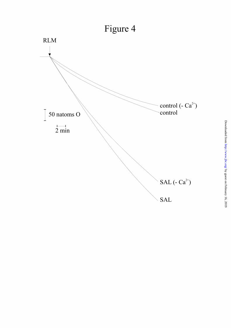
In the absence of Ca2+, salicylate causes a considerable increase in mitochondrial oxygen uptake when compared with controls (Fig. 4). Ca2+, which alone has only a negligible effect, causes a further increase in oxygen uptake (Fig. 4).
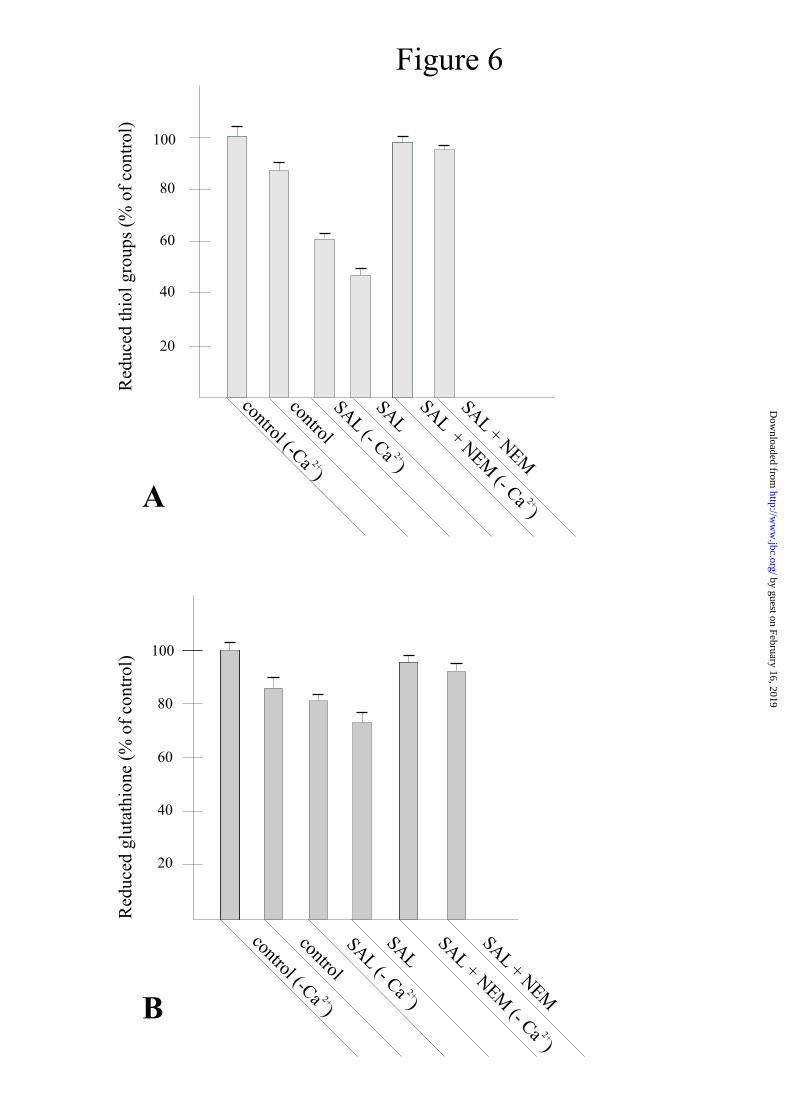
These observations, particularly notable in the absence of Ca2+, mean that salicylate, alone, can cause the generation of hydrogen peroxide or other ROS. Fig. 5A shows that the measurement of H2O2 production by mitochondria operating in a resting state (see controls in the absence or presence of Ca2+, about 0.2 nmol/mg prot.), greatly increases up to 1.5 nmol/mg prot. in the presence of salicylate but in the absence of Ca2+. With Ca2+, the measurement of H2O2 generation by salicylate further increases to 2.2 nmol/mg prot. (Fig. 5B). CsA or BKA are ineffective in inducing inhibition when Ca2+ is absent (Fig. 5A); instead, in the presence of Ca2+, both the inhibitors reduce the level of H2O2 generation to the same value as in the absence of the cation (Fig. 5B). The real amounts of H2O2 generated by salicylate are most probably higher than those reported because, as soon it is generated, H2O2 reacts with its targets and may also be transformed into other ROS (see also the description of the assay in Materials and Methods). The production of hydrogen peroxide and, most probably, of other ROS, explains the onset of oxidative stress, the effects of which are shown in Fig. 6. This figure shows that, without Ca2+, salicylate induces a drop of
about 40% in the total contents of reduced thiol groups and of 55% with the cation (panel A). However, Ca2+ alone induces a drop in reduced thiols of about 15%. NEM almost completely prevents the oxidizing effect of salicylate.
Salicylate can also oxidize glutathione in the absence and presence of Ca2+ by about 20% and 30% respectively, whereas Ca2+ alone causes oxidation of 17-18% (Fig. 5, panel B). Also in this case, NEM strongly inhibits glutathione oxidation in both conditions.
Besides sulphydryl groups and glutathione, another target of pro-oxidants agents in mitochondria are pyridine nucleotides. Fig. 7 shows that salicylate can induce strong pyridine nucleotide oxidation, but this effect is observable only in the presence of Ca2+ - in its absence, no oxidation is observable, and Ca2+ alone is ineffective. NEM and DTT prevent oxidation. Oxidation of pyridine nucleotides probably occurs after pore opening, as they are not oxidized in the absence of Ca2+. It is noteworthy that oxidized pyridine nucleotides are released from mitochondria (see Fig. 2C). This accounts for the results of Fig. 2D, in which palmitoylcarnitine oxidation is blocked in RLM undergoing MPT induction.
It is generally agreed that MPT is the first step of the pro-apoptotic pathway. This characteristic is due to the release of soluble pro-apoptotic factors from mitochondria following massive swelling and consequent outer membrane rupture. Western blot analyses (Fig. 8) show that, in the presence of Ca2+, salicylate induces efflux of Cyt c (panel A) and AIF (panel B) from RLM. In the absence of Ca2+, no efflux is observable. CsA and NEM are able to prevent both releases.
Comparisons between the effects of salicylate and acetylsalicylate in inducing MPT are shown in Fig. 9, and highlight the greater efficacy of salicylate in causing mitochondrial swelling (Fig. 9). Instead, benzoate is completely ineffective. The reduced effect of acetylsalicylate with respect to salicylate and the complete inefficacy of benzoate were also observed when measuring thiol, glutathione and pyridine nucleotide
by guest on February 16, 2019http://w
ww
.jbc.org/D
ownloaded from

5
oxidation and in the release of pro-apoptotic factors (results not reported). As acetylsalicylate is known to hydrolyze to salicylate in the cellular environment, the reduced efficacy observed in the experiment of Fig. 9 is due to incomplete hydrolysis of acetylsalicylate by mitochondrial esterases. The complete inefficacy of benzoate means that the reactive center of salicylate is the hydroxyl group.
For information about the site(s) of action of salicylate on the mitochondrial membrane, we considered the possibility of its interaction with some transition metal of the respiratory chain, in the oxidized state, using the same approach used for flavonoids (29) and triterpenoids (41). The basis of this strategy was to induce various redox levels in the respiratory complexes, in order to identify their involvement in the interaction. Fig. 10 shows that RLM incubated with salicylate in the presence of β-hydroxybutyrate as energizing substrate, which gives electrons to Complex I, exhibits considerable swelling (panel A), accompanied by rapid and complete ∆Ψ collapse (panel B). The addition to the medium of the inhibitor rotenone, which blocks β-hydroxybutyrate oxidation, and succinate, which restores electron flux at the level of Complex II, produces almost complete inhibition of the mitochondrial swelling induced by salicylate (panel C), whereas ∆Ψ undergoes only a slight transient drop (panel D).
When RLM are energized with ascorbate plus trimethyl-para-phenylendiamine (TMPD) (which gives electrons to Cyt c) and are again incubated in the presence of β-hydroxybutyrate plus rotenone and succinate plus antimycin A (which maintain reduced Complexes II and III) (42), the inhibition of swelling is slightly less marked than in the previous experiment and is accompanied by a small ∆Ψ drop (results not reported).
When β-hydroxybutyrate acts as energizing substrate, the production of H2O2 by salicylate, determined in the absence of Ca2+, increases by about 0.5nmol/mg prot. when compared with that normally obtained when the substrate is succinate plus rotenone
(Fig. 5A). Instead, the presence of β-hydroxybutyrate, when used as a reducing agent of Complex I, together with succinate plus rotenone, almost completely inhibits H2O2 generation (Fig. 11).
The variable amounts of H2O2 produced by salicylate in the various experiment conditions of Fig. 11 exactly reflect its effect at the level of MPT induction shown in Fig. 10. In the presence of Ca2+, H2O2 production is increased when β-hydroxybutyrate is the energizing substrate (results not reported).
DISCUSSION
The results reported here demonstrate
that the well-known induction of MPT by salicylate (see Figs. 1, 2) is the result of oxidative stress triggered by the interaction of salicylate with the respiratory chain in the presence of Ca2+.
The first indication of the involvement of such a process is given here by the results shown in Fig. 3, i.e., that several reducing, antioxidizing or alkylating agents (DTT, BHT, Trolox, NEM) can inhibit the induction of MPT by salicylate. The significant increase in oxygen uptake caused by salicylate in the absence of Ca2+ (Fig. 4), correlated with the production of hydrogen peroxide in the same condition (Fig. 5A), is a clear demonstration of the pro-oxidant effect of salicylate. This action is further confirmed by the oxidation undergone by sulphydryl groups and glutathione and the complete prevention exhibited by NEM (Figs. 6A, 6B) when RLM are again subjected to salicylate in the absence of Ca2+. It should be noted that, in these conditions, although oxidative stress is established, the permeability transition is not triggered (Fig. 1, curves (SAL (-Ca2+)). The results reported in Fig. 5A showing that MPT inhibitors CsA and BKA do not induce any inhibition of H2O2 generation, unequivocally confirm that the observed oxidative stress is not due to pore opening. This phenomenon takes place only if Ca2+ is also present (Fig. 1). The increase in oxygen uptake (Fig. 4), H2O2 generation (Fig. 5B) and enhanced oxidation of thiol groups, glutathione (Fig. 6)
by guest on February 16, 2019http://w
ww
.jbc.org/D
ownloaded from

6
and pyridine nucleotides (Fig. 7), observed in the presence of Ca2+, are the result of the opening of the transition pore. That is, with pore opening, the electrochemical gradient collapses, and the corresponding increase in electron flux along the respiratory chain to restore it results in an increase in oxygen uptake and, consequently, of ROS production by the chain. This is responsible for the increase in thiol and glutathione oxidation and oxidation of pyridine nucleotides. The maintenance of the reduced state - as controls showed by the presence of NEM (Figs. 6-7) and DTT (Fig. 7) - is further confirmation of increased oxidative stress in the presence of Ca2+.
The observation that, in this condition, CsA and BKA diminish H2O2 generation to the same level as that seen in the absence of Ca2+ - that is, they block H2O2 generation due to the opening of the pore - further confirms that MPT is responsible only for enhanced oxidation of thiols and glutathione and oxidation and efflux of pyridine nucleotides. In conclusion, the sequence of the events is: ROS production by salicylate; MPT induction by ROS; further ROS production by pore opening.
One proposable mechanism for pore opening is that H2O2, or other ROS produced by salicylate, act at the level of critical thiol groups (43) located on adenine nucleotide translocase. This is generally known to be the main protein involved, the oxidation of which forms dithiols, and is responsible for pore opening in the presence of Ca2+. The inefficacy in inhibiting MPT by catalase seems to demonstrate that H2O2 is not directly involved in the phenomenon. However, it must be noted that the enzyme cannot cross the membrane and that peroxide generation does not always rapidly diffuse out of the mitochondrion. Instead, glutathione oxidation (Fig. 6B), which demonstrates the involvement of the glutathione peroxidase/glutathione reductase system in the induction of the phenomenon, confirms that H2O2 is also an active agent (44). Fig. 7 clearly demonstrates that oxidation of pyridine nucleotides may be considered as one effect of pore opening, rather than as a
primary event for induction of the phenomenon, as no oxidation is observable in the absence of Ca2+.
Ca2+ alone can also induce the oxidation of sulphydryl groups and glutathione (Fig. 6), although without inducing MPT (Fig. 1). This effect is most probably due to the generation of hydrogen peroxide induced by the cation (see Fig. 5), like the results of its interaction with membrane cardiolipin. The change in membrane organization which follows this interaction may affect coenzyme Q mobility and favor ROS production (45). But the critical thiol groups responsible for pore opening are very probably not oxidized in this condition.
In considering MPT induction by salicylate as the result of oxidative stress, the generation of hydrogen peroxide probably follows the same pathway previously observed with the isoflavonoid genistein (29). Fig. 9 demonstrates that the reactive group of salicylate responsible for the observed effects is the hydroxyl group. As the reactive group of genistein and other flavonoids is also a hydroxyl group (41, 46), the generation of hydrogen peroxide may be explained by the reaction sequence reported in works in which a transition metal in the oxidized form, Fe3+ or Cu2+, was involved in the first step (e.g., 46).
Fig. 10 shows that the maximum extent of MPT induction by salicylate, evaluated as mitochondrial swelling (panel A) and ∆Ψ collapse (panel B), is obtained when β-hydroxybutyrate is used as substrate, as the result of H2O2 generation. This effect is almost completely abolished by the addition of succinate plus rotenone (panels C and D), which triggers the reduction of all the compounds of Complex I and prevents the production of H2O2 (Fig. 11). This indicates that the main and, most probably, the only target of salicylate is an Fe3+ of some Fe-S cluster of Complex I. That is, if some transition metal of all the other complexes were involved in the interaction with salicylate, the addition of succinate plus rotenone would not cause complete inhibition of MPT. Bearing in mind that the redox
by guest on February 16, 2019http://w
ww
.jbc.org/D
ownloaded from

7
couple of the hydroxyl group of salicylate is of the ubiquinone/semiquinone type, the standard redox potential of which is about –60mV (46), one hypothesis is that the interacting Fe3+ belongs to the N-2 (Fe-S) center of Complex I, as it has a redox potential in the range –20 to –160mV (47). A control experiment with ascorbate as energizing substrate, in which Complexes I, II and III were in a completely reduced state (results not reported), only demonstrates the induction of MPT to a very low extent. This effect was most probably due to the presence of antimycin (48) and may be considered negligible.
As discussed above, the hypothesized target of the reactive group of salicylate is the N-2 center of Complex I. This indicates that the compounds which react by means of a redox couple of ubiquinone/semiquinone type interact with the respiratory chain at the level of the last part of Complex I or bc1 Complex. Genistein has also been proposed to interact with bH heme (28) and the cyclic triterpene, glycyrrhetinic acid, with the N-2 center (41).
In conclusion, the results reported here highlight important data regarding the interaction mechanism of salicylates with the mitochondrial membrane responsible for induction of MPT. One of the most important functions of salicylates is their well-known anti-inflammatory and anti-oxidizing capacity, exhibited on a physiological level. Thus, the main result of this study – i.e., induction of MPT by oxidative stress - apparently seems to be a contradiction. However, it should be borne in mind that other drugs such as flavonoids (46), isoflavonoids (29), cyclic terpenoids (41) and cynnamil ketones (49), which normally behave as ROS scavengers, in particular conditions may also exert pro-oxidizing activity, as observed with salicylate. This possibility is due, as mentioned above, to the characteristic of the reactive redox couple of these compounds to interact with particular transition metals of the respiratory chain.
The close correlation between oxidative stress, MPT and apoptosis, as also confirmed by the release of cytochrome c and AIF, inhibited by NEM and CsA (Fig. 8),
indicates that salicylate is a pro-apoptotic agent and, in particular conditions, has an anti-neoplastic action. In this regard, it has been observed that salicylate is able to cause apoptosis in cultured hepatocytes (12). Indeed, aspirin and other non-steroid anti-inflammatory compounds exert their chemio-preventive action against cancer by inducing apoptosis in transforming cells (50).
In the exhaustive study performed by Trost and Lemasters (39) it was emphasized that a cardinal feature of Reye’s syndrome is the formation of microvesicular steatosis. This is a typical pathological event in liver, in which small fatty droplets accumulate in the hepatocytic cytosol without displacing the control nucleus. The above authors also point out that salicylate causes Reye’s syndrome by inhibiting mitochondrial energy production and inducing mitochondrial damage. The results reported here, besides giving a general picture regarding the induction of MPT by salicylate and the mechanism of its action, may also provide further elucidations regarding the involvement of MPT in Reye’s syndrome. The main goal of our study was to demonstrate that salicylate induces MPT by means of oxidative stress in the presence of Ca2+. However, it is noteworthy that the opening of the transition pore provokes further oxidative stress leading to increases in thiol and glutathione oxidation. Indeed, in this condition, pyridine nucleotides are also oxidized (Fig. 7) and released from the mitochondria (Fig. 2C). This finding accounts for the observed block of NAD-dependent substrate oxidation (Fig. 2A, b) and consequently the suppression of the citric acid cycle and gluconeogenesis, which are consistent with clinical pathology changes in Reye’s syndrome (39). The depletion of oxidized pyridine nucleotides also explains the complete inhibition of palmitoylcarnitine oxidation (Fig. 2D) and, more generally, the suppression of β-oxidation. As a consequence of this impairment, there is an increased cytoplasmic level of fatty acids, with the generation of microvesicular steatosis. Other metabolic alterations in Reye’s syndrome are consistent with mitochondrial uncoupling and suppression of ATP synthesis by oxidative
by guest on February 16, 2019http://w
ww
.jbc.org/D
ownloaded from

8
phosphorylation. These events are consistent with the observed effect of MPT in the complete lowering of RCI and P/O ratio (Fig. 2A).
As reported in the literature (39) and also confirmed by the results shown in Figs. 1-2, CsA and some its analogs may be considered as potential therapeutic agents against Reye’s syndrome. However, other compounds with anti-oxidizing properties and able to inhibit MPT induction, e.g., propargylamines and indolalkylamines, used in Parkinson’s disease (51-53), may also exert effective therapeutic action in the treatment of
Reye’s syndrome. Very recently the compound MitoQ has been proposed as a potent antioxidant, as its mitochondrial transport is driven by ∆Ψ and it concentrates into the matrix several hundred-folds (54). This characteristic overcomes the limited efficacy of conventional antioxidants, due to the fact that thet are difficult to deliver to mitochondria “in situ”. The use of targeting antioxidants to mitochondria “in vivo” may be a new therapeutic strategy for Reye’s syndrome, as well as for other diseases.
REFERENCES
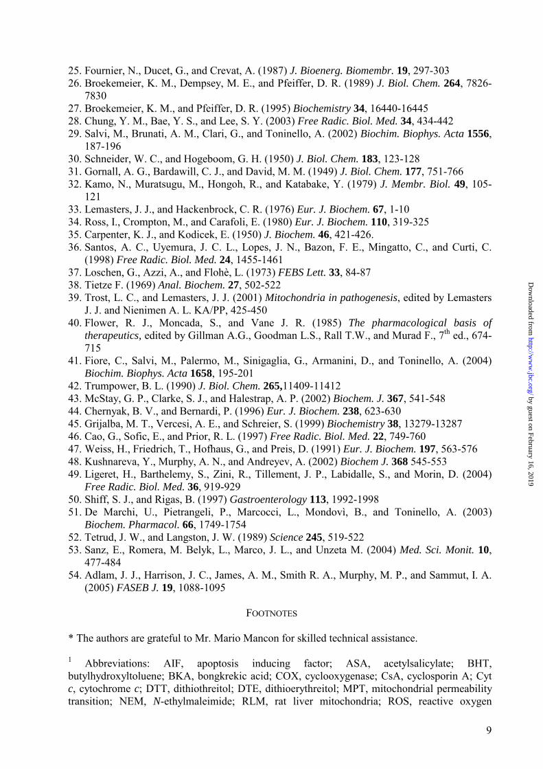
1. Weissmann, G. (1991) Sci. Am. 264, 84-90 2. Vane, J. R. (1971) Nat. New Biol. 231, 232-235 3. Vane, J. R. (1994) Nature 367, 215-216 4. Taketo, M. M. (1998) J. Natl. Cancer Inst. 90, 1529-1536 5. Taketo, M. M. (1998 B) J. Natl. Cancer Inst. 90, 1609-1620 6. Sheng, H., Shao, J., Morrow, J. D., Beauchamp, R. D., and DuBois, R. N. (1998) Cancer
Res. 58, 363-366 7. Sawaoka, H., Kawano, S., Tsuji, M., Gunawan, E. S., Takei, Y., Nagano, K., and Hori, M.
(1998) Am. J. Physiol. 273, G1061-G1067 8. Hanif, R., Pittas, A., Feng, Y., Koutsos, M. I., Qiao, L., StaianoCoico, L., Shiff, S. J., and
Rigas, B. (1996) Biochem. Pharmacol. 52, 237-245 9. Rigas, B., and Shiff, S. J. (2000) Med. Hypotheses 54, 210-215 10. Stark, L. A., Din, F. V. N., Zwacka, R. M., and Dunlop, M. G. (2001) FASEB J. 15, 1273-
1275 11. Kroemer, G, Dalla Porta, B., and Resche-Rigon, M. (1998) Annu. Rev. Physiol. 60, 619-642 12. Trost, L. C., and Lemasters, J. J. (1997) Toxicol. Appl. Pharmacol. 147, 431-441 13. Segalman, T. Y., and Lee, C. P. (1995) Arch. Biochem. Biophys. 317, 79-84 14. Martens, M. E., and Lee, C. P. (1984) Biochem. Pharmacol. 33, 2869-2876 15. Tonsgard, J. H., and Getz, G. S. (1985) J. Clin. Invest. 76, 816-825 16. Martens, M. E., Chang, C. H., and Lee, C. P. (1986) Arch. Biochem. Biophys. 244, 773-786 17. Tomoda, T., Takeda, K., Kurashige, T., Enzan, H., and Miyahara, M. (1994) Liver 14, 103-
108 18. Nulton-Presson, A. C., Szweda, L. I., and Sadek, H. A. (2004) J. Cardiovasc. Pharmacol.
44, 591-595 19. Biban, C., Tassani, V., Toninello, A., Siliprandi, D., and Siliprandi, N. (1995) Biochem.
Pharmacol. 50, 497-500 20. Starko, K. M., Ray, C. G., Dominguez, L. B. Stromberg, W. L., and Woodall, D. F. (1980).
Pediatrics 66, 859-864 21. Waldman, R. J., Hall, W. N., McGee, H., and VanAmburg, G. (1982) J. Am. Med. Assoc.
247, 3089-3094 22. Partin, J. C., Shubert, W. K., and Partin, J. S. (1971) New Engl. J. Med. 285, 1339-1343 23. Gunter, T. E., and Pfeiffer, D. R. (1990) Am. J. Physiol. 258, C755-C786 24. Zoratti, M., and Szabò, I. (1995) Biochim. Biophys. Acta 1241, 139-176
by guest on February 16, 2019http://w
ww
.jbc.org/D
ownloaded from
9
25. Fournier, N., Ducet, G., and Crevat, A. (1987) J. Bioenerg. Biomembr. 19, 297-303 26. Broekemeier, K. M., Dempsey, M. E., and Pfeiffer, D. R. (1989) J. Biol. Chem. 264, 7826-
7830 27. Broekemeier, K. M., and Pfeiffer, D. R. (1995) Biochemistry 34, 16440-16445 28. Chung, Y. M., Bae, Y. S., and Lee, S. Y. (2003) Free Radic. Biol. Med. 34, 434-442 29. Salvi, M., Brunati, A. M., Clari, G., and Toninello, A. (2002) Biochim. Biophys. Acta 1556,
187-196 30. Schneider, W. C., and Hogeboom, G. H. (1950) J. Biol. Chem. 183, 123-128 31. Gornall, A. G., Bardawill, C. J., and David, M. M. (1949) J. Biol. Chem. 177, 751-766 32. Kamo, N., Muratsugu, M., Hongoh, R., and Katabake, Y. (1979) J. Membr. Biol. 49, 105-
121 33. Lemasters, J. J., and Hackenbrock, C. R. (1976) Eur. J. Biochem. 67, 1-10 34. Ross, I., Crompton, M., and Carafoli, E. (1980) Eur. J. Biochem. 110, 319-325 35. Carpenter, K. J., and Kodicek, E. (1950) J. Biochem. 46, 421-426. 36. Santos, A. C., Uyemura, J. C. L., Lopes, J. N., Bazon, F. E., Mingatto, C., and Curti, C.
(1998) Free Radic. Biol. Med. 24, 1455-1461 37. Loschen, G., Azzi, A., and Flohè, L. (1973) FEBS Lett. 33, 84-87 38. Tietze F. (1969) Anal. Biochem. 27, 502-522 39. Trost, L. C., and Lemasters, J. J. (2001) Mitochondria in pathogenesis, edited by Lemasters
J. J. and Nienimen A. L. KA/PP, 425-450 40. Flower, R. J., Moncada, S., and Vane J. R. (1985) The pharmacological basis of
therapeutics, edited by Gillman A.G., Goodman L.S., Rall T.W., and Murad F., 7th ed., 674-715
41. Fiore, C., Salvi, M., Palermo, M., Sinigaglia, G., Armanini, D., and Toninello, A. (2004) Biochim. Biophys. Acta 1658, 195-201
42. Trumpower, B. L. (1990) J. Biol. Chem. 265,11409-11412 43. McStay, G. P., Clarke, S. J., and Halestrap, A. P. (2002) Biochem. J. 367, 541-548 44. Chernyak, B. V., and Bernardi, P. (1996) Eur. J. Biochem. 238, 623-630 45. Grijalba, M. T., Vercesi, A. E., and Schreier, S. (1999) Biochemistry 38, 13279-13287 46. Cao, G., Sofic, E., and Prior, R. L. (1997) Free Radic. Biol. Med. 22, 749-760 47. Weiss, H., Friedrich, T., Hofhaus, G., and Preis, D. (1991) Eur. J. Biochem. 197, 563-576 48. Kushnareva, Y., Murphy, A. N., and Andreyev, A. (2002) Biochem J. 368 545-553 49. Ligeret, H., Barthelemy, S., Zini, R., Tillement, J. P., Labidalle, S., and Morin, D. (2004)
Free Radic. Biol. Med. 36, 919-929 50. Shiff, S. J., and Rigas, B. (1997) Gastroenterology 113, 1992-1998 51. De Marchi, U., Pietrangeli, P., Marcocci, L., Mondovì, B., and Toninello, A. (2003)
Biochem. Pharmacol. 66, 1749-1754 52. Tetrud, J. W., and Langston, J. W. (1989) Science 245, 519-522 53. Sanz, E., Romera, M. Belyk, L., Marco, J. L., and Unzeta M. (2004) Med. Sci. Monit. 10,
477-484 54. Adlam, J. J., Harrison, J. C., James, A. M., Smith R. A., Murphy, M. P., and Sammut, I. A.
(2005) FASEB J. 19, 1088-1095
FOOTNOTES
* The authors are grateful to Mr. Mario Mancon for skilled technical assistance. 1 Abbreviations: AIF, apoptosis inducing factor; ASA, acetylsalicylate; BHT, butylhydroxyltoluene; BKA, bongkrekic acid; COX, cyclooxygenase; CsA, cyclosporin A; Cyt c, cytochrome c; DTT, dithiothreitol; DTE, dithioerythreitol; MPT, mitochondrial permeability transition; NEM, N-ethylmaleimide; RLM, rat liver mitochondria; ROS, reactive oxygen
by guest on February 16, 2019http://w
ww
.jbc.org/D
ownloaded from

10
species; SAL, salicylate; TMPD, trimethyl-para-phenylendiamine; TPP+, tetraphenylphosphonium; β-OH, β-hydroxybutyrate; ∆Ψ, membrane potential.
FIGURE LEGENDS
Fig. 1. Mitochondrial swelling (A) and ∆Ψ collapse (B) induced by salicylate in rat liver mitochondria. Effect of cyclosporin A and bongkrekic acid. RLM were incubated in standard medium in conditions described in Materials and Methods. Salicylate (SAL) was present at 0.5 mM concentration. When added to medium, 1 µM CsA or 5 µM BKA were present. Control traces refer to RLM incubated in standard medium or in standard medium deprived of Ca2+ in presence of salicylate. Downward deflection (panel A): mitochondrial swelling. ∆Ε value (panel B): electrode potential. Both assays were performed four times, with comparable results. Fig. 2. Effect of salicylate on succinate and β-hydroxybutyrate oxidation and oxidative phosphorylation (A), adenine (B) and pyridine (C) nucleotide mitochondrial content, and palmitoylcarnitine oxidation (D). Panel A-C: RLM were incubated in standard medium as described in Materials and Methods, in presence of 1 mM phosphate. Salicylate (SAL) and CsA were present at 0.5 mM and 1 µM, respectively. Dashed lines in panel A, b: presence of 5 mM β-hydroxybutyrate as substrate instead of succinate plus rotenone. Where indicated in panel A, a: additions of 0.2 mM ADP for RCI and P/O calculations. Adenine nucleotides (AdN) (panel B) and pyridine nucleotides (panel C) were measured in mitochondrial pellet and supernatant, respectively. Panel D: RLM pre-incubated for 10 min as reported for panels A-C and treated as described in Materials and Methods, were incubated in assay medium for β-oxidation (see Materials and Methods), in presence of 30 µM palmitoylcarnitine as substrate. Fig. 3. Effect of antioxidizing agents on mitochondrial swelling (A) and ∆Ψ collapse (B) induced by salicylate. Incubation conditions and salicylate concentration as in Fig. 1. When added to medium, 1 mM DTT, 10 µM NEM, 25 µM BHT and 100 µM Trolox were present. Both assays were performed four times, with comparable results. Fig. 4. Increased oxygen uptake induced by salicylate. Incubation conditions and salicylate concentration as in Fig. 1. Where indicated, standard medium was deprived of Ca2+. Data are representative of four similar experiments. Fig. 5. Hydrogen peroxide generation by mitochondria induced by salicylate in absence (A) or presence (B) of Ca2+. Incubation conditions and salicylate concentration as in Fig. 1. Where indicated, 1 µM CsA and 5 µM BKA were present. Mean values + SD of four experiments. Fig. 6. Mitochondrial thiol (A) and glutathione (B) oxidation by salicylate. Incubation conditions and salicylate concentration as in Fig. 1. When present, NEM was 10 µM. Mean values + SD of three experiments. Fig. 7. Endogenous pyridine nucleotide oxidation by salicylate. Inhibition by antioxidizing agents. RLM incubated as in Fig. 1. Salicylate present at 0.5 mM concentration. When added to medium, 10 µM NEM, and 1 mM DTT were present. Where indicated, standard medium was deprived of Ca2+. Three additional experiments gave comparable results.
by guest on February 16, 2019http://w
ww
.jbc.org/D
ownloaded from
11
Fig. 8. Release of cytochrome c (A) and AIF (B) induced by salicylate. Inhibitory effect by CsA, and NEM. RLM incubated for 15 min in standard medium and, where indicated, deprived of Ca2+. Salicylate at 0.5 mM concentration. When added to medium, 1 µM CsA, 10 µM NEM was present. Data are representative of three separate experiments. Fig. 9. Mitochondrial swelling induced by salicylate, acetylsalicylate and benzoate. RLM were incubated in standard medium, as described in Materials and Methods. Where indicated, medium was deprived of Ca2+. Salicylate, acetylsalicylate (ASA) and benzoate present at 0.5 mM. Experiment was performed four times, with identical results. Fig. 10. Effect of various redox states of respiratory chain complexes and differing membrane energizing substrates on mitochondrial swelling (A, C) and ∆Ψ collapse (B, D) induced by salicylate. All incubations carried out as described in incubation procedures in a medium containing 250 mM sucrose, 10 mM Hepes (pH 7.4) and 40 µM Ca2+. When present, salicylate was at 0.5 mM concentration. Panels A, B: 5 mM β-hydroxybutyrate (β-OH) as energizing substrate. Panels C, D: 5 mM succinate (energizing substrate), 1,25 µM rotenone, 5 mM β-OH. Three additional experiments gave comparable results. Fig. 11. Hydrogen peroxide generation by mitochondria treated with salicylate, in absence of Ca2+, in differing redox states of respiratory chain complexes. General incubation conditions as in Fig. 10, except that medium was deprived of Ca2+. Where indicated, medium was energized by β-OH (trace a) or succinate plus rotenone in presence of β-OH (trace b), at concentrations as shown in Fig. 9. Control traces refer to both energizing conditions.
by guest on February 16, 2019http://w
ww
.jbc.org/D
ownloaded from

T = 1'
DE = 10mV
SAL
SAL + CsAcontrol / SAL (-Ca )
2 +
SAL + BKA
170
050
100
150
Dy
(mV
)
B
SAL + CsAcontrol / SAL (-Ca )
2+
SAL + BKA
SAL
DA = 0.200
T = 1'
A
Figure 1
by guest on February 16, 2019http://w
ww
.jbc.org/D
ownloaded from

D
RL
M
contr
ol
SA
L
SA
L(p
re-t
reat
edw
ith C
sAor
BK
A)
50 n
atom
s O
2 m
in
B
contr
ol
Tim
e (m
in)
SA
L+
CsA
SA
L
[ C]AdN in pellet (nmol/mg prot)14
5
510
10
15
C
contr
ol
SA
L
0
Tim
e (m
in)
510
15
48
Pyridine nucleotides in medium (nmol/ml)
SA
L+
CsA
contr
ol
SA
L
50 n
atom
s O
RL
M
2 m
in
AD
P
AD
P
RL
M
contr
ol
SA
L
SA
L+
CsA
SA
L+
CsA
Tab
leA
condit
ions
RC
IP
/O
contr
ol
SA
L
SA
L+
CsA
4.3
1.0
3.4
1.7
5
1.6
8
-
AF
igure
2a
b
by guest on February 16, 2019http://w
ww
.jbc.org/D
ownloaded from

SAL + NEM
control
SAL
DA = 0.200
T = 1'
SAL + DTT
T = 1'
DE = 10mV
SAL
SAL + NEM
control170
050
100
150
Dy
(mV
)
B
A
SAL + DTT
Figure 3
SAL + BHT
SAL + Trolox
SAL + BHT
SAL + Trolox
by guest on February 16, 2019http://w
ww
.jbc.org/D
ownloaded from
control (- Ca )2+
control
SAL (- Ca )2+
SAL
2 min
50 natoms O
RLM
Figure 4
by guest on February 16, 2019http://w
ww
.jbc.org/D
ownloaded from

Control
SAL
0
0,5 1 1,5 2 2,5 3 3,5 4
0,2
0,4
0,6
0,8
1
1,2
1,4
1,6
1,8
2
2,2
2,4
nm
oli
H O
/mg p
rot.
22
Time (min)
Figure 5
SAL
(-Ca )2+
SAL + BKA
SAL + CsA
Control
0
0,5 1 1,5 2 2,5 3 3,5 4
0,2
0,4
0,6
0,8
1
1,2
1,4
1,6
1,8
2
2,2
2,4
nm
oli
H O
/mg p
rot.
22
Time (min)
(+Ca )2+
SAL + BKASAL + CsA
A
B
by guest on February 16, 2019http://w
ww
.jbc.org/D
ownloaded from
100
80
60
40
20
SAL
+N
EM(- C
a)2+
SAL
SAL
(- Ca
)2+
control
control (-Ca
)2+
Red
uce
dth
iol
gro
ups
(%of
contr
ol)
SAL
+N
EM
20
40
60
80
100
control (-Ca
)2+
SAL
SAL
(- Ca
)2+
control
A
B
Red
uce
dglu
tath
ione
(%of
contr
ol)
SAL
+ NEM
SAL
+ NEM
(- Ca
)2+
Figure 6
by guest on February 16, 2019http://w
ww
.jbc.org/D
ownloaded from

SAL
Flu
ore
scen
ce c
han
ge
(arb
itra
ry u
nit
)N
AD
+(P
)
N
AD
(P)H
2 min
control
SAL + NEM
SAL (-Ca )2+
Figure 7
SAL + DTT
0
100
200
300
by guest on February 16, 2019http://w
ww
.jbc.org/D
ownloaded from

control (-Ca
)2 +
control
SAL
(- Ca
)2 +
SAL
+ CsA
SAL
A
SAL
+ CsA
SAL
SAL
(- Ca
)2 +
control
control (-Ca
)2 +
B
SAL
+ NEM
SAL
+ NEM
Figure 8
by guest on February 16, 2019http://w
ww
.jbc.org/D
ownloaded from

control
SAL (- Ca )2 +
ASA
SAL
DA = 0.200
T = 1'
ASA (- Ca )2 +
Benzoate (- Ca )2 +
Benzoate
Salicylate BenzoateAcetylsalicylate
OH
COO-
OCOCH3
COO-
COO-
Figure 9
by guest on February 16, 2019http://w
ww
.jbc.org/D
ownloaded from

control ( -OH)b
SAL (b-OH)
DA = 0,200
T = 1'
A
controlSAL ( -OH +Succinate +Rotenone)
b
C
control ( -OH)b
T = 1'
DE = 10mV
170
050
100
150
Dy
(mV
)
B
SAL
DE = 10mV
170
050
100
150
Dy
(mV
)
D
SALcontrol
DA = 0,200
T = 1'
T = 1'
SAL (b-OH)
SAL ( -OH +Succinate +Rotenone)
b
Figure 10
by guest on February 16, 2019http://w
ww
.jbc.org/D
ownloaded from

controls
(different substrates)(-Ca )2 +
SAL ( -OH +Succinate + Rotenone)
(-Ca )
b
2 +
00,5 1 1,5 2 2,5 3 3,5 4
0,2
0,4
0,6
0,8
1
1,2
1,4
1,6
1,8
2
2,2
2,4
SAL ( -OH)
(- Ca )
b2 +
Time (min)
nm
ol
H O
/m
g p
rot.
22
a
b
Figure 11
by guest on February 16, 2019http://w
ww
.jbc.org/D
ownloaded from

Valentina Battaglia, Mauro Salvi and Antonio Toninelloby salicylate in liver mitochondria
Oxidative stress is responsible for mitochondrial permeability transition induction
published online August 12, 2005J. Biol. Chem.
10.1074/jbc.M502391200Access the most updated version of this article at doi:
Alerts:
When a correction for this article is posted•
When this article is cited•
to choose from all of JBC's e-mail alertsClick here
by guest on February 16, 2019http://w
ww
.jbc.org/D
ownloaded from