Overview: They’re (Almost) Everywhere! Most prokaryotes are microscopic, but what they lack in...
63
Overview: They’re (Almost) Everywhere! • Most prokaryotes are microscopic, but what they lack in size they make up for in numbers • There are more in a handful of fertile soil than the number of people who ever lived • Prokaryotes thrive almost everywhere, including places too acidic, too salty, too cold, or too hot for most other organisms • They have an astonishing genetic diversity
-
Upload
gaige-gibbens -
Category
Documents
-
view
216 -
download
0
Transcript of Overview: They’re (Almost) Everywhere! Most prokaryotes are microscopic, but what they lack in...
Overview: They’re (Almost) Everywhere!
• Most prokaryotes are microscopic, but what they lack in size they make up for in numbers
• There are more in a handful of fertile soil than the number of people who ever lived
• Prokaryotes thrive almost everywhere, including places too acidic, too salty, too cold, or too hot for most other organisms
• They have an astonishing genetic diversity
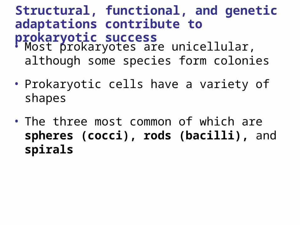
Structural, functional, and genetic adaptations contribute to prokaryotic success
• Most prokaryotes are unicellular, although some species form colonies
• Prokaryotic cells have a variety of shapes
• The three most common of which are spheres (cocci), rods (bacilli), and spirals
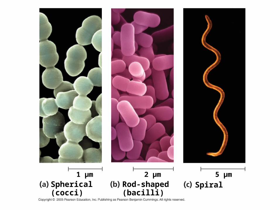
Spherical (cocci)
Rod-shaped (bacilli)
Spiral5 µm2 µm1 µm
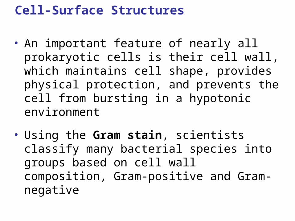
Cell-Surface Structures
• An important feature of nearly all prokaryotic cells is their cell wall, which maintains cell shape, provides physical protection, and prevents the cell from bursting in a hypotonic environment
• Using the Gram stain, scientists classify many bacterial species into groups based on cell wall composition, Gram-positive and Gram-negative

• Most bacterial cell walls contain peptidoglycan, a polymer of modified sugars cross-linked by short polypeptides.
• The walls of archaea lack peptidoglycan.
• The Gram stain is a valuable tool for identifying specific bacteria based on differences in their cell walls.
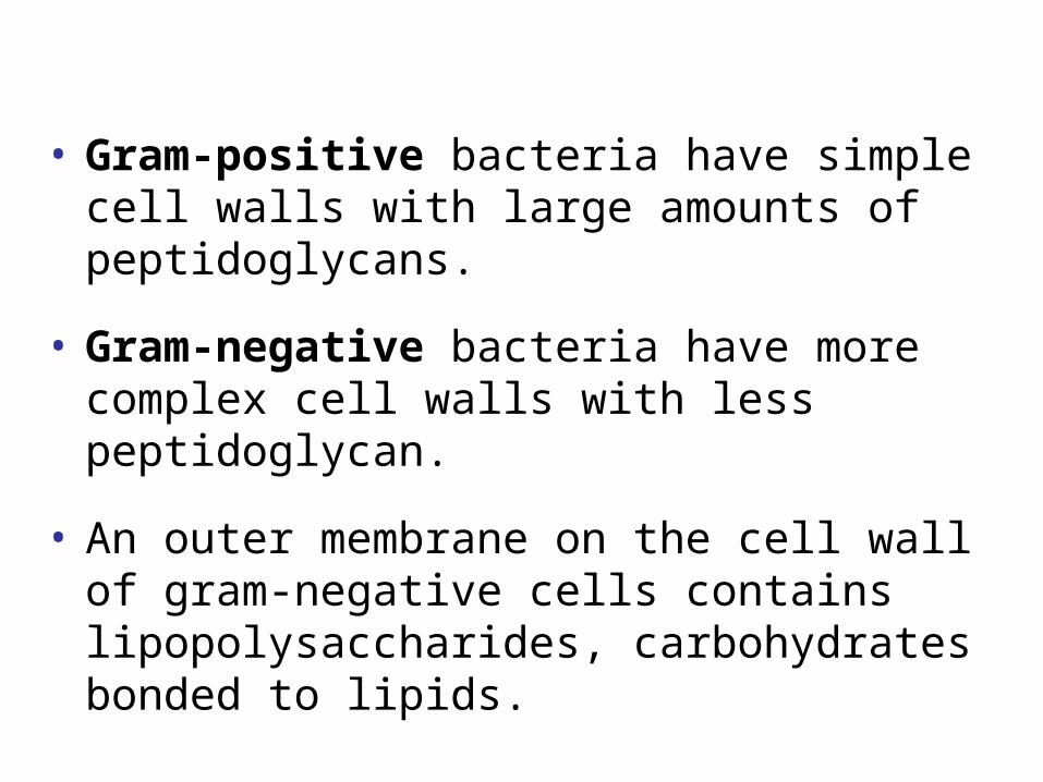
• Gram-positive bacteria have simple cell walls with large amounts of peptidoglycans.
• Gram-negative bacteria have more complex cell walls with less peptidoglycan.
• An outer membrane on the cell wall of gram-negative cells contains lipopolysaccharides, carbohydrates bonded to lipids.

Peptidoglycanlayer
Cell wall
Protein
Gram-positivebacteria
Gram-positive Gram-negative
Gram-negativebacteria
Peptidoglycanlayer
Cell wall
Plasma membrane
Lipopolysaccharide
Plasma membrane
Protein
Outermembrane
20 µm
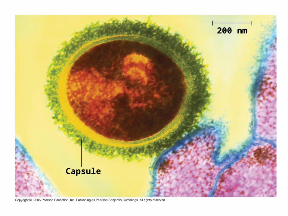
Capsule
200 nm
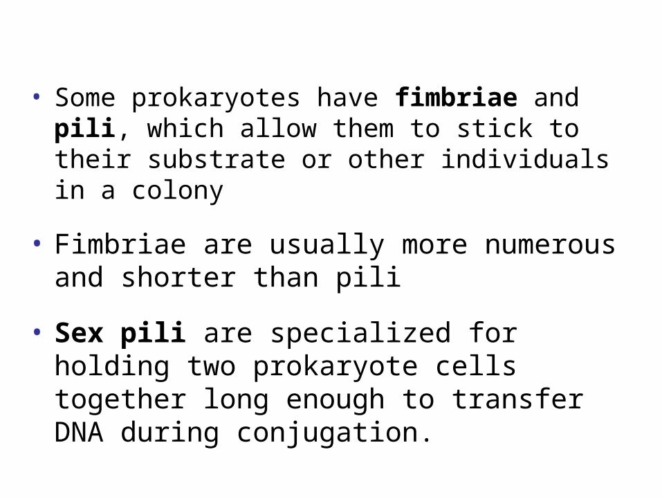
• Some prokaryotes have fimbriae and pili, which allow them to stick to their substrate or other individuals in a colony
• Fimbriae are usually more numerous and shorter than pili
• Sex pili are specialized for holding two prokaryote cells together long enough to transfer DNA during conjugation.
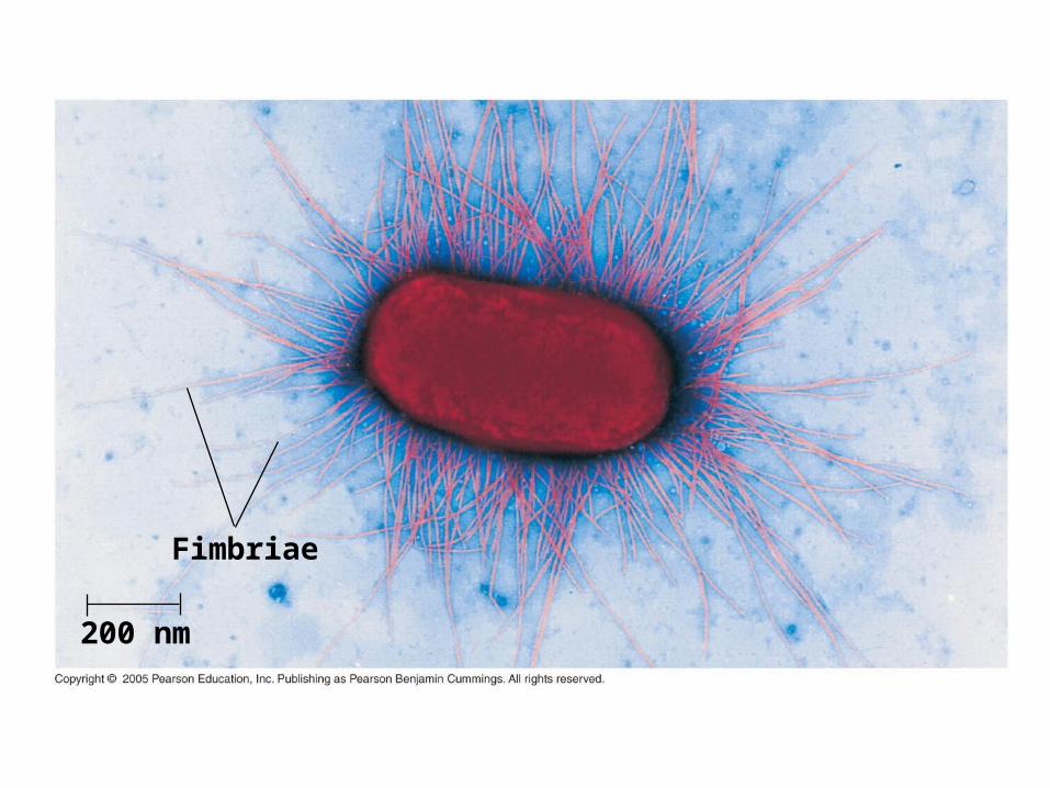
Fimbriae
200 nm
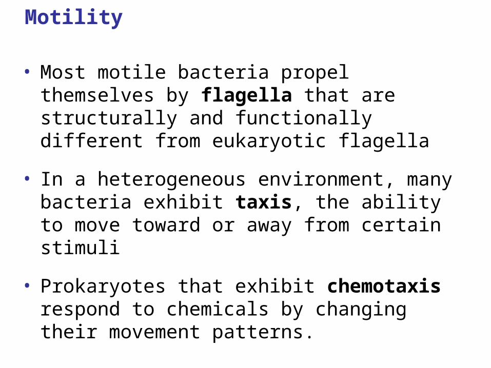
Motility
• Most motile bacteria propel themselves by flagella that are structurally and functionally different from eukaryotic flagella
• In a heterogeneous environment, many bacteria exhibit taxis, the ability to move toward or away from certain stimuli
• Prokaryotes that exhibit chemotaxis respond to chemicals by changing their movement patterns.
Internal and Genomic Organization
• Prokaryotic cells usually lack complex compartmentalization
• Some prokaryotes do have specialized membranes that perform metabolic functions
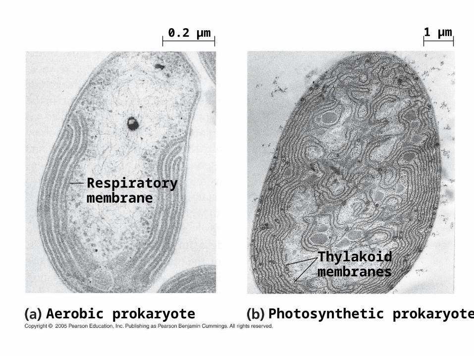
Thylakoidmembranes
Respiratorymembrane
Photosynthetic prokaryoteAerobic prokaryote
0.2 µm 1 µm

• The typical prokaryotic genome is a ring of DNA that is not surrounded by a membrane and that is located in a nucleoid region
• Some species of bacteria also have smaller rings of DNA called plasmids

Chromosome
1 µm
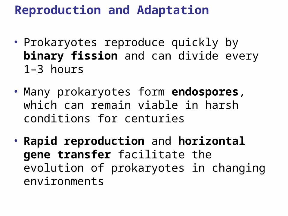
Reproduction and Adaptation
• Prokaryotes reproduce quickly by binary fission and can divide every 1–3 hours
• Many prokaryotes form endospores, which can remain viable in harsh conditions for centuries
• Rapid reproduction and horizontal gene transfer facilitate the evolution of prokaryotes in changing environments
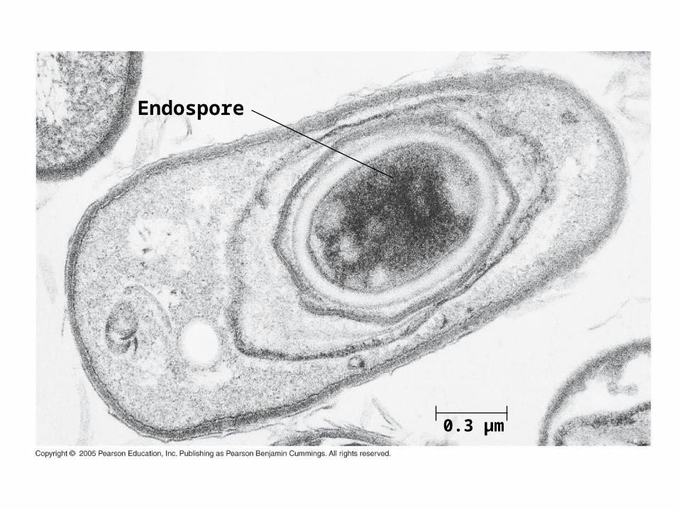
Endospore
0.3 µm
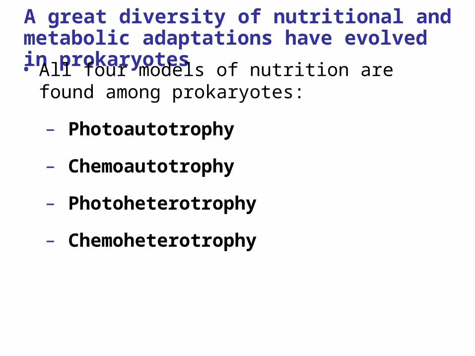
A great diversity of nutritional and metabolic adaptations have evolved in prokaryotes
• All four models of nutrition are found among prokaryotes:
– Photoautotrophy
– Chemoautotrophy
– Photoheterotrophy
– Chemoheterotrophy
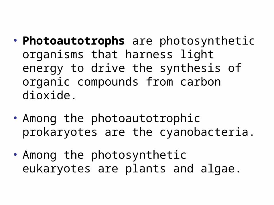
• Photoautotrophs are photosynthetic organisms that harness light energy to drive the synthesis of organic compounds from carbon dioxide.
• Among the photoautotrophic prokaryotes are the cyanobacteria.
• Among the photosynthetic eukaryotes are plants and algae.

• Chemoautotrophs need only CO2 as a carbon source but obtain energy by oxidizing inorganic substances.
• These substances include hydrogen sulfide (H2S), ammonia (NH3), and ferrous ions (Fe2+) among others.
• This nutritional mode is unique to prokaryotes.
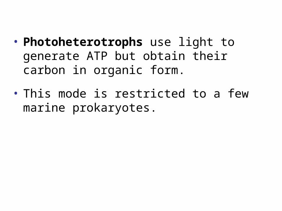
• Photoheterotrophs use light to generate ATP but obtain their carbon in organic form.
• This mode is restricted to a few marine prokaryotes.
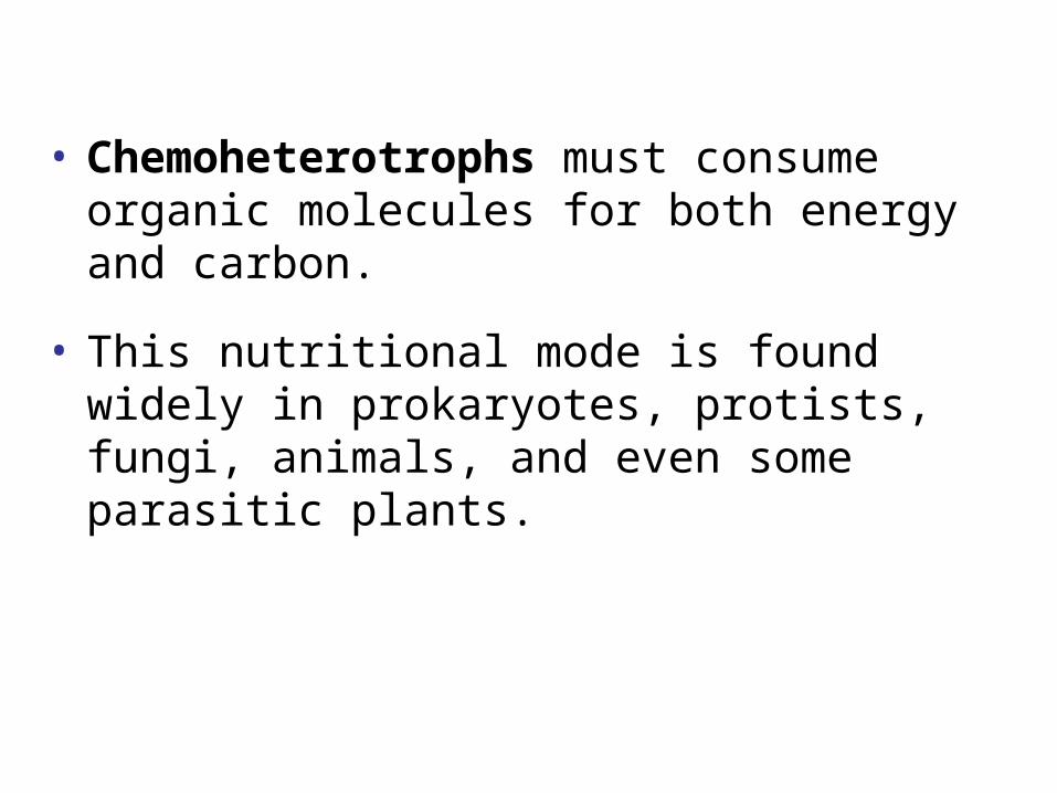
• Chemoheterotrophs must consume organic molecules for both energy and carbon.
• This nutritional mode is found widely in prokaryotes, protists, fungi, animals, and even some parasitic plants.
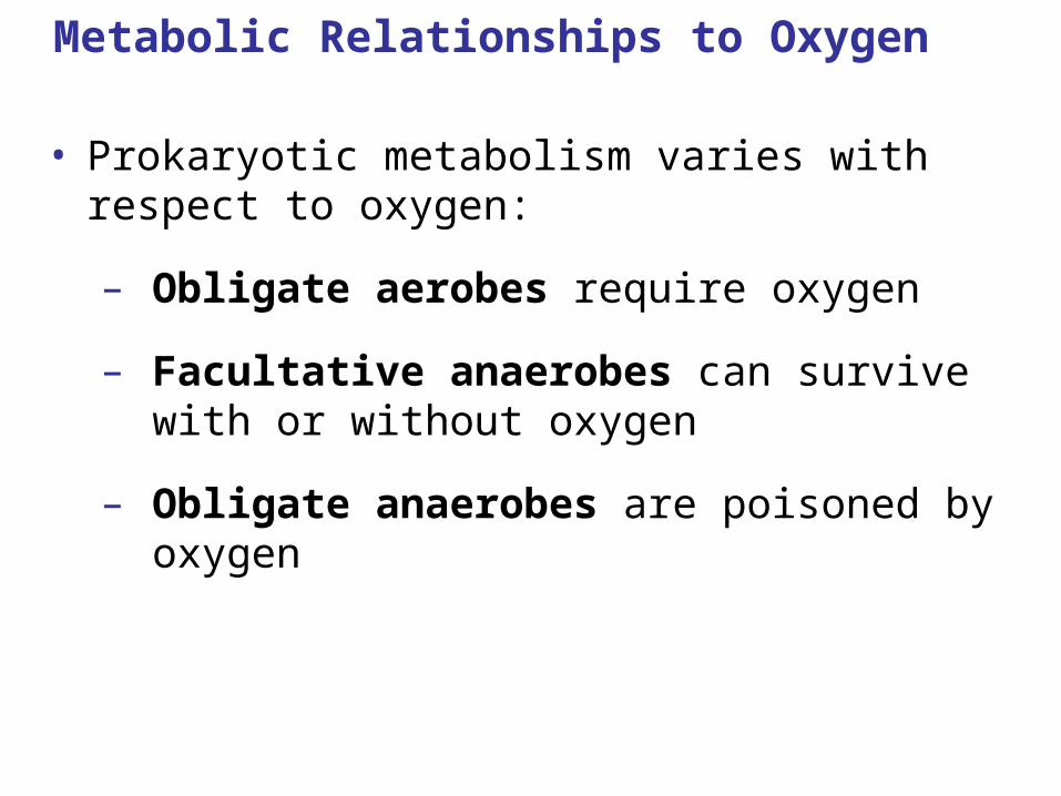
Metabolic Relationships to Oxygen
• Prokaryotic metabolism varies with respect to oxygen:
– Obligate aerobes require oxygen
– Facultative anaerobes can survive with or without oxygen
– Obligate anaerobes are poisoned by oxygen
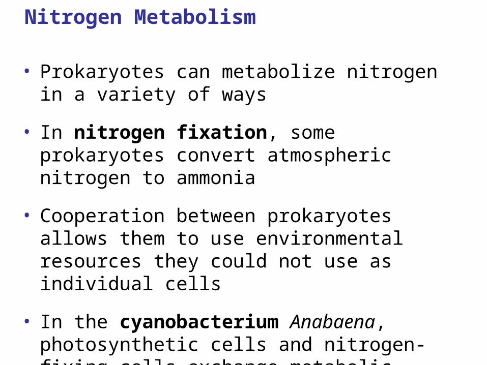
Nitrogen Metabolism
• Prokaryotes can metabolize nitrogen in a variety of ways
• In nitrogen fixation, some prokaryotes convert atmospheric nitrogen to ammonia
• Cooperation between prokaryotes allows them to use environmental resources they could not use as individual cells
• In the cyanobacterium Anabaena, photosynthetic cells and nitrogen-fixing cells exchange metabolic products
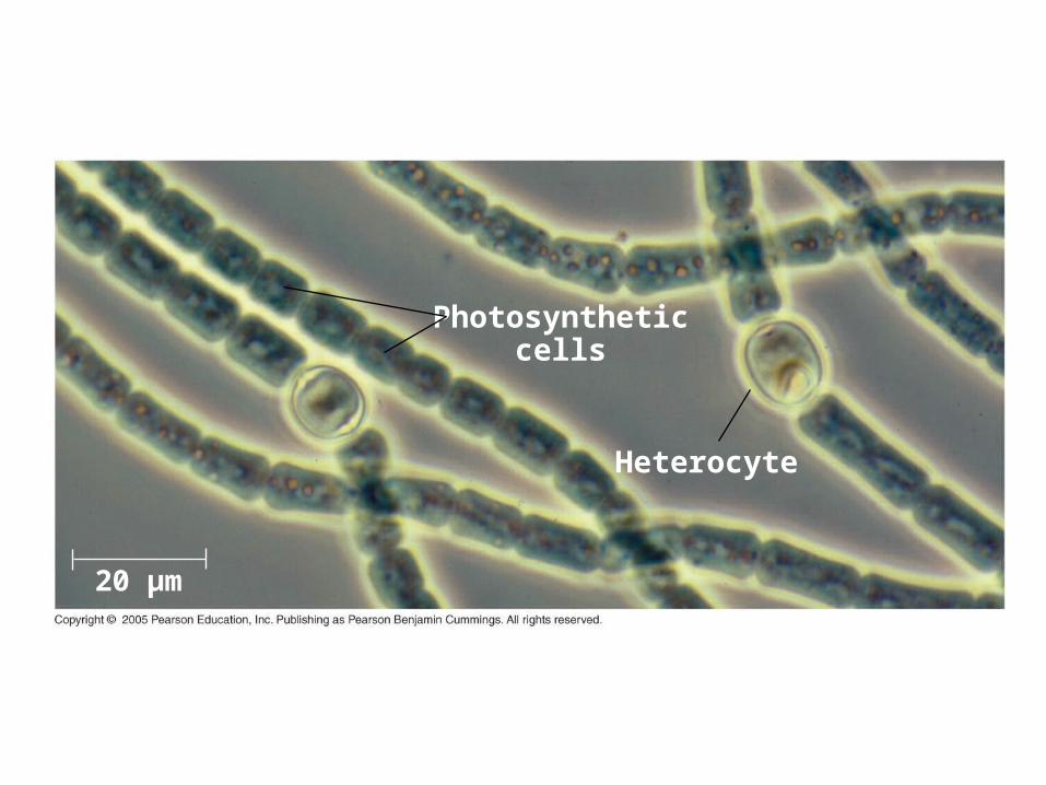
Heterocyte
Photosyntheticcells
20 µm
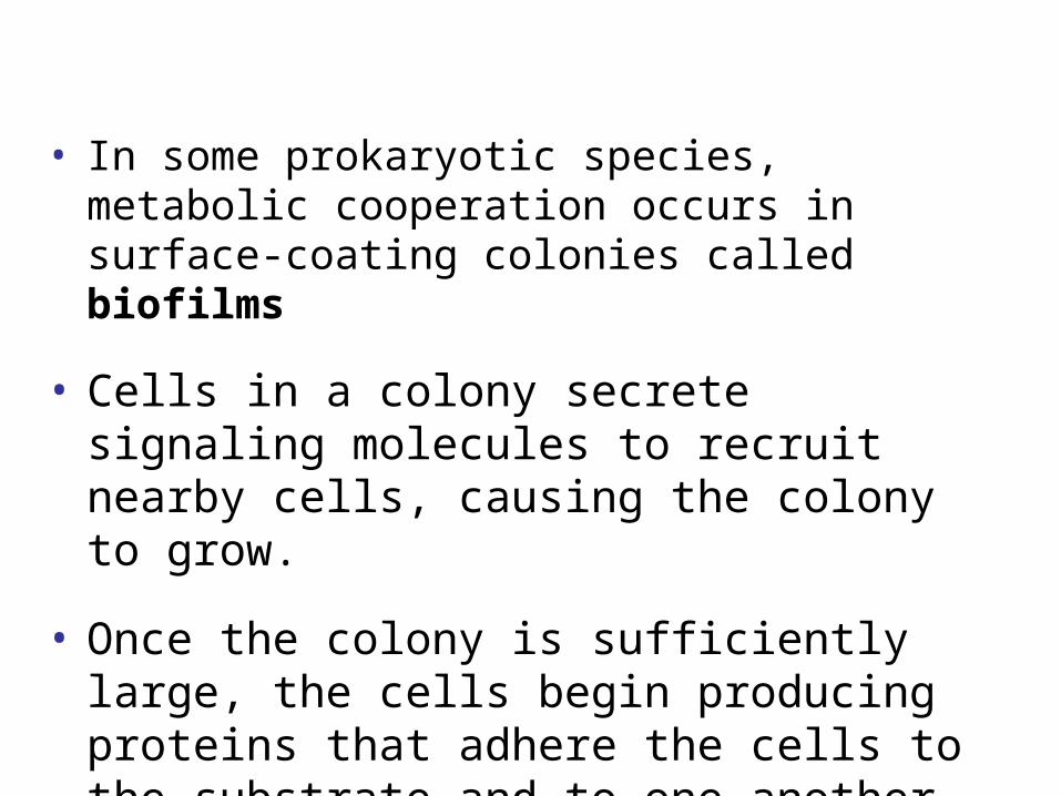
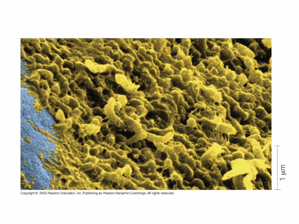
• In some prokaryotic species, metabolic cooperation occurs in surface-coating colonies called biofilms
• Cells in a colony secrete signaling molecules to recruit nearby cells, causing the colony to grow.
• Once the colony is sufficiently large, the cells begin producing proteins that adhere the cells to the substrate and to one another.
• For example, sulfate-consuming bacteria and methane-consuming archaea coexist in ball-shaped aggregates in the mud of the ocean floor.
• The bacteria use the archaea’s waste products.
• In turn, the bacteria produce compounds that facilitate methane consumption by the archaea.
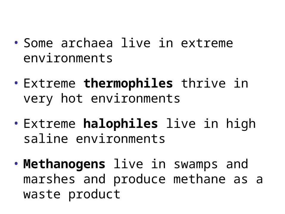
• Some archaea live in extreme environments
• Extreme thermophiles thrive in very hot environments
• Extreme halophiles live in high saline environments
• Methanogens live in swamps and marshes and produce methane as a waste product
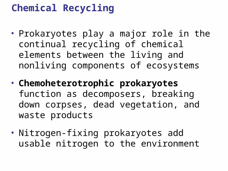
Chemical Recycling
• Prokaryotes play a major role in the continual recycling of chemical elements between the living and nonliving components of ecosystems
• Chemoheterotrophic prokaryotes function as decomposers, breaking down corpses, dead vegetation, and waste products
• Nitrogen-fixing prokaryotes add usable nitrogen to the environment
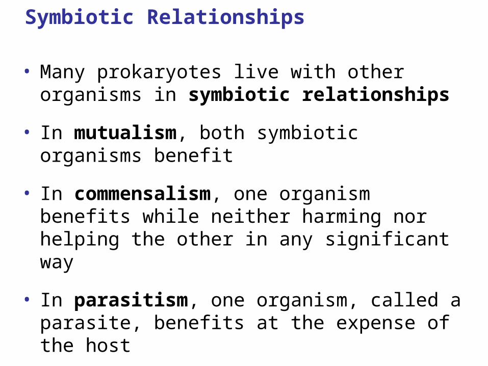
Symbiotic Relationships
• Many prokaryotes live with other organisms in symbiotic relationships
• In mutualism, both symbiotic organisms benefit
• In commensalism, one organism benefits while neither harming nor helping the other in any significant way
• In parasitism, one organism, called a parasite, benefits at the expense of the host
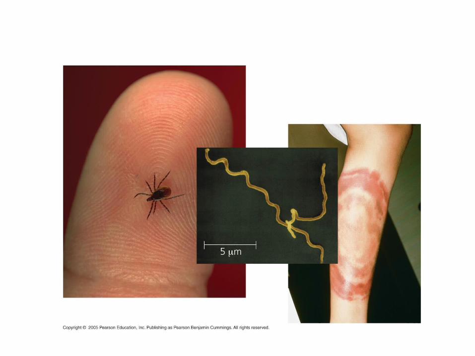
Pathogenic Prokaryotes
• Prokaryotes cause about half of all human diseases
• Lyme disease is an example
• Pathogenic prokaryotes typically cause disease by releasing exotoxins or endotoxins
• Exotoxins cause disease even if the prokaryotes that produce them are not present
• Endotoxins are released only when bacteria die and their cell walls break down
• Many pathogenic bacteria are potential weapons of bioterrorism
Prokaryotes in Research and Technology
• Experiments using prokaryotes have led to important advances in DNA technology
• Prokaryotes are the principal agents in bioremediation, the use of organisms to remove pollutants from the environment
• Some other uses of prokaryotes:
– Recovery of metals from ores
– Synthesis of vitamins
– Production of antibiotics, hormones, and other products

Animations and Videos
• Gram Staining
• Bozeman – Archaea
• Bozeman – Bacteria
• Chapter Quiz Questions – 1
• Chapter Quiz Questions – 2
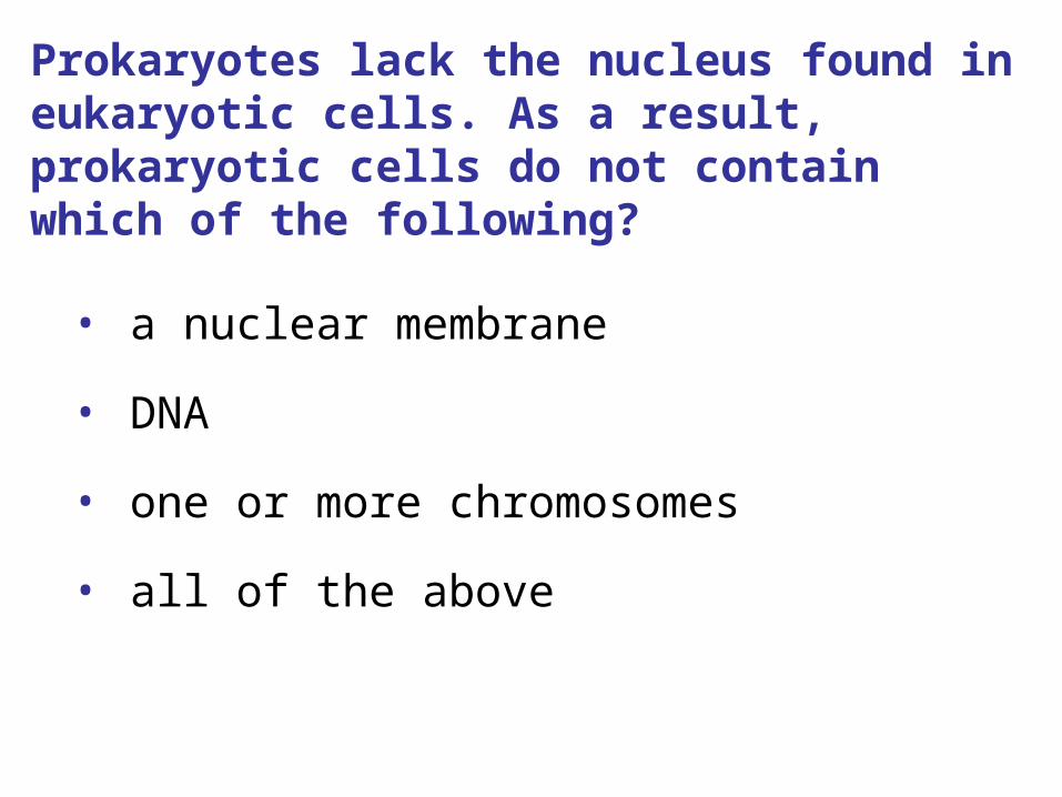
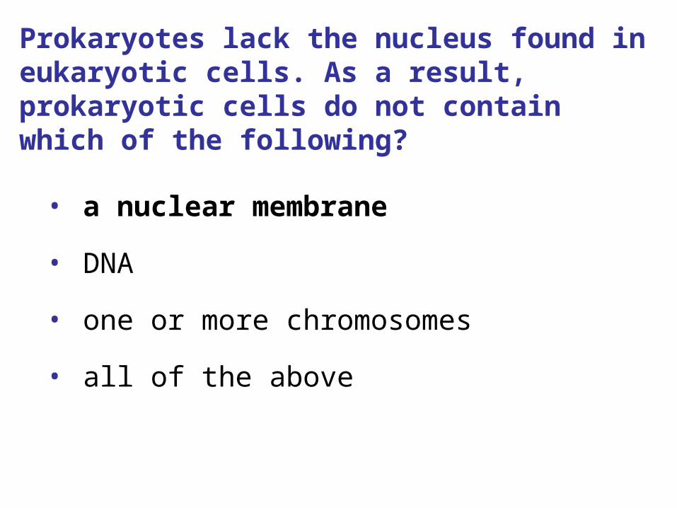
Prokaryotes lack the nucleus found in eukaryotic cells. As a result, prokaryotic cells do not contain which of the following?
• a nuclear membrane
• DNA
• one or more chromosomes
• all of the above
Prokaryotes lack the nucleus found in eukaryotic cells. As a result, prokaryotic cells do not contain which of the following?
• a nuclear membrane
• DNA
• one or more chromosomes
• all of the above
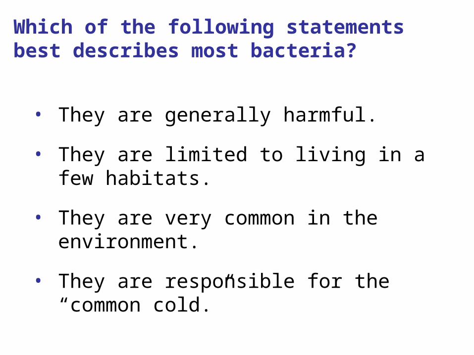
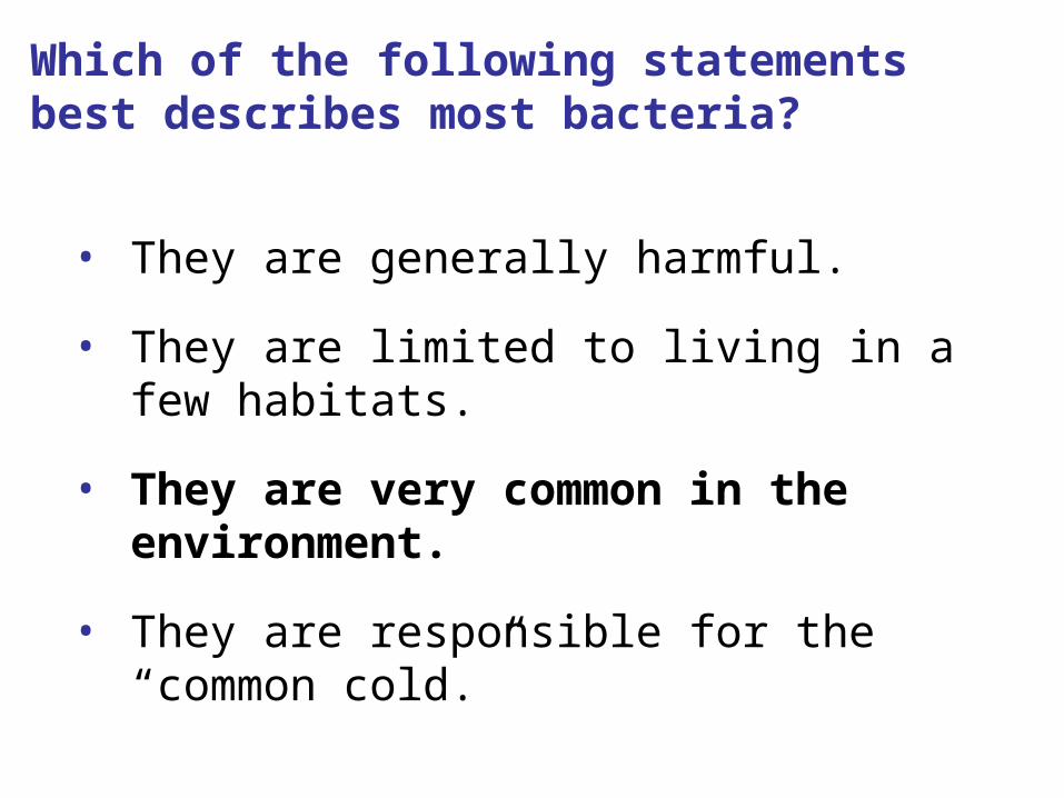
Which of the following statements best describes most bacteria?
• They are generally harmful.
• They are limited to living in a few habitats.
• They are very common in the environment.
• They are responsible for the “common cold.”
Which of the following statements best describes most bacteria?
• They are generally harmful.
• They are limited to living in a few habitats.
• They are very common in the environment.
• They are responsible for the “common cold.”
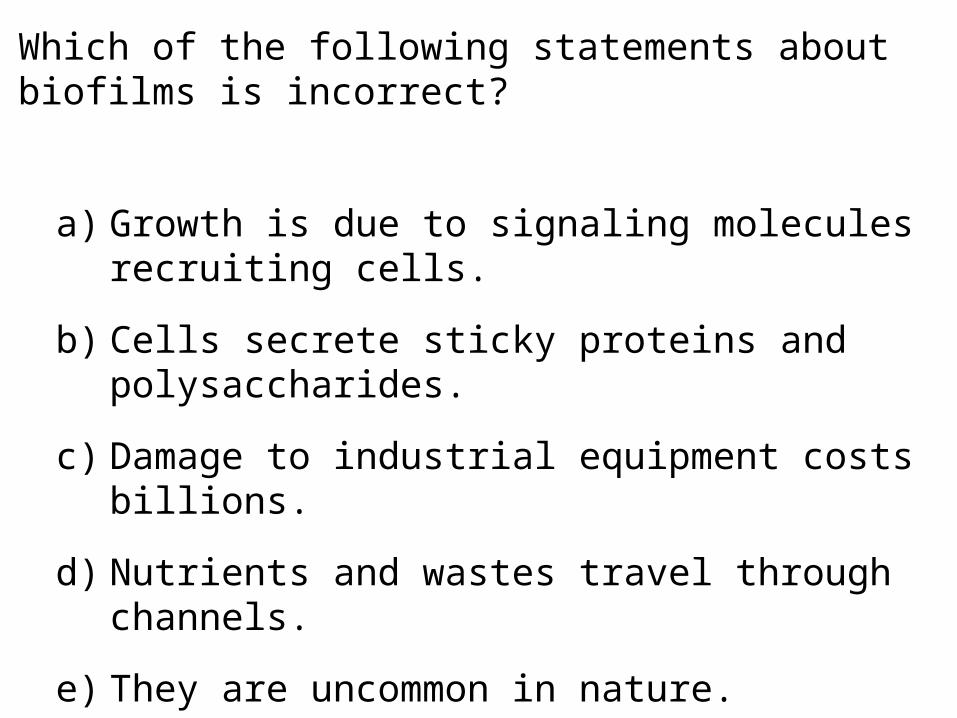
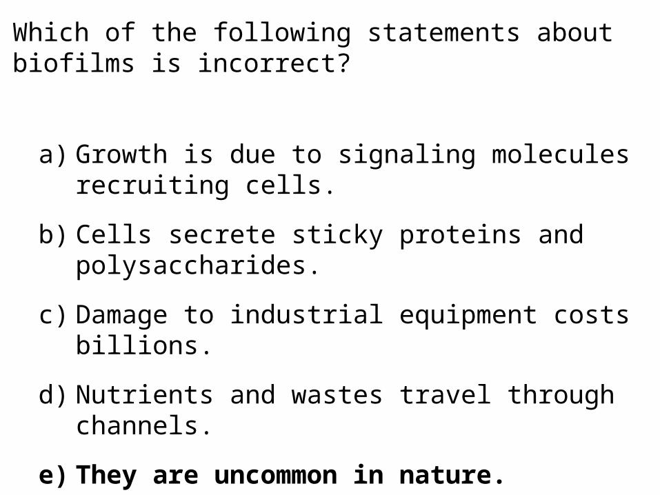
Which of the following statements about biofilms is incorrect?
a) Growth is due to signaling molecules recruiting cells.
b) Cells secrete sticky proteins and polysaccharides.
c) Damage to industrial equipment costs billions.
d) Nutrients and wastes travel through channels.
e) They are uncommon in nature.
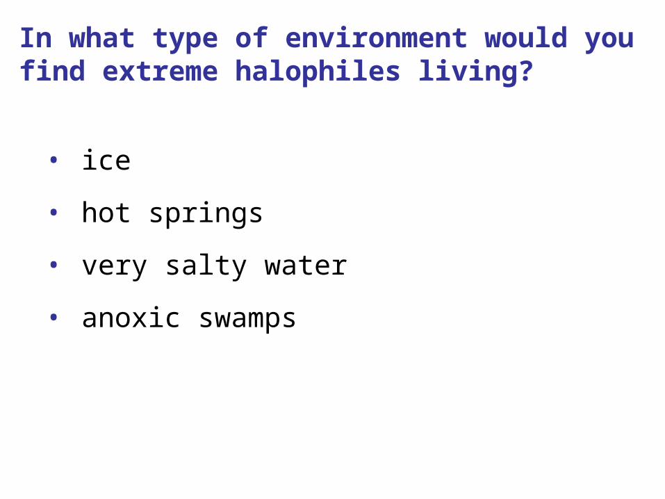
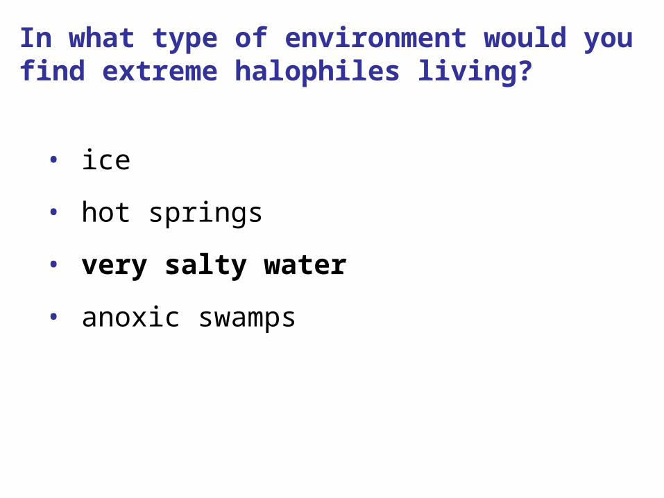
In what type of environment would you find extreme halophiles living?
• ice
• hot springs
• very salty water
• anoxic swamps
In what type of environment would you find extreme halophiles living?
• ice
• hot springs
• very salty water
• anoxic swamps

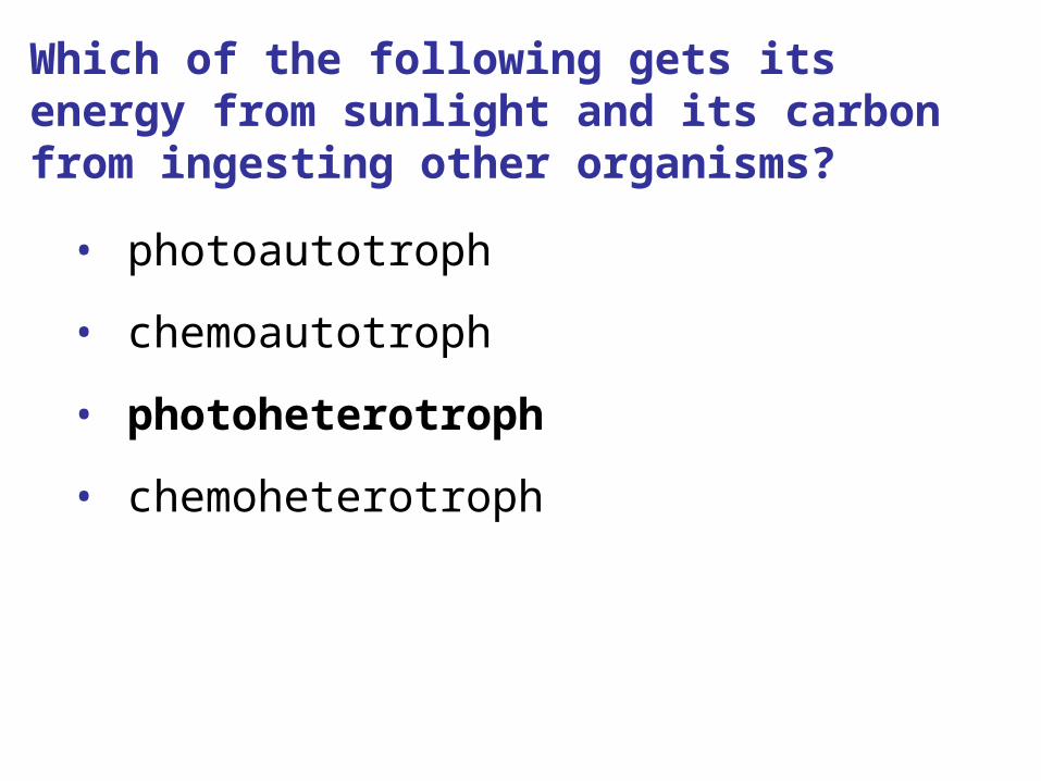
Which of the following gets its energy from sunlight and its carbon from ingesting other organisms?
• photoautotroph
• chemoautotroph
• photoheterotroph
• chemoheterotroph
Which of the following gets its energy from sunlight and its carbon from ingesting other organisms?
• photoautotroph
• chemoautotroph
• photoheterotroph
• chemoheterotroph
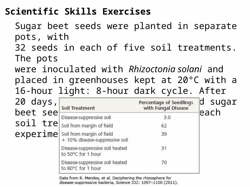
Sugar beet seeds were planted in separate pots, with 32 seeds in each of five soil treatments. The pots were inoculated with Rhizoctonia solani and placed in greenhouses kept at 20°C with a 16-hour light: 8-hour dark cycle. After 20 days, the percentage of infected sugar beet seedlings was determined for each soil treatment. The results of the experiment are shown in the table.
Scientific Skills Exercises
What hypothesis were the researchers testing in this study?
a) Growth is due to signaling molecules recruiting cells.
b) Cells secrete sticky proteins and polysaccharides.
c) Damage to industrial equipment costs billions.
d) Nutrients and wastes travel through channels.
e) They are uncommon in nature.
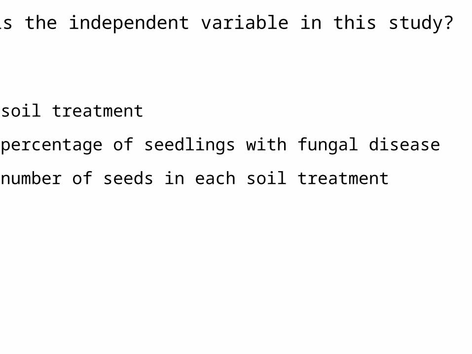
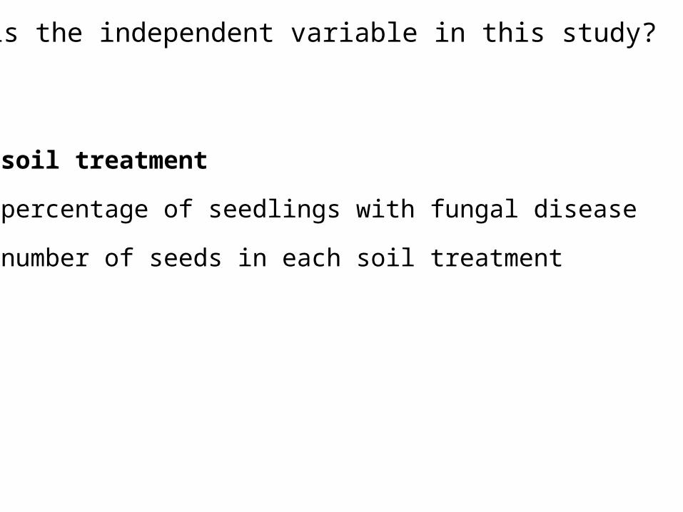
What is the independent variable in this study?
a) soil treatment
b) percentage of seedlings with fungal disease
c) number of seeds in each soil treatment
What is the independent variable in this study?
a) soil treatment
b) percentage of seedlings with fungal disease
c) number of seeds in each soil treatment
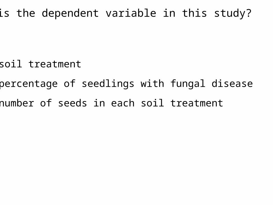
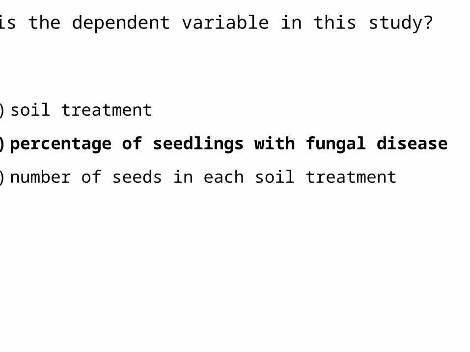
What is the dependent variable in this study?
a) soil treatment
b) percentage of seedlings with fungal disease
c) number of seeds in each soil treatment
What is the dependent variable in this study?
a) soil treatment
b) percentage of seedlings with fungal disease
c) number of seeds in each soil treatment
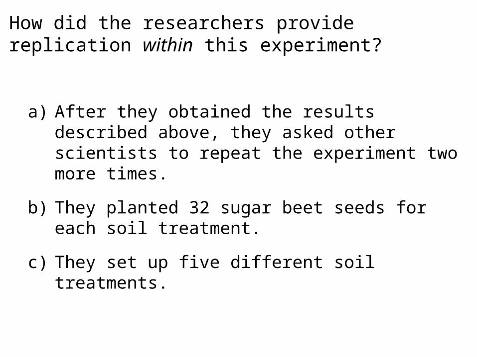
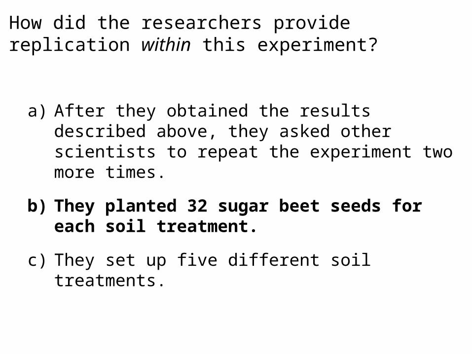
How did the researchers provide replication within this experiment?
a) After they obtained the results described above, they asked other scientists to repeat the experiment two more times.
b) They planted 32 sugar beet seeds for each soil treatment.
c) They set up five different soil treatments.
How did the researchers provide replication within this experiment?
a) After they obtained the results described above, they asked other scientists to repeat the experiment two more times.
b) They planted 32 sugar beet seeds for each soil treatment.
c) They set up five different soil treatments.
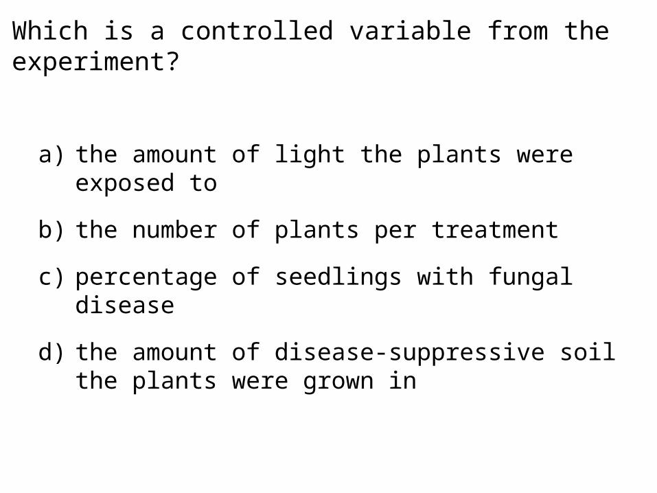
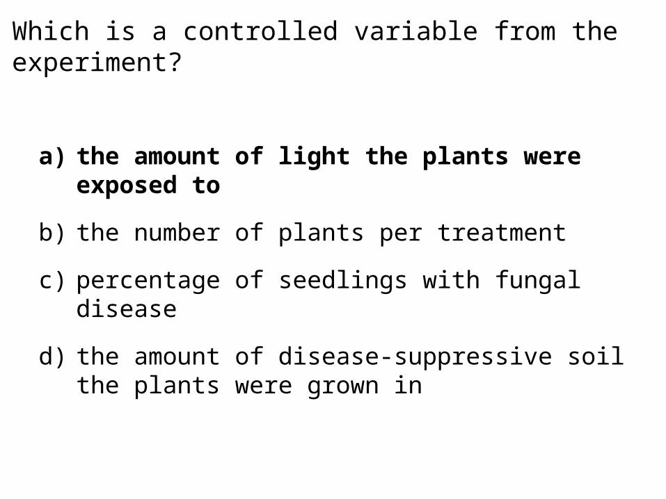
Which is a controlled variable from the experiment?
a) the amount of light the plants were exposed to
b) the number of plants per treatment
c) percentage of seedlings with fungal disease
d) the amount of disease-suppressive soil the plants were grown in
Which is a controlled variable from the experiment?
a) the amount of light the plants were exposed to
b) the number of plants per treatment
c) percentage of seedlings with fungal disease
d) the amount of disease-suppressive soil the plants were grown in
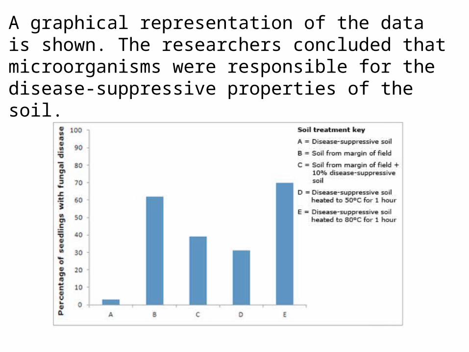
A graphical representation of the data is shown. The researchers concluded that microorganisms were responsible for the disease-suppressive properties of the soil.

How do the results shown in the figure on the previous slide support this conclusion?
a) The lowest rate of fungal infection was found in seedlings grown in disease-suppressive soil.
b) A high rate of fungal infection was found in seedlings grown in soil from the margin of the field.
c) Fungal infection rate increased in seedlings grown in disease-suppressive soil that had been heated.
d) Fungal infection rate decreased in seedlings grown in soil from the margin of the field mixed with disease-suppressive soil.
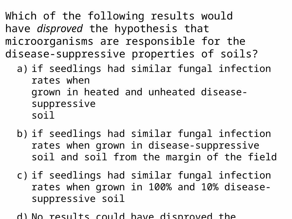
Which of the following results would have disproved the hypothesis that microorganisms are responsible for the disease-suppressive properties of soils?
a) if seedlings had similar fungal infection rates when grown in heated and unheated disease-suppressive soil
b) if seedlings had similar fungal infection rates when grown in disease-suppressive soil and soil from the margin of the field
c) if seedlings had similar fungal infection rates when grown in 100% and 10% disease-suppressive soil
d) No results could have disproved the hypothesis that microorganisms are responsible for the disease-suppressive properties of soil.