Cancer Clinical Trials A treatment option for cancer patients.
© Journal of the National Comprehensive Cancer Network | Volume 7 Number 8 | September 2009
838
OverviewIn 2009 an estimated 40,870 new cases of rectal cancer will occur in the United States (23,580 cases in men; 17,290 cases in women). During the same year, an estimated 49,920 people will die from rectal and colon cancers.1 Although colorectal cancer is ranked as the fourth most frequently diagnosed can-cer and the second leading cause of cancer death in the United States, mortality from colorectal cancer has decreased during the past 30 years. This decrease may be due to earlier diagnosis through screening and better treatment modalities.
The recommendations in these clinical prac-tice guidelines are classified as category 2A except where noted, meaning that there is uniform NCCN consensus, based on lower-level evidence (including
The NCCN
Rectal CancerClinical Practice Guidelines in OncologyTM
Paul F. Engstrom, MD; Juan Pablo Arnoletti, MD; Al B. Benson III, MD; Yi-Jen Chen, MD, PhD; Michael A. Choti, MD; Harry S. Cooper, MD; Anne Covey, MD; Raza A. Dilawari, MD; Dayna S. Early, MD; Peter C. Enzinger, MD; Marwan G. Fakih, MD; James Fleshman, Jr., MD; Charles Fuchs, MD; Jean L. Grem, MD; Krystyna Kiel, MD; James A. Knol, MD; Lucille A. Leong, MD; Edward Lin, MD; Mary F. Mulcahy, MD; Sujata Rao, MD; David P. Ryan, MD; Leonard Saltz, MD; David Shibata, MD; John M. Skibber, MD; Constantinos Sofocleous, MD, PhD; James Thomas, MD, PhD; Alan P. Venook, MD; and Christopher Willett, MD
Rectal Cancer Clinical Practice Guidelines in Oncology
Key WordsNCCN Clinical Practice Guidelines, rectal neoplasms, colorectal surgery, adjuvant chemotherapy, adjuvant radiotherapy, fluorouracil, neoplasm staging, neoplasm recurrence, irinotecan, oxaliplatin (JNCCN 2009;7:838-881)
NCCN Categories of Evidence and ConsensusCategory 1: The recommendation is based on high-level evidence (e.g., randomized controlled trials) and there is uniform NCCN consensus.Category 2A: The recommendation is based on lower-level evidence and there is uniform NCCN consensus.Category 2B: The recommendation is based on lower-level evidence and there is nonuniform NCCN consensus (but no major disagreement).Category 3: The recommendation is based on any level of evidence but reflects major disagreement.
All recommendations are category 2A unless otherwise noted.
Clinical trials: The NCCN believes that the best management for any cancer patient is in a clinical trial. Participation in clinical trials is especially encouraged.
Please NoteThese guidelines are a statement of consensus of the
authors regarding their views of currently accepted ap-proaches to treatment. Any clinician seeking to apply or consult these guidelines is expected to use independent medical judgment in the context of individual clinical cir-cumstances to determine any patient’s care or treatment. The National Comprehensive Cancer Network makes no representation or warranties of any kind regarding their content, use, or application and disclaims any responsibil-ity for their applications or use in any way.
These guidelines are copyrighted by the National Comprehensive Cancer Network. All rights reserved. These guidelines and the illustrations herein may not be reproduced in any form without the express written per-mission of the NCCN © 2009.Disclosures for the NCCN Rectal Cancer Guidelines Panel
At the beginning of each NCCN guidelines panel meeting, panel members disclosed any financial support they have received from industry. Through 2008, this information was published in an aggregate statement in JNCCN and on-line. Furthering NCCN’s commitment to public transparency, this disclosure process has now been expanded by listing all potential conflicts of interest respective to each individual expert panel member.
Individual disclosures for the NCCN Rectal Cancer Guidelines Panel members can be found on page 881. (To view the most recent version of these guidelines and accompanying disclo-sures, visit the NCCN Web site at www.nccn.org.)
These guidelines are also available on the Internet. For the latest update, please visit www.nccn.org.
Rectal Cancer
NCCNClinical Practice Guidelines
© Journal of the National Comprehensive Cancer Network | Volume 7 Number 8 | September 2009
839
Journal of the National Comprehensive Cancer Network
Text continues on p. 859
clinical experience), that the recommendation is ap-propriate. The panel unanimously endorses patient participation in clinical trials over standard or ac-cepted therapy. This is especially true for cases of ad-vanced disease and for patients with locally aggres-sive colorectal cancer who are receiving combined modality treatment. These guidelines overlap con-siderably with the NCCN Clinical Practice Guide-lines in Oncology: Colon Cancer (in this issue; to view the most recent version, visit the NCCN Web site at www.nccn.org). First-degree relatives of pa-tients with newly diagnosed adenomas2 or invasive carcinoma3 are at increased risk for colorectal can-cer. Therefore, all rectal cancer patients should be counseled regarding their family history as outlined in the NCCN Clinical Practice Guidelines in On-
cology: Colorectal Cancer Screening (to view the most recent version of these guidelines, visit the NCCN Web site at www.nccn.org).
TNM StagingThe NCCN Clinical Practice Guidelines in On-
cology: Rectal Cancer adhere to the current TNM staging system included in the 6th edition of the American Joint Committee on Cancer’s (AJCC) Cancer Staging Manual (available online, in these guidelines, at www.nccn.org [ST-1]).4,5 Stage I rectal cancer is defined as T1-T2, N0, M0. Stage II disease is subdivided into IIA (if the primary tumor is T3, N0, M0) and IIB (T4, N0, M0). Stage III disease is subdivided into IIIA (T1-2, N1, M0), IIIB (T3-4,
NCCN Rectal Cancer Panel Members*Paul F. Engstrom, MD/Chair†
Fox Chase Cancer CenterJuan Pablo Arnoletti, MD¶
University of Alabama at Birmingham Comprehensive Cancer Center
*Al B. Benson III, MD†Robert H. Lurie Comprehensive Cancer Center of Northwestern University
Yi-Jen Chen, MD, PhD§City of Hope Comprehensive Cancer Center
Michael A. Choti, MD¶The Sidney Kimmel Comprehensive Cancer Center at Johns Hopkins
Harry S. Cooper, MD≠Fox Chase Cancer Center
Anne Covey, MDфMemorial Sloan-Kettering Cancer Center
Raza A. Dilawari, MD¶St. Jude Children’s Research Hospital/ University of Tennessee Cancer Institute
Dayna S. Early, MD¤Siteman Cancer Center at Barnes-Jewish Hospital and Washington University School of Medicine
Peter C. Enzinger, MD†Dana-Farber/Brigham and Women’s Cancer Center
Marwan G. Fakih, MD†Roswell Park Cancer Institute
James Fleshman, Jr., MD¶Siteman Cancer Center at Barnes-Jewish Hospital and Washington University School of Medicine
Charles Fuchs, MD†Dana-Farber/Brigham and Women’s Cancer Center
Jean L. Grem, MD†UNMC Eppley Cancer Center at The Nebraska Medical Center
Krystyna Kiel, MD§Robert H. Lurie Comprehensive Cancer Center of Northwestern University
James A. Knol, MD¶University of Michigan Comprehensive Cancer Center
Lucille A. Leong, MD†City of Hope Comprehensive Cancer Center
Edward Lin, MD†Fred Hutchinson Cancer Research Center/ Seattle Cancer Care Alliance
Mary F. Mulcahy, MD‡Robert H. Lurie Comprehensive Cancer Center of Northwestern University
Sujata Rao, MD†Fred Hutchinson Cancer Research Center/ Seattle Cancer Care Alliance
David P. Ryan, MD¤Massachusetts General Hospital Cancer Center
*Leonard Saltz, MD†‡ÞMemorial Sloan-Kettering Cancer Center
David Shibata, MD¶H. Lee Moffitt Cancer Center & Research Institute
John M. Skibber, MD¶The University of Texas M. D. Anderson Cancer Center
Constantinos Sofocleous, MD, PhDфMemorial Sloan-Kettering Cancer Center
James Thomas, MD, PhD‡The Ohio State University Comprehensive Cancer Center – James Cancer Hospital and Solove Research Institute
Alan P. Venook, MD†‡UCSF Helen Diller Family Comprehensive Cancer Center
Christopher Willett, MD§Duke Comprehensive Cancer Center
KEY:Specialties: †Medical Oncology; ¶Surgery/Surgical Oncology; §Radiotherapy/Radiation Oncology; ≠Pathology; фDiagnostic/Interventional Radiology; ¤Gastroenterology; ‡Hematology/Hematology Oncology; ÞInternal Medicine

© Journal of the National Comprehensive Cancer Network | Volume 7 Number 8 | September 2009
840
Rectal Cancer Version 3:2009
Clinical trials: The NCCN believes that the best management for any cancer patient is in a clinical trial. Participation in clinical trials is especially encouraged. All recommendations are category 2A unless otherwise noted.
Single specimen, completelyremoved with favorablehistological features andclear margins (T1 only)
d Observe
Pathology reviewColonoscopyMarking of cancerous polypsite (at time of colonoscopyor within 2 wk)
b,c
CLINICALPRESENTATIONa
Pedunculated polyp(adenoma [tubular,tubulovillous, orvillous]) withinvasive cancer
WORKUP FINDINGS
Fragmented specimen ormargin cannot be assessedor unfavorable histologicalfeaturesd
See Primary andAdjuvant Treatment(page 842)
a
bc
d
All patients with colon cancer should be counseled for family history. Patients with suspected hereditary non-polyposis colon cancer (HNPCC), familialadenomatous polyposis (FAP), and attenuated FAP should see the NCCN Clinical Practice Guidelines in Oncology: Colorectal Cancer Screening
Confirm the presence of invasive cancer (pT1). pTis has no biological potential to metastasize.It has not been established if molecular markers are useful in treatment determination (predictive markers) and prognosis. College of American PathologistsConsensus Statement 1999. Prognostic factors in colorectal cancer. Arch Pathol Lab Med 2000;124:979-994.
See Principles of Pathologic Review (page 848): Endoscopically Removed Malignant Polyp.
(to viewthe most recent version of these guidelines, visit the NCCN Web site at www.nccn.org).
Single specimen, completelyremoved with favorablehistological features andclear margins (T1 only)
d
ObserveorSee PrimaryTreatment onpage page 842
Pathology reviewColonoscopyMarking of cancerous polypsite (at time of colonoscopyor within 2 wk)
b,cSessile polyp(Adenoma [tubular,tubulovillous, orvillous]) withinvasive cancer
Fragmented specimen ormargin cannot be assessedor unfavorable histologicalfeaturesd
See Primary andAdjuvant Treatment(page 842)
WORKUP CLINICAL STAGE
T1-2, N0e
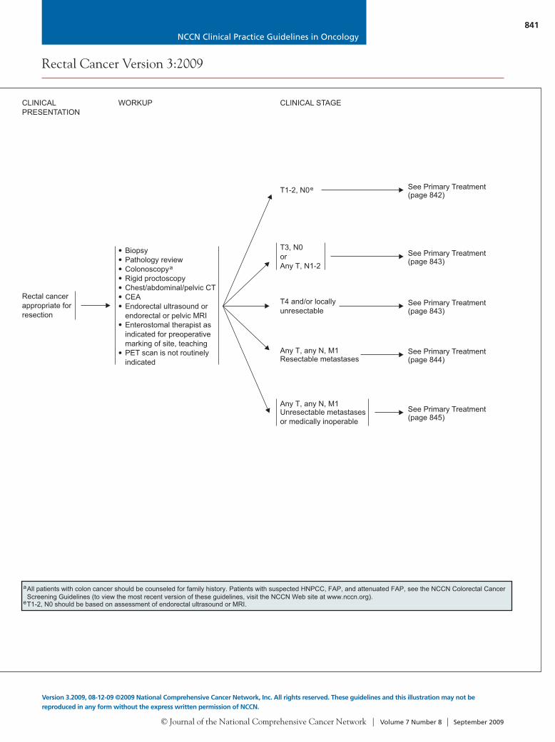
BiopsyPathology reviewColonoscopyRigid proctoscopyChest/abdominal/pelvic CTCEAEndorectal ultrasound orendorectal or pelvic MRIEnterostomal therapist asindicated for preoperativemarking of site, teachingPET scan is not routinelyindicated
a
T3, N0orAny T, N1-2
CLINICALPRESENTATION
Rectal cancerappropriate forresection
See Primary Treatment(page 845)
See Primary Treatment(page 843)
See Primary Treatment(page 842)
T4 and/or locallyunresectable
See Primary Treatment(page 843)
See Primary Treatment(page 844)
Any T, any N, M1Resectable metastases
Any T, any N, M1Unresectable metastasesor medically inoperable
aAll patients with colon cancer should be counseled for family history. Patients with suspected HNPCC, FAP, and attenuated FAP, see the NCCN Colorectal CancerScreening Guidelines (to view the most recent version of these guidelines, visit the NCCN Web site at www.nccn.org).T1-2, N0 should be based on assessment of endorectal ultrasound or MRI.e
NCCN Clinical Practice Guidelines in Oncology
© Journal of the National Comprehensive Cancer Network | Volume 7 Number 8 | September 2009
841
Rectal Cancer Version 3:2009
Version 3.2009, 08-12-09 ©2009 National Comprehensive Cancer Network, Inc. All rights reserved. These guidelines and this illustration may not be reproduced in any form without the express written permission of NCCN.
Single specimen, completelyremoved with favorablehistological features andclear margins (T1 only)
d Observe
Pathology reviewColonoscopyMarking of cancerous polypsite (at time of colonoscopyor within 2 wk)
b,c
CLINICALPRESENTATIONa
Pedunculated polyp(adenoma [tubular,tubulovillous, orvillous]) withinvasive cancer
WORKUP FINDINGS
Fragmented specimen ormargin cannot be assessedor unfavorable histologicalfeaturesd
See Primary andAdjuvant Treatment(page 842)
a
bc
d
All patients with colon cancer should be counseled for family history. Patients with suspected hereditary non-polyposis colon cancer (HNPCC), familialadenomatous polyposis (FAP), and attenuated FAP should see the NCCN Clinical Practice Guidelines in Oncology: Colorectal Cancer Screening
Confirm the presence of invasive cancer (pT1). pTis has no biological potential to metastasize.It has not been established if molecular markers are useful in treatment determination (predictive markers) and prognosis. College of American PathologistsConsensus Statement 1999. Prognostic factors in colorectal cancer. Arch Pathol Lab Med 2000;124:979-994.
See Principles of Pathologic Review (page 848): Endoscopically Removed Malignant Polyp.
(to viewthe most recent version of these guidelines, visit the NCCN Web site at www.nccn.org).
Single specimen, completelyremoved with favorablehistological features andclear margins (T1 only)
d
ObserveorSee PrimaryTreatment onpage page 842
Pathology reviewColonoscopyMarking of cancerous polypsite (at time of colonoscopyor within 2 wk)
b,cSessile polyp(Adenoma [tubular,tubulovillous, orvillous]) withinvasive cancer
Fragmented specimen ormargin cannot be assessedor unfavorable histologicalfeaturesd
See Primary andAdjuvant Treatment(page 842)
WORKUP CLINICAL STAGE
T1-2, N0e
BiopsyPathology reviewColonoscopyRigid proctoscopyChest/abdominal/pelvic CTCEAEndorectal ultrasound orendorectal or pelvic MRIEnterostomal therapist asindicated for preoperativemarking of site, teachingPET scan is not routinelyindicated
a
T3, N0orAny T, N1-2
CLINICALPRESENTATION
Rectal cancerappropriate forresection
See Primary Treatment(page 845)
See Primary Treatment(page 843)
See Primary Treatment(page 842)
T4 and/or locallyunresectable
See Primary Treatment(page 843)
See Primary Treatment(page 844)
Any T, any N, M1Resectable metastases
Any T, any N, M1Unresectable metastasesor medically inoperable
aAll patients with colon cancer should be counseled for family history. Patients with suspected HNPCC, FAP, and attenuated FAP, see the NCCN Colorectal CancerScreening Guidelines (to view the most recent version of these guidelines, visit the NCCN Web site at www.nccn.org).T1-2, N0 should be based on assessment of endorectal ultrasound or MRI.e

© Journal of the National Comprehensive Cancer Network | Volume 7 Number 8 | September 2009
842
Rectal Cancer Version 3:2009
Clinical trials: The NCCN believes that the best management for any cancer patient is in a clinical trial. Participation in clinical trials is especially encouraged. All recommendations are category 2A unless otherwise noted.
CLINICALSTAGE
PRIMARY TREATMENT
T1-2, N0e
Transabdominalresection
or
Transanalexcision, ifappropriate(category 2Bfor T2)
f
fT1-T2, NX;high riskfeaturesg
Trans-abdominalresectionf
T1, NX;marginsnegative
Observe
T2, NX;marginsnegative
Trans-abdominalresectionor5-FU/RT
f
eT1-2, N0 should be based on assessment of endorectal ultrasound or MRI.See Principles of Surgery (pages 851-853).High risk features include positive margins, lymphovascular invasion, and poorly differentiated tumors.See Principles of Adjuvant Therapy (pages 854 and 855).
See Principles of Radiation Therapy (page 856).The use of FOLFOX or capecitabine is an extrapolation from the available data in colon cancer. Trials are still pending in rectal cancer.Data regarding the use of capecitabine/RT is limited and no phase III randomized data are available. Trials are pending. Kim J-Sang, Kim J-Sung, Cho, M,et al. Preoperative chemoradiation using oral capecitabine in locally advanced rectal cancer. Int J Radiat Oncol Biol Phys 2002;54:403-408.
fghijk
ADJUVANT TREATMENTh,i
pT3, N0, M0orpT1-3, N1-2
pT1-2,N0, M0 Observe
5-FU ± leucovorin or FOLFOX (category 2B) orcapecitabine (category 2B),then continuous 5-FU/RT or bolus 5-FU + leucovorin/RT(category 2B) or capecitabine/RT (category 2B),then 5-FU ± leucovorin or FOLFOX (category 2B) orcapecitabine (category 2B)
jj
kj
j
pT3, N0,M0 orpT1-3,N1-2
pT1–2,N0, M0 Observe
5-FU ± leucovorin or FOLFOX(category 2B) or capecitabine(category 2B),then continuous 5-FU/RT or bolus5-FU + leucovorin/RT (category 2B)or capecitabine/RT (category 2B),then 5-FU ± leucovorin or FOLFOX(category 2B) or capecitabine(category 2B)
jj
kj
j
Consider systemic chemotherapy
Surveillance(Seepage 846)
Surveillance(See page 846)
pT3, N0, M0or pT1-3, N1-2
l,m
pT1–2, N0, M0 Observe
5-FU ± leucovorin (category 1)orFOLFOX (category 2B)orCapecitabine (category 2B)
j,o
j
fhijk
lm
n
See Principles of Surgery (pages 851-853).See Principles of Adjuvant Therapy (pages 854 and 855).
See Principles of Radiation Therapy (page 856).The use of FOLFOX or capecitabine is an extrapolation from the available data in colon cancer. Trials are still pending in rectal cancer.Data regarding the use of capecitabine/RT is limited and no phase III randomized data are available. Trials are pending. Kim J-Sang, Kim J-Sung, Cho, M,et al. Preoperative chemoradiation using oral capecitabine in locally advanced rectal cancer. Int J Radiat Oncol Biol Phys 2002;54:403-408.
The use of agents other than fluoropyrimidines are not recommended concurrently with RT.For patients with proximal T3, N0 disease with clear margins and favorable prognostic features, the incremental benefit of RT is likely to be small. Considerchemotherapy alone.
Postoperative therapy is indicated in all patients who undergo preoperative therapy, regardless of the surgical pathology results.An ongoing Intergroup trial compares 5-FU/leucovorin, FOLFOX, and FOLFIRI after surgery.o
T3, N0orAny T, N1-2
Preoperative continuous5-FU/RT (preferred; category 1 for node positive disease) orbolus 5-FU + leucovorin/RT orcapecitabine/RT (category 2B)k
Transabdominalresectionf
Reconsider:5-FU ± leucovorin or FOLFOX(category 2B) or capecitabine(category 2B),then continuous 5-FU/RT orbolus 5-FU + leucovorin/RT(category 2B) orcapecitabine/RT (category 2B)then 5-FU ± leucovorin orFOLFOX (category 2B) orcapecitabine (category 2B)
j,oj
k
j,oj
5-FU ± leucovorinorFOLFOX (category 2B)j,o
orCapecitabine (category 2B)j
Continuous IV 5-FU/RT orbolus 5-FU + leucovorin/RTor capecitabine/RT(category 2B)
kResection,if possible
T4 and/orlocallyunresectable
Any T
CLINICALSTAGE
PRIMARY TREATMENT ADJUVANT TREATMENTh,i,n
Transabdominalresectionf
Patients with medicalcontraindication to combinedmodality therapy

NCCN Clinical Practice Guidelines in Oncology
© Journal of the National Comprehensive Cancer Network | Volume 7 Number 8 | September 2009
843
Rectal Cancer Version 3:2009
Version 3.2009, 08-12-09 ©2009 National Comprehensive Cancer Network, Inc. All rights reserved. These guidelines and this illustration may not be reproduced in any form without the express written permission of NCCN.
CLINICALSTAGE
PRIMARY TREATMENT
T1-2, N0e
Transabdominalresection
or
Transanalexcision, ifappropriate(category 2Bfor T2)
f
fT1-T2, NX;high riskfeaturesg
Trans-abdominalresectionf
T1, NX;marginsnegative
Observe
T2, NX;marginsnegative
Trans-abdominalresectionor5-FU/RT
f
eT1-2, N0 should be based on assessment of endorectal ultrasound or MRI.See Principles of Surgery (pages 851-853).High risk features include positive margins, lymphovascular invasion, and poorly differentiated tumors.See Principles of Adjuvant Therapy (pages 854 and 855).
See Principles of Radiation Therapy (page 856).The use of FOLFOX or capecitabine is an extrapolation from the available data in colon cancer. Trials are still pending in rectal cancer.Data regarding the use of capecitabine/RT is limited and no phase III randomized data are available. Trials are pending. Kim J-Sang, Kim J-Sung, Cho, M,et al. Preoperative chemoradiation using oral capecitabine in locally advanced rectal cancer. Int J Radiat Oncol Biol Phys 2002;54:403-408.
fghijk
ADJUVANT TREATMENTh,i
pT3, N0, M0orpT1-3, N1-2
pT1-2,N0, M0 Observe
5-FU ± leucovorin or FOLFOX (category 2B) orcapecitabine (category 2B),then continuous 5-FU/RT or bolus 5-FU + leucovorin/RT(category 2B) or capecitabine/RT (category 2B),then 5-FU ± leucovorin or FOLFOX (category 2B) orcapecitabine (category 2B)
jj
kj
j
pT3, N0,M0 orpT1-3,N1-2
pT1–2,N0, M0 Observe
5-FU ± leucovorin or FOLFOX(category 2B) or capecitabine(category 2B),then continuous 5-FU/RT or bolus5-FU + leucovorin/RT (category 2B)or capecitabine/RT (category 2B),then 5-FU ± leucovorin or FOLFOX(category 2B) or capecitabine(category 2B)
jj
kj
j
Consider systemic chemotherapy
Surveillance(Seepage 846)
Surveillance(See page 846)
pT3, N0, M0or pT1-3, N1-2
l,m
pT1–2, N0, M0 Observe
5-FU ± leucovorin (category 1)orFOLFOX (category 2B)orCapecitabine (category 2B)
j,o
j
fhijk
lm
n
See Principles of Surgery (pages 851-853).See Principles of Adjuvant Therapy (pages 854 and 855).
See Principles of Radiation Therapy (page 856).The use of FOLFOX or capecitabine is an extrapolation from the available data in colon cancer. Trials are still pending in rectal cancer.Data regarding the use of capecitabine/RT is limited and no phase III randomized data are available. Trials are pending. Kim J-Sang, Kim J-Sung, Cho, M,et al. Preoperative chemoradiation using oral capecitabine in locally advanced rectal cancer. Int J Radiat Oncol Biol Phys 2002;54:403-408.
The use of agents other than fluoropyrimidines are not recommended concurrently with RT.For patients with proximal T3, N0 disease with clear margins and favorable prognostic features, the incremental benefit of RT is likely to be small. Considerchemotherapy alone.
Postoperative therapy is indicated in all patients who undergo preoperative therapy, regardless of the surgical pathology results.An ongoing Intergroup trial compares 5-FU/leucovorin, FOLFOX, and FOLFIRI after surgery.o
T3, N0orAny T, N1-2
Preoperative continuous5-FU/RT (preferred; category 1 for node positive disease) orbolus 5-FU + leucovorin/RT orcapecitabine/RT (category 2B)k
Transabdominalresectionf
Reconsider:5-FU ± leucovorin or FOLFOX(category 2B) or capecitabine(category 2B),then continuous 5-FU/RT orbolus 5-FU + leucovorin/RT(category 2B) orcapecitabine/RT (category 2B)then 5-FU ± leucovorin orFOLFOX (category 2B) orcapecitabine (category 2B)
j,oj
k
j,oj
5-FU ± leucovorinorFOLFOX (category 2B)j,o
orCapecitabine (category 2B)j
Continuous IV 5-FU/RT orbolus 5-FU + leucovorin/RTor capecitabine/RT(category 2B)
kResection,if possible
T4 and/orlocallyunresectable
Any T
CLINICALSTAGE
PRIMARY TREATMENT ADJUVANT TREATMENTh,i,n
Transabdominalresectionf
Patients with medicalcontraindication to combinedmodality therapy

© Journal of the National Comprehensive Cancer Network | Volume 7 Number 8 | September 2009
844
Rectal Cancer Version 3:2009
Clinical trials: The NCCN believes that the best management for any cancer patient is in a clinical trial. Participation in clinical trials is especially encouraged. All recommendations are category 2A unless otherwise noted.
Surveillance(Seepage 846)
CLINICALSTAGE
PRIMARY TREATMENT ADJUVANT THERAPY(resected metastatic disease;6 mo perioperative treatment)
h,i
Any T,Any N, M1Resectablesynchronousmetastasesp
pT1-2, N0, M1
pT3-4, any N,or Any T, N1-2
Staged orsynchronousresection ofmetastases andrectal lesion
f
Active chemotherapy regimen for advanceddisease (pages 793-797; category 2B)r
fhijk
opq
s
See Principles of Surgery (pages 851-853).See Principles of Adjuvant Therapy (pages 854 and 855).
See Principles of Radiation Therapy (page 856).The use of FOLFOX or capecitabine is an extrapolation from the available data in colon cancer. Trials are still pending in rectal cancer.Data regarding the use of capecitabine/RT is limited and no phase III randomized data are available. Trials are pending. Kim J-Sang, Kim J-Sung, Cho, Met al. Preoperative chemoradiation using oral capecitabine in locally advanced rectal cancer. Int J Radiat Oncol Biol Phys 2002;54:403-408.
An ongoing Intergroup trial compares 5-FU/leucovorin, FOLFOX, and FOLFIRI after surgery.Determination of tumor KRAS gene status. See Principles of Pathologic Review: KRAS Mutation TestingThe safety of administering bevacizumab pre- or postoperatively, in combination with 5-FU-based regimens, has not been adequately evaluated. Thereshould be at least a 6 wk interval between the last dose of bevacizumab and elective surgery. There is an increased risk of stroke and other arterialevents especially in patients ≥ 65 years of age. The use of bevacizumab may interfere with wound healing.
rFOLFOXIRI is not recommended in this setting.RT only recommended for patients at relative risk for pelvic recurrence.
(page 849).
5-FU ± leucovorin or FOLFOX (category 2B)or capecitabine (category 2B),then continuous 5-FU/RT or bolus 5-FU +leucovorin/RT (category 2B) orcapecitabine/RT (category 2B),then 5-FU ± leucovorin or FOLFOX(category 2B) or capecitabine (category 2B)
j,oj
sr
k,sj,o
j
5-FU/RT orCapecitabine/RT (category 2B)orResection of involved rectal segmentorLaser recanalizationorDiverting colostomyorStentingorChemotherapy alone
k
t
See Chemotherapy for Advancedor Metastatic Disease (pages 793-797)
Any T, any N, M1Unresectable synchronousmetastasesor medically inoperable
p
CLINICAL STAGE PRIMARY TREATMENT
k
pt
Data regarding the use of capecitabine/RT is limited and no phase III randomized data are available. Trials are pending. Kim J-Sang, Kim J-Sung, Cho, Met al. Preoperative chemoradiation using oral capecitabine in locally advanced rectal cancer. Int J Radiat Oncol Biol Phys 2002;54:403-408.
Determination of tumor KRAS gene status. See Principles of Pathologic Review: KRAS Mutation Testing .See Chemotherapy for Advanced or Metastatic Disease (pages 793-797).
(page 849)
Symptomatic
Asymptomatic Reassess response todetermine resectability
Staged or synchronousresection of metastases+ rectal lesion
f
Continuous IV 5-FU/pelvic RT or bolus 5-FU+ leucovorin/pelvic RT orcapecitabine/RT(category 2B)
k
or
Combination chemotherapy(2-3 mo)FOLFIRI or FOLFOXor CapeOX ± bevacizumabor FOLFIRI or FOLFOX orCapeOX ± cetuximab (KRASwild-type gene only)
q
p
or
Staged orsynchronousresection ofmetastases andrectal lesion
f
Consider continuous IV 5-FU/pelvic RTor bolus 5-FU + leucovorin/pelvic RTor capecitabine/RT (category 2B)k
Staged or synchronousresection of metastasesand rectal lesion
f
Continuous IV 5-FU/pelvic RTor bolus 5-FU +leucovorin/pelvic RT orcapecitabine/RT (category 2B)k
or
Active chemotherapy regimen for advanceddisease (pages 793-797; category 2B)r
SeeChemotherapyfor Advanced orMetastaticDisease (pages793-797)

NCCN Clinical Practice Guidelines in Oncology
© Journal of the National Comprehensive Cancer Network | Volume 7 Number 8 | September 2009
845
Rectal Cancer Version 3:2009
Version 3.2009, 08-12-09 ©2009 National Comprehensive Cancer Network, Inc. All rights reserved. These guidelines and this illustration may not be reproduced in any form without the express written permission of NCCN.
Surveillance(Seepage 846)
CLINICALSTAGE
PRIMARY TREATMENT ADJUVANT THERAPY(resected metastatic disease;6 mo perioperative treatment)
h,i
Any T,Any N, M1Resectablesynchronousmetastasesp
pT1-2, N0, M1
pT3-4, any N,or Any T, N1-2
Staged orsynchronousresection ofmetastases andrectal lesion
f
Active chemotherapy regimen for advanceddisease (pages 793-797; category 2B)r
fhijk
opq
s
See Principles of Surgery (pages 851-853).See Principles of Adjuvant Therapy (pages 854 and 855).
See Principles of Radiation Therapy (page 856).The use of FOLFOX or capecitabine is an extrapolation from the available data in colon cancer. Trials are still pending in rectal cancer.Data regarding the use of capecitabine/RT is limited and no phase III randomized data are available. Trials are pending. Kim J-Sang, Kim J-Sung, Cho, Met al. Preoperative chemoradiation using oral capecitabine in locally advanced rectal cancer. Int J Radiat Oncol Biol Phys 2002;54:403-408.
An ongoing Intergroup trial compares 5-FU/leucovorin, FOLFOX, and FOLFIRI after surgery.Determination of tumor KRAS gene status. See Principles of Pathologic Review: KRAS Mutation TestingThe safety of administering bevacizumab pre- or postoperatively, in combination with 5-FU-based regimens, has not been adequately evaluated. Thereshould be at least a 6 wk interval between the last dose of bevacizumab and elective surgery. There is an increased risk of stroke and other arterialevents especially in patients ≥ 65 years of age. The use of bevacizumab may interfere with wound healing.
rFOLFOXIRI is not recommended in this setting.RT only recommended for patients at relative risk for pelvic recurrence.
(page 849).
5-FU ± leucovorin or FOLFOX (category 2B)or capecitabine (category 2B),then continuous 5-FU/RT or bolus 5-FU +leucovorin/RT (category 2B) orcapecitabine/RT (category 2B),then 5-FU ± leucovorin or FOLFOX(category 2B) or capecitabine (category 2B)
j,oj
sr
k,sj,o
j
5-FU/RT orCapecitabine/RT (category 2B)orResection of involved rectal segmentorLaser recanalizationorDiverting colostomyorStentingorChemotherapy alone
k
t
See Chemotherapy for Advancedor Metastatic Disease (pages 793-797)
Any T, any N, M1Unresectable synchronousmetastasesor medically inoperable
p
CLINICAL STAGE PRIMARY TREATMENT
k
pt
Data regarding the use of capecitabine/RT is limited and no phase III randomized data are available. Trials are pending. Kim J-Sang, Kim J-Sung, Cho, Met al. Preoperative chemoradiation using oral capecitabine in locally advanced rectal cancer. Int J Radiat Oncol Biol Phys 2002;54:403-408.
Determination of tumor KRAS gene status. See Principles of Pathologic Review: KRAS Mutation Testing .See Chemotherapy for Advanced or Metastatic Disease (pages 793-797).
(page 849)
Symptomatic
Asymptomatic Reassess response todetermine resectability
Staged or synchronousresection of metastases+ rectal lesion
f
Continuous IV 5-FU/pelvic RT or bolus 5-FU+ leucovorin/pelvic RT orcapecitabine/RT(category 2B)
k
or
Combination chemotherapy(2-3 mo)FOLFIRI or FOLFOXor CapeOX ± bevacizumabor FOLFIRI or FOLFOX orCapeOX ± cetuximab (KRASwild-type gene only)
q
p
or
Staged orsynchronousresection ofmetastases andrectal lesion
f
Consider continuous IV 5-FU/pelvic RTor bolus 5-FU + leucovorin/pelvic RTor capecitabine/RT (category 2B)k
Staged or synchronousresection of metastasesand rectal lesion
f
Continuous IV 5-FU/pelvic RTor bolus 5-FU +leucovorin/pelvic RT orcapecitabine/RT (category 2B)k
or
Active chemotherapy regimen for advanceddisease (pages 793-797; category 2B)r
SeeChemotherapyfor Advanced orMetastaticDisease (pages793-797)
© Journal of the National Comprehensive Cancer Network | Volume 7 Number 8 | September 2009
846
Rectal Cancer Version 3:2009
Clinical trials: The NCCN believes that the best management for any cancer patient is in a clinical trial. Participation in clinical trials is especially encouraged. All recommendations are category 2A unless otherwise noted.
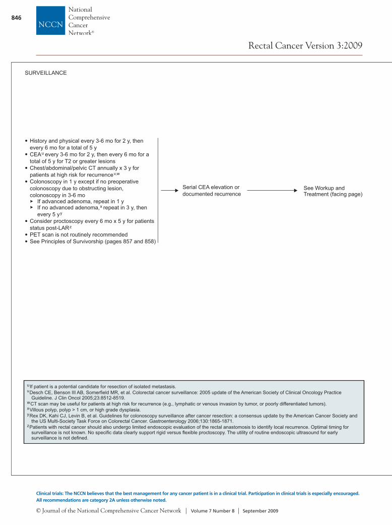
History and physical every 3-6 mo for 2 y, thenevery 6 mo for a total of 5 yCEA every 3-6 mo for 2 y, then every 6 mo for atotal of 5 y for T2 or greater lesionsChest/abdominal/pelvic CT annually x 3 y forpatients at high risk for recurrenceColonoscopy in 1 y except if no preoperativecolonoscopy due to obstructing lesion,colonoscopy in 3-6 mo
If advanced adenoma, repeat in 1 yIf no advanced adenoma, repeat in 3 y, thenevery 5 y
Consider proctoscopy every 6 mo x 5 y for patientsstatus post-LARPET scan is not routinely recommendedSee Principles of Survivorship (pages 857 and 858)
u
v,w
xy
z
SURVEILLANCE
Serial CEA elevation ordocumented recurrence
See Workup andTreatment (facing page)
uv
wxy
z
If patient is a potential candidate for resection of isolated metastasis.Desch CE, Benson III AB, Somerfield MR, et al. Colorectal cancer surveillance: 2005 update of the American Society of Clinical Oncology PracticeGuideline. J Clin Oncol 2005;23:8512-8519.CT scan may be useful for patients at high risk for recurrence (e.g., lymphatic or venous invasion by tumor, or poorly differentiated tumors).Villous polyp, polyp > 1 cm, or high grade dysplasia.Rex DK, Kahi CJ, Levin B, et al. Guidelines for colonoscopy surveillance after cancer resection: a consensus update by the American Cancer Society andthe US Multi-Society Task Force on Colorectal Cancer. Gastroenterology 2006;130:1865-1871.
Patients with rectal cancer should also undergo limited endoscopic evaluation of the rectal anastomosis to identify local recurrence. Optimal timing forsurveillance is not known. No specific data clearly support rigid versus flexible proctoscopy. The utility of routine endoscopic ultrasound for earlysurveillance is not defined.
WORKUPRECURRENCE
iSee Principles of Radiation Therapy (page 856).pDetermination of tumor KRAS gene status. See Principles of Pathologic Review: KRAS Mutation Testing (page 849).
SerialCEAelevation
Negativefindings
Positivefindings
Physical examColonoscopyChest/abdominal/pelvic CTConsider PET scan
Reevaluate chest/abdominal/pelvic CTin 3 mo
PET scan
Isolated pelvic/anastomoticrecurrence
Preoperative continuous5-FU IV + RT, if not givenpreviously
Resection, if feasible± IORTi
Negativefindings
Positivefindings
TREATMENT
Documentedmetachronousmetastases by CT,MRI, and/or biopsy
p
All othermetastases
See treatment forDocumented MetachronousMetastases (page 786)
See treatmentfor DocumentedMetachronousMetastases(page 786)
See treatment forDocumented MetachronousMetastases (page 786)

NCCN Clinical Practice Guidelines in Oncology
© Journal of the National Comprehensive Cancer Network | Volume 7 Number 8 | September 2009
847
Rectal Cancer Version 3:2009
Version 3.2009, 08-12-09 ©2009 National Comprehensive Cancer Network, Inc. All rights reserved. These guidelines and this illustration may not be reproduced in any form without the express written permission of NCCN.
History and physical every 3-6 mo for 2 y, thenevery 6 mo for a total of 5 yCEA every 3-6 mo for 2 y, then every 6 mo for atotal of 5 y for T2 or greater lesionsChest/abdominal/pelvic CT annually x 3 y forpatients at high risk for recurrenceColonoscopy in 1 y except if no preoperativecolonoscopy due to obstructing lesion,colonoscopy in 3-6 mo
If advanced adenoma, repeat in 1 yIf no advanced adenoma, repeat in 3 y, thenevery 5 y
Consider proctoscopy every 6 mo x 5 y for patientsstatus post-LARPET scan is not routinely recommendedSee Principles of Survivorship (pages 857 and 858)
u
v,w
xy
z
SURVEILLANCE
Serial CEA elevation ordocumented recurrence
See Workup andTreatment (facing page)
uv
wxy
z
If patient is a potential candidate for resection of isolated metastasis.Desch CE, Benson III AB, Somerfield MR, et al. Colorectal cancer surveillance: 2005 update of the American Society of Clinical Oncology PracticeGuideline. J Clin Oncol 2005;23:8512-8519.CT scan may be useful for patients at high risk for recurrence (e.g., lymphatic or venous invasion by tumor, or poorly differentiated tumors).Villous polyp, polyp > 1 cm, or high grade dysplasia.Rex DK, Kahi CJ, Levin B, et al. Guidelines for colonoscopy surveillance after cancer resection: a consensus update by the American Cancer Society andthe US Multi-Society Task Force on Colorectal Cancer. Gastroenterology 2006;130:1865-1871.
Patients with rectal cancer should also undergo limited endoscopic evaluation of the rectal anastomosis to identify local recurrence. Optimal timing forsurveillance is not known. No specific data clearly support rigid versus flexible proctoscopy. The utility of routine endoscopic ultrasound for earlysurveillance is not defined.
WORKUPRECURRENCE
iSee Principles of Radiation Therapy (page 856).pDetermination of tumor KRAS gene status. See Principles of Pathologic Review: KRAS Mutation Testing (page 849).
SerialCEAelevation
Negativefindings
Positivefindings
Physical examColonoscopyChest/abdominal/pelvic CTConsider PET scan
Reevaluate chest/abdominal/pelvic CTin 3 mo
PET scan
Isolated pelvic/anastomoticrecurrence
Preoperative continuous5-FU IV + RT, if not givenpreviously
Resection, if feasible± IORTi
Negativefindings
Positivefindings
TREATMENT
Documentedmetachronousmetastases by CT,MRI, and/or biopsy
p
All othermetastases
See treatment forDocumented MetachronousMetastases (page 786)
See treatmentfor DocumentedMetachronousMetastases(page 786)
See treatment forDocumented MetachronousMetastases (page 786)

© Journal of the National Comprehensive Cancer Network | Volume 7 Number 8 | September 2009
848
Rectal Cancer Version 3:2009
Clinical trials: The NCCN believes that the best management for any cancer patient is in a clinical trial. Participation in clinical trials is especially encouraged. All recommendations are category 2A unless otherwise noted.
PRINCIPLES OF PATHOLOGIC REVIEW
Endoscopically Removed Malignant PolypsA malignant polyp is defined as one with cancer invading through the muscularis mucosae and into the submucosa (pT1).pTIS is not considered a “malignant polyp.”Favorable histologic features: grade 1 or 2, no angiolymphatic invasion, and negative margin of resection. No consensusexists regarding the definition of what constitutes a positive margin of resection. A positive margin has been defined as1) tumor < 1 mm from the transected margin, 2) tumor < 2 mm from the transected margin, or 3) tumor cells present withinthe diathermy of the transected margin.Unfavorable histologic features: grade 3 or 4, angiolymphatic invasion, or a “positive margin.” See above for definition of apositive margin.Controversy exists as to whether malignant colorectal polyps with a sessile configuration can be successfully treated byendoscopic removal. The literature seems to indicate that endoscopically removed sessile malignant polyps have asignificantly greater incidence of adverse outcome (residual disease, recurrent disease, mortality, hematogenousmetastasis, but not lymph node metastasis) than polypoid malignant polyps. However, when closely examining the data,configuration by itself is not a significant variable for adverse outcome, and endoscopically removed malignant sessilepolyps with grade I or II histology, negative margin, and no lymphovascular invasion can be successfully treated withendoscopic polypectomy.
Transanal ExcisionFavorable histopathologic features: < 3 cm size, T1 or T2 (use caution in T2 because of high recurrence rate; see pages 851-853), grade I or II, no lymphatic or venous invasion, or negative margins.Unfavorable histopathologic features: > 3 cm in size, T1 or T2, with grade III, lymphovascular invasion, or positive margin.
Rectal Cancer Appropriate for ResectionHistologic confirmation of primary malignant rectal neoplasm.
Pathologic StageThe following parameters should be reported:
Grade of the cancer.Depth of penetration, (T) the T stage is based on viable tumor. Acellular mucin pools are not considered residual tumor incases treated with neoadjuvant therapy.Number of lymph nodes evaluated and number positive (N). Acellular mucin pools are not considered residual tumor inthose cases treated with neoadjuvant therapy.Status of proximal, distal, and circumferential (radial) margins.A positive circumferential resection margin (CRM) has been defined as < 1 mm or < 2 mm depending on thepublication.
1-4
3-7
8,98-10
11-12
13-14
See Staging Table (available online, in these guidelines, at www.nccn.org [ST-1])
See footnotes on page 850
Sentinel lymph node and detection of micrometastasis by immunohistochemistryExamination of the sentinal lymph node allows an intense histologic and/or immunohistochemical investigation to detect thepresence of metastatic carcinoma. Studies in the literature have been reported using multiple H & E sections and/orimmunohistochemistry (IHC) to detect cytokeratin positive cells. Although studies to date seem promising, there is nouniformity in the definition of what constitutes "true metastatic carcinoma." Confusion arises when isolated tumors cells(ITCs) have been considered micrometastatic disease in contraindication to true micrometastasis (tumor aggregates > 0.2mm to < 2 mm in size). The significance of detection of single cells by IHC alone is controversial. Some studies haveconsidered these to be micrometastasis; however, “consensus” recommends these be considered ITC and notmicrometastatic disease. While the 6th edition of the AJCC Cancer Staging manual considers "tumor clusters" < 0.2mm as ITCs (pN0) and not metastatic carcinoma, some have challenged this. Some investigators believe that size shouldnot effect the diagnosis of metastatic cancer. They believe that tumor foci that show evidence of growth (e.g., glandulardifferentiation, distension of sinus, stromal reaction) should be diagnosed as a lymph node metastasis, regardless of size.Hermanek et al. proposed ITCs be defined as single tumor cells or small clusters (never more than a few cells clumpedtogether) without evidence of extrasinusoidal stromal proliferation or reaction and no contact with or invasion of the vessel(lymphatic) wall.Some studies have shown that the detection of IHC cytokeratin positive cells in stage II (N0) colon cancer (defined by H & E)has a worse prognosis, whereas others have failed to show this survival difference. In these studies, ITCs were consideredmicrometastasis.Currently, the use of sentinel lymph nodes and detection of cancer cells using IHC alone should be considered investigational,and results used with caution in clinical management decisions.
26-28 29
3031
32-36
26-28,32-36
See footnotes on page 850
PRINCIPLES OF PATHOLOGIC REVIEW (Cont.)
KRAS Mutation TestingMutations in codons 12 and 13 in exon 2 of the coding region of the KRAS gene predict lack of response to therapy with antibodiestargeted to the epidermal growth factor receptor.
Testing for mutations in codons 12 and 13 should be performed only in laboratories that are certified under the Clinical LaboratoryImprovement Amendments of 1988 (CLIA – 88) as qualified to perform high complex clinical laboratory (molecular pathology) testing.No specific methodology is recommended (sequencing, hybridization, etc.).
The testing can be performed on formalin-fixed paraffin embedded tissue. The testing can be performed on the primary colorectalcancers and/or the metastasis, as literature has shown that the KRAS mutations are similar in both specimen types.
Evaluation of Mesorectum (TME)The pathologist should evaluate the quality (completeness) of the mesorectum (only for low rectal cancer - distal 2/3).
37,38
39
40-42
Lymph Node EvaluationThe AJCC and College of American Pathologists recommend examination of a minimum of 12 lymph nodes to accuratelyidentify stage II colorectal cancers. The literature lacks consensus as to what is the minimal number of lymph nodes toaccurately identify stage II cancer. The minimal number of nodes has been reported as > 7, > 9, > 13, > 20, and > 30. Mostof these studies have combined rectal and colon cancers and reflect those cases with surgery as the initial treatment. Twostudies confined only to rectal cancer have reported 14 and > 10 lymph nodes as the minimal number to accurately identifystage II rectal cancer. The number of lymph nodes retrieved can vary with age, gender, tumor grade, and tumor site.For stage II (pN0) colon cancer, if < 12 lymph nodes are initially identified, it is recommended that the pathologist go backto the specimen and resubmit more tissue of potential lymph nodes. If 12 lymph nodes are still not identified, a comment in the report should indicate that an extensive search for lymph nodes was undertaken. The mean number of lymph nodesretrieved from rectal cancers treated with neoadjuvant therapy is significantly less than those treated with surgery alone(13 vs. 19; < .05; 7 vs. 10; < .001). If 12 lymph nodes is considered the number needed to accurately identify stage IItumors, then only 20% of cases treated with neoadjuvant therapy had adequate lymph node sampling. To date, the number oflymph nodes needed to accurately stage neoadjuvant-treated cases is unknown. However, the clinical significance of this in theneoadjuvant setting is unknown because postoperative therapy is indicated in all patients who undergo preoperative therapy,regardless of the surgical pathology results.
11,12,1516-23
19,22 16
24,2525
P P

NCCN Clinical Practice Guidelines in Oncology
© Journal of the National Comprehensive Cancer Network | Volume 7 Number 8 | September 2009
849
Rectal Cancer Version 3:2009
Version 3.2009, 08-12-09 ©2009 National Comprehensive Cancer Network, Inc. All rights reserved. These guidelines and this illustration may not be reproduced in any form without the express written permission of NCCN.
PRINCIPLES OF PATHOLOGIC REVIEW
Endoscopically Removed Malignant PolypsA malignant polyp is defined as one with cancer invading through the muscularis mucosae and into the submucosa (pT1).pTIS is not considered a “malignant polyp.”Favorable histologic features: grade 1 or 2, no angiolymphatic invasion, and negative margin of resection. No consensusexists regarding the definition of what constitutes a positive margin of resection. A positive margin has been defined as1) tumor < 1 mm from the transected margin, 2) tumor < 2 mm from the transected margin, or 3) tumor cells present withinthe diathermy of the transected margin.Unfavorable histologic features: grade 3 or 4, angiolymphatic invasion, or a “positive margin.” See above for definition of apositive margin.Controversy exists as to whether malignant colorectal polyps with a sessile configuration can be successfully treated byendoscopic removal. The literature seems to indicate that endoscopically removed sessile malignant polyps have asignificantly greater incidence of adverse outcome (residual disease, recurrent disease, mortality, hematogenousmetastasis, but not lymph node metastasis) than polypoid malignant polyps. However, when closely examining the data,configuration by itself is not a significant variable for adverse outcome, and endoscopically removed malignant sessilepolyps with grade I or II histology, negative margin, and no lymphovascular invasion can be successfully treated withendoscopic polypectomy.
Transanal ExcisionFavorable histopathologic features: < 3 cm size, T1 or T2 (use caution in T2 because of high recurrence rate; see pages 851-853), grade I or II, no lymphatic or venous invasion, or negative margins.Unfavorable histopathologic features: > 3 cm in size, T1 or T2, with grade III, lymphovascular invasion, or positive margin.
Rectal Cancer Appropriate for ResectionHistologic confirmation of primary malignant rectal neoplasm.
Pathologic StageThe following parameters should be reported:
Grade of the cancer.Depth of penetration, (T) the T stage is based on viable tumor. Acellular mucin pools are not considered residual tumor incases treated with neoadjuvant therapy.Number of lymph nodes evaluated and number positive (N). Acellular mucin pools are not considered residual tumor inthose cases treated with neoadjuvant therapy.Status of proximal, distal, and circumferential (radial) margins.A positive circumferential resection margin (CRM) has been defined as < 1 mm or < 2 mm depending on thepublication.
1-4
3-7
8,98-10
11-12
13-14
See Staging Table (available online, in these guidelines, at www.nccn.org [ST-1])
See footnotes on page 850
Sentinel lymph node and detection of micrometastasis by immunohistochemistryExamination of the sentinal lymph node allows an intense histologic and/or immunohistochemical investigation to detect thepresence of metastatic carcinoma. Studies in the literature have been reported using multiple H & E sections and/orimmunohistochemistry (IHC) to detect cytokeratin positive cells. Although studies to date seem promising, there is nouniformity in the definition of what constitutes "true metastatic carcinoma." Confusion arises when isolated tumors cells(ITCs) have been considered micrometastatic disease in contraindication to true micrometastasis (tumor aggregates > 0.2mm to < 2 mm in size). The significance of detection of single cells by IHC alone is controversial. Some studies haveconsidered these to be micrometastasis; however, “consensus” recommends these be considered ITC and notmicrometastatic disease. While the 6th edition of the AJCC Cancer Staging manual considers "tumor clusters" < 0.2mm as ITCs (pN0) and not metastatic carcinoma, some have challenged this. Some investigators believe that size shouldnot effect the diagnosis of metastatic cancer. They believe that tumor foci that show evidence of growth (e.g., glandulardifferentiation, distension of sinus, stromal reaction) should be diagnosed as a lymph node metastasis, regardless of size.Hermanek et al. proposed ITCs be defined as single tumor cells or small clusters (never more than a few cells clumpedtogether) without evidence of extrasinusoidal stromal proliferation or reaction and no contact with or invasion of the vessel(lymphatic) wall.Some studies have shown that the detection of IHC cytokeratin positive cells in stage II (N0) colon cancer (defined by H & E)has a worse prognosis, whereas others have failed to show this survival difference. In these studies, ITCs were consideredmicrometastasis.Currently, the use of sentinel lymph nodes and detection of cancer cells using IHC alone should be considered investigational,and results used with caution in clinical management decisions.
26-28 29
3031
32-36
26-28,32-36
See footnotes on page 850
PRINCIPLES OF PATHOLOGIC REVIEW (Cont.)
KRAS Mutation TestingMutations in codons 12 and 13 in exon 2 of the coding region of the KRAS gene predict lack of response to therapy with antibodiestargeted to the epidermal growth factor receptor.
Testing for mutations in codons 12 and 13 should be performed only in laboratories that are certified under the Clinical LaboratoryImprovement Amendments of 1988 (CLIA – 88) as qualified to perform high complex clinical laboratory (molecular pathology) testing.No specific methodology is recommended (sequencing, hybridization, etc.).
The testing can be performed on formalin-fixed paraffin embedded tissue. The testing can be performed on the primary colorectalcancers and/or the metastasis, as literature has shown that the KRAS mutations are similar in both specimen types.
Evaluation of Mesorectum (TME)The pathologist should evaluate the quality (completeness) of the mesorectum (only for low rectal cancer - distal 2/3).
37,38
39
40-42
Lymph Node EvaluationThe AJCC and College of American Pathologists recommend examination of a minimum of 12 lymph nodes to accuratelyidentify stage II colorectal cancers. The literature lacks consensus as to what is the minimal number of lymph nodes toaccurately identify stage II cancer. The minimal number of nodes has been reported as > 7, > 9, > 13, > 20, and > 30. Mostof these studies have combined rectal and colon cancers and reflect those cases with surgery as the initial treatment. Twostudies confined only to rectal cancer have reported 14 and > 10 lymph nodes as the minimal number to accurately identifystage II rectal cancer. The number of lymph nodes retrieved can vary with age, gender, tumor grade, and tumor site.For stage II (pN0) colon cancer, if < 12 lymph nodes are initially identified, it is recommended that the pathologist go backto the specimen and resubmit more tissue of potential lymph nodes. If 12 lymph nodes are still not identified, a comment in the report should indicate that an extensive search for lymph nodes was undertaken. The mean number of lymph nodesretrieved from rectal cancers treated with neoadjuvant therapy is significantly less than those treated with surgery alone(13 vs. 19; < .05; 7 vs. 10; < .001). If 12 lymph nodes is considered the number needed to accurately identify stage IItumors, then only 20% of cases treated with neoadjuvant therapy had adequate lymph node sampling. To date, the number oflymph nodes needed to accurately stage neoadjuvant-treated cases is unknown. However, the clinical significance of this in theneoadjuvant setting is unknown because postoperative therapy is indicated in all patients who undergo preoperative therapy,regardless of the surgical pathology results.
11,12,1516-23
19,22 16
24,2525
P P
© Journal of the National Comprehensive Cancer Network | Volume 7 Number 8 | September 2009
850
Rectal Cancer Version 3:2009
Clinical trials: The NCCN believes that the best management for any cancer patient is in a clinical trial. Participation in clinical trials is especially encouraged. All recommendations are category 2A unless otherwise noted.
PRINCIPLES OF PATHOLOGIC REVIEWReferences
1
2
3
4
5
6
7
8
9
10
11
12
13
14
15
16
17
18
19
20
21
22
23
24
25
26
27
28
29
30
31
32
33
34
35
36
37
38
39
40
41
42
Volk EE, Goldblum JR, Petras RE, et al. Management and outcome ofpatients with invasive carcinoma arising in colorectal polyps.Gastroenterology 1995;109:1801-1807.
Cooper HS, Deppisch LM, Gourley WK, et al. Endoscopically removedmalignant colorectal polyps: clinical pathological correlations.Gastroenterology 1995;108:1657-1665.
Ueno H, Mochizuki H, Hashiguchi Y, et al. Risk factors for an adverseoutcome in early invasive colorectal carcinoma. Gastroenterology2004;127:385-394.
Seitz U, Bohnacker S, Seewald S, et al. Is endoscopic polypectomy anadequate therapy for malignant colorectal polyps? Presentation of 114patients and review of the literature. Dis Colon Rectum 2004;47:1789-1797.
Morson BC, Whiteway JE, Jones EA, et al. Histopathology and prognosisof malignant colorectal polyps treated by endoscopic polypectomy. Gut1984;25:437-444.
Haggitt RC, Glotzbach RE, Soffer EE, Wruble LD. Prognostic factors incolorectal carcinomas arising in adenomas: implications for lesionsremoved by endoscopic polypectomy. Gastroenterology 1985;89:328-336.
Netzer P, Binck J, Hammer B, et al. Significance of histological criteria forthe management of patients with malignant colorectal polyps. Scand JGastroenterol 1997;323:915-916.
Hager T, Gall FP, Hermanek P. Local excision of cancer of the rectum.Dis Colon Rect 1983;26:149-151.
Willett, CG, Tepper JE, Donnelly S, et al. Patterns of failure followinglocal excision and local excision and postoperative radiation therapy forinvasive rectal adenocarcinoma. J Clin Oncol 1989;7:1003-1008.Nascimbeni R, Burgart LJ, Nivatvongs S, Larson DR. Risk of lymphnode metastasis in T1 carcinoma of the colon and rectum. Dis ColonRectum 2002;45:2001-2006.Compton CC, Greene FL. The staging of colorectal cancer: 2004 andbeyond. CA Cancer J Clin 2004;54:295-308.Compton CC, Fielding LP, Burkhardt LJ, et al. Prognostic factors incolorectal cancer. College of American pathologists consensusstatement. Arch Pathol Lab Med 2000;124:979-994.Nagtegaal ID, Merijnenca M, Kranenbarg EK, et al. Circumferentialmargin involvement is still an important predictive local occurrence inrectal carcinoma. Not one millimeter but two millimeters is the limit. Am JSurg Pathol 2002;26:350-357.Wibe A, Rendedal PR, Svensson E, et al. Prognostic significance of thecircumferential resection margin following total mesorectal excision forrectal cancer. Br J Surg 2002;89;327-334.Sobin HL, Green EFL. TNM classification. Clarification of number ofregional lymph node for PN0. Cancer 2001;92:452.Sarli L, Bader G, Lusco D, et al. Number of lymph nodes examined andprognosis of TNM stage II colorectal cancer. Eur J Cancer 2005;41:272-279.Chaplin S, Scerottini GP, Bosman FT, et al. For patients with Duke's B(TNM stage II) colorectal carcinoma, examination of six or fewer lymphnodes is related to poor prognosis. Cancer 1998;83:666-672.Maurel J, Launoy G, Grosclaude P, et al. Lymph node harvest reportingin patients with carcinoma of the large bowel. A French population-based study. Cancer 1998;82:1482-1486.Pocard M, Panis Y, Malassagane B, et al. Assessing the effectivenessof mesorectal excision in rectal cancer. Dis Colon Rectum 1998;41:839-845.Joseph NE, Sigurdson ER, Hanlon AL, et al. Accuracy of determiningnodal negativity in colorectal cancer on the basis of number of nodesretrieved on resection. Ann Surg Oncol 2003;10:213-218.Goldstein NS. Lymph node recurrences from 2427 PT3 colorectalresection specimens spanning 45 years. Recommendations for aminimum number of recovered lymph nodes based on predictiveprobabilities. Am J Surg Pathol 2002;26:179-189.
Tepper JE, O'Connell MJ, Niedzwiecki D, et al. Impact of number of nodesretrieved on outcome in patients with rectal cancer. J Clin Oncol2001;19:157-162.Scott KWM, Grace RH. Detection of lymph node metastasis and colorectalcarcinoma before and after fat clearance. Br J Surg 1989;76:1165-1167.Wichmann MW, Mollar C, Meyer G, et al. Effect of pre-operativeradiochemotherapy on lymph node retrieval after resection of rectal cancer.Arch Surg 2002;137:206-210.Baxter NN, Morris AM, Rothenberger DA, Tepper JE. Impact of pre-operative radiation for rectal cancer on subsequent lymph node evaluation:population based analysis. Int J Radiat Oncol Biol Phys 2005;61:426-431.Turner RR, Nora DT, Trochas D, Bilchik AJ. Colorectal carcinoma in nodalstaging. Frequency and nature of cytokeratin positive cells in sentinal andnonsentinal lymph nodes. Arch Pathol Lab Med 2003;127:673-679.Wood TF, Nora DT, Morton DL, et al. One hundred consecutive cases ofsentinal node mapping in early colorectal carcinoma. Detection of missedmicrometastasis. J Gastroinest Surg 2002;6:322-330.Wiese DA, Sha S, Badin J, et al. Pathological evaluation of sentinel lymphnodes in colorectal carcinoma. Arch Pathol Lab Med 2000;124:1759-1763.AJCC Cancer Staging Manual, 6th ed. Greene FL, Page D, Balch C, et al.,eds. New York: Springer; 2002.Jass JB, O'Brien MJ, Riddell RH, Snover DC. Recommendations for the reporting of surgically resected specimens of colorectal carcinoma. HumPathol 2007;38:537-545. Hermanek P, Hutter RVP, Sobin LH, Wittekind CH. Classification of isolatedtumor cells and micrometastasis. Cancer 1999;86:2668-2673.Noura S, Yamamoto H, Ohnishi T, et al. Comparative detection of lymphnode micrometastasis of stage II colorectal cancer by reverse transcriptasepolymerase chain reaction in immunohistochemistry. J Clin Oncol2002;20:4232-4241.Yasuda K, Adachi Y, Shiraishi N, et al. Pattern of lymph nodemicrometastasis and prognosis of patients with colorectal cancer. Ann SurgOncol 2001;8:300-304.Noura S, Yamamoto H, Miyake Y, et al. Immunohistochemical assessmentof localization of frequency of micrometastasis in lymph nodes of colorectalcancer. Clin Cancer Res 2002;8:759-767.Oberg A, Stenling R, Tavelin B, Lindmark G. Are lymph nodemicrometastasis of any clinical significance in Duke stages A and Bcolorectal cancer? Dis Colon Rectum 1998;41:1244-1249.Greenson JK, Isenhart TCE, Rice R, et al. Identification of occultmicrometastasis in pericolonic lymph nodes of Duke's B colorectal cancer.Patient's using monoclonal antibodies against cytokeratin and CC49.Correlation with long term survival. Cancer 1994;73:563-569.Lievre A, Bachatte JB, Blige V, et al. KRAS mutations as an independentprognostic factor in patients with advanced colorectal cancer treated withCetuximab. J Clin Oncol 2008;26:374-379.Amado IG, Wolf M, Peters M, et al. Wild-type KRAS is required forpanitunumab efficacy in patients with metastatic colorectal cancer. J ClinOncol 2008;26:1626-1634.Etienne-Grimaldi MC, Formento JL, Francoual M, et al. K-Ras mutationsand treatment outcome in colorectal cancer in patients receiving exclusivefluoropyrimidine. Clin Cancer Res 2008;14:4830-4835.Parfitt JR, Driman KR. Total mesorectal excision specimen for rectalcancer: a review of its pathological assessment. J Clin Pathol 2007;60:849-855.Jass JR, O'Brien MJ, Riddell RH, Snover DC. Recommendations for thereporting of surgically resected specimens in colorectal carcinoma. HumPathol 38:537-545.Nagtegaal ID, Vandevelde CJA, Derworp EV, et al. Macroscopic evaluationof the rectal cancer resection margin: clinical significance of the pathologistin quality control. J Clin Oncol 20:1729-1734.
2007;
2002;
PRINCIPLES OF SURGERY
Criteria< 30% circumference of bowel< 3 cm in sizeMargin clear (> 3 mm)Mobile, nonfixedWithin 8 cm of anal vergeT1 or T2 (use caution in T2, due to high recurrence rate)Endoscopically removed polyp with cancer or indeterminate pathologyNo lymphovascular (LVI) or perineural invasionWell to moderately differentiatedNo evidence of lymphadenopathy on pretreatment imaging
When the lesion can be adequately identified in the rectum, transanal microsurgery may be used.
Transabdominal resection: abdominoperineal resection or low anterior resection or coloanal anastomosis using totalmesorectal excision.
Management principlesThe treating surgeon should perform an endoscopy before initiating treatmentRemoval of primary tumor with adequate marginsLaparoscopic surgery is not recommended outside of a clinical trialTreatment of draining lymphatics by total mesorectal excisionRestoration of organ integrity, if possibleSurgery should occur 5-10 weeks following full-dose 5-1/2 wk neoadjuvant chemoradiation
Total mesorectal excisionReduces positive radial margin rateExtend 4-5 cm below distal edge of tumors for an adequate mesorectal excision. In distal rectal cancers (i.e., < 5 cm fromanal verge), negative distal bowel wall margin of 1-2 cm may be acceptable. This must be confirmed to be tumor free byfrozen sectionFull rectal mobilization allows for a negative distal margin and adequate mesorectal excision
Lymph node dissectionBiopsy or remove clinically suspicious nodes beyond the field of resection if possibleExtended resection not indicated in the absence of clinically suspected nodes
1,2
1
2
Gunderson LL, Sargent DJ, Tepper JB, et al. Impact of T and N stage and treatment on survival and relapse in adjuvant rectal cancer: a pooled analysis. JClin Oncol 2004;22:1785-1796.
Greene FL, Stewart AK, Norton HJ. New tumor-node-metastasis staging strategy for node-positive (stage III) rectal cancer: an analysis. J Clin Oncol2004;22:1778-1784.
See Criteria for Resectability of Metastases on page 852
Transanal excision

NCCN Clinical Practice Guidelines in Oncology
© Journal of the National Comprehensive Cancer Network | Volume 7 Number 8 | September 2009
851
Rectal Cancer Version 3:2009
Version 3.2009, 08-12-09 ©2009 National Comprehensive Cancer Network, Inc. All rights reserved. These guidelines and this illustration may not be reproduced in any form without the express written permission of NCCN.
PRINCIPLES OF PATHOLOGIC REVIEWReferences
1
2
3
4
5
6
7
8
9
10
11
12
13
14
15
16
17
18
19
20
21
22
23
24
25
26
27
28
29
30
31
32
33
34
35
36
37
38
39
40
41
42
Volk EE, Goldblum JR, Petras RE, et al. Management and outcome ofpatients with invasive carcinoma arising in colorectal polyps.Gastroenterology 1995;109:1801-1807.
Cooper HS, Deppisch LM, Gourley WK, et al. Endoscopically removedmalignant colorectal polyps: clinical pathological correlations.Gastroenterology 1995;108:1657-1665.
Ueno H, Mochizuki H, Hashiguchi Y, et al. Risk factors for an adverseoutcome in early invasive colorectal carcinoma. Gastroenterology2004;127:385-394.
Seitz U, Bohnacker S, Seewald S, et al. Is endoscopic polypectomy anadequate therapy for malignant colorectal polyps? Presentation of 114patients and review of the literature. Dis Colon Rectum 2004;47:1789-1797.
Morson BC, Whiteway JE, Jones EA, et al. Histopathology and prognosisof malignant colorectal polyps treated by endoscopic polypectomy. Gut1984;25:437-444.
Haggitt RC, Glotzbach RE, Soffer EE, Wruble LD. Prognostic factors incolorectal carcinomas arising in adenomas: implications for lesionsremoved by endoscopic polypectomy. Gastroenterology 1985;89:328-336.
Netzer P, Binck J, Hammer B, et al. Significance of histological criteria forthe management of patients with malignant colorectal polyps. Scand JGastroenterol 1997;323:915-916.
Hager T, Gall FP, Hermanek P. Local excision of cancer of the rectum.Dis Colon Rect 1983;26:149-151.
Willett, CG, Tepper JE, Donnelly S, et al. Patterns of failure followinglocal excision and local excision and postoperative radiation therapy forinvasive rectal adenocarcinoma. J Clin Oncol 1989;7:1003-1008.Nascimbeni R, Burgart LJ, Nivatvongs S, Larson DR. Risk of lymphnode metastasis in T1 carcinoma of the colon and rectum. Dis ColonRectum 2002;45:2001-2006.Compton CC, Greene FL. The staging of colorectal cancer: 2004 andbeyond. CA Cancer J Clin 2004;54:295-308.Compton CC, Fielding LP, Burkhardt LJ, et al. Prognostic factors incolorectal cancer. College of American pathologists consensusstatement. Arch Pathol Lab Med 2000;124:979-994.Nagtegaal ID, Merijnenca M, Kranenbarg EK, et al. Circumferentialmargin involvement is still an important predictive local occurrence inrectal carcinoma. Not one millimeter but two millimeters is the limit. Am JSurg Pathol 2002;26:350-357.Wibe A, Rendedal PR, Svensson E, et al. Prognostic significance of thecircumferential resection margin following total mesorectal excision forrectal cancer. Br J Surg 2002;89;327-334.Sobin HL, Green EFL. TNM classification. Clarification of number ofregional lymph node for PN0. Cancer 2001;92:452.Sarli L, Bader G, Lusco D, et al. Number of lymph nodes examined andprognosis of TNM stage II colorectal cancer. Eur J Cancer 2005;41:272-279.Chaplin S, Scerottini GP, Bosman FT, et al. For patients with Duke's B(TNM stage II) colorectal carcinoma, examination of six or fewer lymphnodes is related to poor prognosis. Cancer 1998;83:666-672.Maurel J, Launoy G, Grosclaude P, et al. Lymph node harvest reportingin patients with carcinoma of the large bowel. A French population-based study. Cancer 1998;82:1482-1486.Pocard M, Panis Y, Malassagane B, et al. Assessing the effectivenessof mesorectal excision in rectal cancer. Dis Colon Rectum 1998;41:839-845.Joseph NE, Sigurdson ER, Hanlon AL, et al. Accuracy of determiningnodal negativity in colorectal cancer on the basis of number of nodesretrieved on resection. Ann Surg Oncol 2003;10:213-218.Goldstein NS. Lymph node recurrences from 2427 PT3 colorectalresection specimens spanning 45 years. Recommendations for aminimum number of recovered lymph nodes based on predictiveprobabilities. Am J Surg Pathol 2002;26:179-189.
Tepper JE, O'Connell MJ, Niedzwiecki D, et al. Impact of number of nodesretrieved on outcome in patients with rectal cancer. J Clin Oncol2001;19:157-162.Scott KWM, Grace RH. Detection of lymph node metastasis and colorectalcarcinoma before and after fat clearance. Br J Surg 1989;76:1165-1167.Wichmann MW, Mollar C, Meyer G, et al. Effect of pre-operativeradiochemotherapy on lymph node retrieval after resection of rectal cancer.Arch Surg 2002;137:206-210.Baxter NN, Morris AM, Rothenberger DA, Tepper JE. Impact of pre-operative radiation for rectal cancer on subsequent lymph node evaluation:population based analysis. Int J Radiat Oncol Biol Phys 2005;61:426-431.Turner RR, Nora DT, Trochas D, Bilchik AJ. Colorectal carcinoma in nodalstaging. Frequency and nature of cytokeratin positive cells in sentinal andnonsentinal lymph nodes. Arch Pathol Lab Med 2003;127:673-679.Wood TF, Nora DT, Morton DL, et al. One hundred consecutive cases ofsentinal node mapping in early colorectal carcinoma. Detection of missedmicrometastasis. J Gastroinest Surg 2002;6:322-330.Wiese DA, Sha S, Badin J, et al. Pathological evaluation of sentinel lymphnodes in colorectal carcinoma. Arch Pathol Lab Med 2000;124:1759-1763.AJCC Cancer Staging Manual, 6th ed. Greene FL, Page D, Balch C, et al.,eds. New York: Springer; 2002.Jass JB, O'Brien MJ, Riddell RH, Snover DC. Recommendations for the reporting of surgically resected specimens of colorectal carcinoma. HumPathol 2007;38:537-545. Hermanek P, Hutter RVP, Sobin LH, Wittekind CH. Classification of isolatedtumor cells and micrometastasis. Cancer 1999;86:2668-2673.Noura S, Yamamoto H, Ohnishi T, et al. Comparative detection of lymphnode micrometastasis of stage II colorectal cancer by reverse transcriptasepolymerase chain reaction in immunohistochemistry. J Clin Oncol2002;20:4232-4241.Yasuda K, Adachi Y, Shiraishi N, et al. Pattern of lymph nodemicrometastasis and prognosis of patients with colorectal cancer. Ann SurgOncol 2001;8:300-304.Noura S, Yamamoto H, Miyake Y, et al. Immunohistochemical assessmentof localization of frequency of micrometastasis in lymph nodes of colorectalcancer. Clin Cancer Res 2002;8:759-767.Oberg A, Stenling R, Tavelin B, Lindmark G. Are lymph nodemicrometastasis of any clinical significance in Duke stages A and Bcolorectal cancer? Dis Colon Rectum 1998;41:1244-1249.Greenson JK, Isenhart TCE, Rice R, et al. Identification of occultmicrometastasis in pericolonic lymph nodes of Duke's B colorectal cancer.Patient's using monoclonal antibodies against cytokeratin and CC49.Correlation with long term survival. Cancer 1994;73:563-569.Lievre A, Bachatte JB, Blige V, et al. KRAS mutations as an independentprognostic factor in patients with advanced colorectal cancer treated withCetuximab. J Clin Oncol 2008;26:374-379.Amado IG, Wolf M, Peters M, et al. Wild-type KRAS is required forpanitunumab efficacy in patients with metastatic colorectal cancer. J ClinOncol 2008;26:1626-1634.Etienne-Grimaldi MC, Formento JL, Francoual M, et al. K-Ras mutationsand treatment outcome in colorectal cancer in patients receiving exclusivefluoropyrimidine. Clin Cancer Res 2008;14:4830-4835.Parfitt JR, Driman KR. Total mesorectal excision specimen for rectalcancer: a review of its pathological assessment. J Clin Pathol 2007;60:849-855.Jass JR, O'Brien MJ, Riddell RH, Snover DC. Recommendations for thereporting of surgically resected specimens in colorectal carcinoma. HumPathol 38:537-545.Nagtegaal ID, Vandevelde CJA, Derworp EV, et al. Macroscopic evaluationof the rectal cancer resection margin: clinical significance of the pathologistin quality control. J Clin Oncol 20:1729-1734.
2007;
2002;
PRINCIPLES OF SURGERY
Criteria< 30% circumference of bowel< 3 cm in sizeMargin clear (> 3 mm)Mobile, nonfixedWithin 8 cm of anal vergeT1 or T2 (use caution in T2, due to high recurrence rate)Endoscopically removed polyp with cancer or indeterminate pathologyNo lymphovascular (LVI) or perineural invasionWell to moderately differentiatedNo evidence of lymphadenopathy on pretreatment imaging
When the lesion can be adequately identified in the rectum, transanal microsurgery may be used.
Transabdominal resection: abdominoperineal resection or low anterior resection or coloanal anastomosis using totalmesorectal excision.
Management principlesThe treating surgeon should perform an endoscopy before initiating treatmentRemoval of primary tumor with adequate marginsLaparoscopic surgery is not recommended outside of a clinical trialTreatment of draining lymphatics by total mesorectal excisionRestoration of organ integrity, if possibleSurgery should occur 5-10 weeks following full-dose 5-1/2 wk neoadjuvant chemoradiation
Total mesorectal excisionReduces positive radial margin rateExtend 4-5 cm below distal edge of tumors for an adequate mesorectal excision. In distal rectal cancers (i.e., < 5 cm fromanal verge), negative distal bowel wall margin of 1-2 cm may be acceptable. This must be confirmed to be tumor free byfrozen sectionFull rectal mobilization allows for a negative distal margin and adequate mesorectal excision
Lymph node dissectionBiopsy or remove clinically suspicious nodes beyond the field of resection if possibleExtended resection not indicated in the absence of clinically suspected nodes
1,2
1
2
Gunderson LL, Sargent DJ, Tepper JB, et al. Impact of T and N stage and treatment on survival and relapse in adjuvant rectal cancer: a pooled analysis. JClin Oncol 2004;22:1785-1796.
Greene FL, Stewart AK, Norton HJ. New tumor-node-metastasis staging strategy for node-positive (stage III) rectal cancer: an analysis. J Clin Oncol2004;22:1778-1784.
See Criteria for Resectability of Metastases on page 852
Transanal excision

© Journal of the National Comprehensive Cancer Network | Volume 7 Number 8 | September 2009
852
Rectal Cancer Version 3:2009
Clinical trials: The NCCN believes that the best management for any cancer patient is in a clinical trial. Participation in clinical trials is especially encouraged. All recommendations are category 2A unless otherwise noted.
Liver
Lung
Evaluation for conversion to resectable disease
Complete resection must be feasible based on anatomic grounds and the extent of disease; maintenance of adequate hepaticfunction is required.The primary tumor must have been resected for cure (R0). No unresectable extrahepatic sites of disease should be present.Plan for a debulking resection (less than an R0 resection) is not recommended.Patients with resectable metastatic disease and primary tumor in place should have both sites resected with curative intent.These can be resected in one operation or as a staged approach, depending on the complexity of the hepatectomy orcolectomy, comorbid diseases, surgical exposure, and surgeon expertise.When hepatic metastatic disease is not optimally resectable based on insufficient remnant liver volume, approaches usingpreoperative portal vein embolization or staged liver resections can be considered.Hepatic resection is the preferred treatment for resectable liver metastases from colorectal cancer.Ablative techniques may be considered alone or in conjunction with resection. All original sites of disease must be amenableto ablation or resection.Some institutions use intra-arterial embolization in select patients with chemotherapy resistant/refractory disease, withoutobvious systemic disease, with predominant hepatic metastases (category 3).Conformal external beam radiation therapy should not be used unless the patient is symptomatic or in a clinical trial.Re-resection can be considered in select patients.
Complete resection based on the anatomic location and extent of disease with maintenance of adequate functionis required.The primary tumor must have been resected for cure (R0).Resectable extrapulmonary metastases do not preclude resection.Reresection can be considered in select patients.Ablative techniques can be considered when disease is unresectable and amenable to complete ablation.Patients with resectable synchronous metastases can be resected synchronously or using a staged approach.
Re-evaluation for resection should be considered in otherwise unresectable patients after 2 months of preoperativechemotherapy and every 2 months thereafter.Diseases with a higher likelihood of being converted to resectable are those with initially convertible disease distributed withinlimited sites.When considering whether disease has been converted to resectable, all original sites must be amenable to resection.Preoperative chemotherapy regimens with high response rates should be considered for patients with potentially convertibledisease.
1,23-5
66
7
8-11
12-1516
17-20
21
22
PRINCIPLES OF SURGERYCRITERIA FOR RESECTABILITY OF METASTASES
1
2
345
6
78
101112
131415
1617
19
20
21
22
Resection of the liver for colorectal carcinoma metastases: a multi-institutional study of indications for resection. Registry of Hepatic Metastases. Surgery1988;103:278-288.
Hughes KS, Simon R, Songhorabodi S, et al. Resection of the liver for colorectal carcinoma metastases: a multi-institutional study of patterns of recurrence.Surgery 1986;100:278-284.
Fong Y, Cohen AM, Fortner JG, et al. Liver resection for colorectal metastases. J Clin Oncol 1997;15:938-946.Nordlinger B, Quilichini MA, Parc R, et al. Surgical resection of liver metastases from colo-rectal cancers. Int Surg 1987;72:70-72.Fong Y, Fortner J, Sun RL, et al. Clinical score for predicting recurrence after hepatic resection for metastatic colorectal cancer: analysis of 1001consecutive cases. Ann Surg 1999;230:309-318; discussion 318-321.
Abdalla EK, Vauthey JN, Ellis LM, et al. Recurrence and outcomes following hepatic resection, radiofrequency ablation, and combined resection/ablation forcolorectal liver metastases. Ann Surg 2004;239:818-825; discussion 825-827.
Adam R, Bismuth H, Castaing D, et al. Repeat hepatectomy for colorectal liver metastases. Ann Surg 1997;225:51-62.McAfee MK, Allen MS, Trastek VF, et al. Colorectal lung metastases: results of surgical excision. Ann Thorac Surg 1992;53:780-785; discussion 785-786.Regnard JF, Grunenwald D, Spaggiari L, et al. Surgical treatment of hepatic and pulmonary metastases from colorectal cancers. Ann Thorac Surg1998;66:214-218; discussion 218-219.Inoue M, Kotake Y, Nakagawa K, et al. Surgery for pulmonary metastases from colorectal carcinoma. Ann Thorac Surg 2000;70:380-383.Sakamoto T, Tsubota N, Iwanaga K, et al. Pulmonary resection for metastases from colorectal cancer. Chest 2001;119:1069-1072.Rena O, Casadio C, Viano F, et al. Pulmonary resection for metastases from colorectal cancer: factors influencing prognosis. Twenty-year experience. EurJ Cardiothorac Surg 2002;21:906-912.Irshad K, Ahmad F, Morin JE, Mulder DS. Pulmonary metastases from colorectal cancer: 25 years of experience. Can J Surg 2001;44:217-221.Ambiru S, Miyazaki M, Ito H, et al. Resection of hepatic and pulmonary metastases in patients with colorectal carcinoma. Cancer 1998;82:274-278.Yano T, Hara N, Ichinose Y, et al. Results of pulmonary resection of metastatic colorectal cancer and its application. J Thorac Cardiovasc Surg1993;106:875-879.Hendriks JM, Romijn S, Van Putte B, et al. Long-term results of surgical resection of lung metastases. Acta Chir Belg 2001;101:267-272.Adam R, Avisar E, Ariche A, et al. Five-year survival following hepatic resection after neoadjuvant therapy for nonresectable colorectal. Ann Surg Oncol2001;8:347-353.Rivoire M, De Cian F, Meeus P, et al. Combination of neoadjuvant chemotherapy with cryotherapy and surgical resection for the treatment of unresectableliver metastases from colorectal carcinoma. Cancer 2002;95:2283-2292.Vauthey JN, Pawlik TM, Ribero D, et al. Chemotherapy regimen predicts steatohepatitis and an increase in 90-day mortality after surgery for hepaticcolorectal metastases. J Clin Oncol 2006;24:2065-2072.Pawlik TM, Olino K, Gleisner AL, et al. Preoperative chemotherapy for colorectal liver metastases: impact on hepatic histology and postoperative outcome.J Gastrointest Surg 2007;11:860-868.Benoist S, Brouquet A, Penna C, et al. Complete response of colorectal liver metastases after chemotherapy: does it mean cure? J Clin Oncol2006;24:3939-3945.Bartlett DL, Berlin J, Lauwers GY, et al. Chemotherapy and regional therapy of hepatic colorectal metastases: expert consensus statement. Ann SurgOncol 2006;13:1284-1292.
9
18
PRINCIPLES OF SURGERYCRITERIA FOR RESECTABILITY OF METASTASES - REFERENCES

NCCN Clinical Practice Guidelines in Oncology
© Journal of the National Comprehensive Cancer Network | Volume 7 Number 8 | September 2009
853
Rectal Cancer Version 3:2009
Version 3.2009, 08-12-09 ©2009 National Comprehensive Cancer Network, Inc. All rights reserved. These guidelines and this illustration may not be reproduced in any form without the express written permission of NCCN.
Liver
Lung
Evaluation for conversion to resectable disease
Complete resection must be feasible based on anatomic grounds and the extent of disease; maintenance of adequate hepaticfunction is required.The primary tumor must have been resected for cure (R0). No unresectable extrahepatic sites of disease should be present.Plan for a debulking resection (less than an R0 resection) is not recommended.Patients with resectable metastatic disease and primary tumor in place should have both sites resected with curative intent.These can be resected in one operation or as a staged approach, depending on the complexity of the hepatectomy orcolectomy, comorbid diseases, surgical exposure, and surgeon expertise.When hepatic metastatic disease is not optimally resectable based on insufficient remnant liver volume, approaches usingpreoperative portal vein embolization or staged liver resections can be considered.Hepatic resection is the preferred treatment for resectable liver metastases from colorectal cancer.Ablative techniques may be considered alone or in conjunction with resection. All original sites of disease must be amenableto ablation or resection.Some institutions use intra-arterial embolization in select patients with chemotherapy resistant/refractory disease, withoutobvious systemic disease, with predominant hepatic metastases (category 3).Conformal external beam radiation therapy should not be used unless the patient is symptomatic or in a clinical trial.Re-resection can be considered in select patients.
Complete resection based on the anatomic location and extent of disease with maintenance of adequate functionis required.The primary tumor must have been resected for cure (R0).Resectable extrapulmonary metastases do not preclude resection.Reresection can be considered in select patients.Ablative techniques can be considered when disease is unresectable and amenable to complete ablation.Patients with resectable synchronous metastases can be resected synchronously or using a staged approach.
Re-evaluation for resection should be considered in otherwise unresectable patients after 2 months of preoperativechemotherapy and every 2 months thereafter.Diseases with a higher likelihood of being converted to resectable are those with initially convertible disease distributed withinlimited sites.When considering whether disease has been converted to resectable, all original sites must be amenable to resection.Preoperative chemotherapy regimens with high response rates should be considered for patients with potentially convertibledisease.
1,23-5
66
7
8-11
12-1516
17-20
21
22
PRINCIPLES OF SURGERYCRITERIA FOR RESECTABILITY OF METASTASES
1
2
345
6
78
101112
131415
1617
19
20
21
22
Resection of the liver for colorectal carcinoma metastases: a multi-institutional study of indications for resection. Registry of Hepatic Metastases. Surgery1988;103:278-288.
Hughes KS, Simon R, Songhorabodi S, et al. Resection of the liver for colorectal carcinoma metastases: a multi-institutional study of patterns of recurrence.Surgery 1986;100:278-284.
Fong Y, Cohen AM, Fortner JG, et al. Liver resection for colorectal metastases. J Clin Oncol 1997;15:938-946.Nordlinger B, Quilichini MA, Parc R, et al. Surgical resection of liver metastases from colo-rectal cancers. Int Surg 1987;72:70-72.Fong Y, Fortner J, Sun RL, et al. Clinical score for predicting recurrence after hepatic resection for metastatic colorectal cancer: analysis of 1001consecutive cases. Ann Surg 1999;230:309-318; discussion 318-321.
Abdalla EK, Vauthey JN, Ellis LM, et al. Recurrence and outcomes following hepatic resection, radiofrequency ablation, and combined resection/ablation forcolorectal liver metastases. Ann Surg 2004;239:818-825; discussion 825-827.
Adam R, Bismuth H, Castaing D, et al. Repeat hepatectomy for colorectal liver metastases. Ann Surg 1997;225:51-62.McAfee MK, Allen MS, Trastek VF, et al. Colorectal lung metastases: results of surgical excision. Ann Thorac Surg 1992;53:780-785; discussion 785-786.Regnard JF, Grunenwald D, Spaggiari L, et al. Surgical treatment of hepatic and pulmonary metastases from colorectal cancers. Ann Thorac Surg1998;66:214-218; discussion 218-219.Inoue M, Kotake Y, Nakagawa K, et al. Surgery for pulmonary metastases from colorectal carcinoma. Ann Thorac Surg 2000;70:380-383.Sakamoto T, Tsubota N, Iwanaga K, et al. Pulmonary resection for metastases from colorectal cancer. Chest 2001;119:1069-1072.Rena O, Casadio C, Viano F, et al. Pulmonary resection for metastases from colorectal cancer: factors influencing prognosis. Twenty-year experience. EurJ Cardiothorac Surg 2002;21:906-912.Irshad K, Ahmad F, Morin JE, Mulder DS. Pulmonary metastases from colorectal cancer: 25 years of experience. Can J Surg 2001;44:217-221.Ambiru S, Miyazaki M, Ito H, et al. Resection of hepatic and pulmonary metastases in patients with colorectal carcinoma. Cancer 1998;82:274-278.Yano T, Hara N, Ichinose Y, et al. Results of pulmonary resection of metastatic colorectal cancer and its application. J Thorac Cardiovasc Surg1993;106:875-879.Hendriks JM, Romijn S, Van Putte B, et al. Long-term results of surgical resection of lung metastases. Acta Chir Belg 2001;101:267-272.Adam R, Avisar E, Ariche A, et al. Five-year survival following hepatic resection after neoadjuvant therapy for nonresectable colorectal. Ann Surg Oncol2001;8:347-353.Rivoire M, De Cian F, Meeus P, et al. Combination of neoadjuvant chemotherapy with cryotherapy and surgical resection for the treatment of unresectableliver metastases from colorectal carcinoma. Cancer 2002;95:2283-2292.Vauthey JN, Pawlik TM, Ribero D, et al. Chemotherapy regimen predicts steatohepatitis and an increase in 90-day mortality after surgery for hepaticcolorectal metastases. J Clin Oncol 2006;24:2065-2072.Pawlik TM, Olino K, Gleisner AL, et al. Preoperative chemotherapy for colorectal liver metastases: impact on hepatic histology and postoperative outcome.J Gastrointest Surg 2007;11:860-868.Benoist S, Brouquet A, Penna C, et al. Complete response of colorectal liver metastases after chemotherapy: does it mean cure? J Clin Oncol2006;24:3939-3945.Bartlett DL, Berlin J, Lauwers GY, et al. Chemotherapy and regional therapy of hepatic colorectal metastases: expert consensus statement. Ann SurgOncol 2006;13:1284-1292.
9
18
PRINCIPLES OF SURGERYCRITERIA FOR RESECTABILITY OF METASTASES - REFERENCES

© Journal of the National Comprehensive Cancer Network | Volume 7 Number 8 | September 2009
854
Rectal Cancer Version 3:2009
Clinical trials: The NCCN believes that the best management for any cancer patient is in a clinical trial. Participation in clinical trials is especially encouraged. All recommendations are category 2A unless otherwise noted.
PRINCIPLES OF ADJUVANT THERAPY
See footnotes on facing page
PRINCIPLES OF ADJUVANT THERAPYREFERENCES
1
23
4
5
6
11
Tepper JE, O'Connell M, Niedzwiecki D, et al. Adjuvant therapy in rectal cancer: analysis of stage, sex, and local control--final report of Intergroup0114. J Clin Oncol 2002;20:1744-1750.
Sauer R, Becker H, Hohenberger W, et al. Preoperative versus postoperative chemoradiotherapy for rectal cancer. N Engl J Med 2004;351:1731-1740.Petrelli N, Herrera L, Rustum Y, et al. A prospective randomized trial of 5-fluorouracil versus 5-fluorouracil and high-dose leucovorin versus 5-fluorouracil and methotrexate in previously untreated patients with advanced colorectal carcinoma. J Clin Oncol 1987;5:1559-1565.
Petrelli N, Douglass HO Jr, Herrare L, et al. The modulation of fluorouracil with leucovorin in metastatic colorectal carcinoma: a prospectiverandomized phase III trial. J Clin Oncol 1989;7:1419-1426.
Goldberg R, Sargent DJ, Morton RF, et al. A randomized controlled trial of fluorouracil plus leucovorin, irinotecan, and oxaliplatin combinations inpatients with previously untreated metastatic colorectal cancer. J Clin Oncol 2004; 22:23-30.
Cheeseman S, Joel S, Chester J, et al. A “modified de Gramont” regimen of fluorouracil, alone and with oxaliplatin, for advanced colorectal cancer. BrJ Cancer 2002;87:393-399.
.Das P, Lin E, Bhatia S, et al. Neoadjuvant chemoradiation with capecitabine versus infusional 5-fluorouracil (5-FU) for locally advanced rectal cancer:a matched pair analysis. Int J Radiat Oncol Biol Phys 2006;66:1378-1383.
7
89
10
Welles L, Hochster H, Ramanathan R, et al. Preliminary results of a randomized study of safety and tolerability of three oxaliplatin-based regimens asfirst-line treatment for advanced colorectal cancer (”Tree” study) [abstract]. J Clin Oncol 2004;22(Suppl):Abstract 3537.
Twelves C, Wong A, Nowacki MP, et al. Capecitabine as adjuvant treatment for stage III colon cancer. N Engl J Med 2005;352:2696-2704.O'Connell MJ, Martenson JA, Wieand HS, et al. Improving adjuvant therapy for rectal cancer by combining protracted-infusion fluorouracil withradiation therapy after curative surgery. N Engl J Med 1994;331:502-507.Krishnan S, Janjan N, Skibber J, et al. Phase II study of capecitabine and radiation plus concomitant boost in the treatment of locally advanced rectalcancer. Int J Radiat Oncol Biol Phys 2006;66:762-771
Adjuvant therapy for rectal cancer consists of regimens that include both concurrent chemotherapy/RT and adjuvant chemotherapy.The chemotherapy/RT may be administered either pre- or postoperatively.
:5-FU 380 mg/m /d on days 1-5 ± leucovorin IV 20 mg/m on days 1-5 every 28 d x 4 cycles5-FU 500 mg/m IV bolus injection 1 h after the start of leucovorin infusion, once a wk for 6 wk x 3 cyclesLeucovorin 500 mg/m IV over 2 h once a wk for 6 wk x 3 cycles
A cycle is comprised of 6 wk followed by 2 wk of rest
:5-FU + leucovorin x 1 cycle, then concurrent chemotherapy/XRT (see below for regimens), then 5-FU/leucovorin x 2 cycles
5-FU 500 mg/m IV bolus injection 1 h after the start of the leucovorin infusion, once a wk for 6 wk +leucovorin 500 mg/m IV over 2 h once a wk for 6 wkA cycle consists of 6 wk followed by 2 wk of rest
5-FU ± leucovorin x 2 cycles, then concurrent chemotherapy/RT (see below for regimens), then 5-FU ± leucovorin x 2 cycles5-FU 425 mg/m /d and leucovorin 20 mg/m /d, days 1-5 and 29-33 before RT. After RT, the regimen is 5-FU 380 mg/m /d andleucovorin 20 mg/m /d for 5 consecutive days x 2 cycles
Capecitabine (category 2B)Capecitabine 1250 mg/m twice daily d 1-14 every 3 wk
XRT + continuous infusion 5-FU5-FU 225 mg/m over 24 h, 7 d/wk during XRTXRT + 5-FU/leucovorin5-FU 400 mg/m IV bolus + leucovorin 20 mg/m IV bolus for 4 d during wk 1 and 5 of XRTXRT + Capecitabine (category 2B)Capecitabine 825 mg/m twice daily 5 or 7 d/wk + XRT x 5 wk
Postoperative adjuvant chemotherapy for patients undergoing preoperative chemotherapy/RT
Postoperative adjuvant regimens for patients not undergoing preoperative therapy
Dosing Schedules for concurrent chemotherapy/RT:
2 2 1,22
2 3,4
3,4
2 22
22
12
82
92
12 2
10,112
to a total of 6 mo perioperative therapy
FOLFOX (category 2B)FOLFOX 4Oxaliplatin 85 mg/m IV over 2 h, d 1Leucovorin 200 mg/m IV over 2 h, d 1 and 2 Followed on d 1 and 2 by 5-FU 400 mg/m IV bolus,then 600 mg/m IV over 22 h continuous infusionRepeat every 2 wk to a total of 6 mo perioperative therapy
mFOLFOX 6Oxaliplatin 85 mg/m IV over 2 h, d 1Leucovorin* 400 mg/m IV over 2 h, d 15-FU 400 mg/m IV bolus on d 1, then 1200 mg/m /d x 2 days(total 2400 mg/m over 46-48 h)† continuous infusionRepeat every 2 wk
22
22
5
22
2 22
6,7 to a total of 6 mo perioperative therapy
*Levo-leucovorin dose is 200 mg/m of levo-leucovorin. The equivalent dose of leucovorin is 400 mg/m .2 2
†NCCN recommends limiting chemotherapy orders to 24-h units (i.e., 1200 mg/m /d NOT 2400 mg/m /d over 46 h) tominimize medication errors.
2 2
NCCN Clinical Practice Guidelines in Oncology
© Journal of the National Comprehensive Cancer Network | Volume 7 Number 8 | September 2009
855
Rectal Cancer Version 3:2009
Version 3.2009, 08-12-09 ©2009 National Comprehensive Cancer Network, Inc. All rights reserved. These guidelines and this illustration may not be reproduced in any form without the express written permission of NCCN.
PRINCIPLES OF ADJUVANT THERAPY
See footnotes on facing page
PRINCIPLES OF ADJUVANT THERAPYREFERENCES
1
23
4
5
6
11
Tepper JE, O'Connell M, Niedzwiecki D, et al. Adjuvant therapy in rectal cancer: analysis of stage, sex, and local control--final report of Intergroup0114. J Clin Oncol 2002;20:1744-1750.
Sauer R, Becker H, Hohenberger W, et al. Preoperative versus postoperative chemoradiotherapy for rectal cancer. N Engl J Med 2004;351:1731-1740.Petrelli N, Herrera L, Rustum Y, et al. A prospective randomized trial of 5-fluorouracil versus 5-fluorouracil and high-dose leucovorin versus 5-fluorouracil and methotrexate in previously untreated patients with advanced colorectal carcinoma. J Clin Oncol 1987;5:1559-1565.
Petrelli N, Douglass HO Jr, Herrare L, et al. The modulation of fluorouracil with leucovorin in metastatic colorectal carcinoma: a prospectiverandomized phase III trial. J Clin Oncol 1989;7:1419-1426.
Goldberg R, Sargent DJ, Morton RF, et al. A randomized controlled trial of fluorouracil plus leucovorin, irinotecan, and oxaliplatin combinations inpatients with previously untreated metastatic colorectal cancer. J Clin Oncol 2004; 22:23-30.
Cheeseman S, Joel S, Chester J, et al. A “modified de Gramont” regimen of fluorouracil, alone and with oxaliplatin, for advanced colorectal cancer. BrJ Cancer 2002;87:393-399.
.Das P, Lin E, Bhatia S, et al. Neoadjuvant chemoradiation with capecitabine versus infusional 5-fluorouracil (5-FU) for locally advanced rectal cancer:a matched pair analysis. Int J Radiat Oncol Biol Phys 2006;66:1378-1383.
7
89
10
Welles L, Hochster H, Ramanathan R, et al. Preliminary results of a randomized study of safety and tolerability of three oxaliplatin-based regimens asfirst-line treatment for advanced colorectal cancer (”Tree” study) [abstract]. J Clin Oncol 2004;22(Suppl):Abstract 3537.
Twelves C, Wong A, Nowacki MP, et al. Capecitabine as adjuvant treatment for stage III colon cancer. N Engl J Med 2005;352:2696-2704.O'Connell MJ, Martenson JA, Wieand HS, et al. Improving adjuvant therapy for rectal cancer by combining protracted-infusion fluorouracil withradiation therapy after curative surgery. N Engl J Med 1994;331:502-507.Krishnan S, Janjan N, Skibber J, et al. Phase II study of capecitabine and radiation plus concomitant boost in the treatment of locally advanced rectalcancer. Int J Radiat Oncol Biol Phys 2006;66:762-771
Adjuvant therapy for rectal cancer consists of regimens that include both concurrent chemotherapy/RT and adjuvant chemotherapy.The chemotherapy/RT may be administered either pre- or postoperatively.
:5-FU 380 mg/m /d on days 1-5 ± leucovorin IV 20 mg/m on days 1-5 every 28 d x 4 cycles5-FU 500 mg/m IV bolus injection 1 h after the start of leucovorin infusion, once a wk for 6 wk x 3 cyclesLeucovorin 500 mg/m IV over 2 h once a wk for 6 wk x 3 cycles
A cycle is comprised of 6 wk followed by 2 wk of rest
:5-FU + leucovorin x 1 cycle, then concurrent chemotherapy/XRT (see below for regimens), then 5-FU/leucovorin x 2 cycles
5-FU 500 mg/m IV bolus injection 1 h after the start of the leucovorin infusion, once a wk for 6 wk +leucovorin 500 mg/m IV over 2 h once a wk for 6 wkA cycle consists of 6 wk followed by 2 wk of rest
5-FU ± leucovorin x 2 cycles, then concurrent chemotherapy/RT (see below for regimens), then 5-FU ± leucovorin x 2 cycles5-FU 425 mg/m /d and leucovorin 20 mg/m /d, days 1-5 and 29-33 before RT. After RT, the regimen is 5-FU 380 mg/m /d andleucovorin 20 mg/m /d for 5 consecutive days x 2 cycles
Capecitabine (category 2B)Capecitabine 1250 mg/m twice daily d 1-14 every 3 wk
XRT + continuous infusion 5-FU5-FU 225 mg/m over 24 h, 7 d/wk during XRTXRT + 5-FU/leucovorin5-FU 400 mg/m IV bolus + leucovorin 20 mg/m IV bolus for 4 d during wk 1 and 5 of XRTXRT + Capecitabine (category 2B)Capecitabine 825 mg/m twice daily 5 or 7 d/wk + XRT x 5 wk
Postoperative adjuvant chemotherapy for patients undergoing preoperative chemotherapy/RT
Postoperative adjuvant regimens for patients not undergoing preoperative therapy
Dosing Schedules for concurrent chemotherapy/RT:
2 2 1,22
2 3,4
3,4
2 22
22
12
82
92
12 2
10,112
to a total of 6 mo perioperative therapy
FOLFOX (category 2B)FOLFOX 4Oxaliplatin 85 mg/m IV over 2 h, d 1Leucovorin 200 mg/m IV over 2 h, d 1 and 2 Followed on d 1 and 2 by 5-FU 400 mg/m IV bolus,then 600 mg/m IV over 22 h continuous infusionRepeat every 2 wk to a total of 6 mo perioperative therapy
mFOLFOX 6Oxaliplatin 85 mg/m IV over 2 h, d 1Leucovorin* 400 mg/m IV over 2 h, d 15-FU 400 mg/m IV bolus on d 1, then 1200 mg/m /d x 2 days(total 2400 mg/m over 46-48 h)† continuous infusionRepeat every 2 wk
22
22
5
22
2 22
6,7 to a total of 6 mo perioperative therapy
*Levo-leucovorin dose is 200 mg/m of levo-leucovorin. The equivalent dose of leucovorin is 400 mg/m .2 2
†NCCN recommends limiting chemotherapy orders to 24-h units (i.e., 1200 mg/m /d NOT 2400 mg/m /d over 46 h) tominimize medication errors.
2 2

© Journal of the National Comprehensive Cancer Network | Volume 7 Number 8 | September 2009
856
Rectal Cancer Version 3:2009
Clinical trials: The NCCN believes that the best management for any cancer patient is in a clinical trial. Participation in clinical trials is especially encouraged. All recommendations are category 2A unless otherwise noted.
PRINCIPLES OF RADIATION THERAPY
RT fields should include the tumor or tumor bed, with a 2- to 5-cm margin, presacral nodes, and internal iliac nodes.The external iliac nodes should also be included for T4 tumors involving anterior structures. Consider inguinal nodes fortumors invading into the distal anal canal.Multiple RT fields should be used (generally a 3- or 4-field technique). Positioning and other techniques to minimize thevolume of small bowel in the fields should be encouraged.For postoperative patients treated by abdominoperineal resection, the perineal wound should be included within the fields.Intensity modulated RT (IMRT) or tomotherapy should only be used in the setting of a clinical trial.Radiation doses:
45-50 Gy in 25-28 fractions to the pelvis.For resectable cancers, after 45 Gy, a tumor bed boost with a 2 cm margin of 5.4 Gy in 3 fractions could be consideredfor preoperative radiation and 5.4 to 9.0 Gy in 3-5 fractions for postoperative radiation.Small bowel dose should be limited to 45 Gy.
Intraoperative RT (IORT), if available, should be considered for very close or positive margins after resection, as anadditional boost, especially for patients with T4 or recurrent cancers. If IORT is not available, 10-20 Gy external beamradiation to a limited volume could be considered soon after surgery, before adjuvant chemotherapy.For unresectable cancers, doses higher than 54 Gy may be required.5-FU-based chemotherapy should be delivered as continuous infusion 5 to 7 days with radiation.
1American Cancer Society Guidelines for Early Detection of Cancer. Available at:http://www.cancer.org/docroot/PED/content/PED_2_3X_ACS_Cancer_Detection_Guidelines_36.asp. Accessed September 21, 2008.234
Schneider EC, Malin JL, Kahn KL, et al. Surviving colorectal cancer. Cancer 2007;110: 2075-2082.Sprangers MAG, Taal BG, Aaronson NK, et al. Quality of life in colorectal cancer: stoma vs. nonstoma patients. Dis Colon Rectum 1995;38:361-369.Baxter NN, Habermann EB, Tepper JE, et al. Risk of pelvic fractures in older women following pelvic irradiation. JAMA 2005; 294:2587-2593.
PRINCIPLES OF SURVIVORSHIPColorectal Long-term Follow-up Care
Colorectal Cancer Surveillance:History and physical every 3-6 mo for 2 y, then every 6 mo for 3 y.CEA every 3-6 mo for 2 y, then every 6 mo for 3 y.CT scan of abdomen and pelvis annually for 3 y.Colonoscopy at 1 y, then as clinically indicated.
Cancer Screening Recommendations:Breast Cancer:
Periodic self breast exam (SBE) encouraged (optional)Clinical breast exam (CBE) every 1-3 years between ages 20 and 40.Annual mammogram with clinical breast exam beginning at age 40.Women at high risk (> 20% lifetime risk) should get breast MRI and mammogram annually.See NCCN Clinical Practice Guidelines in Oncology: Breast Cancer Screening and Diagnosis.*
Cervical Cancer:Annual cervical cytology testing with conventional smears or every 2 years with liquid-based cytology for women up to age 30.After age 30, screening may be every 2-3 y if 3 negative/satisfactory annually cervical cytology tests documented.Alternatively, human papilloma virus (HPV) DNA testing for women age 30 and older, combined with cervical cytology.If cervical cytology and HPV DNA testing both negative, testing may be performed every 3 y.Counseling regarding HPV infection.Women older than 70 y with no abnormal testing in past 10 years and 3 normal tests in a row may discontinue screening.Women without a cervix from a total abdominal hysterectomy do not need to be screened.See NCCN Clinical Practice Guidelines in Oncology: Cervical Cancer Screening.*
Prostate Cancer:Annual prostate-specific antigen (PSA) testing and digital rectal exam (DRE) beginning at age 50.For high-risk men (African-American men and those with a family history of prostate cancer): PSA testing and DRE beginningat age 40.See NCCN Clinical Practice Guidelines in Oncology: Prostate Cancer Early Detection.*
1
Management of Late Sequelae of Disease or Treatment:Chronic diarrhea or incontinence
Consider anti-diarrheal agents, bulk-forming agents, diet manipulation, and protective undergarments.Oxaliplatin-induced neuropathy
Consider the use of analgesics or referral to a pain specialist.Bone health after pelvic radiation
Consider monitoring of bone density or evaluation for pelvic fractures with pelvic pain if previously received pelvic radiation.Sexual dysfunction after pelvic radiation
Screen for erectile dysfunction and dyspareunia in those who received pelvic radiationConsider referral to urologist or gynecologist for persistent symptoms.
2-4
*To view the most recent version of these guidelines, visit the NCCN Web site at www.nccn.org.

NCCN Clinical Practice Guidelines in Oncology
© Journal of the National Comprehensive Cancer Network | Volume 7 Number 8 | September 2009
857
Rectal Cancer Version 3:2009
Version 3.2009, 08-12-09 ©2009 National Comprehensive Cancer Network, Inc. All rights reserved. These guidelines and this illustration may not be reproduced in any form without the express written permission of NCCN.
PRINCIPLES OF RADIATION THERAPY
RT fields should include the tumor or tumor bed, with a 2- to 5-cm margin, presacral nodes, and internal iliac nodes.The external iliac nodes should also be included for T4 tumors involving anterior structures. Consider inguinal nodes fortumors invading into the distal anal canal.Multiple RT fields should be used (generally a 3- or 4-field technique). Positioning and other techniques to minimize thevolume of small bowel in the fields should be encouraged.For postoperative patients treated by abdominoperineal resection, the perineal wound should be included within the fields.Intensity modulated RT (IMRT) or tomotherapy should only be used in the setting of a clinical trial.Radiation doses:
45-50 Gy in 25-28 fractions to the pelvis.For resectable cancers, after 45 Gy, a tumor bed boost with a 2 cm margin of 5.4 Gy in 3 fractions could be consideredfor preoperative radiation and 5.4 to 9.0 Gy in 3-5 fractions for postoperative radiation.Small bowel dose should be limited to 45 Gy.
Intraoperative RT (IORT), if available, should be considered for very close or positive margins after resection, as anadditional boost, especially for patients with T4 or recurrent cancers. If IORT is not available, 10-20 Gy external beamradiation to a limited volume could be considered soon after surgery, before adjuvant chemotherapy.For unresectable cancers, doses higher than 54 Gy may be required.5-FU-based chemotherapy should be delivered as continuous infusion 5 to 7 days with radiation.
1American Cancer Society Guidelines for Early Detection of Cancer. Available at:http://www.cancer.org/docroot/PED/content/PED_2_3X_ACS_Cancer_Detection_Guidelines_36.asp. Accessed September 21, 2008.234
Schneider EC, Malin JL, Kahn KL, et al. Surviving colorectal cancer. Cancer 2007;110: 2075-2082.Sprangers MAG, Taal BG, Aaronson NK, et al. Quality of life in colorectal cancer: stoma vs. nonstoma patients. Dis Colon Rectum 1995;38:361-369.Baxter NN, Habermann EB, Tepper JE, et al. Risk of pelvic fractures in older women following pelvic irradiation. JAMA 2005; 294:2587-2593.
PRINCIPLES OF SURVIVORSHIPColorectal Long-term Follow-up Care
Colorectal Cancer Surveillance:History and physical every 3-6 mo for 2 y, then every 6 mo for 3 y.CEA every 3-6 mo for 2 y, then every 6 mo for 3 y.CT scan of abdomen and pelvis annually for 3 y.Colonoscopy at 1 y, then as clinically indicated.
Cancer Screening Recommendations:Breast Cancer:
Periodic self breast exam (SBE) encouraged (optional)Clinical breast exam (CBE) every 1-3 years between ages 20 and 40.Annual mammogram with clinical breast exam beginning at age 40.Women at high risk (> 20% lifetime risk) should get breast MRI and mammogram annually.See NCCN Clinical Practice Guidelines in Oncology: Breast Cancer Screening and Diagnosis.*
Cervical Cancer:Annual cervical cytology testing with conventional smears or every 2 years with liquid-based cytology for women up to age 30.After age 30, screening may be every 2-3 y if 3 negative/satisfactory annually cervical cytology tests documented.Alternatively, human papilloma virus (HPV) DNA testing for women age 30 and older, combined with cervical cytology.If cervical cytology and HPV DNA testing both negative, testing may be performed every 3 y.Counseling regarding HPV infection.Women older than 70 y with no abnormal testing in past 10 years and 3 normal tests in a row may discontinue screening.Women without a cervix from a total abdominal hysterectomy do not need to be screened.See NCCN Clinical Practice Guidelines in Oncology: Cervical Cancer Screening.*
Prostate Cancer:Annual prostate-specific antigen (PSA) testing and digital rectal exam (DRE) beginning at age 50.For high-risk men (African-American men and those with a family history of prostate cancer): PSA testing and DRE beginningat age 40.See NCCN Clinical Practice Guidelines in Oncology: Prostate Cancer Early Detection.*
1
Management of Late Sequelae of Disease or Treatment:Chronic diarrhea or incontinence
Consider anti-diarrheal agents, bulk-forming agents, diet manipulation, and protective undergarments.Oxaliplatin-induced neuropathy
Consider the use of analgesics or referral to a pain specialist.Bone health after pelvic radiation
Consider monitoring of bone density or evaluation for pelvic fractures with pelvic pain if previously received pelvic radiation.Sexual dysfunction after pelvic radiation
Screen for erectile dysfunction and dyspareunia in those who received pelvic radiationConsider referral to urologist or gynecologist for persistent symptoms.
2-4
*To view the most recent version of these guidelines, visit the NCCN Web site at www.nccn.org.
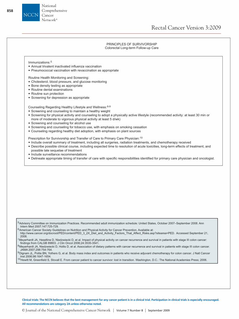
© Journal of the National Comprehensive Cancer Network | Volume 7 Number 8 | September 2009
858
Rectal Cancer Version 3:2009
Clinical trials: The NCCN believes that the best management for any cancer patient is in a clinical trial. Participation in clinical trials is especially encouraged. All recommendations are category 2A unless otherwise noted.
Immunizations:Annual trivalent inactivated influenza vaccinationPneumococcal vaccination with revaccination as appropriate
Routine Health Monitoring and Screening:Cholesterol, blood pressure, and glucose monitoringBone density testing as appropriateRoutine dental examinationsRoutine sun protectionScreening for depression as appropriate
5
Counseling Regarding Healthy Lifestyle and Wellness:Screening and counseling to maintain a healthy weightScreening for physical activity and counseling to adopt a physically active lifestyle (recommended activity: at least 30 min ormore of moderate to vigorous physical activity at least 5 d/wk)Screening and counseling for alcohol useScreening and counseling for tobacco use, with emphasis on smoking cessationCounseling regarding healthy diet adoption, with emphasis on plant sources
Prescription for Survivorship and Transfer of Care to Primary Care Physician:Include overall summary of treatment, including all surgeries, radiation treatments, and chemotherapy receivedDescribe possible clinical course, including expected time to resolution of acute toxicities, long-term effects of treatment, andpossible late sequelae of treatmentInclude surveillance recommendationsDelineate appropriate timing of transfer of care with specific responsibilities identified for primary care physician and oncologist.
6-9
10
PRINCIPLES OF SURVIVORSHIPColorectal Long-term Follow-up Care
5
6
8
9
10
Advisory Committee on Immunization Practices. Recommended adult immunization schedule: United States, October 2007–September 2008. AnnIntern Med 2007;147:725-729.
American Cancer Society Guidelines on Nutrition and Physical Activity for Cancer Prevention. Available at:http://www.cancer.org/docroot/PED/content/PED_3_2X_Diet_and_Activity_Factors_That_Affect_Risks.asp?sitearea=PED. Accessed September 21,2008.
Meyerhardt JA, Heseltine D, Niedzwiecki D, et al. Impact of physical activity on cancer recurrence and survival in patients with stage III colon cancer:findings from CALGB 89803. J Clin Oncol 2006;24:3535-3541.
Meyerhardt JA, Niedzwiecki D, Hollis D, et al. Association of dietary patterns with cancer recurrence and survival in patients with stage III colon cancer.JAMA 2007;298:754-764.
Dignam JL, Polite BN, Yothers G, et al. Body mass index and outcomes in patients who receive adjuvant chemotherapy for colon cancer. J Natl CancerInst 2006;98:1647-1654.Hewitt M, Greenfield S, Stovall E. From cancer patient to cancer survivor: lost in transition. Washington, D.C.: The National Academies Press; 2006.
7
NCCN Clinical Practice Guidelines in Oncology
Rectal Cancer
© Journal of the National Comprehensive Cancer Network | Volume 7 Number 8 | September 2009
859
Text continued from p. 839
N1, M0), and IIIC (any T, N2, M0). Stage IV dis-ease is defined as any T, any N, and the presence of 1 or more distant metastases (M1). The difference between N1 and N2 disease is the number of nodes involved; N1 lesions have 1 to 3 positive regional lymph nodes, whereas N2 tumors have 4 or more. In this version of the staging system, smooth metastatic nodules in the pericolic or perirectal fat are consid-ered lymph node metastases and should be included in N staging. Irregularly contoured metastatic nod-ules in the peritumoral fat are considered vascular invasion. In addition, the 6th edition of the AJCC staging manual6 suggests that surgeons mark the area of the specimen with the deepest tumor penetration so the pathologist can directly evaluate the status of the resection margins. The surgeon is encouraged to score the completeness of the resection as 1) R0 for complete tumor resection with all margins negative; 2) R1 for incomplete tumor resection with micro-scopic involvement of a margin; and 3) R2 for in-complete tumor resection with gross residual tumor that was not resected.
PathologyPathologic staging information is provided by exami-nation of the surgical specimen (see pages 848–850). Some information that should be detailed in the re-port of the pathologic evaluation of rectal cancer in-cludes 1) gross description of the tumor and specimen; 2) grade of cancer; 3) depth of penetration and exten-sion to adjacent structures (T); 4) number of regional lymph nodes evaluated; 5) number of positive region-al lymph nodes (N); 6) presence of distant metasta-ses to other organs, the peritoneum of an abdominal structure, or non-regional lymph nodes (M); and 7) status of the proximal, distal, and circumferential (ra-dial) margins.5,7 Prefixes “p” and “yp” denote patho-logic staging and pathologic staging after neoadjuvant therapy, respectively.8
The circumferential margin or circumferential resection margin (CRM) is an important pathologic staging parameter in rectal cancer. Although the ra-dial margin for resected segments of the colon that are completely encased by a peritonealized (serosal) surface is also referred to as the peritoneal margin, the CRM is very important in segments of the colon or rectum that are either not encased or only partially encased in peritoneum.5 The CRM is the closest ra-
dial margin between the deepest penetration of the tumor and the edge of resected soft tissue around the rectum (i.e., the retroperitoneal or subperitoneal as-pect of the tumor) and should be measured in mil-limeters. Identification of the CRM is determined through evaluation of the outer circumference of the rectal and mesorectal specimen which often requires inking of the outer surfaces and “bread-loaf” slicing of the specimen.9
A positive CRM has been defined as tumor with-in 1 to 2 mm from the transected margin.10–13 Ac-curate pathologic assessment of the CRM of resected rectal tumor specimens is very important because the CRM has been shown to be a strong predictor of both local recurrence and overall survival, and is an important consideration when post-operative treatment decisions are made.8,14,15 Furthermore, in a retrospective study of more than 17,000 patients with rectal cancer, CRM was found to be a better predictor of local recurrence for patients who had received preoperative therapy compared with those undergoing surgery as initial therapy.16 Additional components of the pathological evaluation of the surgical specimen after a total mesorectal excision (TME) are described under “Surgical Approaches”.
The AJCC and College of American Patholo-gists (CAP) recommend evaluating a minimum of 12 lymph nodes to accurately identify stage II colorectal cancers.5,6 The number of lymph nodes re-trieved can vary with age, gender, and tumor grade or site.17,18 The extent and quality of surgical resec-tion and pathologic review of the specimen can also have an impact on the node harvest.19 The literature lacks consensus regarding the minimal number of lymph nodes needed to accurately identify stage II rectal cancer. Most of these studies have combined rectal and colon cancers and reflect those cases with surgery as the initial treatment. Two studies confined only to rectal cancer reported 14 and more than 10 lymph nodes as the minimal number to accurately identify stage II rectal cancer.20,21
Furthermore, the mean number of lymph nodes retrieved from rectal cancers treated with neo-adjuvant therapy is significantly less than those treated with surgery alone (13 vs. 19, P < .05; 7 vs. 10, P ≤ .0001).22,23 A recent retrospective analysis of data from patients with T3 or T4 and/or lymph node-positive rectal cancer in the Intergroup 0114 trial showed lymph node ratio (LNR), the number of
NCCN Clinical Practice Guidelines in Oncology
Rectal Cancer
© Journal of the National Comprehensive Cancer Network | Volume 7 Number 8 | September 2009
860
positive lymph nodes divided by the total number, to be a strong predictor of survival.24 Nevertheless, the panel does not consider determination of LNR to be a substitute for an adequate lymph node evaluation.
Results of studies evaluating the sentinel node for micrometastatic disease through hematoxylin and eosin (H&E) staining to identify small foci of tumor cells, or identification of particular tumor antigens through immunohistochemical (IHC) analysis, have been reported.25,26 Although results of some of these studies seem promising, there is no uniformity in the definition of “true” clinically relevant metastatic carcinoma. Some studies have considered detection of single cells by IHC as well as isolated tumor cells (ITC) to be micrometastasis.27,28 In addition, results of one study demonstrated that, after neoadjuvant radiotherapy for rectal cancer, the sensitivity for the sentinel node procedure was only 40%.29 Pres-ently, the use of sentinel lymph nodes and detection of cancer cells by IHC alone should be considered investigational, and the results should be used with caution in clinical management decisions.
A sizable body of literature has shown that mu-tations in codons 12 and 13 in exon 2 of the coding region of the KRAS gene predict lack of response to cetuximab or panitumumab therapy.30–40 Therefore, the panel strongly recommends genotyping tumor tissue (either primary tumor or metastasis) in all patients with stage IV metastatic colorectal cancer. The recommendation for KRAS testing at this point is not meant to indicate a preference regarding regi-men selection in the first-line setting but, this early establishment of KRAS status is appropriate to plan for the treatment continuum, so that the informa-tion may be obtained in a non-time–sensitive man-ner. Thus, the patient and provider can discuss the implications of a KRAS mutation, if present, while other treatment options still exist. KRAS mutations are early events in colorectal cancer formation, and a tight correlation exists between mutation status in the primary tumor and metastases.41,42 For this reason, KRAS genotyping can be done on archived speci-mens of either the primary tumor or metastasis. Fresh biopsies should not be obtained solely for KRAS ge-notyping if an archived specimen from either the primary tumor or metastasis is available. The panel recommends that KRAS gene testing be performed only in laboratories that are certified under the clini-cal laboratory improvement amendments of 1988
(CLIA-88) as qualified to perform highly complex molecular pathology testing (see pages 848–850).
Clinical Presentation and Treatment
Management of Polypoid CancerText describing the management of polypoid cancer is available in the Colon Cancer Guidelines, in this issue, on page 804 or online in these guidelines at www.nccn.org.
Management of Rectal CancerRectal cancer has been defined as a cancerous lesion located within 12 cm of the anal verge by rigid proc-toscopy.50 Some support for this definition comes from the study of Kapiteijn et al.51 which included a subgroup analysis of the risk of recurrence of rectal cancer based on tumor location. Univariate analy-ses indicated that local recurrence rates were low for patients who had tumors with an inferior margin of 10.1 cm or more from the anal verge and that no significant differences between patients receiving radiotherapy and surgery were seen compared with those undergoing surgery alone. A recent retrospec-tive review of patients with rectal or rectosigmoid cancer showed that treatment options were impact-ed by whether the location of the rectal lesion was characterized by rigid proctoscopy or colonoscopy.52
Determination of an optimal treatment plan for an individual patient with rectal cancer is a com-plex process. In addition to decisions relating to the intent of rectal cancer surgery (i.e., curative or palliative), consideration must also be given to the likely functional results of treatment, including the probability of maintaining or restoring normal bowel function and anal continence and preserving geni-tourinary functions. For patients with distal rectal cancer, in particular, the simultaneous achievement of the goals of cure and minimal impact on quality of life can be challenging.53 Furthermore, the risk of pelvic recurrence is higher in patients with rectal cancer compared with those with colon cancer, and locally recurrent rectal cancer has frequently been associated with a poor prognosis.54,55 Careful patient selection for particular treatment options and the use of sequenced multimodality therapy for selected patients that combines chemoradiation (chemoRT) with operative treatment as part of the treatment regimen is recommended.

NCCN Clinical Practice Guidelines in Oncology
Rectal Cancer
© Journal of the National Comprehensive Cancer Network | Volume 7 Number 8 | September 2009
861
Clinical Evaluation/Staging: The initial clinical workup of patients with rectal cancer provides impor-tant preoperative information on the clinical stage of disease (see page 841). Because the clinical stage of disease is used to direct decisions on primary treat-ment, including surgical intent (e.g., curative or pal-liative) and approaches, and whether to recommend preoperative chemoRT, the implications of clinically under- or overstaging rectal cancer can be substantial.
Patients who present with rectal cancer appro-priate for resection require complete staging evalua-tion, including total colonoscopy to evaluate for syn-chronous lesions or other pathologic conditions of the colon and rectum, rigid proctoscopy to provide a determination of the location of the cancer (i.e., measurement of the distance of the tumor from the anal verge should be performed by the responsible surgeon using rigid proctoscopy), and a complete physical examination, including assessment of per-formance status to determine operative risk, carcino-embryonic antigen (CEA) and baseline CT scans of the chest, abdomen, and pelvis (see page 841). The panel consensus is that a PET scan is not routinely indicated at baseline in the absence of evidence of synchronous metastatic disease. In addition, the ac-cessibility of rectal cancer to evaluation by certain imaging modalities, such as endoscopic ultrasound and MRI, makes preoperative assessments of tumor penetration depth and presence of local lymph nodal metastases possible.56
Additional information regarding the extent of disease and the occurrence of distant metastases can be determined preoperatively through CT scans. Thus, endorectal ultrasound or endorectal or pelvic MRI and CT scans of the chest, abdomen, and pel-vis are recommended for the preoperative staging of rectal cancer.
Results from a meta-analysis of 90 studies involv-ing the accuracy of endoscopic ultrasound, MRI, and CT in preoperatively staging rectal cancer showed that endoscopic ultrasound and MRI have similarly high sensitivities for evaluating the depth of tumor penetration into the muscularis propia (94%), al-though endoscopic ultrasound was found to be more specific than MRI in the evaluation of local tumor invasion (86% vs. 69%).57 Only a limited number of studies using CT for T-staging have been performed, and it is not currently considered to be an optimal method for staging the extent of tumor penetra-
tion.57,58 Accurate assessment of nodal status is one of the greatest challenges in the preoperative stag-ing of rectal cancer. In the meta-analysis by Bipat et al.,57 the sensitivities and specificities of the 3 imag-ing modalities for accurately evaluating lymph node involvement were 55% and 74% for CT, 67% and 78% for endoscopic ultrasound, and 66% and 76% for MRI.
Results from another recent meta-analysis of 84 articles indicated that none of the 3 imaging modali-ties were significantly superior to another for accu-rately determining tumor N-stage.59 Disadvantages of endoscopic ultrasound and MRI include a high degree of operator dependence.57 An advantage of MRI is its ability to provide accurate images of soft tissue structures in the mesorectum, including the mesorectal fascia. Hence, MRI evaluation of patients with more advanced rectal cancer has the potential to provide information useful in the prediction of the CRM before radical surgery.58–60
Clinical staging is also based on histopathologic examination of the specimen obtained via biopsy or local excision (e.g., excised polyps). Endoscopic bi-opsy specimens of the lesion should undergo careful pathology review for evidence of invasion into the muscularis mucosa. If removal of the rectum is con-templated, early consultation with an enterostomal therapist is recommended for preoperative marking of the site and patient teaching purposes.Surgical Approaches: A variety of surgical ap-proaches, depending on the location and extent of disease, are used to treat the primary rectal cancer le-sion.61 These methods include local procedures, such as polypectomy, transanal excision and transanal mi-crosurgery, and radical procedures involving a trans-abdominal resection (e.g., low anterior resection [LAR], TME with coloanal anastomosis, or abdomi-noperineal resection [APR]; see pages 851–853).
Transanal excision may be appropriate for se-lected early stage cancers. Small (< 3 cm), well to moderately differentiated tumors that are within 8 cm of the anal verge, limited to less than 30% of the rectal circumference, and for which no evidence of nodal involvement (category 2A) is seen can be approached with transanal excision with negative margins. Transanal endoscopic microsurgery (TEM) can facilitate excision of small tumors through the anus that are located higher up in the rectum. Both transanal excision and TEM involve a full thick-

NCCN Clinical Practice Guidelines in Oncology
Rectal Cancer
© Journal of the National Comprehensive Cancer Network | Volume 7 Number 8 | September 2009
862
ness excision performed perpendicularly through the bowel wall into the perirectal fat. Negative (> 3 mm) deep and mucosal margins are required. Tumor frag-mentation should be avoided. The excised specimen should be oriented and pinned before fixation and brought to the pathologist by the surgeon (i.e., to facilitate an oriented histopathologic evaluation of the specimen).
Advantages of a local procedure include mini-mal morbidity (e.g., a sphincter-sparing procedure) and mortality and rapid postoperative recovery.53,62 If pathologic examination reveals adverse features such as high grade, positive margins, lymphovascular inva-sion (LVI), or perineural invasion, a more radical resec-tion is recommended. Data are limited on long-term patient outcomes, including risk of local recurrence, for patients undergoing local excision for T2 tumors.62
Limitations of a transanal excision include the absence of pathologic staging of nodal involvement. Further, there is evidence to indicate that lymph node micrometastases are both more common in early rectal lesions and unlikely to be identified by endorectal ultrasound.63 These observations may underlie the findings of a recent retrospective study of 282 patients undergoing either transanal excision or radical resection for T1 rectal cancer from 1985 through 2004, which showed respective local recur-rence rates of 13.2% and 2.7% for these 2 groups.64
Patients with rectal cancer who do not meet re-quirements for local surgery should be treated with transabdominal resection. Organ-preserving proce-dures that maintain sphincter function are preferable but not possible in all cases. For lesions in the mid to upper rectum, an LAR extended 4 to 5 cm below the distal edge of tumor, followed by creation of a colorectal anastomosis, is the treatment of choice. Where creation of an anastomosis is not possible, co-lostomy is required.
Data from randomized studies evaluating use of laparoscopic surgery in the treatment of patients with rectal cancer are limited.65,66 In the CLASICC trial comparing laparoscopically-assisted resection to open resection, nearly half of the 794 patients were diagnosed with rectal cancer.65 No significant differences in local recurrence, disease-free survival (DFS), or overall survival were seen between the 2 groups of patients with rectal cancer based on surgi-cal approach. However, factors that may confound conclusions drawn from randomized studies compar-
ing open surgery with laparoscopically-assisted sur-gery for colorectal cancer have been described,67 and laparoscopic surgery for rectal cancer is not recom-mended by the panel outside of a clinical trial.
For low rectal lesions, APR or TME with colo-anal anastomosis is required. A TME involves an en bloc removal of the mesorectum, including associ-ated vascular and lymphatic structures, fatty tissue, and mesorectal fascia as a “tumor package” through sharp dissection and is designed to spare the auto-nomic nerves.53,68 In cases in which anal function is intact and distal clearance is adequate, the TME may be followed by creation of a coloanal anastomosis. Pathologists play a key role in evaluating the surgical specimen after TME, which includes a macroscopic assessment of both external appearance/complete-ness and the CRM (see pages 848–850).69,70 Detailed descriptions of how the quality of the mesorectal specimens should be scored were provided in the Dutch Rectal Cancer Trial and those guidelines are endorsed by the NCCN panel.70
An APR involves en bloc resection of the recto-sigmoid, rectum, and anus, as well as the surround-ing mesentery, mesorectum, and perianal soft tissue and necessitates creation of a colostomy.71 An APR is necessary in cases in which a margin-negative resection of the tumor would result in loss of anal sphincter function resulting in incontinence. Al-though preoperative chemoRT may result in tumor downsizing and a decrease in tumor bulk (see follow-ing section), tumor location is not altered. Although sphincter preservation may become possible in cases in which initial tumor bulk prevented consideration of such surgery but exposure to the tumor is im-proved by chemoRT, an APR should be performed when tumor directly involves the anal sphincter or the levator muscles.
Recent comparisons of the outcomes of patients undergoing an APR versus a LAR in the treatment of rectal cancer have shown those treated with an APR to have worse local control and overall survival.72,73
Whether these differences can be attributed to the surgical procedure alone, patient- and tumor-related characteristics, or some combination of these factors is presently unclear. However, results from a recent retrospective study of 3633 patients with T3 or 4 rec-tal cancer tumors included in 5 large European trials suggest an association between the APR procedure it-self and the increased risks for recurrence and death.73
NCCN Clinical Practice Guidelines in Oncology
Rectal Cancer
© Journal of the National Comprehensive Cancer Network | Volume 7 Number 8 | September 2009
863
The lymphatic drainage regions of rectal tumors are influenced by their position in the rectum. More distal tumors are more likely to be characterized by upward and lateral lymphatic drainage, whereas the likelihood of only upward mesorectal drainage is much higher for more proximal tumors.74 The TME approach is designed to radically remove lymphatic drainage regions of tumors located above the level of the levator muscles.75 The panel does not recom-mend extension of nodal dissection beyond the field of resection (e.g., into the distribution of iliac lymph nodes) unless these nodes are clinically suspicious.Neoadjuvant/Adjuvant Therapy: Neoadjuvant/adjuvant therapy of rectal cancer often includes lo-coregional treatment due to the relatively high risk of locoregional recurrence. This risk is associated with the close proximity of the rectum to pelvic structures and organs, the absence of a serosa surrounding the rectum, and technical difficulties associated with ob-taining wide surgical margins at resection. In contrast, adjuvant treatment of colon cancer is more focused on preventing distant metastases since this disease is characterized by lower rates of local recurrence.
Combined-modality therapy consisting of sur-gery, radiation therapy (RT), and chemotherapy is recommended for most patients with stage II (node-negative disease with tumor penetration through the muscle wall) or stage III rectal cancer (node-positive disease without distant metastasis). Use of periopera-tive pelvic RT in the treatment of patients with stage II to III rectal cancer continues to evolve. Concur-rent fluoropyrimidine-based chemotherapy is recom-mended with radiation.
Ionizing radiation to the pelvis provides local tumoricidal therapy. Putative advantages to preop-erative radiation are related to both tumor response and normal tissue.76,77 Reducing tumor volume may facilitate resection and increase the likelihood of a sphincter-sparing procedure. Irradiating tissue that is surgery-naïve and thus better oxygenated may result in increased sensitivity to RT. Preoperative radiation can avoid the occurrence of radiation-induced injury to small bowel trapped in the pelvis by post-surgi-cal adhesions. Preoperative radiation that includes structures that will be resected increases the likeli-hood that an anastomosis with healthy colon can be performed (i.e., the anastomosis remains unaffected by the effects of RT because irradiated tissue is re-sected). One disadvantage of using preoperative RT
is the possibility of over-treating early stage tumors that do not require adjuvant radiation.77–79 Improve-ments in preoperative staging techniques, such as endoscopic ultrasound and CT scans, allow for more accurate staging, although the risk of overstaging dis-ease has not been eliminated.80
The results of the Swedish Rectal Cancer Trial evaluating the use of short course (5-day) RT admin-istered preoperatively for resectable rectal cancer showed a survival advantage and a decreased rate of local recurrence compared with surgery alone.81 However, although a number of other studies inves-tigating the effectiveness of preoperative or postop-erative RT in patients with rectal cancer staged as T1 to 3 have shown improvements in local control of disease, overall survival was not shown to be sig-nificantly affected.51,82,83 A multicenter, randomized study of 1350 patients with stage II to III rectal can-cer compared short-course preoperative RT with a postoperative approach that included chemoRT in selected patients (i.e., those with a positive CRM after resection) and no RT in patients without evi-dence of residual disease after surgery. The study in-dicated that patients in the preoperative RT arm had significantly lower local recurrence rates and a 6% absolute improvement in 3-year DFS (P = .03).84 No difference in overall survival has been observed be-tween the 2 arms. Currently, however, short-course RT for the treatment of rectal cancer is not widely practiced in the United States.
A number of randomized trials evaluated the ef-fectiveness of chemoRT administered either preop-eratively after clinical evaluation and staging (e.g., T3-4 by endoscopic ultrasound) or postoperatively after pathologic staging of rectal cancer as T3 and/or N1 or 2. Putative benefits of addition of chemother-apy concurrent with either pre- or postoperative RT include local RT sensitization and systemic control of disease (i.e., eradication of micrometastases). Pre-operative chemoRT also has the potential to increase rates of pathologic complete response and sphincter preservation. In a study of patients with T3 or 4 rec-tal cancer without evidence of distant metastases who were randomly assigned to receive either preoperative RT alone or preoperative concurrent chemoRT and 5-fluorouracil (5-FU)/leucovorin (LV), no difference in overall survival or sphincter preservation was seen, although patients receiving chemoRT were signifi-cantly more likely to exhibit a pathologic complete

NCCN Clinical Practice Guidelines in Oncology
Rectal Cancer
© Journal of the National Comprehensive Cancer Network | Volume 7 Number 8 | September 2009
864
response (11.4% vs. 3.6%; P < .05) and grade 3 to 4 toxicity (14.6% vs. 2.7%; P < .05) and less likely to exhibit local recurrence of disease (8.1% vs. 16.5%; P < .05).85 These conclusions have been supported in a recent systematic review which included 4 random-ized controlled trials.86
A large prospective, randomized trial from The German Rectal Cancer Study Group compared preoperative versus postoperative chemoRT in the treatment of clinical stage II or III rectal cancer.77 Results of this study indicated that preoperative therapy was associated with a significant reduction in local recurrence (6% vs. 13%; P = .006) and treat-ment-associated toxicity, although overall survival was similar in the 2 groups. Preliminary results of a phase III trial that included an evaluation of the addition of chemotherapy to preoperative RT in pa-tients with T3 or T4 resectable rectal cancer showed that 5-FU/LV chemotherapy enhanced the tumoro-cidal effect of RT when used concurrently. Signifi-cant reductions in tumor size, pTN stage, and lym-phatic, vascular, and perineural invasion rates were seen with combined-modality therapy compared with RT and surgery without chemotherapy.87,88 More mature results from this trial, which included 4 treatment groups (preoperative RT; preoperative chemoRT; preoperative RT plus postoperative che-motherapy; and preoperative chemoRT plus postop-erative chemotherapy), indicated that no significant differences in overall survival were associated with adding 5-FU–based chemotherapy pre- or postopera-tively.89 Although local recurrence rates were signifi-cantly lower in the groups receiving RT followed by chemotherapy, concurrent chemoRT, or concurrent chemoRT plus chemotherapy compared with the group receiving preoperative RT alone, the addition of chemotherapy after concurrent chemoRT did not significantly impact local recurrence rates.
In subsequent exploratory analyses of data from the group of patients in this trial who underwent com-plete tumor resection without evidence of distant dis-ease before or at surgery, those with ypT0-2 showed significant benefit in DFS and overall survival from adjuvant chemotherapy.90 These findings may indi-cate that patients are more likely to benefit from adju-vant therapy if their disease can be downstaged using chemoRT. Although reports from at least one of these studies has indicated that preoperative chemoRT is associated with increased rates of sphincter preserva-
tion in rectal cancer patients,77 this conclusion was not supported by 2 recent meta-analyses of random-ized trials involving preoperative chemoRT in the treatment of rectal cancer.91,92
Combined-modality therapy has been associated with decreased rates of local recurrence for rectal can-cer but with increased toxicity (e.g., radiation-induced injury, hematologic toxicities) relative to surgery alone.9,93 It has been suggested that some patients with disease at lower risk of local recurrence (e.g., proximal rectal cancer staged as T3, N0, M0, characterized by clear margins and favorable prognostic features) may be adequately treated with surgery and adjuvant che-motherapy.9,94,95 Nevertheless, results from a recent retrospective analysis showed the risk of locoregional recurrence to be significantly higher in patients with pT3N0 rectal cancer who did not undergo RT.96 In addition, 22% of 188 patients clinically staged with T3N0 rectal cancer using either esophageal ultra-sound or MRI who subsequently received preoperative chemoRT had positive lymph nodes after pathologic review of the surgical specimens, according to results of a recent retrospective multicenter study.80
Regarding the type of chemotherapy adminis-tered concurrently with RT, results from the Inter-group 0114 trial showed bolus 5-FU as part of ad-juvant therapy for rectal cancer to be non-inferior to bolus 5-FU plus LV.94 After a median follow-up of 4 years, neither the rate of local control nor sur-vival differed among 3 different combinations of modulated 5-FU chemotherapy. The equivalence of bolus 5-FU/LV and infusional 5-FU in concurrent chemoRT for rectal cancer is supported by the re-sults of a phase III trial (median followup, 5.7 years) in which similar outcomes in overall and relapse-free survival were seen when a continuous infusion of 5-FU or bolus 5-FU plus LV was administered con-currently with postoperative RT, although hemato-logic toxicity was greater in the group of patients receiving bolus 5-FU.97 However, results from an earlier trial from the North Central Cancer Treat-ment Group (NCCTG) showed that postoperative administration of continuous infusion 5-FU during pelvic irradiation was associated with longer overall survival when compared with bolus 5-FU.98 Most of the patients in this study had node-positive disease.
Postoperative chemoRT regimens commonly employ a “sandwich” approach whereby chemother-apy (typically 5-FU based) is administered before

NCCN Clinical Practice Guidelines in Oncology
Rectal Cancer
© Journal of the National Comprehensive Cancer Network | Volume 7 Number 8 | September 2009
865
and after the chemoRT regimen.94,97,98 The use of FOLFOX or capecitabine chemotherapy before and after postoperative chemoRT is an extrapolation of the available data in colon cancer.99,100
During administration of RT, multiple RT fields should include the tumor or tumor bed with a 2- to 5-cm margin, presacral nodes, and internal iliac nodes. The external iliac nodes should also be in-cluded for T4 tumors involving anterior structures, and clinicians should also consider inclusion of the inguinal nodes for tumors invading into the distal anal canal. Recommended doses of radiation are typically 45 to 50 Gy, with the exceptions of unre-sectable cancers for which doses higher than 54 Gy may be required and irradiation of the small bowel for which the dose should be limited to 45 Gy. In-tensity-modulated radiotherapy which uses comput-er-imaging to focus RT to the tumor site and poten-tially decrease toxicity to normal tissue,101–103 should be used in the context of a clinical trial only. As an additional boost, intraoperative radiotherapy,104–106
which involves direct exposure of tumors to RT dur-ing surgery while removing normal structures from the field of treatment, should be considered preop-eratively for patients with T4 tumors or recurrent cancers to facilitate resection.
Coordination of preoperative therapy, surgery, and adjuvant chemotherapy is important. For pa-tients treated with preoperative chemoRT, the panel recommends an interval of 5 to 10 weeks af-ter completion of full-dose 5.5-week chemoRT and before performance of surgical resection to allow patient recuperation from chemoRT-associated tox-icities. Although longer intervals from completion of chemoRT to surgery have been shown to be as-sociated with an increase in pathologic complete re-sponse rates,107–109 it is unclear whether this is associ-ated with clinical benefit. Nevertheless, when longer intervals are clinically necessary, they do not appear to increase the blood loss, time associated with sur-gery, or positive margin rate.110
Adjuvant chemotherapy for approximately 4 months is recommended for all patients with stage II or III rectal cancer after neoadjuvant chemoRT/surgery regardless of the surgical pathology results. However, few studies have evaluated the effect of adjuvant chemotherapy in patients with rectal can-cer, and its role is not well defined. Evaluation of adjuvant chemotherapy with 5-FU/LV alone ver-
sus postoperative RT followed by adjuvant chemo-therapy with 5-FU/LV in patients with stage II to III rectal cancer in the National Surgical Breast and Bowel Project (NSABP) R-02 trial showed a significant decrease in local recurrence rate in the group receiving adjuvant chemotherapy after RT compared with the group receiving adjuvant che-motherapy alone.111 However, no benefit of adding 5-FU–based adjuvant chemotherapy to preopera-tive chemoRT with respect to rate of local recur-rence was observed in the EORTC Radiotherapy Group trial 22921 (hazard ratio = 0.87; 95% CI, 0.72–1.04; P = .13) when the DFS of patients re-ceiving adjuvant chemotherapy after preoperative RT (with or without 5-FU–based chemotherapy) was compared with DFS of patients who underwent preoperative RT (with or without 5-FU–based che-motherapy) but did not receive adjuvant 5-FU–based chemotherapy.89 However, patients respond-ing to preoperative chemoRT had a survival benefit with adjuvant chemotherapy.
Most of the support for use of FOLFOX or capecitabine as adjuvant chemotherapy in rectal cancer is an extrapolation from the data available for colon cancer.99,100 The phase III ECOG E3201 trial is investigating the effect of adding either oxalipla-tin (FOLFOX) or irinotecan (FOLFIRI) to 5-FU/LV–based adjuvant chemotherapy administered to stage II to III rectal cancer patients after either pre-operative or postoperative chemoRT. Early reports indicate that adjuvant FOLFOX can be safely used in this patient population.112 Nevertheless, the dura-tion of treatment with adjuvant FOLFOX in rectal cancer is still unclear.113,114
In the MOSAIC trial, patients with stage II or III colon cancer were treated with 6 months of adjuvant FOLFOX. Some justification for a shorter course of adjuvant FOLFOX in rectal cancer (i.e., 4 months) can be provided when preoperative chemoRT is ad-ministered. In addition, the NSABP-07 trial showed similar DFS benefits to those reported in the MO-SAIC trial with only 9 cycles of an oxaliplatin-containing adjuvant regimen.114 A summary of on-going clinical trials in early-stage rectal cancer has been presented.115
Treatment of Nonmetastatic Rectal Cancer: Rec-ommendations for Patients With T1 and T2 Lesions: Node-negative T1 and T2 lesions are treated using transabdominal resection or transanal excision (cat-
NCCN Clinical Practice Guidelines in Oncology
Rectal Cancer
© Journal of the National Comprehensive Cancer Network | Volume 7 Number 8 | September 2009
866
egory 2B for T2), if appropriate (see page 842). This recommendation is category 2B for node-negative T2 tumors because local recurrence rates of 11% to 45% have been observed for T2 lesions after local excision alone.53,116,117 In selected lesions that are staged using endoscopic ultrasound or MRI as T1 to 2, N0 and without adverse pathologic features (e.g., no LVI or perineural invasion, < 3 cm, well to mod-erately differentiated), local excision with negative margins may give results comparable to transabdom-inal resection.118 No additional therapy is recom-mended for patients with well-differentiated T1 can-cers. If pathology review after local excision reveals a poorly differentiated histology, positive margins, or LVI, then a transabdominal re-resection should be performed. T2 cancers excised with negative mar-gins and no poor prognostic factors should be treated with transabdominal resection or adjuvant 5-FU/RT. Systemic chemotherapy should be considered as an adjuvant treatment for those patients who receive adjuvant chemoRT without additional surgery to avoid the risk of undertreatment as the lymph node status is unknown.
For patients with T1 to T2 lesions not amenable to local excision, a transabdominal resection is re-quired. No adjuvant therapy is indicated for patients with pathologic findings of T1 or T2 lesions. Patients with pathologic lymph node-negative T3 lesions (pT3, N0, M0) or pathologic lymph node-positive lesions (pT1-3, N1-2) should receive a “sandwich regimen” consisting of adjuvant chemotherapy with 5-FU with or without LV or FOLFOX (category 2B) or capecitabine (category 2B), followed by concur-rent 5-FU/RT (continuous infusion [category 2A] or bolus infusion along with LV [category 2B]) or capecitabine/RT (category 2B), then 5-FU with or without LV or FOLFOX (category 2B) or capecitabi-ne (category 2B; see pages 854 and 855, and 856). The panel recommends postoperative therapy for a total duration of approximately 6 months. For pa-tients with pathologic evidence of proximal T3, N0, M0 disease with clear margins and favorable prog-nostic features after an upfront resection, the incre-mental benefit of RT is likely to be small, and che-motherapy alone can be considered. However, most patients are not likely to be part of this subset.Recommendations for Patients with T3 Lesions and Lesions with Nodal Involvement: Patients clinically staged as having resectable T3, N0 or any T, N1-2 lesions should
initially be treated with preoperative combined-mo-dality therapy (see pages 843, 854 and 855, and 856). Upfront surgery should be reserved for patients with medical contraindications to chemoRT. Preoperative continuous infusional 5-FU/RT is the preferred treat-ment option (category 1 for node positive disease). Alternative regimens include bolus 5-FU/LV/RT (cat-egory 2A) or capecitabine/RT (category 2B). Patients who receive preoperative radiotherapy should undergo transabdominal resection 5 to 10 weeks after comple-tion of neoadjuvant therapy. The panel recommends approximately 6 months total duration of pre- and postoperative chemotherapy (regardless of surgical pa-thology results) with 5-FU with or without LV (cat-egory 1 for T3, N0 or any T, N1-2 tumors), FOLFOX (category 2B), or capecitabine (category 2B).
Patients with disease characterized as T3, N0 or any T, N1-2 disease initially treated by transab-dominal resection with subsequent pathologic stag-ing of disease as pT1-2, N0, M0 can be followed up with observation only. Patients with disease staged as pT3, N0, M0 or pT1-3, N1-2, M0 after initial treat-ment by transabdominal resection should receive ap-proximately 6 months postoperative chemotherapy with 5-FU with or without LV, FOLFOX (category 2B), or capecitabine (category 2B), followed by con-current 5-FU/RT (5-FU as continuous infusion [cat-egory 2A] or bolus infusion with LV [category 2B]) or capecitabine/RT (category 2B), then 5-FU with or without LV (category 2A), FOLFOX (category 2B), or capecitabine (category 2B). For some patients with pathologic evidence of proximal T3, N0, M0 disease with clear margins and favorable prognostic features after transabdominal resection, the incre-mental benefit of RT is probably small, and chemo-therapy alone can be considered, although this sub-set of patients is small.Recommendations for Patients with T4 Lesions and/or Locally Unresectable Disease: Patients with T4 or locally unresectable disease are treated with preop-erative continuous infusional 5-FU/RT (category 2A) or bolus 5-FU with LV/RT (category 2A) or capecitabine/RT (category 2B; see pages 843, 854 and 855, and 856). If possible, resection should be considered after preoperative chemoRT. Adjuvant therapy to complete 6 months with either 5-FU with or without LV (category 2A), FOLFOX (category 2B), or capecitabine (category 2B) is recommended regardless of the surgical pathology results.

NCCN Clinical Practice Guidelines in Oncology
Rectal Cancer
© Journal of the National Comprehensive Cancer Network | Volume 7 Number 8 | September 2009
867
Treatment of Metastatic Disease: Approximately 50% to 60% of patients diagnosed with colorectal cancer will develop colorectal metastases.119,120 Pa-tients with stage IV (any T, any N, M1) colorectal cancer or recurrent disease can present with syn-chronous liver or lung metastases or abdominal peritoneal metastases. Approximately 15% to 25% of patients with colorectal cancer present with syn-chronous liver metastases, although 80% to 90% of these patients are initially evaluated to have unre-sectable metastatic liver disease.119,121–123 Metastatic disease more frequently develops metachronously af-ter treatment for colorectal cancer, with the liver as a common site of involvement.124 Some evidence is available that indicates that synchronous metastatic colorectal liver disease is associated with a more dis-seminated disease state and a worse prognosis than metastatic colorectal disease that develops meta-chronously. In one retrospective study of 155 patients who underwent hepatic resection for colorectal liver metastases, patients with synchronous liver metasta-ses had more sites of liver involvement (P = .008) and bilobar metastases (P = .016) compared with patients diagnosed with metachronous liver metas-tases.125 For patients presenting with synchronous metastases and an intact primary that is not acute-ly obstructed, palliative resection of the primary is rarely indicated, and systemic chemotherapy is the preferred initial maneuver.126
It has been estimated that over half of patients
who die of colorectal cancer have liver metastases at autopsy and that metastatic liver disease is the cause of death in the majority of these patients.127 Results from reviews of autopsy reports of patients dying from colorectal cancer showed that the liver was the only site of metastatic disease in one third of patients.121 Furthermore, in a number of studies, 5-year survival rates for patients with metastatic liver disease not undergoing surgery have been quite low.119,128 How-ever, studies of selected patients undergoing surgery to remove colorectal liver metastases have demon-strated that cure is possible in this population and should be the goal for many patients with colorectal metastatic liver disease.119,129
Recent reports have shown 5-year survival rates following resection of hepatic colorectal metastases exceeding 50%.130,131 Therefore, decisions relating to patient suitability, or potential suitability, and subse-quent selection for metastatic colorectal surgery are
critical junctures in the management of metastatic colorectal liver disease.132
The criteria for determining patient suitability for resection or surgical cure of metastatic disease are evolving, and the emphasis is increasingly on the like-lihood of achieving negative surgical margins while maintaining adequate liver reserve, as opposed to other criteria, such as the number of liver metastases present.133–136 Resectability differs fundamentally from end points that focus more on palliative measures such as response and DFS. Instead, the resectability end point is focused on the potential of surgery to cure the disease,137 since partial liver resection or debulking has not been shown to be beneficial.120,135 Approach-es used in the surgical treatment of liver metastases include simultaneous resections of colorectal cancer and synchronous liver metastases,138 preoperative por-tal vein embolization for the purpose of increasing the volume and function of the portion of the liver which will remain postsurgically,139 and hepatic resection performed in 2 stages for bilobar disease.140
Resection is the standard of care for local treat-ment of metastatic disease that is initially resectable or converted to a potentially curable status after che-motherapy.141 However, some patients in this group who cannot undergo resection due to comorbidity, location of the metastatic lesions (i.e., adjacent to a major hepatic vein or the vena cava), or an estimate of inadequate liver volume after resection may be can-didates for ablation therapy.142 A number of retrospec-tive studies have compared radiofrequency ablation (RFA) and liver resection in the treatment of liver me-tastases,143–145 although RFA has not been well studied in this setting. Most of these studies have shown RFA to be inferior to resection with respect to rates of local recurrence and 5-year overall survival.141 Whether the differences in outcome observed for patients with liver metastases treated with RFA versus resection alone are due to patient selection bias, technologic limita-tions of RFA, or a combination is currently unclear.144 Nevertheless, the panel does not consider RFA to be a substitute for resection in patients with completely resectable disease. In addition, resection or RFA (ei-ther alone or in combination with resection) should be reserved for patients with disease that is completely amenable to local therapy. Use of surgery, RFA, or the combination for “debulking procedures” with a goal of less than complete resection or ablation of all known sites of disease is not recommended.

NCCN Clinical Practice Guidelines in Oncology
Rectal Cancer
© Journal of the National Comprehensive Cancer Network | Volume 7 Number 8 | September 2009
868
The panel’s consensus is that patients diagnosed with potentially resectable metastatic colorectal cancer should undergo an upfront evaluation by a multidisciplinary team, including surgical con-sultation (i.e., with an experienced hepatic sur-geon in cases involving liver metastases) to assess resectability status.
Most patients diagnosed with metastatic colorec-tal disease are initially classified as having unresect-able disease. For those with liver-limited unresectable disease, however, preoperative chemotherapy is in-creasingly used to downsize colorectal metastases and convert them to a resectable status (i.e., conversion chemotherapy); it has also been administered to pa-tients with metastatic disease considered to be resect-able (i.e., neoadjuvant therapy). Potential advantages include earlier treatment of micrometastatic disease, determination of responsiveness to chemotherapy (which can be prognostic and help plan postoperative therapy), and avoidance of local therapy in those who progress early. Potential disadvantages include chemo-therapy-induced liver injury and missing the “window of opportunity” for resection through the possibility of either disease progression or achievement of a com-plete response, thereby making it difficult to identify areas for resection.121,146
Furthermore, results from a recent study of colorectal cancer patients receiving preoperative chemotherapy indicated that cancer cells were still present in most of the original sites of metastases when these sites were examined pathologically de-spite achievement of a complete response as evaluat-ed on CT scan.147 It is therefore essential that during treatment with preoperative chemotherapy, frequent evaluations are undertaken and close communica-tion is maintained among medical oncologists, radi-ologists, surgeons, and patients so that a treatment strategy can be developed that optimizes exposure to the preoperative regimen and facilitates an appro-priately timed surgical intervention.148 When pre-operative chemotherapy is planned for patients with initially unresectable disease, the panel recommends that a surgical re-evaluation should be planned 2 months after initiation of preoperative chemothera-py, and that patients who continue to receive preop-erative chemotherapy undergo surgical re-evaluation every 2 months thereafter.149–152
Certain clinicopathologic factors, such as the presence of extrahepatic metastases and a disease-free
interval of less than 12 months, have been associated with a poor prognosis in patients with colorectal can-cer,130,131,153–155 although the ability of these factors to predict outcome after resection may be limited.119 However, the decision whether to offer preopera-tive chemotherapy begins with an initial evaluation of the degree of resectability of metastatic disease. Benefits of initial surgery in patients with clearly re-sectable disease characterized by generally favorable prognostic characteristics may outweigh the benefits of downsizing the disease with neoadjuvant chemo-therapy. Alternatively, preoperative chemotherapy would be more appropriate in patients with border-line resectable or initially unresectable but poten-tially convertible after response to chemotherapy. In addition, preoperative chemotherapy may be more beneficial in patients who have not been exposed to prior chemotherapy or who have not received che-motherapy in the previous 12 months.
The most important benefit of the preoperative approach is the potential to convert patients with initially unresectable metastatic disease to a resect-able state. The study by Pozzo et al.134 reported that preoperative therapy with irinotecan combined with 5-FU/LV enabled a significant portion (32.5%) of patients with initially unresectable liver metastases to undergo liver resection. Median time to progres-sion was 14.3 months, and all these patients were alive at a median follow-up time of 19 months. In a NCCTG phase II study,123 44 patients with unresect-able liver metastases were treated with FOLFOX4. Twenty five patients (60%) had tumor reduction and 17 (40%; 68% of the responders) were able to un-dergo resection after a median period of 6 months of chemotherapy.
In another study of 1439 initially unresectable patients with colorectal liver disease, 1104 patients were treated with chemotherapy and 335 (23%) were able to undergo primary hepatic resection. Of the 1104 patients receiving chemotherapy, 138 pa-tients (12.5%) classified as “good responders” under-went secondary hepatic resection after preoperative chemotherapy, which included oxaliplatin in most cases.156 The 5-year survival rate for these 138 pa-tients overall was 33%. In addition, results from a retrospective analysis of 795 previously untreated patients with metastatic colorectal cancer enrolled in the Intergroup N9741 randomized phase III trial evaluating the efficacy of mostly oxaliplatin-con-

NCCN Clinical Practice Guidelines in Oncology
Rectal Cancer
© Journal of the National Comprehensive Cancer Network | Volume 7 Number 8 | September 2009
869
taining chemotherapy regimens indicated that 24 patients (3.3%) were able to undergo curative liver resection after treatment.157 The median overall sur-vival time in this group was 42.4 months.
The choice of chemotherapy regimen in the preoperative setting depends on a number of factors, including whether the patient has resectable or po-tentially convertible metastatic disease and the re-sponse rates and safety/toxicity issues associated with the regimens. Although the benefits of pre- or post-operative chemotherapy for patients with liver me-tastases have not yet been fully validated in random-ized clinical trials, a recent EORTC phase III study evaluating use of perioperative FOLFOX4 (6 cycles before and 6 cycles after surgery) for patients with initially resectable liver metastases demonstrated ab-solute improvements in 3-year progression-free sur-vival of 8.1% (P = .041) and 9.2% (P = .025) for all eligible and all resected patients, respectively, when chemotherapy in conjunction with surgery was com-pared with surgery alone.158 The partial response rate after preoperative FOLFOX was 40% and operative mortality was less than 1% in both treatment groups.
There have been recent reports of randomized clinical trials evaluating preoperative FOLFIRI or FOLFOX as conversion therapies in combination with anti-epidermal growth factor receptors (EGFR) inhibitors.159,160 However, a number of randomized studies have investigated the efficacy and safety of FOLFOX, CapeOX, or FOLFIRI with and without bevacizumab or cetuximab in the first-line treat-ment of patients with metastatic colorectal cancer (see “Chemotherapy for Advanced or Metastatic Disease”). In addition, first-line FOLFOXIRI (infu-sional 5-FU, LV, oxaliplatin, irinotecan) has been compared with FOLFIRI in 2 randomized clinical trials.161,162 Significantly improved rates of response and overall survival were reported for patients in the FOLFOXIRI arm of one of the studies,162 but not in the other.161
The efficacy of bevacizumab in combination with FOLFOX and FOLFIRI (infusional 5-FU, LV, irinotecan) in the treatment of unresectable meta-static disease (see “Chemotherapy for Advanced or Metastatic Disease”) has led to its use in combina-tion with these regimens in the preoperative setting, although the safety of administering bevacizumab pre- or postoperatively in combination with 5-FU–based regimens has not been adequately evaluated.
A retrospective evaluation of data from 2 random-ized trials of 1132 patients receiving chemotherapy with or without bevacizumab as initial therapy for metastatic colorectal cancer indicated that the inci-dence of wound healing complications was increased for the group of patients undergoing a major surgical procedure while receiving a bevacizumab-containing regimen when this population was compared with the group receiving chemotherapy alone while un-dergoing major surgery (13% vs. 3.4%, respectively; P = .28).163 However, when chemotherapy plus be-vacizumab or chemotherapy alone was administered before surgery, the incidence of wound healing com-plications in either group of patients was low (1.3% vs. 0.5%; P = .63).
The panel recommends at least a 6-week interval (which corresponds to 2 half-lives of the drug164) be-tween the last dose of bevacizumab and elective sur-gery. Further support for this recommendation comes from results of a single-center, non-randomized phase II trial of patients with potentially resectable liver metastases that showed no increase in bleed-ing or wound complications when the bevacizumab component of CapeOX plus bevacizumab therapy was stopped 5 weeks before surgery (i.e., bevaci-zumab excluded from the sixth cycle of therapy).165 In addition, no significant differences in bleeding, wound, or hepatic complications were observed in a retrospective trial evaluating effects of preoperative bevacizumab stopped 8 weeks or prior compared to more than 8 weeks before resection of liver colorec-tal metastases for patients receiving oxaliplatin- or irinotecan-containing regimens.166
Other reported risks associated with the preop-erative approach include the potential for develop-ment of liver steatosis or steatohepatitis when oxali-platin- or irinotecan-containing chemotherapeutic regimens are administered.148 To limit the develop-ment of hepatotoxicity, it is therefore recommended that surgery should be performed as soon as possible after the patient becomes resectable and usually not more than 3 to 4 months following initiation of pre-operative treatment.
As mentioned previously, colorectal metastatic disease can also occur in the lung.167 Most of the treatment recommendations discussed for metastatic colorectal liver disease also apply to the treatment of colorectal pulmonary metastases. Combined pulmo-nary and hepatic resections of resectable metastatic

NCCN Clinical Practice Guidelines in Oncology
Rectal Cancer
© Journal of the National Comprehensive Cancer Network | Volume 7 Number 8 | September 2009
870
disease have been performed in selected cases.168 The goal of treatment of most abdominal/peritoneal me-tastases is palliative, rather than curative.
It is important to note that some of the treat-ment approaches for patients diagnosed with rectal cancer and potentially resectable synchronous lung or liver metastases differ from those for patients di-agnosed with stage IV colon cancer characterized as potentially resectable metastatic disease. In particu-lar, initial treatment options for potentially resect-able rectal cancer include preoperative chemoRT directed toward treatment of the primary cancer; preoperative combination chemotherapy regimen plus a biologic agent to target metastatic disease; and a surgical approach (i.e., staged or synchronous re-section of metastases and rectal lesion). Advantages of an initial chemoRT approach include a possible decreased risk of pelvic failure following surgery, al-though preoperative pelvic RT may decrease toler-ance to systemic bevacizumab-containing adjuvant regimens, thereby limiting subsequent treatment of systemic disease. However, data to guide deci-sions regarding optimal treatment approaches in this population of patients are very limited. Of note, patients with stage II/III rectal cancer enrolled in a large randomized trial evaluating the effect of adding chemotherapy to preoperative RT were found to be 3 times more likely to develop distant metastases than local recurrence of disease after a median follow-up of more than 5 years.89
Only limited data exist regarding the efficacy of adjuvant chemotherapy following resection for metastatic colorectal liver or lung disease. Never-theless, the panel recommends administration of a course of an active systemic chemotherapy regimen for metastatic disease for some patients following liver or lung resection who have received preop-erative chemoRT or no preoperative therapy after staged or synchronous resection of metastases and rectal lesion to increase the likelihood that residu-al microscopic disease will be eradicated for a total perioperative treatment time of approximately 6 months. Postoperative chemoRT is recommended for patients with synchronous metastases who have not received prior chemoRT and who are at higher risk for pelvic recurrence after staged or synchro-nous resection of metastases and rectal lesion (i.e., patients with disease staged as pT3-4, any N, or any T,N1-2).
Placement of a hepatic arterial port or implant-able pump during surgical intervention for liver re-section with subsequent administration of chemo-therapy directed to the liver metastases through the hepatic artery (i.e., hepatic arterial infusion [HAI]) is listed in the guidelines as an option (category 2B). In a randomized study of patients who had un-dergone hepatic resection, administration of floxu-ridine (with dexamethasone and with or without LV) by HAI in addition to systemic chemotherapy was shown to be superior to systemic chemotherapy alone with respect to 2-year survival and time to progression of hepatic disease.169,170 However, the dif-ference in survival between the 2 arms was not sig-nificant at later follow-up periods.169,171 A number of other clinical trials have shown significant improve-ment in response or time to hepatic disease progres-sion when HAI therapy was compared with systemic chemotherapy, although most have not shown a sur-vival benefit of HAI therapy.169 Some of the uncer-tainties regarding patient selection for preoperative chemotherapy are also relevant to the application of HAI.129 However, limitations of HAI therapy include the potential for biliary toxicity169 and the require-ment for specific technical expertise. The consensus of the panel is that HAI therapy should be consid-ered only at institutions with extensive experience in both the surgical and medical oncologic aspects of the procedure.
Finally, a number of liver-directed therapies are available for the treatment of unresectable meta-static disease in highly select patients, although their role in the treatment of colorectal metastases is controversial. These therapies include arterial ra-dioembolization with yttrium-90 microspheres,172,173 arterial chemoembolization,173 and conformal radia-tion therapy.174 Use of intra-arterial embolization is a category 3 recommendation for select patients with predominant hepatic metastases, and conformal ex-ternal beam radiation therapy is not recommended unless the patient is symptomatic or it is used in a clinical trial. (See “Workup and Management of Synchronous Metastatic Disease” and “Workup and Management of Metachronous Metastatic Disease”).
Locally recurrent rectal cancer is characterized by isolated pelvic/anastomotic recurrence of disease. In a single-center study at The University of Texas M. D. Anderson Cancer Center, rates of 5-year lo-cal recurrence were reported to be low (i.e., 5-year

NCCN Clinical Practice Guidelines in Oncology
Rectal Cancer
© Journal of the National Comprehensive Cancer Network | Volume 7 Number 8 | September 2009
871
locoregional control rate of 91%) for patients with rectal cancer treated with surgery and either RT or chemoRT, and 78% of recurrences occurred in the low pelvic and presacral regions.175 Patients with disease recurrence at the anastomotic site are more likely than those with an isolated pelvic recurrence to be cured following re-resection.176,177 In a study of 43 consecutive patients with advanced pelvic recur-rence of colorectal cancer who had not undergone prior RT, treatment with 5 weeks of 5-FU by con-tinuous infusion concurrent with RT enabled the majority of patients (77%) to undergo re-resection with curative intent.176
Recommendations for Treatment of Synchronous Metas-tases/Resectable Disease: As part of the pre-treatment work-up, the panel recommends tumor KRAS gene status testing for all patients with metastatic colorec-tal cancer at the time of diagnosis of metastatic disease (see previous discussion of KRAS testing). Initial treatment options for patients with stage IV disease (any T, any N, M1) and resectable liver or lung metastases include combination chemotherapy for 2 to 3 months (e.g., FOLFOX, CapeOX, or FOL-FIRI regimens with or without bevacizumab or ce-tuximab for KRAS wild-type tumors only); staged or synchronous resection of metastases and rectal lesion; treatment with continuous infusional 5-FU/pelvic RT (category 2A) or bolus 5-FU with LV/pelvic RT (category 2A) or capecitabine/RT (category 2B); or 2 to 3 months of upfront combination chemotherapy with FOLFOX, CapeOX, or FOLFIRI regimens with or without bevacizumab or cetuximab (KRAS wild-type tumors only) followed by chemoRT (see pages 793–797, 844, and 856). The impetus for inclusion of the latter option is upfront systemic treatment with a goal of early eradication of micrometastases followed by consolidating chemoRT for local con-trol of disease prior to surgery (see page 844). For the 3 groups of patients receiving neoadjuvant therapy, surgery should be performed 5 to 10 weeks following completion of such treatment.
Adjuvant therapy for patients undergoing ini-tial surgery depends on pathologic staging of dis-ease. For patients undergoing initial surgical treat-ment, the panel recommends that those at higher risk for pelvic failure relative to systemic disease (e.g., disease pathologically staged as pT3-4, any N or any T, N1-2) undergo postoperative chemoRT using the “sandwich” approach (i.e., chemotherapy
followed by concurrent chemoRT followed by che-motherapy for 6 months total duration).97,98 The panel acknowledged that not all patients with rec-tal cancer and resectable liver or lung metastases need to be treated with chemoRT. For example, in patients with pT1-2, N0 disease, the competing risk of distant metastases is considered to be higher than that of locoregional recurrence. Therefore, the panel recommends that these patients receive an active adjuvant chemotherapy regimen (for 6 months) as described on pages 793–797, with the exception of FOLFOXIRI.
Adjuvant therapy recommendations for patients who have received neoadjuvant chemoRT only is as described for patients with pT1-2, N0 disease (ex-cept total duration of pre- plus postoperative che-motherapy should be 6 months), whereas patients who have undergone preoperative bevacizumab- or cetuximab (KRAS wild-type tumors only) -contain-ing therapy should receive postoperative chemoRT as described previously for patients with pT3-4, any N, or any T, N1-2 disease (except total duration of pre- plus postoperative chemotherapy should be 6 months). Those patients undergoing preoperative bevacizumab- or cetuximab-containing therapy fol-lowed by preoperative chemoRT should not receive postoperative chemotherapy (see page 844).Recommendations for Treatment of Synchronous Me-tastases/Unresectable Disease: Patients with any un-resectable or medically inoperable metastases are treated according to whether they are symptomatic or asymptomatic. Symptomatic patients are treated with chemotherapy alone or combined modality therapy with 5-FU/RT or capecitabine/RT (category 2B), resection of the involved rectal segment or laser canalization or diverting colostomy or stenting (see page 845). Primary treatment should be followed by an active chemotherapy regimen for metastatic dis-ease (see pages 793–797).
For patients with asymptomatic liver or lung disease that is deemed to be unresectable, the panel recommends chemotherapy corresponding to initial therapy for metastatic disease (e.g., choice of FOL-FIRI, FOLFOX, or CapeOX chemotherapy with or without bevacizumab or cetuximab [KRAS wild-type tumors only], or the same chemotherapy regi-mens with or without cetuximab [KRAS wild-type tumors only], or FOLFOXIRI [category 2B for FOLF-OXIRI]) to attempt to render these patients candi-

NCCN Clinical Practice Guidelines in Oncology
Rectal Cancer
© Journal of the National Comprehensive Cancer Network | Volume 7 Number 8 | September 2009
872
dates for resection (see pages 793–797). Preoperative chemotherapy regimens with high response rates should be considered for patients with potentially convertible disease,178 and these patients should be re-evaluated for resection after 2 months of preop-erative chemotherapy and every 2 months thereafter while undergoing such therapy.
Primary treatment of unresectable synchronous liver or lung metastases by palliative surgery to re-move the primary tumor should be considered only if the patient has an unequivocal imminent risk of ob-struction or acute significant bleeding. It should be noted that symptomatic improvement in the primary is often seen with first-line systemic chemotherapy, even within the first 1 to 2 weeks, and routine palli-ate resection of a synchronous primary lesion should not be done in the absence of overt, serious symp-toms.126 Complications from the primary lesion are uncommon in these circumstances, and its removal delays initiation of systemic chemotherapy. An in-tact primary is not a contraindication to bevacizum-ab use. The risk of gastrointestinal perforation in the setting of bevacizumab is not decreased by removal of the primary tumor, as large bowel perforations, in general, and perforation of the primary lesion, in particular, are rare (see section on “Chemotherapy for Advanced or Metastatic Disease” in the NCCN Colon Cancer Guidelines).
Ablative therapy of metastatic disease, either alone or in combination with resection, can also be considered when all measurable metastatic disease can be treated (see “Principles of the Management of Metastatic Disease”). Post-treatment follow-up for patients classified as stage IV and no evidence of disease (NED) is described in the section on “Post-Treatment Surveillance.”
Patients with unresectable metastatic disease not responding to preoperative therapy should receive chemotherapy for advanced or metastatic disease as outlined on pages 793–797 with treatment selection based, in part, on whether the patient is or is not an appropriate candidate for intensive therapy.
There was no panel consensus regarding the use of liver-directed therapies such as arterial radioem-bolization therapy and arterial chemoembolization therapy. For select patients with chemotherapy resis-tant/refractory disease characterized by predominant liver metastases and no obvious systemic disease, use of these interventions was supported by some panel
members but not others (category 3). The consen-sus of the panel is that conformal external radia-tion therapy should not be used unless the patient is symptomatic or it is administered in the context of a clinical trial.Recommendations for Treatment of Metachronous Me-tastases: Routine use of PET to monitor for disease recurrence is not recommended. It should be noted that the CT that accompanies a “PET/CT” is a non-contrast CT and thus not of ideal quality for rou-tine surveillance. On documentation on dedicated contrast-enhanced CT or MRI of metachronous metastases in which disease is or may become po-tentially resectable, characterization of the extent of disease by PET scan is recommended. PET is used at this juncture to promptly characterize the extent of metastatic disease, and to identify possible sites of extrahepatic disease which could preclude surgery (see pages 786, 793–797, and 847).179 As with other first identifications of metastatic disease, a tumor sample (metastases or original primary) should be sent for KRAS genotyping to define whether anti-EGFR agents can be considered in the list of poten-tial options for this patient (see previous discussion of KRAS testing). Close communication between members of the multidisciplinary treatment team is recommended, including upfront evaluation by a surgeon experienced in the resection of hepatobili-ary and lung metastases (see pages 786 and 847).
The management of metachronous metastatic disease is further distinguished from that of syn-chronous disease by also including an evaluation of the chemotherapy history of the patient, and by the absence of transabdominal resection. Resect-able patients are classified according to whether they have received no previous chemotherapy or chemo-therapy within or prior to the previous 12 months (see page 787). For patients who have not received prior chemotherapy and who have resectable meta-static disease, primary treatment options include initial resection followed by chemotherapy with an active chemotherapy regimen (see pages 793–797) for 6 months or neoadjuvant chemotherapy for 2 to 3 months followed by resection and additional postoperative chemotherapy for a total duration of pre- plus postoperative chemotherapy for up to 6 months based on response to the neoadjuvant regi-men; observation is also an option for patients with-out a response to neoadjuvant therapy. For example,
NCCN Clinical Practice Guidelines in Oncology
Rectal Cancer
© Journal of the National Comprehensive Cancer Network | Volume 7 Number 8 | September 2009
873
the same chemotherapy regimen used in the neo-adjuvant setting should be repeated postoperatively for patients with a preoperative disease response to such therapy. However either an alternative active chemotherapy regimen (see pages 793–797) or ob-servation is an option in the postoperative setting for patients not responding to neoadjuvant therapy.
Patients determined by cross-sectional imaging or PET scan to have unresectable (including those considered to potentially convertible or unconvert-ible) disease should receive an active chemotherapy regimen based on prior chemotherapy history (see pages 786, 787, and 793–797). Specifically, patients exhibiting disease progression on FOLFOX admin-istered within the previous 12 months should be switched to a FOLFIRI regimen with the option of inclusion of bevacizumab or cetuximab (KRAS wild type only). Patients potentially convertible to resectability should be re-evaluated for disease con-version to a resectable status every 2 months; those with chemotherapy-responsive disease who are con-verted to a resectable state should undergo resection followed by postoperative therapy as described above for patients with resectable disease and a history of previous chemotherapy. In the case of liver metas-tases only, HAI therapy with or without systemic 5-FU/LV (category 2B) or continuous IV 5-FU in-fusion remains at option at centers with experience with the surgical and medical oncologic aspects of this procedure.
Patients with unresectable metastatic disease not responding to preoperative therapy should receive chemotherapy for advanced or metastatic disease as outlined on pages 793–797, with treatment selection based, in part, on whether the patient is or is not an appropriate candidate for intensive therapy. Patients receiving palliative chemotherapy should be moni-tored with CT or MRI scans approximately every 2 to 3 months. PET scans are not recommended for routine monitoring of the progression of metastatic disease.
Isolated pelvic/anastomotic recurrence is opti-mally managed by preoperative RT and concurrent infusional 5-FU, if full course RT was not given pre-viously. Resection followed by the option of IORT should be considered if it can be safely delivered (see page 847).180 However, debulking, resulting in gross residual cancer, is discouraged. Patients with unre-sectable lesions are treated according to their abil-ity to tolerate therapy. The treatment goal for most
abdominal/peritoneal metastases is palliative rather than curative. The panel currently considers the treatment of disseminated carcinomatosis with cyto-reductive surgery (i.e., peritoneal stripping surgery) and perioperative hyperthermic intraperitoneal che-motherapy181,182 to be investigational and does not endorse such therapy outside of a clinical trial. How-ever, the panel recognizes the need for randomized clinical trials to address the risks and benefits associ-ated with each of these modalities.Chemotherapy for Advanced or Metastatic Dis-ease: The continuum of care approach to the man-agement of patients with metastatic rectal cancer is the same as described for patients with metastatic co-lon cancer. Please refer to the corresponding section in the Colon Cancer Guidelines.
Post-Treatment SurveillanceThe approach to monitoring and surveillance of pa-tients with rectal cancer is similar to that described for colon cancer with the addition of proctoscopy to evaluate the rectal anastomosis for local recurrence for patients who have undergone an LAR (see page 846). Anastomotic recurrence of rectal cancer has a much more favorable prognosis than local recur-rence at other locations in the pelvis,176,177 although the optimal timing for surveillance of the rectal anastomosis is not known.
Additional information on Post-treatement Sur-veillance is available in the Colon Cancer Guide-lines, on page 820 in this issue or online in these guidelines at www.nccn.org.
Post-treatment surveillance also includes a sur-vivorship care plan involving disease preventive measures such as immunizations against influenza and pneumococcal infections at prescribed intervals and regular dental care, and early disease detection through periodic screening for second primary cancers (e.g., breast, cervical, or prostrate cancers) and routine health monitoring to screen for comorbid conditions including psychosocial distress associated with colorec-tal cancer and its treatment (see pages 857 and 858).
Other recommendations include monitoring for late sequelae of rectal cancer or the treatment of rec-tal cancer, such as chronic diarrhea or incontinence (e.g., patients with stoma);199 persistent neuropathy (a well known side effect of oxaliplatin treatment);99 pelvic pain or pelvic fractures; and urogenital dys-

NCCN Clinical Practice Guidelines in Oncology
Rectal Cancer
© Journal of the National Comprehensive Cancer Network | Volume 7 Number 8 | September 2009
874
function after resection or pelvic irradiation.200–203 Specific management interventions to address these side effects are described on pages 857 and 858 and in a recent review.204
There is also evidence to indicate that certain lifestyle characteristics, such as smoking cessation, maintaining a healthy body mass index, engaging in regular exercise, and making certain dietary choices are associated with improved outcomes after treat-ment for colon cancer. For example, a retrospective study of patients with stage II and III colon cancer enrolled in NSABP trials from 1989 to 1994 showed that patients with a body mass index of 35 kg/m2 or greater had an increased risk of disease recurrence and death.205 In a prospective observational study of patients with stage III colon cancer enrolled in the CALGB 89803 adjuvant chemotherapy trial, DFS was found to be directly dependent on how much ex-ercise these patients received.206 Furthermore, a diet consisting of more fruits, vegetables, poultry and fish, and less red meat, as well as diets higher in whole grains and lower in refined grains and concentrated sweets was found to be associated with an improved outcome in terms of cancer recurrence or death.207 A discussion of lifestyle characteristics that may be associated with a decreased risk of colorectal cancer recurrence also provides “a teachable moment” for the promotion of overall health and an opportunity to encourage patients to make choices and changes compatible with a healthy lifestyle.
Managing an Increasing CEA LevelThe approach to managing an increased CEA level for patients with rectal cancer is the same as that de-scribed for patients with colon cancer. It is available in the colon cancer guidelines in this issue (page 822) or in these guidelines online at www.nccn.org.
SummaryThe NCCN Rectal Cancer Guidelines panel believes that a multidisciplinary approach, including repre-sentation from gastroenterology, medical oncology, surgical oncology, radiation oncology, and radiology is necessary for treating patients with rectal cancer. Adequate pathologic assessment of the resected lymph nodes is important with a goal of evaluating at least 12 nodes when possible. Patients with very early stage tumors lesions that are node-negative by endorectal ultrasound or endorectal or pelvic MRI
and who meet carefully defined criteria can be man-aged with a transanal excision. A transabdominal resection is appropriate for all other rectal lesions. Preoperative chemoRT is preferred for the majority of patients with suspected or proven T3/T4 disease and/or regional node involvement and adjuvant che-motherapy is recommended. Patients with recurrent localized disease should be considered for resection with or without radiotherapy.
A patient with metastatic disease in the liver or lung should be considered for surgical resection if he or she is a candidate for surgery and if complete resection (R0) or ablation can be achieved. Preop-erative chemotherapy can be considered as initial therapy in patients with synchronous or metachro-nous resectable metastatic disease (i.e., neoadjuvant therapy) or when a response to chemotherapy may convert a patient from an unresectable to resect-able state (i.e., conversion therapy). Other options for patients with resectable synchronous metastases are initial treatment with chemoRT or chemother-apy with or without a bevacizumab or cetuximab (KRAS wild type tumor only) followed by consoli-dating chemoRT. Resection should be followed by adjuvant therapy based on prior therapy received. The recommended post-treatment surveillance program for rectal cancer patients includes serial CEA determinations, as well as periodic chest, ab-dominal and pelvic CT scans, and periodic evalua-tions by colonoscopy and proctoscopy.
Recommendations for patients with previously untreated disseminated metastatic disease represent a continuum of care in which lines of treatment are blurred rather than discrete. Principles to consider at the start of therapy include pre-planned strategies for altering therapy for patients in both the presence and absence of disease progression, including plans for adjusting therapy for patients who experience certain toxicities. Recommended initial therapy options for advanced or metastatic disease depend on whether or not the patient is appropriate for in-tensive therapy. The more intensive initial therapy options include FOLFOX, FOLFIRI, CapeOX, and FOLFOXIRI (category 2B). Addition of a biologic agent (e.g., bevacizumab or cetuximab) is either rec-ommended, or listed as an option, in combination with some of these regimens, depending on avail-able data. Chemotherapy options for patients with progressive disease are dependent on the choice of

NCCN Clinical Practice Guidelines in Oncology
Rectal Cancer
© Journal of the National Comprehensive Cancer Network | Volume 7 Number 8 | September 2009
875
initial therapy. The panel endorses the concept that treating patients in a clinical trial has priority over standard or accepted therapy.
References 1. Jemal A, Siegel R, Ward E, et al. Cancer statistics, 2009. CA
Cancer J Clin 2009;59:225–229. 2. Bonelli L, Martines H, Conio M, et al. Family history of colorectal
cancer as a risk factor for benign and malignant tumours of the large bowel. A case-control study. Int J Cancer 1988;41:513–517.
3. Ahsan H, Neugut AI, Garbowski GC, et al. Family history of colorectal adenomatous polyps and increased risk for colorectal cancer. Ann Intern Med 1998;128:900–905.
4. Greene F, Page D, Fleming I, Fritz A. AJCC Cancer Staging Manual. New York: Springer-Verlag; 2002.
5. Compton CC, Greene FL. The staging of colorectal cancer: 2004 and beyond. CA Cancer J Clin 2004;54:295–308.
6. Greene FL, Stewart AK, Norton HJ. A new TNM staging strategy for node-positive (stage III) colon cancer: an analysis of 50,042 patients. Ann Surg 2002;236:416–421; discussion 421.
7. Compton CC. Updated protocol for the examination of specimens from patients with carcinomas of the colon and rectum, excluding carcinoid tumors, lymphomas, sarcomas, and tumors of the vermiform appendix: a basis for checklists. Cancer Committee. Arch Pathol Lab Med 2000;124:1016–1025.
8. Compton CC. Key issues in reporting common cancer specimens: problems in pathologic staging of colon cancer. Arch Pathol Lab Med 2006;130:318–324.
9. Lai LL, Fuller CD, Kachnic LA, Thomas CR Jr. Can pelvic radiotherapy be omitted in select patients with rectal cancer? Semin Oncol 2006;33(Suppl 11):S70–74.
10. Nagtegaal ID, Marijnen CA, Kranenbarg EK, et al. Circumferential margin involvement is still an important predictor of local recurrence in rectal carcinoma: not one millimeter but two millimeters is the limit. Am J Surg Pathol 2002;26:350–357.
11. Wibe A, Rendedal PR, Svensson E, et al. Prognostic significance of the circumferential resection margin following total mesorectal excision for rectal cancer. Br J Surg 2002;89:327–334.
12. Stocchi L, Nelson H, Sargent DJ, et al. Impact of surgical and pathologic variables in rectal cancer: a United States community and cooperative group report. J Clin Oncol 2001;19:3895–3902.
13. Glynne-Jones R, Mawdsley S, Novell JR. The clinical significance of the circumferential resection margin following preoperative pelvic chemo-radiotherapy in rectal cancer: why we need a common language. Colorectal Dis 2006;8:800–807.
14. Adam IJ, Mohamdee MO, Martin IG, et al. Role of circumferential margin involvement in the local recurrence of rectal cancer. Lancet 1994;344:707–711.
15. Mawdsley S, Glynne-Jones R, Grainger J, et al. Can histopathologic assessment of circumferential margin after preoperative pelvic chemoradiotherapy for T3-T4 rectal cancer predict for 3-year disease-free survival? Int J Radiat Oncol Biol Phys 2005;63:745–752.
16. Nagtegaal ID, Quirke P. What is the role for the circumferential margin in the modern treatment of rectal cancer? J Clin Oncol 2008;26:303–312.
17. Sarli L, Bader G, Iusco D, et al. Number of lymph nodes examined and prognosis of TNM stage II colorectal cancer. Eur J Cancer
2005;41:272–279. 18. Newland RC, Dent OF, Lyttle MN, et al. Pathologic determinants
of survival associated with colorectal cancer with lymph node metastases. A multivariate analysis of 579 patients. Cancer 1994;73:2076–2082.
19. Wong SL, Hong J, Hollenbeck BK, et al. Hospital lymph node examination rates and survival after resection for colon cancer. JAMA 2007;298:2149–2154.
20. Tepper JE, O’Connell MJ, Niedzwiecki D, et al. Impact of number of nodes retrieved on outcome in patients with rectal cancer. J Clin Oncol 2001;19:157–163.
21. Pocard M, Panis Y, Malassagne B, et al. Assessing the effectiveness of mesorectal excision in rectal cancer: prognostic value of the number of lymph nodes found in resected specimens. Dis Colon Rectum 1998;41:839–845.
22. Wichmann MW, Muller C, Meyer G, et al. Effect of preoperative radiochemotherapy on lymph node retrieval after resection of rectal cancer. Arch Surg 2002;137:206–210.
23. Baxter NN, Morris AM, Rothenberger DA, Teppe JE. Impact of pre-operative radiation for rectal cancer on subsequent lymph node evaluation: population based analysis. Int J Rad Oncol Biol Phys 2005;61:426–431.
24. Meyers MO, Hollis DR, Mayer RJ, et al. Ratio of metastatic to examined lymph nodes is a powerful predictor of overall survival in rectal cancer: an analysis of Intergroup 0114 [abstract]. J Clin Oncol 2007;25(Suppl 1):Abstract 4008.
25. Turner RR, Nora DT, Trocha SD, Bilchik AJ. Colorectal carcinoma nodal staging. Frequency and nature of cytokeratin-positive cells in sentinel and nonsentinel lymph nodes. Arch Pathol Lab Med 2003;127:673–679.
26. Wood TF, Nora DT, Morton DL, et al. One hundred consecutive cases of sentinel node mapping in early colorectal carcinoma. Detection of micrometastasis. J Gastrointest Surg 2002;6:322–330.
27. Noura S, Yamamoto H, Miyake Y, et al. Immunohistochemical assessment of localization and frequency of micrometastases in lymph nodes of colorectal cancer. Clin Cancer Res 2002;8:759–767.
28. Yasuda K, Adachi Y, Shiraishi N, et al. Pattern of lymph node micrometastasis and prognosis of patients with colorectal cancer. Ann Surg Oncol 2001;8:300–304.
29. Braat AE, Oosterhuis JW, Moll FC, et al. Sentinel node detection after preoperative short-course radiotherapy in rectal carcinoma is not reliable. Br J Surg 2005;92:1533–1538.
30. Baselga J, Rosen N. Determinants of RASistance to anti-epidermal growth factor receptor agents. J Clin Oncol 2008;26:1582–1584.
31. Amado RG, Wolf M, Peeters M, et al. Wild-type KRAS is required for panitumumab efficacy in patients with metastatic colorectal cancer. J Clin Oncol 2008;26:1626–1634.
32. Khambata-Ford S, Garrett CR, Meropol NJ, et al. Expression of epiregulin and amphiregulin and K-ras mutation status predict disease control in metastatic colorectal cancer patients treated with cetuximab. J Clin Oncol 2007;25:3230–3237.
33. De Roock W, Piessevaux H, De Schutter J, et al. KRAS wild-type state predicts survival and is associated to early radiological response in metastatic colorectal cancer treated with cetuximab. Ann Oncol 2008;19:508–515.
34. Punt CJ, Tol J, Rodenburg CJ, et al. Randomized phase III study of capecitabine, oxaliplatin, and bevacizumab with or without cetuximab in advanced colorectal cancer, the CAIRO2 study

NCCN Clinical Practice Guidelines in Oncology
Rectal Cancer
© Journal of the National Comprehensive Cancer Network | Volume 7 Number 8 | September 2009
876
of the Dutch Colorectal Cancer Group [abstract]. J Clin Oncol 2008;26(Suppl 1):Abstract LBA 4011.
35. Bokemeyer C, Bondarenko I, Makhson A, et al. Fluorouracil, leucovorin, and oxaliplatin with and without cetuximab in the first-line treatment of metastatic colorectal cancer. J Clin Oncol 2009;27:663–771.
36. Tejpar S, Peeters M, Humblet Y, et al. Relationship of efficacy with KRAS status (wild type versus mutant) in patients with irinotecan-refractory metastatic colorectal cancer, treated with irinotecan and escalating doses of cetuximab: the EVEREST experience (preliminary data) [abstract]. J Clin Oncol 2008;26(Suppl 1):Abstract 4001.
37. Van Cutsem E, Henning-Kohne C, Hitre E, et al. Cetuximab and chemotherapy as initial treatment for metastatic colorectal cancer. N Engl J Med 2009; 36014:1408–1417.
38. Lièvre A, Bachet JB, Boige V, et al. KRAS mutations as an independent prognostic factor in patients with advanced colorectal cancer treated with cetuximab. J Clin Oncol 2008;26:374–379.
39. Karapetis CS, Khambata-Ford S, Jonker DJ, et al. K-ras mutations and benefit from cetuximab in advanced colorectal cancer. N Engl J Med 2008;359:1757–1765.
40. Lievre A, Bachet JB, Boige V, et al. KRAS mutations as an independent prognostic factor in patients with advanced colorectal cancer treatment with cetuximab. J Clin Oncol 2008;26:374–379.
41. Artale S, Sartore-Bianchi A, Veronese SM, et al. Mutations in KRAS and BRAF in primary and matched metastatic sites of colorectal cancer. J Clin Oncol 2008;26:4217–4219.
42. Etienne-Grimaldi MC, Formento JL, Francoual M, et al. KRAS mutations in treatment outcome in colorectal cancer in patients receiving exclusive fluoropyrimidine. Clin Cancer Res 2008;14:4830–4835.
References 43–49 are cited in the section on Management of Polypoid Cancer and are available in this guideline online at www.nccn.org.
50. Nelson H, Petrelli N, Carlin A, et al. Guidelines 2000 for colon and rectal cancer surgery. J Natl Cancer Inst 2001;93:583–596.
51. Kapiteijn E, Marijnen CA, Nagtegaal ID, et al. Preoperative radiotherapy combined with total mesorectal excision for resectable rectal cancer. N Engl J Med 2001;345:638–646.
52. Schoellhammer HF, Gregorian AC, Sarkisyan GG, Petrie BA. How important is rigid proctosigmoidoscopy in localizing rectal cancer. Am J Surg 2008;186:904–908.
53. Baxter NN, Garcia-Aguilar J. Organ preservation for rectal cancer. J Clin Oncol 2007;25:1014–1020.
54. Weiser MR, Landmann RG, Wong WD, et al. Surgical salvage of recurrent rectal cancer after transanal excision. Dis Colon Rectum 2005;48:1169–1175.
55. Wiig JN, Larsen SG, Giercksky KE. Operative treatment of locally recurrent rectal cancer. Recent Results Cancer Res 2005;165:136–147.
56. Bartram C, Brown G. Endorectal ultrasound and magnetic resonance imaging in rectal cancer staging. Gastroenterol Clin North Am 2002;31:827–839.
57. Bipat S, Glas AS, Slors FJ, et al. Rectal cancer: local staging and assessment of lymph node involvement with endoluminal US, CT, and MR imaging—a meta-analysis. Radiology 2004;232:773–783.
58. Klessen C, Rogalla P, Taupitz M. Local staging of rectal cancer: the current role of MRI. Eur Radiol 2007;17:379–389.
59. Lahaye MJ, Engelen SM, Nelemans PJ, et al. Imaging for predicting the risk factors—the circumferential resection margin and nodal disease—of local recurrence in rectal cancer: a meta-analysis. Semin Ultrasound CT MR 2005;26:259–268.
60. Beets-Tan RG, Vliegen RF, Beets GL. Magnetic resonance imaging of rectal cancer: what radiation oncologists need to know. Front Radiat Ther Oncol 2004;38:1–12.
61. Guillem JG, Cohen AM. Current issues in colorectal cancer surgery. Semin Oncol 1999;26:505–513.
62. You YN, Baxter NN, Stewart A, Nelson H. Is the increasing rate of local excision for stage I rectal cancer in the United States justified?: a nationwide cohort study from the National Cancer Database. Ann Surg 2007;245:726–733.
63. Landmann RG, Wong WD, Hoepfl J, et al. Limitations of early rectal cancer nodal staging may explain failure after local excision. Dis Colon Rectum 2007;50:1520–1525.
64. Nash GM, Weiser MR, Guillem JG, et al. Long-term survival after transanal excision of T1 rectal cancer. Dis Colon Rectum 2009;52:577–582.
65. Jayne DG, Guillou, Thorpe H, et al. Randomized trial of laparoscopic-assisted resection of colorectal carcinoma: 3-year results of the UK MRC CLASICC trial group. J Clin Oncol 2007;25:3061–3068.
66. Ng SS, Leung KL, Lee JF, et al. Laparoscopic-assisted versus open abdominoperineal resection for low rectal cancer: a prospective randomized trial. Ann Surg Oncol 2008;15:2418–2425.
67. Wagman LJ. Laparoscopic and open surgery for colorectal cancer: reaching equipoise? J Clin Oncol 2007;25:2996–2998.
68. Heald RJ, Husband EM, Ryall RD. The mesorectum in rectal cancer surgery—the clue to pelvic recurrence? Br J Surg 1982;69:613–616.
69. Parfitt JR, Driman DK. The total mesorectal excision specimen for rectal cancer: a review of its pathological assessment. J Clin Pathol 2007;60:849–855.
70. Nagetgaal ID, van de Velde CJH, van der Worp E, et al. Macroscopic evaluation of rectal cancer resection specimen: clinical significance of the pathologist in quality control. J Clin Oncol 2002;20:1729–1734.
71. Marr R, Birbeck K, Garvican J, et al. The modern abdominoperineal excision: the next challenge after total mesorectal excision. Ann Surg 2005;242:74–82.
72. Pahlman L, Bohe M, Cedermark B, et al. The Swedish rectal cancer registry. Br J Surg 2007;94:1285–1292.
73. den Dulk M, Putter H, Collette L, et al. The abdominoperineal resection itself is associated with an adverse outcome: the European experience based on a pooled analysis of five European randomised clinical trials on rectal cancer. Eur J Cancer 2009;45:1175–1183.
74. Steup WH, Moriya Y, van de velde CJH. Patterns of lymphatic spread in rectal cancer. A topographical analysis on lymph node metastases. Eur J Cancer 2002;38:911–918.
75. Schlag PM. Surgical sphincter preservation in rectal cancer. Oncologist 1996;1:288–292.
76. Wagman R, Minsky BD, Cohen AM, et al. Sphincter preservation in rectal cancer with preoperative radiation therapy and coloanal anastomosis: long term follow-up. Int J Radiat Oncol Biol Phys 1998;42:51–57.
77. Sauer R, Becker H, Hohenberger W, et al. Preoperative versus postoperative chemoradiotherapy for rectal cancer. N Engl J Med 2004;351:1731–1740.
NCCN Clinical Practice Guidelines in Oncology
Rectal Cancer
© Journal of the National Comprehensive Cancer Network | Volume 7 Number 8 | September 2009
877
78. Kachnic LA. Should preoperative or postoperative therapy be administered in the management of rectal cancer? Semin Oncol 2006;33(Suppl 11):S64–69.
79. Madoff RD. Chemoradiotherapy for rectal cancer—when, why, and how? N Engl J Med 2004;351:1790–1792.
80. Guillem JG, Diaz-Gonzolez JA, Minsky B, et al. cT3N0 rectal cancer: potential overtreatment with preoperative chemoradiotherapy is warranted. J Clin Oncol 2008;26:368–373.
81. Improved survival with preoperative radiotherapy in resectable rectal cancer. Swedish Rectal Cancer Trial. N Engl J Med 1997;336:980–987.
82. Colorectal Collaborative Group. Adjuvant radiotherapy for rectal cancer: a systematic overview of 8,507 patients from 22 randomised trials. Lancet 2001;358:1291–1304.
83. Peeters KCMJ, Marijnen CAM, Nagtegaal ID, et al. The TME trial after a median follow-up of 6 years. Ann Surg 2007;246:693–701.
84. Sebag-Montefiore D, Stephens RJ, Steele R, et al. Prooperative radiotherapy versus selective postoperative chemoradiotherapy in patients with rectal cancer (MRC CR07 and NCIC-CTG C016): a multicentre, randomised trial. Lancet 2009;373:811–820.
85. Gerard JP, Conroy T, Bonnetain F, et al. Preoperative radiotherapy with or without concurrent fluorouracil and leucovorin in T3-4 rectal cancers: results of FFCD 9203. J Clin Oncol 2006;24:4620–4625.
86. Ceelen WP, van Nieuwenhove Y, Fierens K. Preoperative chemoradiation versus radiation alone for stage II and III resectable rectal cancer (review). Cochrane Database Syst Rev 2009;CD006041.
87. Glynne-Jones R, Sebag-Montefiore D. Role of neoadjuvant chemotherapy in rectal cancer: interpretation of the EXPERT study. J Clin Oncol 2006;24:4664–4665; author reply 4665–4666.
88. Bosset JF, Calais G, Mineur L, et al. Enhanced tumorocidal effect of chemotherapy with preoperative radiotherapy for rectal cancer: preliminary results—EORTC 22921. J Clin Oncol 2005;23:5620–5627.
89. Bosset JF, Collette L, Calais G, et al. Chemotherapy with preoperative radiotherapy in rectal cancer. N Engl J Med 2006;355:1114–1123.
90. Collette L, Bosset JF, den Dulk M, et al. Patients with curative resection of cT3-4 rectal cancer after preoperative radiotherapy or radiochemotherapy: does anybody benefit from adjuvant fluorouracil-based chemotherapy? A trial of the EORTC Group. J Clin Oncol 2007;25:4379–4386.
91. Bujko K, Kepka L, Michalski W, Nowacki MP. Does rectal cancer shrinkage induced by preoperative radio(chemo)therapy increase the likelihood of anterior resection? A systematic review of randomised trials. Radiother Oncol 2006;80:4–12.
92. Wong RK, Tandan V, De Silva S, Figueredo A. Pre-operative radiotherapy and curative surgery for the management of localized rectal carcinoma. Cochrane Database Syst Rev 2007:CD002102.
93. Peeters KC, van de Velde CJ, Leer JW, et al. Late side effects of short-course preoperative radiotherapy combined with total mesorectal excision for rectal cancer: increased bowel dysfunction in irradiated patients—a Dutch colorectal cancer group study. J Clin Oncol 2005;23:6199–6206.
94. Tepper JE, O’Connell M, Niedzwiecki D, et al. Adjuvant therapy in rectal cancer: analysis of stage, sex, and local control—final report of intergroup 0114. J Clin Oncol 2002;20:1744–1750.
95. Gunderson LL, Sargent DJ, Tepper JE, et al. Impact of T and N
stage and treatment on survival and relapse in adjuvant rectal cancer: a pooled analysis. J Clin Oncol 2004;22:1785–1796.
96. Johal BS, Phang P, McGahan C, et al. Treatment patterns and outcomes of patients with pT3N0 rectal cancer in a population-based setting [abstract]. J Clin Oncol 2007;25(Suppl 1):Abstract 4039.
97. Smalley SR, Benedetti JK, Williamson SK, et al. Phase III trial of fluorouracil-based chemotherapy regimens plus radiotherapy in postoperative adjuvant rectal cancer: GI INT 0144. J Clin Oncol 2006;24:3542–3547.
98. O’Connell MJ, Martenson JA, Wieand HS, et al. Improving adjuvant therapy for rectal cancer by combining protracted-infusion fluorouracil with radiation therapy after curative surgery. N Engl J Med 1994;331:502–507.
99. Andre T, Boni C, Navarro, et al. Improved overall survival with oxaliplatin, fluorouracil, and leucovorin as adjuvant treatment in stage II or III colon cancer in the MOSAIC Trial. J Clin Oncol 2009;27:3109–3116.
100. Twelves C, Wong A, Nowacki MP, et al. Capecitabine as adjuvant treatment for stage III colon cancer. N Engl J Med 2005;352:2696–2704.
101. Hong TS, Ritter MA, Tome WA, Harari PM. Intensity-modulated radiation therapy: emerging cancer treatment technology. Br J Cancer 2005;92:1819–1824.
102. Meyer J, Czito B, Yin FF, Willett C. Advanced radiation therapy technologies in the treatment of rectal and anal cancer: intensity-modulated photon therapy and proton therapy. Clin Colorectal Cancer 2007;6:348–356.
103. American College of Radiology. Practice Guideline for Intensity-modulated radiation therapy (IMRT), 2007; Available at: http://www.acr.org/SecondaryMainMenuCategories/quality_safety/guidelines/ro/imrt.aspx. Accessed July 2009
104. Valenti V, Balducci M, Tororeto F, et al. Intraoperative radiotherapy: current thinking. Eur J Surg Oncol 2002;28:180–185.
105. Hahnloser D, Haddock MG, Nelson H. Intraoperative radiotherapy in the multimodality approach to colorectal cancer. Surg Oncol Clin N Am 2003;12:993–1013.
106. Willett CG, Czito BG, Tyler DS. Intraoperative radiation therapy. J Clin Oncol 2007;25:971–977.
107. Moore HG, Gittleman AE, Minsky BD, et al. Rate of pathologic complete response with increased interval between preoperative combined modality therapy and rectal cancer resection. Dis Colon Rectum 2004;47:279–286.
108. Habr-Gama A, Perez RO, Proscurshim I, et al. Interval between surgery and neoadjuvant chemoradiation therapy for distal rectal cancer: does delayed surgery have an impact on outcome? Int J Radiat Oncol Biol Phys 2008;71:1181–1188.
109. Tulchinsky H, Shmuell E, Giger A, et al. An interval > 7 weeks between neoadjuvant theapy and surgery improves pathologic complete response and disease-free survival in patients with locally advanced rectal cancer. Ann Surg Oncol 2008;15:2661–2667.
110. Tran CL, Udani S, Holt A, Arnell T, et al. Evaluation of safety of increased time interval between chemoradiation and resection for rectal cancer. Am J Surg 2006;192:873–877.
111. Wolmark N, Wieand HS, Hyams DM, et al. Randomized trial of postoperative adjuvant chemotherapy with or without radiotherapy for carcinoma of the rectum: National Surgical Adjuvant Breast and Bowel Project Protocol R-02. J Natl Cancer Inst 2000;92:388–396.

NCCN Clinical Practice Guidelines in Oncology
Rectal Cancer
© Journal of the National Comprehensive Cancer Network | Volume 7 Number 8 | September 2009
878
112. Benson AB III. New approaches to the adjuvant therapy of colon cancer. Oncologist 2006;11:973–980.
113. Minsky BD, Guillem JG. Multidisciplinary management of resectable rectal cancer: new developments and controversies. Oncology (Williston Park) 2008;22:1430–1437.
114. Fakih M.Treating rectal cancer: key issues reconsidered. Oncology (Williston Park) 2008;22:1444–1446.
115. Benson AB III. New approaches to assessing and treating early-stage colon and rectal cancers: cooperative group strategies for assessing optimal approaches in early-stage disease. Clin Cancer Res 2007;13:6913s–6920s.
116. Sengupta S, Tjandra JJ. Local excision of rectal cancer: what is the evidence? Dis Colon Rectum 2001;44:1345–1361.
117. Garcia-Aguilar J, Mellgren A, Sirivongs P, et al. Local excision of rectal cancer without adjuvant therapy: a word of caution. Ann Surg 2000;231:345–351.
118. Willett CG, Compton CC, Shellito PC, Efird JT. Selection factors for local excision or abdominoperineal resection of early stage rectal cancer. Cancer 1994;73:2716–2720.
119. Van Cutsem E, Nordlinger B, Adam R, et al. Towards a pan-European consensus on the treatment of patients with colorectal liver metastases. Eur J Cancer 2006;42:2212–2221.
120. Yoo PS, Lopez-Soler RI, Longo WE, Cha CH. Liver resection for metastatic colorectal cancer in the age of neoadjuvant chemotherapy and bevacizumab. Clin Colorectal Cancer 2006;6:202–207.
121. Kemeny N. Management of liver metastases from colorectal cancer. Oncology (Williston Park) 2006;20:1161–1176, 1179.
122. Muratore A, Zorzi D, Bouzari H, et al. Asymptomatic colorectal cancer with un-resectable liver metastases: immediate colorectal resection or up-front systemic chemotherapy? Ann Surg Oncol 2007;14:766–770.
123. Alberts SR, Horvath WL, Sternfeld WC, et al. Oxaliplatin, fluorouracil, and leucovorin for patients with unresectable liver-only metastases from colorectal cancer: a North Central Cancer Treatment Group phase II study. J Clin Oncol 2005;23:9243–9249.
124. Fong Y, Cohen AM, Fortner JG, et al. Liver resection for colorectal metastases. J Clin Oncol 1997;15:938–946.
125. Tsai M, Su Y, Ho M, et al. Clinicopathological features and prognosis in resectable synchronous and metachronous colorectal liver metastases. Ann Surg Oncol 2007;14:786–794.
126. Poultsides GA, Servais EL, Saltz LB, et al. Outcome of primary tumor in patients with synchronous stage IV colorectal cancer receiving combination chemotherapy without surgery as initial treatment. J Clin Oncol 2009;27:3379–3384.
127. Foster JH. Treatment of metastatic disease of the liver: a skeptic’s view. Semin Liver Dis 1984;4:170–179.
128. Stangl R, Altendorf-Hofmann A, Charnley RM, Scheele J. Factors influencing the natural history of colorectal liver metastases. Lancet 1994;343:1405–1410.
129. Venook AP. The Kemeny article reviewed. Oncology 2006;20:477–484.
130. Choti MA, Sitzmann JV, Tiburi MF, et al. Trends in long-term survival following liver resection for hepatic colorectal metastases. Ann Surg 2002;235:759–766.
131. Pawlik TM, Poon RT, Abdalla EK, et al. Critical appraisal of the clinical and pathologic predictors of survival after resection of large hepatocellular carcinoma. Arch Surg 2005;140:450–457; discussion 457–458.
132. Charnsangavej C, Clary B, Fong Y, et al. Selection of patients for resection of hepatic colorectal metastases: expert consensus statement. Ann Surg Oncol 2006;13:1261–1268.
133. Vauthey JN, Zorzi D, Pawlik TM. Making unresectable hepatic colorectal metastases resectable—does it work? Semin Oncol 2005;32(Suppl 9):S118–122.
134. Pozzo C, Basso M, Cassano A, et al. Neoadjuvant treatment of unresectable liver disease with irinotecan and 5-fluorouracil plus folinic acid in colorectal cancer patients. Ann Oncol 2004;15:933–939.
135. Altendorf-Hofmann A, Scheele J. A critical review of the major indicators of prognosis after resection of hepatic metastases from colorectal carcinoma. Surg Oncol Clin N Am 2003;12:165–192.
136. Pawlik TM, Schulick RD, Choti MA. Expanding criteria for resectability of colorectal liver metastases. Oncologist 2008;13:51–64.
137. Folprecht G, Grothey A, Alberts S, et al. Neoadjuvant treatment of unresectable colorectal liver metastases: correlation between tumour response and resection rates. Ann Oncol 2005;16:1311–1319.
138. Reddy SK, Pawlik TM, Zorzi D, et al. Simultaneous resections of colorectal cancer and synchronous liver metastases: a multi-institutional analysis. Ann Surg Oncol 2007;14:3481–3491.
139. Covey AM, Brown KT, Jarnagin WR, et al. Combined portal vein embolization and neoadjuvant chemotherapy as a treatment strategy for resectable hepatic colorectal metastases. Ann Surg 2008;247:451–455.
140. Adam R, Miller R, Pitombo M, et al. Two-stage hepatectomy approach for initially unresectable colorectal hepatic metastases. Surg Oncol Clin N Am 2007;16:525–536.
141. Abdalla EK. Commentary: radiofrequency ablation for colorectal liver metastases: do not blame the biology when it is the technology. Am J Surg 2009;197:737–739.
142. Abdalla EK, Vauthey JN, Ellis LM, et al. Recurrence and outcomes following hepatic resection, radiofrequency ablation, and combined resection/ablation for colorectal liver metastases. Ann Surg 2004;239:818–825.
143. Hur H, Ko YT, Min BS, et al. Comparative study of resection and radiofrequency ablation in the treatment of solitary colorectal liver metastases. Am J Surg 2009;197:728–736.
144. Gleisner AL, Choti MA, Assumpcao L, et al. Colorectal liver metastases: recurrence and survival following hepatic resectin, radiofrequency ablation, and combined resection-radiofrequency ablation. Arch Surg 2008;143:1204–1212.
145. Reuter NP, Woodall CE, Scoggins CR, et al. Radiofrequency ablation vs. resection for hepatic colorectal metastasis: therapeutically equivalent? J Gastrointest Surg 2009;13:486–491.
146. Leonard GD, Brenner B, Kemeny NE. Neoadjuvant chemotherapy before liver resection for patients with unresectable liver metastases from colorectal carcinoma. J Clin Oncol 2005;23:2038–2048.
147. Benoist S, Brouquet A, Penna C, et al. Complete response of colorectal liver metastases after chemotherapy: does it mean cure? J Clin Oncol 2006;24:3939–3945.
148. Bilchik AJ, Poston G, Curley SA, et al. Neoadjuvant chemotherapy for metastatic colon cancer: a cautionary note. J Clin Oncol 2005;23:9073–9078.
149. Adam R, Avisar E, Ariche A, et al. Five-year survival following hepatic resecton after neoadjuvant therapy for nonresectable colorectal cancer. Ann Surg Oncol 2001;8:347–353.
150. Rivoire M, De Cian F, Meeus P, et al. Combination of neoadjuvant

NCCN Clinical Practice Guidelines in Oncology
Rectal Cancer
© Journal of the National Comprehensive Cancer Network | Volume 7 Number 8 | September 2009
879
chemotherapy with cryotherapy and surgical resection for the treatment of unresectable liver metastases from colorectal carcinoma. Cancer 2002;95:2283–2292.
151. Vauthey JN, Pawlik TM, Ribero D, et al. Chemotherapy regimen predicts steatohepatitis and an increase in 90-day mortality after surgery for hepatic colorectal metastases. J Clin Oncol 2006;24:2065–2072.
152. Pawlik TM, Olino K, Gleisner AL, et al. Preoperative chemotheapy for colorectal liver metastases: impact on hepatic histology and postoperative outcome. J Gastrointest Surg 2007;11:860–888.
153. Pawlik TM, Scoggins CR, Zorzi D, et al. Effect of surgical margin status on survival and site of recurrence after hepatic resection for colorectal metastases. Ann Surg 2005;241:715–722.
154. Elias D, Liberale G, Vernerey D, et al. Hepatic and extrahepatic colorectal metastases: when resectable, their localization does not matter, but their total number has a prognostic effect. Ann Surg Oncol 2005;12:900–909.
155. Fong Y, Salo J. Surgical therapy of hepatic colorectal metastasis. Semin Oncol 1999;26:514–523.
156. Adam R, Delvart V, Pascal G, et al. Rescue surgery for unresectable colorectal liver metastases downstaged by chemotherapy: a model to predict long-term survival. Ann Surg 2004;240:644–657.
157. Delaunoit T, Alberts SR, Sargent DJ, et al. Chemotherapy permits resection of metastatic colorectal cancer: experience from Intergroup N9741. Ann Oncol 2005;16:425–429.
158. Nordlinger B, Sorbye H, Glimelius B, et al. Perioperative chemotherapy with FOLFOX4 and surgery versus surgery alone for resectable liver metastases from colorectal cancer (EORTC Intergroup trial 40983): a randomized controlled trial. Lancet 2008;37:1007–1016.
159. Tan BR, Zubal B, Hawkins w, et al. Preoperative FOLFOX plus cetuximab or panitumumab therapy for patients with potentially resectable hepatic colorectal metastases [abstract]. Prestented at the 2009 ASCO Gastrointestinal Cancers Symposium; January 15–17, 2009; San Francisco, California. Abstract 497.
160. Folprecht G, Gruenberger T, Hartmann JT, et al. Cetuximab plus FOLFOX6 or cetuximab plus FOLFIRI as neoadjuvant treatment of nonresectable colorectal liver metastases: A randomized multicenter study (CELIM-study) [abstract]. Presented at the 2009 ASCO Gastrointestinal Cancers Symposium; January 15–17 2009; San Francisco, California. Abstract 296.
161. Souglakos J, Androulakis N, Syrigos K, et al. FOLFOXIRI vs FOLFIRI as first-line treatment in metastatic colorectal cancer: a multicentre randomised phase III trial from the Hellenic Oncology Research Group. Br J Cancer 2006;94:798–805.
162. Falcone A, Ricci S, Brunetti I, et al. Phase III trial of infusional fluorouracil, leucovorin, oxaliplatin, and irinotecan (FOLFOXIRI) compared with infusional fluorouracil, leucovorin, and irinotecan (FOLFIRI) as first-line treatment for metastatic colorectal cancer: the Gruppo Oncologico Nord Ovest. J Clin Oncol 2007;25:1670–1676.
163. Scappaticci FA, Fehrenbacher L, Cartwright T, et al. Surgical wound healing complications in metastatic colorectal cancer patients treated with bevacizumab. J Surg Oncol 2005;91:173–180.
164. Package Insert. Bevacizumab. South San Francisco, CA, Genentech, Inc. October 2006.
165. Gruenberger B, Tamandl D, Schueller J, et al. Bevacizumab, capecitabine, and oxaliplatin as neoadjuvant therapy for patients with potentially curable metastatic colorectal cancer. J Clin Oncol 2008;26:1830–1835.
166. Reddy SK, Morse MA, Hurwitz HI, et al. Addition of bevacizumab to irinotecan- and oxaliplatin-based preoperative chemotherapy regimens does not increase morbidity after resection of colorectal liver metastases. J Am Coll Surg 2008;206:96–106.
167. Lee WS, Yun SH, Chun HK, et al. Pulmonary resection for metastases from colorectal cancer: prognostic factors and survival. Int J Colorectal Dis 2007;22:699–704.
168. Headrick JR, Miller DL, Nagorney DM, et al. Surgical treatment of hepatic and pulmonary metastases from colon cancer. Ann Thorac Surg 2001;71:975–979; discussion 979–980.
169. Kemeny N. Management of liver metastases from colorectal cancer. Oncology (Williston Park) 2006;20:1161–1176, 1179.
170. Kemeny N, Huang Y, Cohen AM, et al. Hepatic arterial infusion of chemotherapy after resection of hepatic metastases from colorectal cancer. N Engl J Med 1999;341:2039–2048.
171. Kemeny NE, Gonen M. Hepatic arterial infusion after liver resection. N Engl J Med 2005;352:7:734–735.
172. Mulcahy MF, Lewandowski RJ, Ibrahim SM, et al. Radioembolization of colorectal hepatic metastases using yttrium-90 microspheres. Cancer 2009;115:1849–1858.
173. Hong K, McBride JD, Georgiades CS, et al. Salvage therapy for liver-dominant colorectal metastatic adenocarcinoma: comparison between transcatheter arterial chemoembolization versus yttrium-90 radioembolization. J Vasc Interv Radiol 2009;20:360–367.
174. Katz AW, Carey-Sampson M, Muhs AG, et al. Hypofractionated stereotactic body radiation therapy (SBRT) for patients with limited hepatic metastases. Int J Radiat Oncol Biol Phys 2007; 67:793–798.
175. Yu T-K, Bhosale PR, Crane CH, et al. Patterns of locoregional recurrence after surgery and radiotherapy or chemoradiation for rectal cancer. Int J Rad Oncol Biol Phys 2008;71:1175–1180.
176. Lowy AM, Rich TA, Skibber JM, et al. Preoperative infusional chemoradiation, selective intraoperative radiation, and resection for locally advanced pelvic recurrence of colorectal adenocarcinoma. Ann Surg 1996;223:177–185.
177. Hoffman JP, Riley L, Carp NZ, Litwin S. Isolated locally recurrent rectal cancer: a review of incidence, presentation, and management. Semin Oncol 1993;20:506–519.
178. Bartlett DL, Berlin J, Lauwers GY, et al. Chemotherapy and regional therapy of hepatic colorectal metastases: expert consensus statement. Ann Surg Oncol 2006;13:1284–1292.
179. Pelosi E, Deandreis D. The role of 18F-fluoro-deoxy-glucose positron emission tomography (FDG-PET) in the management of patients with colorectal cancer. Eur J Surg Oncol 2007;33:1–6.
180. Dresen RC, Gosens MJ, Martijn H, et al. Radical resection after IORT-containing multimodality treatment is the most important determinant of outcome in patients with locally recurrent rectal cancer. Ann Surg Oncol 2008;15:1937–1947.
181. Yan TD, Black D, Savady R, Sugarbaker PH. Systemic review on the efficacy of cytoreductive surgery combined with perioperative intraperitoneal chemotherapy for peritoneal carcinomatosis from colorectal carcinoma. J Clin Oncol 2006;24:4011–4019.
182. Esquivel J, Sticca R, Sugarbaker P, et al. Cytoreductive surgery and hyperthermic intraperitoneal chemotherapy in the management of peritoneal surface malignancies of colonic origin: a consensus statement. Society of Surgical Oncology. Ann Surg Oncol 2007;14:128–133.
References 183–98 are cited in the section on Post-Treatment Surveillance and are available in this guideline online at www.
NCCN Clinical Practice Guidelines in Oncology
Rectal Cancer
© Journal of the National Comprehensive Cancer Network | Volume 7 Number 8 | September 2009
880
nccn.org.
199. Sprangers MAG, Taal BG, Aaronson NK, et al. Quality of life in colorectal cancer: stoma vs. nonstoma patients. Dis Colon Rectum 1995;38:361–369.
200. Lange MM, Mass CP, Marijnen CAM, et al. Urinary dysfunction after rectal cancer treatment is mainly caused by surgery. Br J Cancer 2008;95:1020–1028.
201. Lange MM, Mass CP, Marijnen CAM, et al. Risk factors for sexual dysfunction after rectal cancer treatment. Eur J Cancer 2009;45:1578–1588.
202. DeSnoo L, Faithfull S. A qualitative study of anterior resection syndrome: the experiences of cancer survivors who have undergone resection surgery. Eur J Cancer Care 2006;15:244–251.
203. Baxter NN, Habermann EB, Tepper JE, et al. Risk of pelvic fractures in older women following pelvic irradiation. JAMA
2005;294:2587–2593. 204. Denlinger CS, Barsevick AM. The Challenges of Colorectal
Cancer Survivorship. J Natl Cancer Netw 2009;7:in press. 205. Dignam JL, Polite BN, Yothers G, et al. Body mass index and
outcomes in patients who receive adjuvant chemotherapy for colon cancer. J Natl Cancer Inst 2006;98:1647–1654.
206. Meyerhardt JA, Heseltine D, Niedzwiecki D, et al. Impact of physical activity on cancer recurrence and survival in patients with stage III colon cancer: findings from CALGB 89803. J Clin Oncol 2006;24:3535–3541.
207. Meyerhardt JA, Niedzwiecki D, Hollis D, et al. Association of dietary patterns with cancer recurrence and survival in patients with stage III colon cancer. JAMA 2007;298:754–764.
Reference 208 is cited in the section on “Managing an Increasing CEA Level” and is available in these guidelines online at www.nccn.org.

NCCN Clinical Practice Guidelines in Oncology
Rectal Cancer
© Journal of the National Comprehensive Cancer Network | Volume 7 Number 8 | September 2009
881
The NCCN guidelines staff have no conflicts to disclose.
Individual Disclosures of the NCCN Rectal Cancer Panel
Panel Member Clinical Research SupportAdvisory Boards, Speakers Bureau, Expert Witness, or Consultant
Patent, Equity, or Royalty
OtherDate Completed
J. Pablo Arnoletti, MD OSI Pharmaceuticals, Inc. None None None 5/19/08
Al B. Benson III, MD Abbott Laboratories; Amgen Inc.; AstraZeneca Pharmaceuticals LP; Bayer HealthCare; Bristol-Myers Squibb Company; Eli Lilly and Company; Enzon Pharmaceuticals; Genentech, Inc.; General Electric; Genomic Health, Inc.; ImClone Systems Incorporated; National Cancer Institute; Novartis Pharmaceuticals Corporation; Onyx Pharmaceuticals, Inc.; Pfizer Inc.; Roche Laboratories, Inc.; sanofi-aventis U.S.; and Taiho Parmaceuticals Co., Ltd.
Abbott Laboratories; Amgen Inc.; AstraZeneca Pharmaceuticals LP; Bayer HealthCare; Bristol-Myers Squibb Company; Eli Lilly and Company; Enzon Pharmaceuticals; Genentech, Inc.; General Electric; Genomic Health, Inc.; ImClone Systems Incorporated; National Cancer Institute; Novartis Pharmaceuticals Corporation;Onyx Pharmaceuticals, Inc.; Pfizer Inc.; Roche Laboratories, Inc.; sanofi-aventis U.S.; and Taiho Parmaceuticals Co., Ltd.
None None 7/20/09
Yi-Jen Chen, MD, PhD None None None None 7/1/09
Michael A. Choti, MD Ipsen Genentech, Inc.; and sanofi-aventis U.S. None None 7/27/08
Harry S. Cooper, MD None None None None 7/7/09
Anne M. Covey, MD None None None None 7/2/09
Raza A. Dilawari, MD None Eisai Inc.; Pfizer Inc.; and Schering-Plough Corporation
None None 7/27/08
Dayna S. Early, MD None None None None 7/23/08
Paul F. Engstrom, MD None None None None 7/2/09
Peter C. Enzinger, MD Genentech, Inc.; ImClone Systems Incorporated; Pfizer Inc.; and sanofi-aventis U.S.
Roche Laboratories, Inc.; and sanofi-aventis U.S.
None None 7/12/09
Marwan G. Fakih, MD Bristol-Myers Squibb Company; and sanofi-aventis U.S.
ImClone Systems Incorporated; and sanofi-aventis U.S.
None None 7/1/09
James W. Fleshman, Jr., MD Covidien AG; ACOSOG; Applied Medical; and SurgRX, Inc.
None None None 6/2/08
Charles S. Fuchs, MD Amgen Inc.; AstraZeneca Pharmaceuticals LP; Genentech, Inc.; ImClone Systems Incorporated; and Roche Laboratories, Inc.
AstraZeneca Pharmaceuticals LP; Genentech, Inc.; and Roche Laboratories, Inc.
None None 7/22/09
Jean L. Grem, MD None Adherex Technologies Inc.; and Amgen Inc.; and Bristol-Myers Squibb Company
None None 9/7/08
Krystyna Kiel, MD None None None None 5/7/08
James A. Knol, MD None None None None 7/28/08
Lucille A. Leong, MD None None None None 6/25/08
Edward Lin, MD None None None None 1/2/09
Mary F. Mulcahy, MD sanofi-aventis U.S. None None None 4/28/08
Sujata Rao, MD None Amgen Inc.; Genentech, Inc.; and sanofi-aventis U.S.
None None 5/15/08
Eric Rohren, MD, PhD None None None None 7/1/09
Leonard Saltz, MD Abbott Laboratories; Amgen Inc.; Bayer HealthCare; Bristol-Myers Squibb Company; Celgene Corporation; Genentech, Inc.; Genomic Health, Inc.; Genzyme Corporation; ImClone Systems Incorporated; Merck & Co., Inc.; Alchemia; Delcath; Pfizer Inc.; Roche Laboratories, Inc.; Taiho Parmaceuticals Co., Ltd.; and YM BioScience Inc.
Abbott Laboratories; Celgene Corporation; Exelixis Inc.; Genentech, Inc.; and ImClone Systems Incorporated
None None 7/1/09
David Shibata, MD None None None None 7/1/09
John M. Skibber, MD None None None None 9/9/08
Constantinos T. Sofocleous, MD, PhD, FSIR
National Cancer Institute None None None 11/7/08
James Thomas, MD, PhD None None None None 7/1/09
Alan P. Venook, MD Amgen Inc.; Bayer HealthCare; Genentech, Inc.; Novartis Pharmaceuticals Corporation; and Pfizer Inc.
Amgen Inc.; Genentech, Inc.; Novartis Pharmaceuticals Corporation; and Pfizer Inc.
None None 7/28/09
Christopher Willett, MD None None None None 9/24/08