Overview of clathrin-mediated endocytosisbich113/PDFs/Lectures/05_06... · Lilly (1993)...
44
Overview of clathrin-mediated endocytosis Doberty & McMahon (2009) Ann Rev Biochem Accessory and adaptor proteins promote clathrin nucleation on the plasma membrane and some help deform membrane. Clathrin assembly into lattices stabilize the membrane curvature. The dynamin GTPase forms a polymerize collar around the endocytic neck and mediates scission of the vesicle. The clathrin coat is disassembled.
Transcript of Overview of clathrin-mediated endocytosisbich113/PDFs/Lectures/05_06... · Lilly (1993)...

Overview of clathrin-mediated endocytosis
Doberty & McMahon (2009) Ann Rev Biochem
Accessory and adaptor proteins promote clathrin nucleation on the plasma membrane and some help deform membrane.
Clathrin assembly into lattices stabilize the membrane curvature.
The dynamin GTPase forms a polymerize collar around the endocytic neck and mediates scission of the vesicle.
The clathrin coat is disassembled.
Lilly (1993) Pathophysiology of Heart Disease
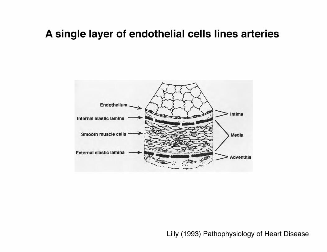
A single layer of endothelial cells lines arteries

Lilly (1993) Pathophysiology of Heart Disease
Fatty streaks: the earliest visible sign of atherosclerosis
Yellowish discoloration on endothelial surface
Lilly (1993) Pathophysiology of Heart Disease
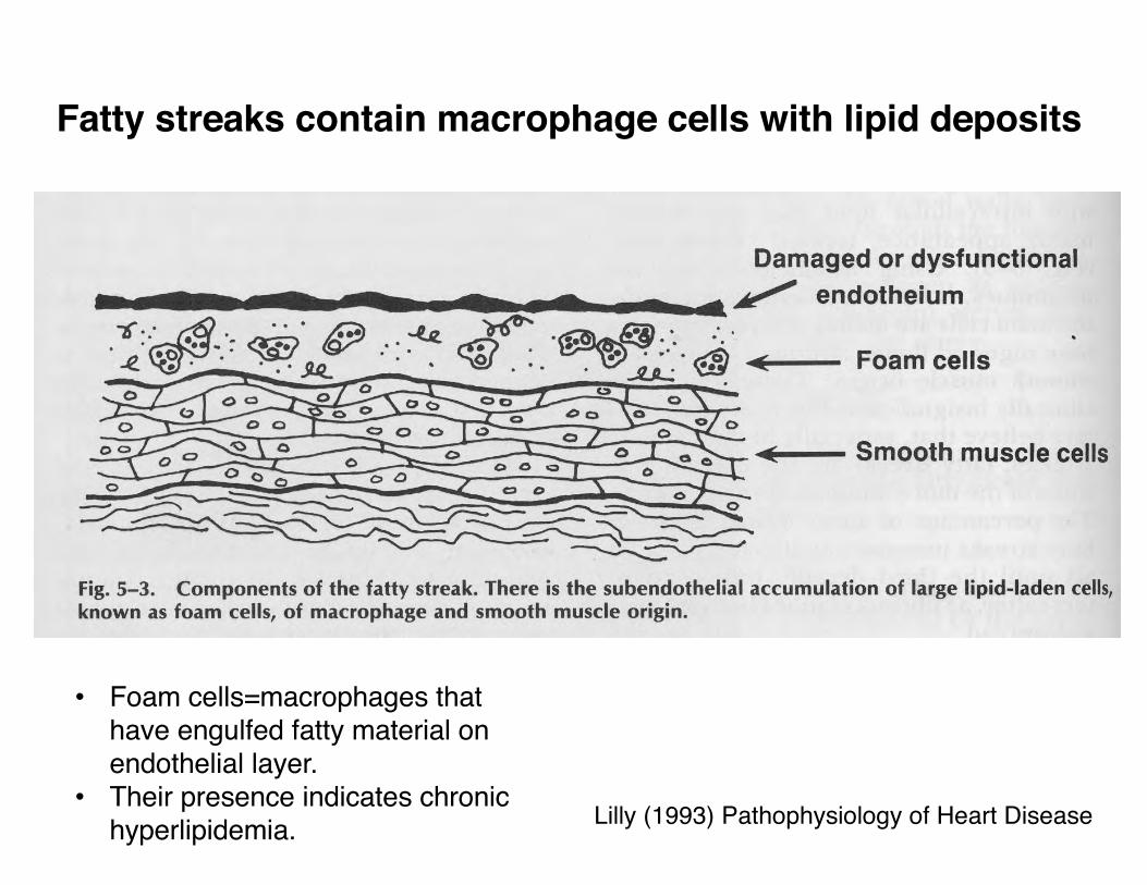
Fatty streaks contain macrophage cells with lipid deposits
• Foam cells=macrophages that have engulfed fatty material on endothelial layer.
• Their presence indicates chronic hyperlipidemia.

Lilly (1993) Pathophysiology of Heart Disease
Fibrous plaque: pathologic atherosclerosis
A swelling in the artery wall made of macrophage cells, debris, lipids, calcium, and connective tissue.Complications:
1) Calcification-rigid and fragile2) Thrombosis
3) Hemorrhage4) Aneurysm (dilatation)
• Myocardial infarction
• Stroke• Claudication

Lilly (1993) Pathophysiology of Heart Disease
Fibrous plaques damage the endothelial layer
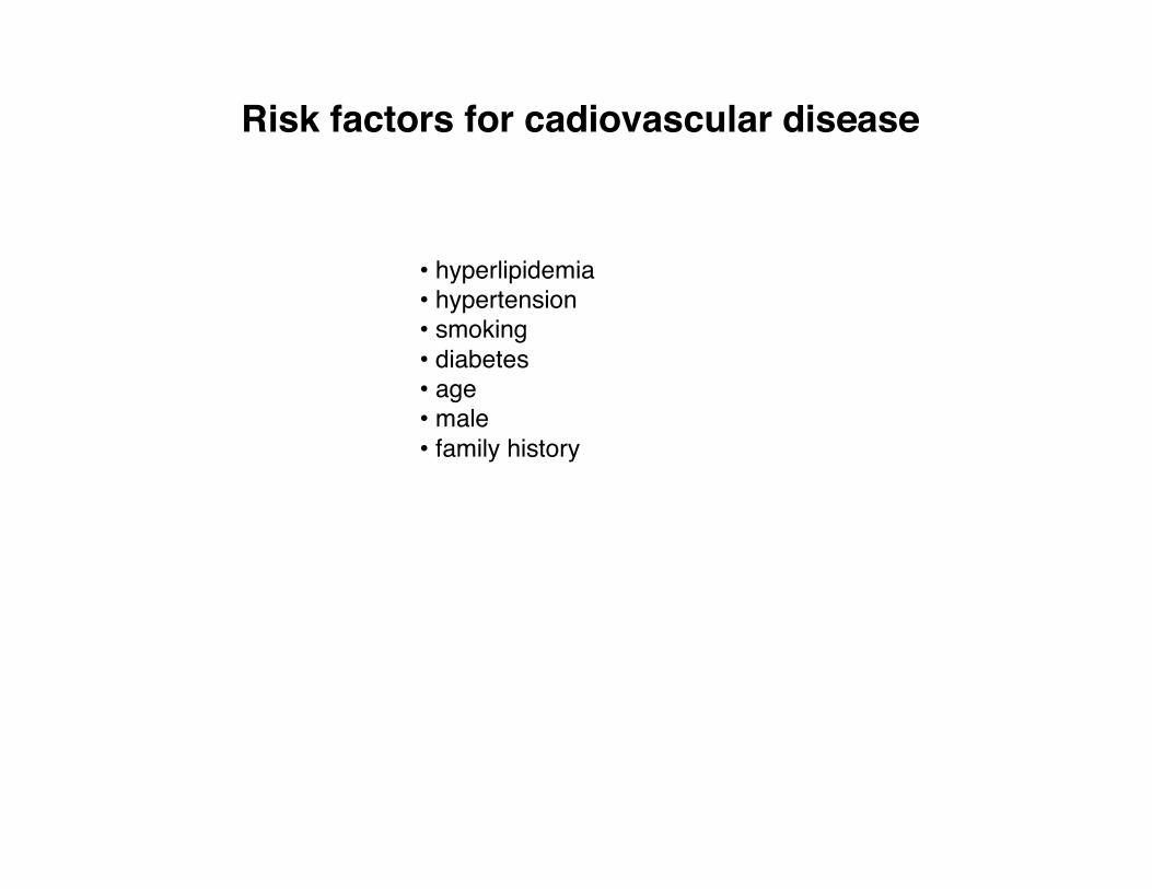
Risk factors for cadiovascular disease
• hyperlipidemia• hypertension• smoking• diabetes• age• male• family history

Brown & Goldstein (1986) Science 232:34
Cholesterol is carried mostly as low density lipoprotein (LDL) particles

HMGCoA controls the rate limiting step in cholesterol synthesis
Zubay, 1998
• Cholesterol is sterol lipid.
• Essential component (30%) of animal cell membrane.
• Precursor for biosynthesis of steroid hormones, bile acid, vitamin D.
• Synthesized by ER of hepatocytes.
• Two thirds of body’s cholesterol is internally synthesized.
Mevalonate is early precursor to cholesterol.
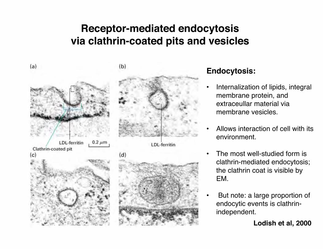
Receptor-mediated endocytosis via clathrin-coated pits and vesicles
Lodish et al, 2000
Endocytosis:
• Internalization of lipids, integral membrane protein, and extraceullar material via membrane vesicles.
• Allows interaction of cell with its environment.
• The most well-studied form is clathrin-mediated endocytosis; the clathrin coat is visible by EM.
• But note: a large proportion of endocytic events is clathrin-independent.

Brown & Goldstein (1986) Science 232:34
LDL receptor recycling pathway: a model system to study receptor-mediated endocytosis
LDL receptor roundtrip: ~10 minutes
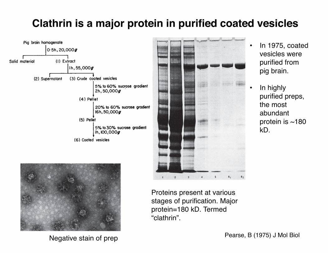
• In 1975, coated vesicles were purified from pig brain.
• In highly purified preps, the most abundant protein is ~180 kD.
Clathrin is a major protein in purified coated vesicles
Negative stain of prep
Proteins present at various stages of purification. Major protein=180 kD. Termed “clathrin”.
Pearse, B (1975) J Mol Biol
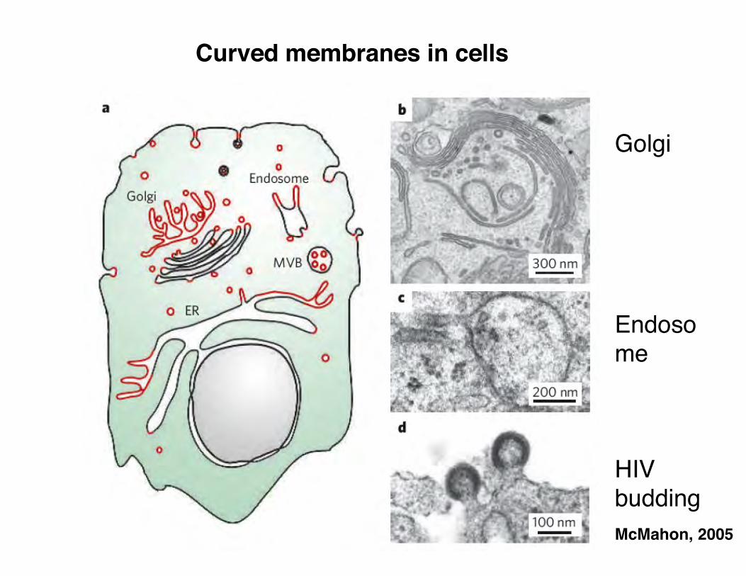
McMahon, 2005
Golgi
Endosome
HIV budding
Curved membranes in cells
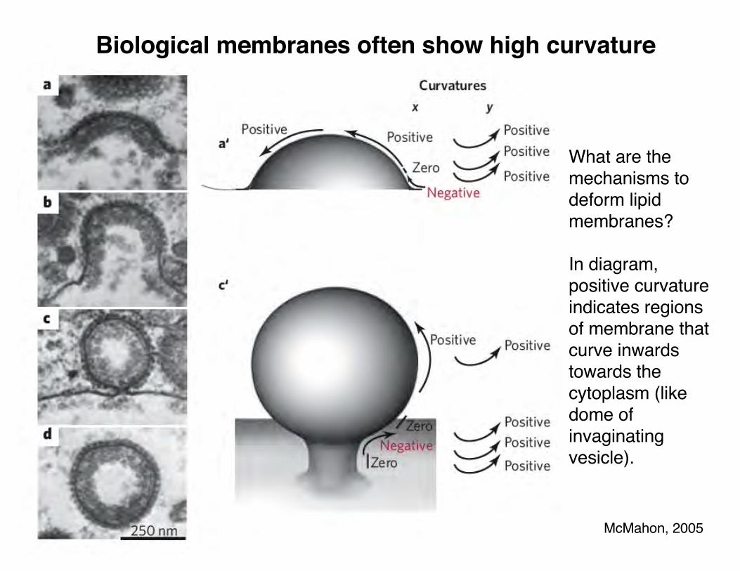
McMahon, 2005
Biological membranes often show high curvature
What are the mechanisms to deform lipid membranes?
In diagram, positive curvature indicates regions of membrane that curve inwards towards the cytoplasm (like dome of invaginatingvesicle).

McMahon, 2005
Ways to impose membrane curvature
Sar1
clathrin and other vesicle coatsdynamin BAR domains
reticulons?
Lipids [e.g., PI(4,5)P2] can also recruit proteins

EM image of clathrin-coated pits from cytosolic side
Lodish et al, 2000
• Budding of vesicles is driven by the polymerization of coat proteins onto the membrane.
• The coat proteins control curvature of the membrane.
• The coat protein helps determine what cargo is carried within the vesicle.
Heuserlab.wustl.edu

Figure 13-7a, b Molecular Biology of the Cell (© Garland Science 2008)
Clathrin is a major vesicle coat protein
• Forms a triskelion shape made of 3 heavy chains and 3 light chains.
• The triskelion is a basic unit forming the polyhedral lattice on the outside of a coated vesicle.
• Triskelions can assembly into a basket.

Fotin et al (2004) Nature
• The clathrin basket has pentagons and hexagons.
• Different size vesicles are possible.
Assembly of clathrin into cages

movie by Allison Bruce
Assembly of clathrin coated vesicle
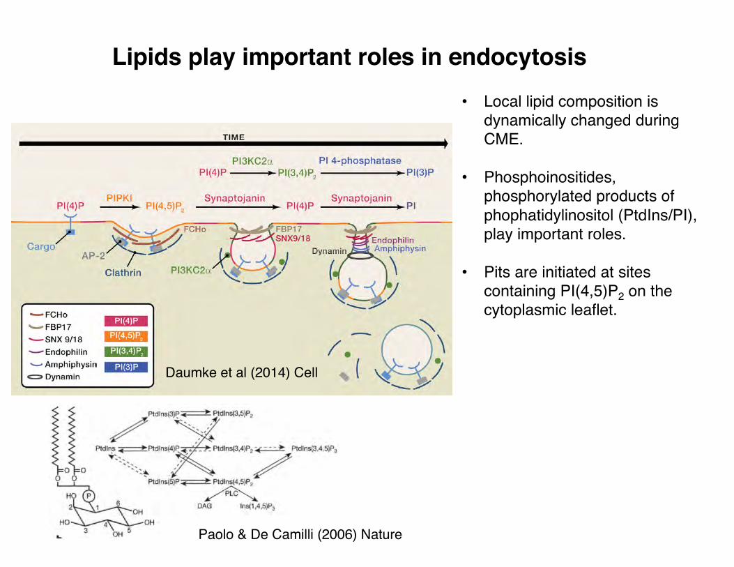
Lipids play important roles in endocytosis
Daumke et al (2014) Cell
• Local lipid composition is dynamically changed during CME.
• Phosphoinositides, phosphorylated products of phophatidylinositol (PtdIns/PI), play important roles.
• Pits are initiated at sites containing PI(4,5)P2 on the cytoplasmic leaflet.
Paolo & De Camilli (2006) Nature

Adaptor proteins bind membranes, clathrin, and cargo
Lodish et al, 2004
1) AP-2 (Adaptor Protein 2) complex is the major clathrin adaptor.2) Nucleation function: mediates binding of clathrin to membrane.3) Contains phospholipid-interacting motifs4) Cargo selection function: Involved in selective recruitment of transmembrane proteins (cargo) into coated pits.5) Heterotetramer: a, b2, µ2, and s2
6) cargo recruitment motifs: •YxxΘ (Θ=bulky hydrophobic)•FxNPxY (in cytoplasmic tail of LDL receptor)•(DE)xxxLL/I) “di-leucine motif”)
7) Crystal structure of µ2 subunit bound to YxxΘ peptide; not accessible in large core structure of core AP-2. Requires conformational change to expose cargo binding domain of µ2.s2 binds dileucine motifs; also requires conformational change.
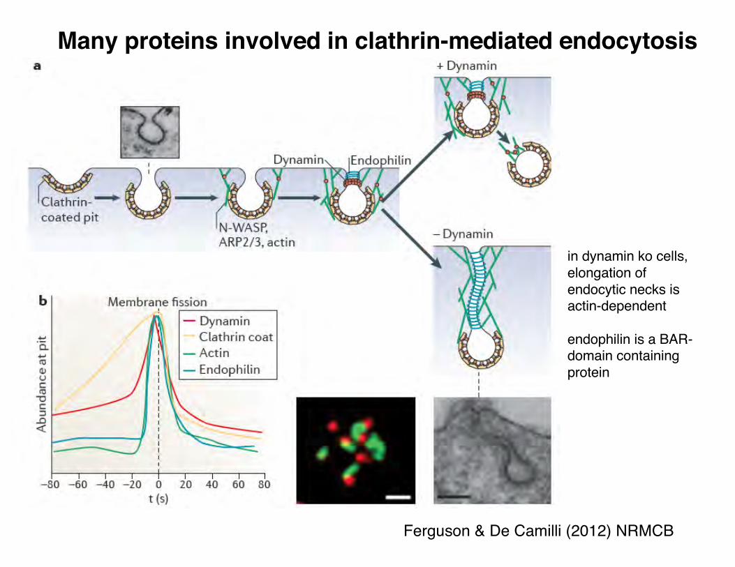
Ferguson & De Camilli (2012) NRMCB
in dynamin ko cells, elongation of endocytic necks is actin-dependent
endophilin is a BAR-domain containing protein
Many proteins involved in clathrin-mediated endocytosis

Merifield, 2004
Studying the timing of endocytic proteins
Zoncu et al (2009) Cell
GFP fusion has PH domain that binds a specific phosphoinositide. RFP fusion has a Rab5 effector.

In addition to clathrin-mediated endocytosis, other types of endocytic events
Doberty &McMahon (2009) Ann Rev Biochem
Caveolae ("little cavities")•Flask-shaped invaginations (60-80 nm diameter).
•Spike-like coat.
•Caveolins are the major structural component; inserted into the cytosolic leaflet; forms large assemblies.
•Invaginations associated with lipid rafts (enriched for cholesterol, glycosphingolipids, GPI-anchored proteins).

What causes scission of the endocytic vesicle?
Lodish et al, 2004
Genetic and biochemical studies implicate the dynamin GTPase

Shibire mutants are paralyzed at high temperatures
Bing Zhang, UT Austin
Praefcke and McMahon, 2004
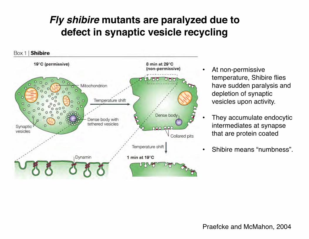
Fly shibire mutants are paralyzed due to defect in synaptic vesicle recycling
• At non-permissive temperature, Shibire flies have sudden paralysis and depletion of synaptic vesicles upon activity.
• They accumulate endocytic intermediates at synapse that are protein coated
• Shibire means “numbness”.

Dynamin: a GTPase enriched on the necks of endocytic vesicles
Lodish et al, 2000
• Dynamin 1: major isoform in brain; dynamin 2: ubiquitous; dynamin 3: brain, testes, lung.
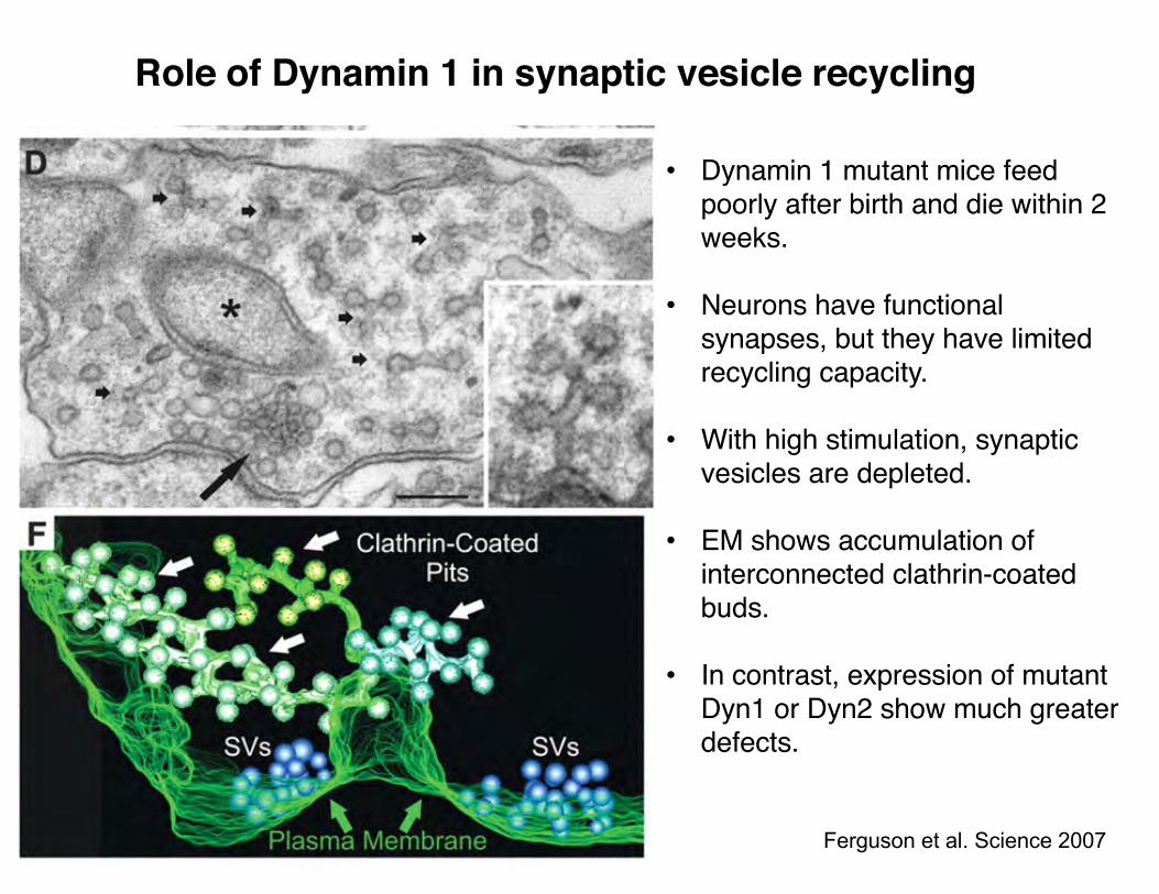
• Dynamin 1 mutant mice feed poorly after birth and die within 2 weeks.
• Neurons have functional synapses, but they have limited recycling capacity.
• With high stimulation, synaptic vesicles are depleted.
• EM shows accumulation of interconnected clathrin-coated buds.
• In contrast, expression of mutant Dyn1 or Dyn2 show much greater defects.
Role of Dynamin 1 in synaptic vesicle recycling
Ferguson et al. Science 2007

Models of the molecular mechanism of dynamin
Model 1: molecular constriction
assembly onliposomes
Model 2: molecular expansion
GTP
Praefcke & McMahon Nature reviews (2004)
Model 3: molecular twisting
Roux et al., Nature (2006)

Dynamin fragments lipid tubules upon addition of GTP
Effect of 1 mM GTP on a netwrk of dynamin-coated lipid tubules. From P. De Camilli
Ferguson & De Camilli (2012)
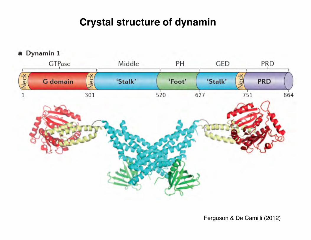
Crystal structure of dynamin
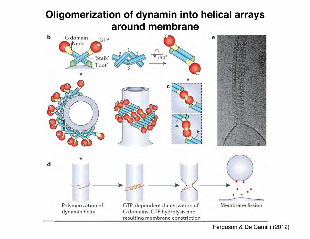
Oligomerization of dynamin into helical arrays around membrane
Ferguson & De Camilli (2012)
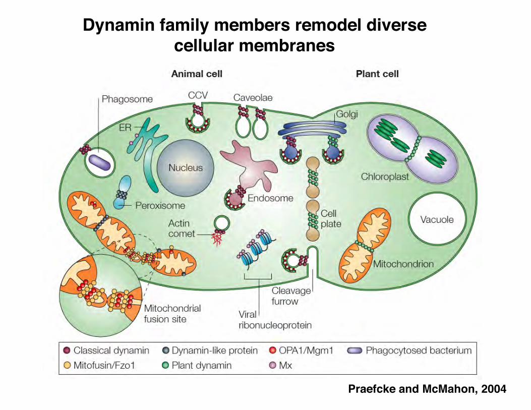
Praefcke and McMahon, 2004
Dynamin family members remodel diverse cellular membranes
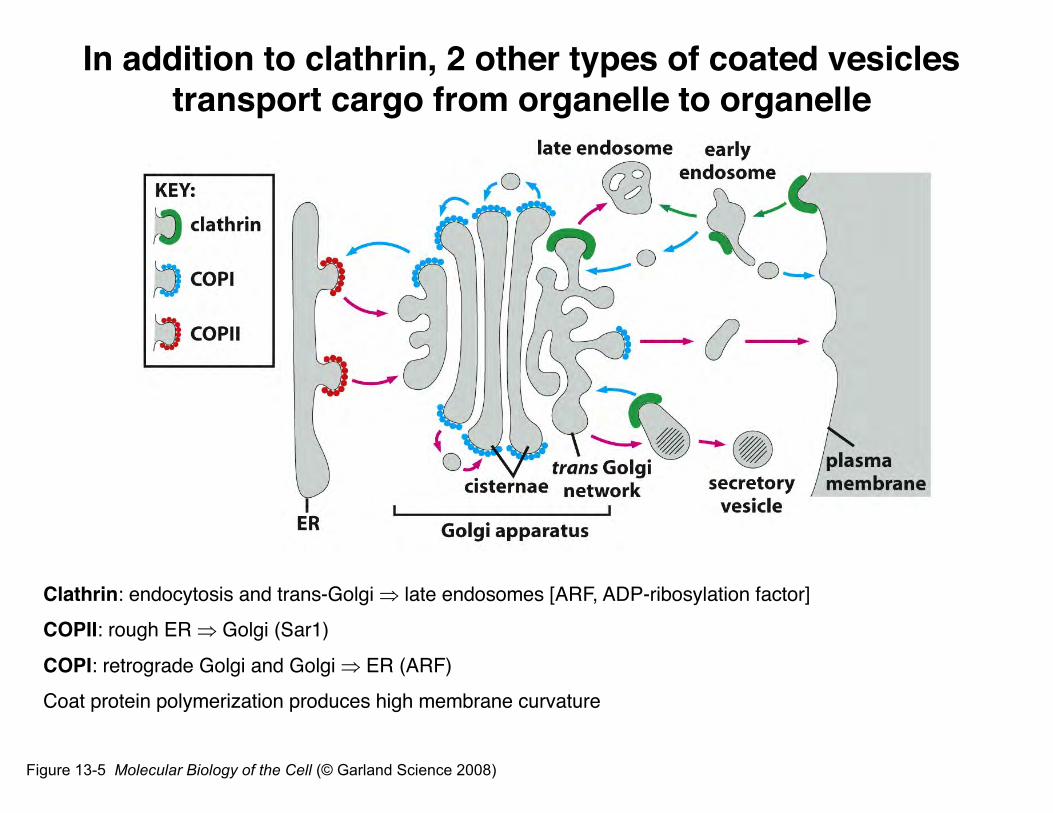
Figure 13-5 Molecular Biology of the Cell (© Garland Science 2008)
Clathrin: endocytosis and trans-Golgi Þ late endosomes [ARF, ADP-ribosylation factor]COPII: rough ER Þ Golgi (Sar1)
COPI: retrograde Golgi and Golgi Þ ER (ARF)Coat protein polymerization produces high membrane curvature
In addition to clathrin, 2 other types of coated vesicles transport cargo from organelle to organelle
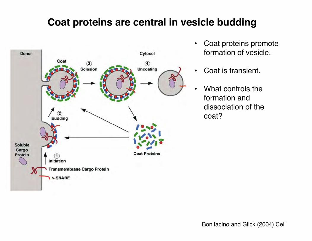
Coat proteins are central in vesicle budding
Bonifacino and Glick (2004) Cell
• Coat proteins promote formation of vesicle.
• Coat is transient.
• What controls the formation and dissociation of the coat?

Small G proteins can regulate timing of molecular interactions
Doberty &McMahon (2009) Ann Rev Biochem
G proteins appear to regulate membrane curvature.
•direct affects on membrane curvature.
•recruitment of coat proteins.
•Example: Sar1 is involved in formation of COPII vesicles.
Lodish et al, 2004
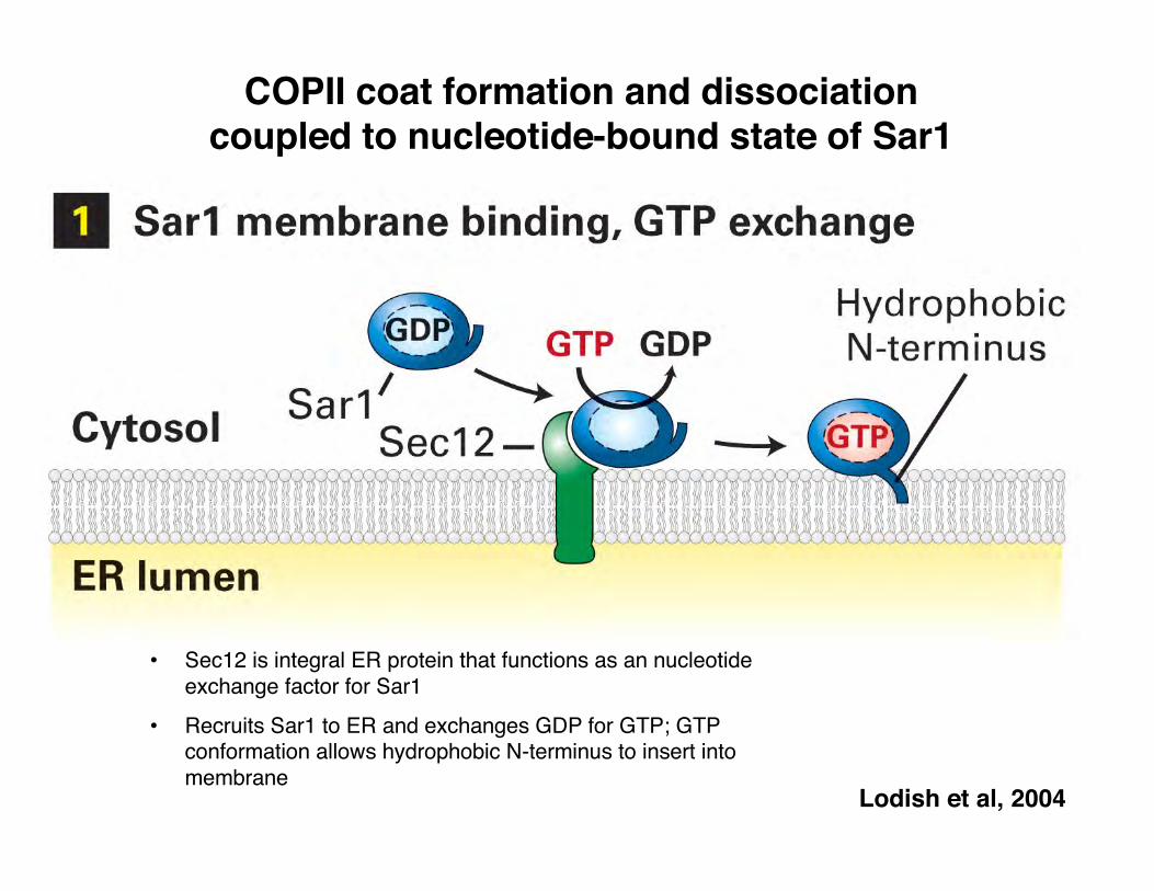
• Sec12 is integral ER protein that functions as an nucleotide exchange factor for Sar1
• Recruits Sar1 to ER and exchanges GDP for GTP; GTP conformation allows hydrophobic N-terminus to insert into membrane
COPII coat formation and dissociation coupled to nucleotide-bound state of Sar1

Lodish et al, 2004
• Sar1-GTP recruits COPII coat;
Coat recruits cargo

Lodish et al, 2004
• Sar1 hydrolysis (promoted by Sec 23 coat subunit) disassembles coat
Summary: Through Sar1, GTP binding recruits vesicle coat, and GTP hydrolysis dissociates coat. The GTPase cycle controls the timing.

Lodish et al, 2004

The Sar1 GTPase can tubulate synthetic liposomes
Lee et al. (2005) Cell
A)• Sar1 (alone) tubulatesmembranes in presence of GTP.
• Average diameter ~26 nm; length up to 1 µm long.
• COPII vesicle ~60-90 nm.
B) GDPC) no Sar1

The Sar1 and COPII coat proteins cooperate
Lee et al. (2005) Cell
• Wt Sar1 reactions contain more vesicles, and tubules.
• D23-Sar1 reactions contain buds. (this Sar1 mutant previously shown to be unable to tubulate membranes)
• suggests that fission is facilitated by Sar1.
COPII consists of Sec23/24 and Sec 13/31
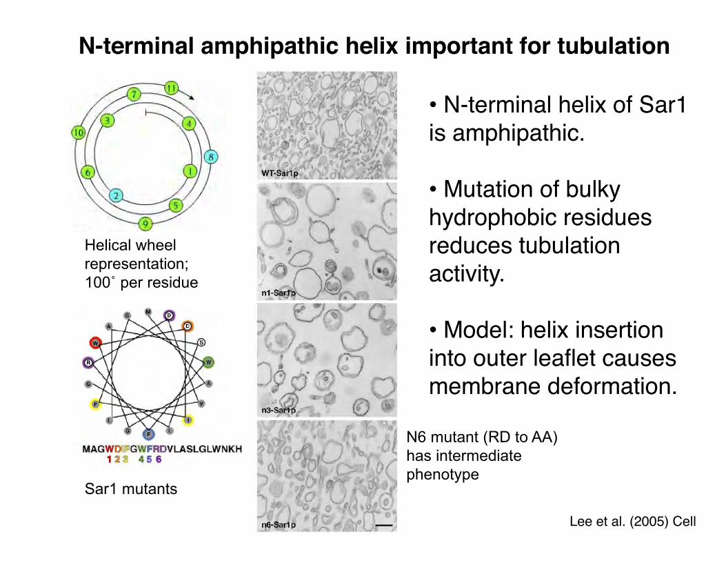
N-terminal amphipathic helix important for tubulation
Lee et al. (2005) Cell
• N-terminal helix of Sar1 is amphipathic.
• Mutation of bulky hydrophobic residues reduces tubulationactivity.
• Model: helix insertion into outer leaflet causes membrane deformation.
Helical wheel representation;100˚ per residue
Sar1 mutants
N6 mutant (RD to AA) has intermediate phenotype