Ovarian tumors I
75
OVARIAN TUMORS-I Dr Aksharaditya Shukla Resident, Department Of Pathology MGM Medical College & M.Y. Hospital, Indore
-
Upload
aksharaditya-shukla -
Category
Health & Medicine
-
view
71 -
download
1
Transcript of Ovarian tumors I
OVARIAN TUMORS-I
Dr Aksharaditya ShuklaResident, Department Of Pathology
MGM Medical College & M.Y. Hospital, Indore
Dr Aksharaditya Shukla
Ovarian tumours
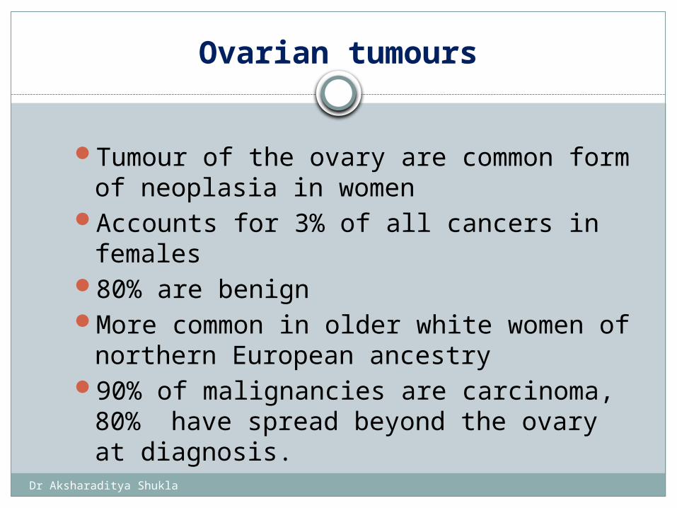
Tumour of the ovary are common form of neoplasia in women
Accounts for 3% of all cancers in females80% are benignMore common in older white women of
northern European ancestry90% of malignancies are carcinoma, 80%
have spread beyond the ovary at diagnosis.
Dr Aksharaditya Shukla
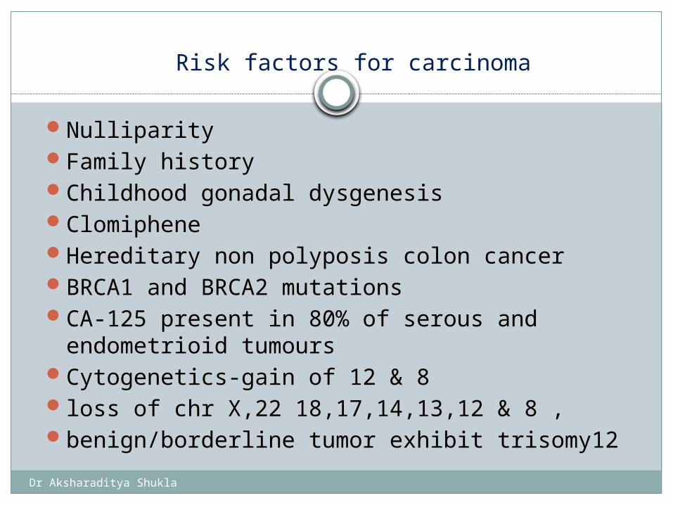
Risk factors for carcinoma
NulliparityFamily historyChildhood gonadal dysgenesisClomipheneHereditary non polyposis colon cancerBRCA1 and BRCA2 mutationsCA-125 present in 80% of serous and
endometrioid tumoursCytogenetics-gain of 12 & 8loss of chr X,22 18,17,14,13,12 & 8 ,benign/borderline tumor exhibit trisomy12
Dr Aksharaditya Shukla
Dr Aksharaditya Shukla
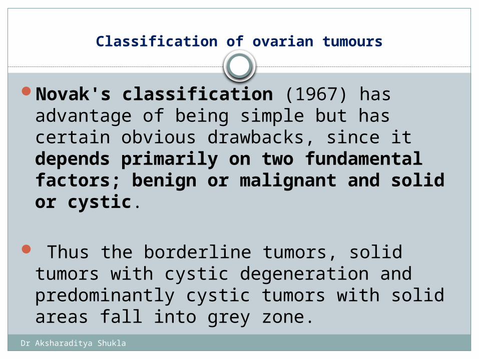
Classification of ovarian tumours
Novak's classification (1967) has advantage of being simple but has certain obvious drawbacks, since it depends primarily on two fundamental factors; benign or malignant and solid or cystic.
Thus the borderline tumors, solid tumors with cystic degeneration and predominantly cystic tumors with solid areas fall into grey zone.
Dr Aksharaditya Shukla
In 1971, the cancer committee of International Federation of Gynecology and Obstetrics (FIGO) proposed a histological classification of common primary epithelial ovarian tumors. Although this classification covered only epithelial tumors, it was a step in the direction of uniformity in classification and it also included the group of tumors of "low potential malignancy".
A significant stride in the direction of a histogenesis-based classification system was made in 1973 with the publication of the World Health Organization (WHO) Classification of Ovarian Tumors. This classification system was updated in 1999 and recently in 2003.
Dr Aksharaditya Shukla
WHO classification of ovarian tumours
1. SURFACE EPITHELIAL TUMOURS2. GERM CELL TUMOURS3. SEX CORD STROMAL TUMOURS 4. GERM CELL SEX CORD STROMAL TUMOURS 5. TUMOUR OF THE RETE OVARII 6. MISCELLANEOUS TUMOURS 7. TUMOUR LIKE CONDITIONS8. LYMPHOID AND HEMATOPOETIC TUMOURS9. SECONDARY TUMOURS
Dr Aksharaditya Shukla
1. Serous tumours2. Mucinous tumours 3. Endometroid tumours including variants of
squamous differentiation4. Clear cell tumours 5. Transitional tumours6. Squamous cell tumours 7. Mixed epithelial tumours 8. Undifferentiated and unclassified tumours.
SURFACE EPITHELIAL TUMOURS
Dr Aksharaditya Shukla
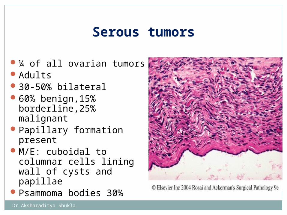
¼ of all ovarian tumorsAdults30-50% bilateral60% benign,15%
borderline,25% malignantPapillary formation
presentM/E: cuboidal to columnar
cells lining wall of cysts and papillae
Psammoma bodies 30%
Serous tumors
Dr Aksharaditya Shukla
BENIGNa) Cystadenoma b) Papillary cystadenomac) Surface papilloma d) Adenofibroma and cystadenofibroma BORDERLINEa) Papillary cystic
tumour b) Surface papillary
tumour.c) Cystadenofibroma
MALIGNANTa) Adenocarcinoma
b) Surface papillary
carcinomac) Adenocarcinofibroma
SURFACE EPITHELIAL TUMOURS (SEROUS TUMORS)
Dr Aksharaditya Shukla
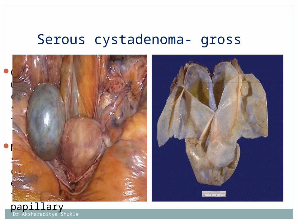
Cystic masses usually unilocular, containg clear but sometimes viscid fluid
Multiloculated smooth glistening cyst wall with no epithelial thickening or papillary
Serous cystadenoma- gross
Dr Aksharaditya Shukla
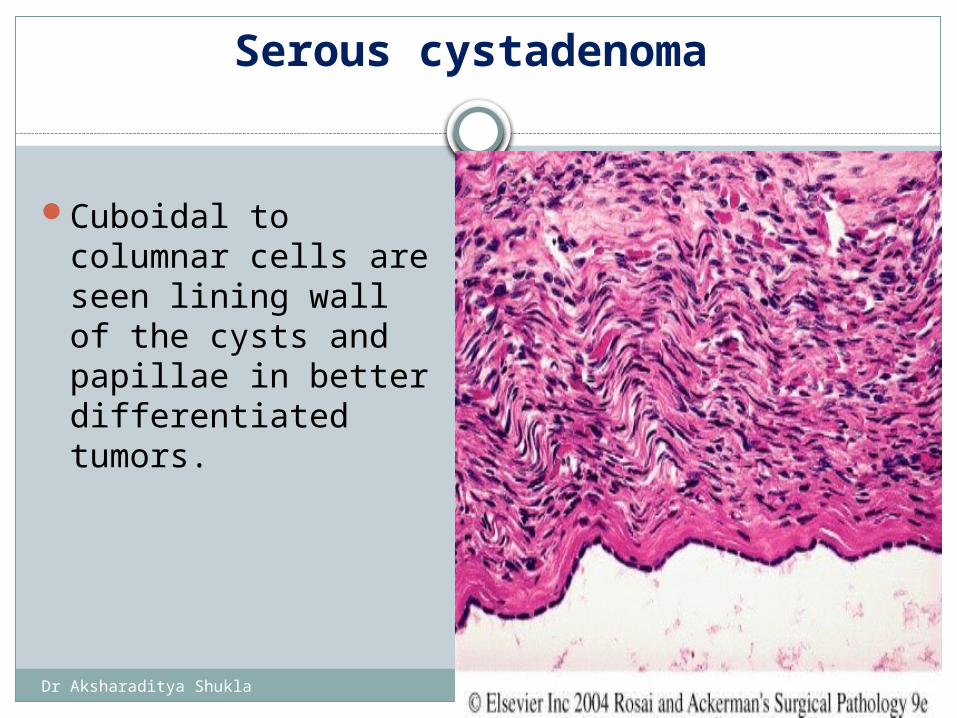
Serous cystadenoma
Cuboidal to columnar cells are seen lining wall of the cysts and papillae in better differentiated tumors.
Dr Aksharaditya Shukla
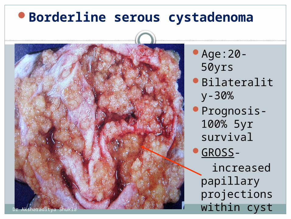
Borderline serous cystadenoma
Age:20-50yrs
Bilaterality-30%
Prognosis-100% 5yr survival
GROSS- increased
papillary projections within cyst
Dr Aksharaditya Shukla
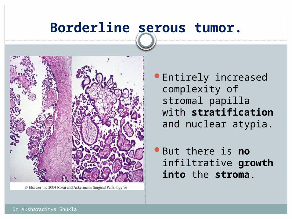
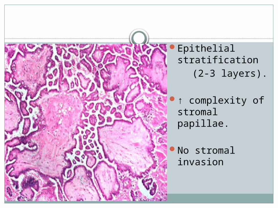
Borderline serous tumor.
Entirely increased complexity of stromal papilla with stratification and nuclear atypia.
But there is no
infiltrative growth into the stroma.
Dr Aksharaditya Shukla
Epithelial stratification
(2-3 layers).
↑ complexity of stromal papillae.
No stromal invasion
Dr Aksharaditya Shukla
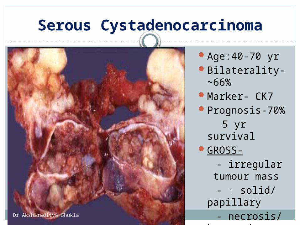
Serous Cystadenocarcinoma
Age:40-70 yrBilaterality-
~66%Marker- CK7Prognosis-70% 5 yr survivalGROSS- - irregular
tumour mass - ↑ solid/
papillary - necrosis/
haemorrhage
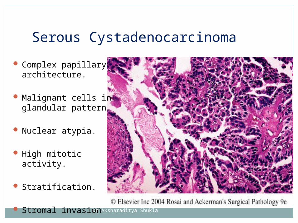
Complex papillary architecture.
Malignant cells in glandular pattern.
Nuclear atypia.
High mitotic activity.
Stratification.
Stromal invasion
Serous Cystadenocarcinoma
Dr Aksharaditya Shukla
Dr Aksharaditya Shukla
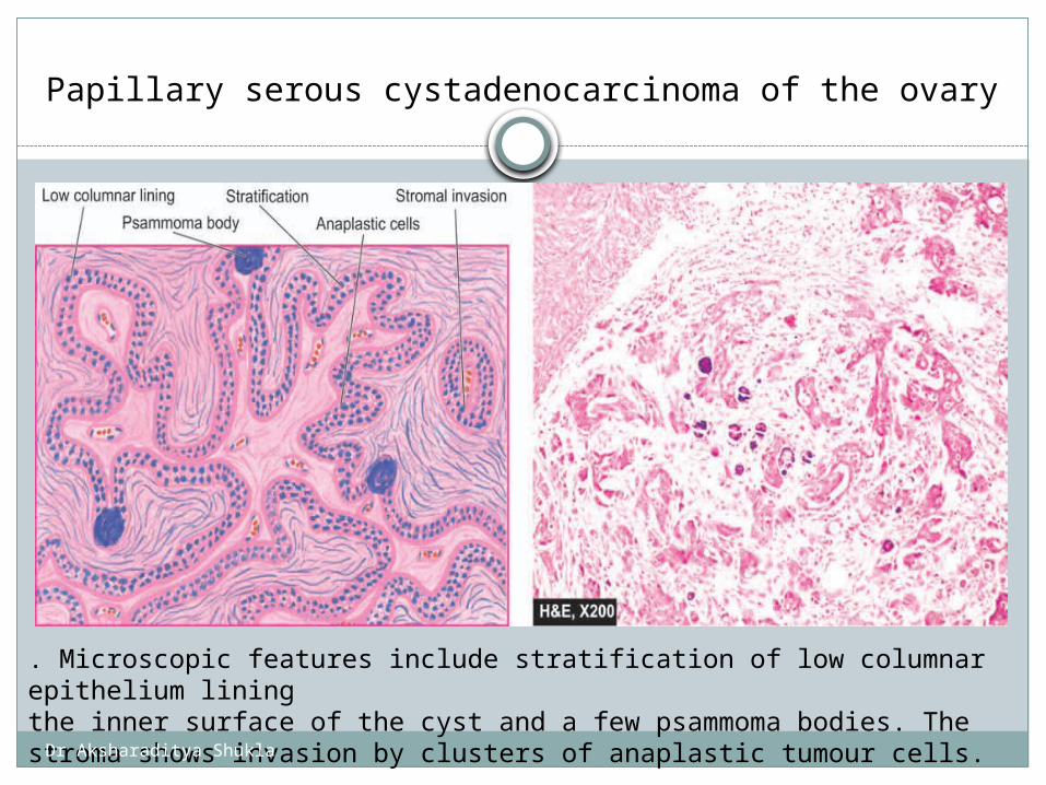
Papillary serous cystadenocarcinoma of the ovary
. Microscopic features include stratification of low columnar epithelium liningthe inner surface of the cyst and a few psammoma bodies. The stroma shows invasion by clusters of anaplastic tumour cells.
Dr Aksharaditya Shukla
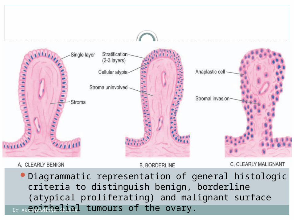
Diagrammatic representation of general histologic criteria to distinguish benign, borderline (atypical proliferating) and malignant surface epithelial tumours of the ovary.
Dr Aksharaditya Shukla
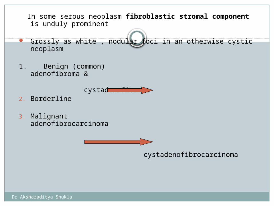
In some serous neoplasm fibroblastic stromal component is unduly prominent
Grossly as white , nodular foci in an otherwise cystic neoplasm
1. Benign (common) adenofibroma &
cystadenofibroma
2. Borderline
3. Malignant adenofibrocarcinoma
cystadenofibrocarcinoma
Dr Aksharaditya Shukla
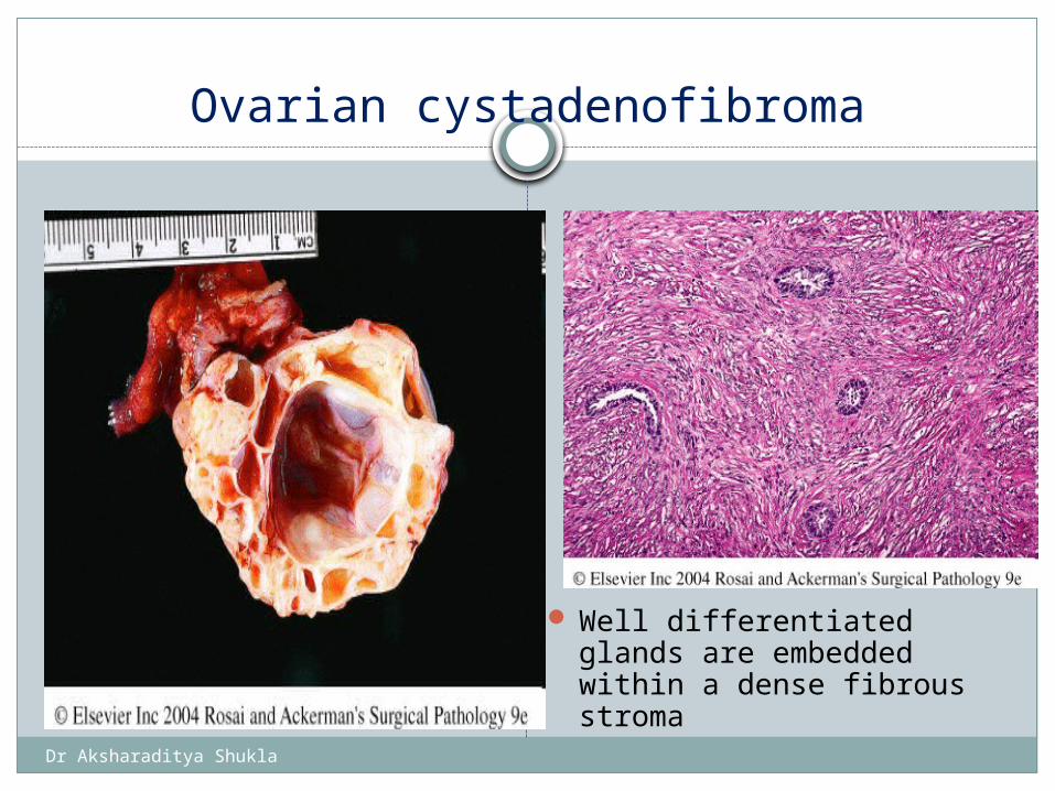
Ovarian cystadenofibroma
Well differentiated glands are embedded within a dense fibrous stroma
Dr Aksharaditya Shukla
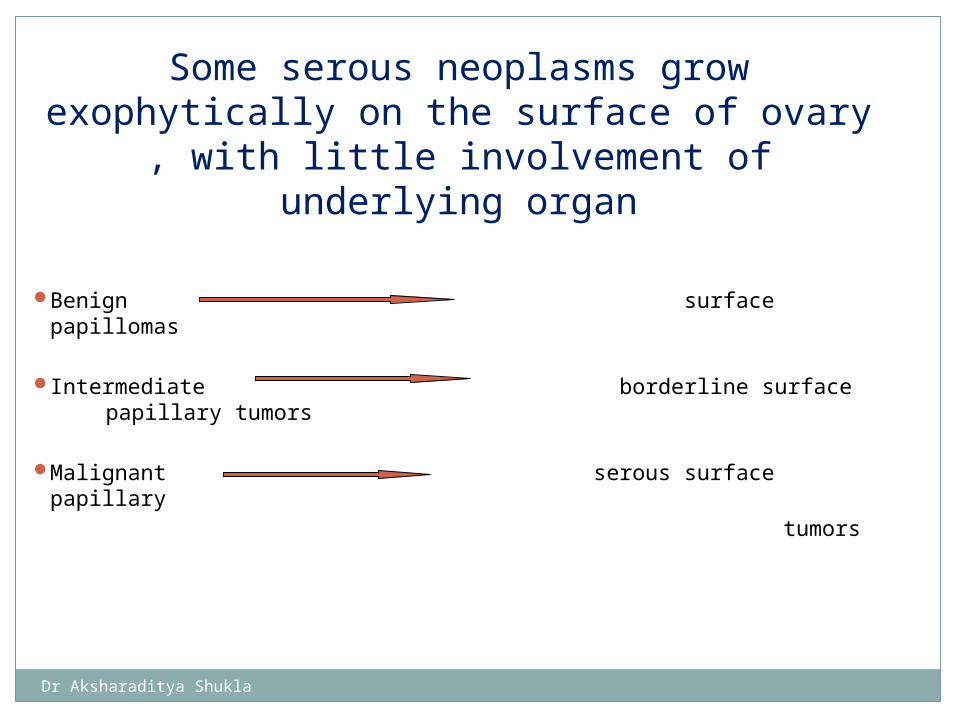
Benign surface papillomas
Intermediate borderline surface papillary tumors
Malignant serous surface papillary
tumors
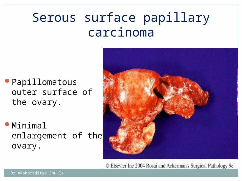
Some serous neoplasms grow exophytically on the surface of ovary , with little involvement of underlying organ
Dr Aksharaditya Shukla
Papillomatous outer surface of the ovary.
Minimal enlargement of the ovary.
Serous surface papillary carcinoma
Dr Aksharaditya Shukla
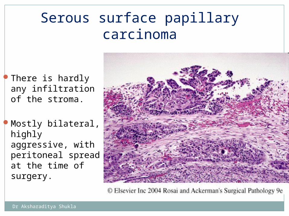
Serous surface papillary carcinoma
There is hardly any infiltration of the stroma.
Mostly bilateral, highly aggressive, with peritoneal spread at the time of surgery.
Dr Aksharaditya Shukla
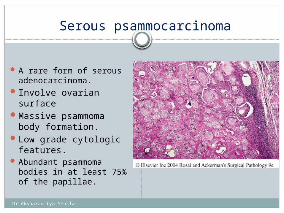
Serous psammocarcinoma
A rare form of serous adenocarcinoma.
Involve ovarian surfaceMassive psammoma
body formation.Low grade cytologic
features. Abundant psammoma
bodies in at least 75% of the papillae.
Dr Aksharaditya Shukla
Immunohistochemistry of serous tumors
keratin profile
CK 7+/ CK20-
Also CK8, CK18, CK19, EMA, S100
WT-1 stains diffusely most serous carcinomas
Dr Aksharaditya Shukla
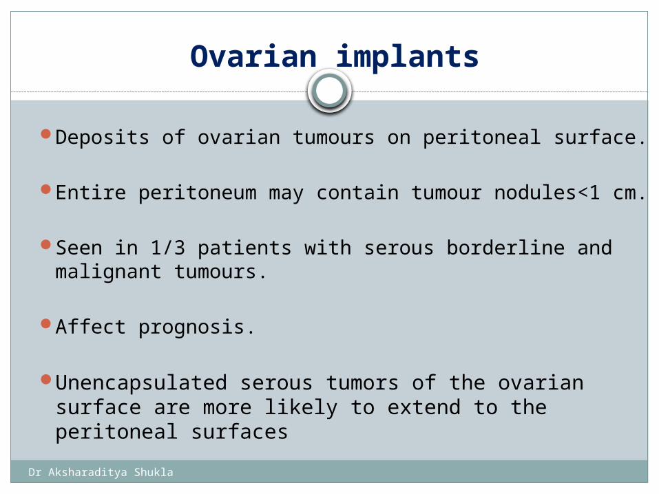
Ovarian implants
Deposits of ovarian tumours on peritoneal surface.
Entire peritoneum may contain tumour nodules<1 cm.
Seen in 1/3 patients with serous borderline and malignant tumours.
Affect prognosis.
Unencapsulated serous tumors of the ovarian surface are more likely to extend to the peritoneal surfaces
Dr Aksharaditya Shukla
Less common. About 25%.
Bilateral 10%-20% (clonal).
80% are benign or borderline type.
MUCINOUS TUMORS
Dr Aksharaditya Shukla
BENIGNa) cystadenoma b) adenofibroma and c) cystadenofibroma BORDERLINEa) intestinal type b) endocervical type MALIGNANT a) adenocarcinoma b) adenocarcinofibroma
MUCINOUS CYSTIC TUMOUR WITH MURAL NODULES
MUCINOUS CYSTIC TUMOUR WITH PSEUDOMYXOMA
PERITONEI
SURFACE EPITHELIAL TUMOURS (MUCINOUS TUMORS)
Dr Aksharaditya Shukla
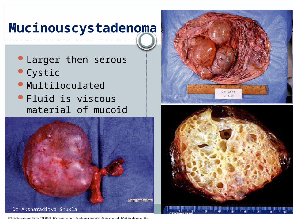
Mucinouscystadenoma
Larger then serousCysticMultiloculatedFluid is viscous
material of mucoid nature present.
Dr Aksharaditya Shukla
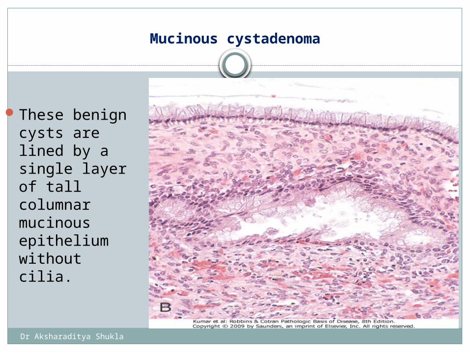
Mucinous cystadenoma
These benign cysts are lined by a single layer of tall columnar mucinous epithelium without cilia.
Dr Aksharaditya Shukla
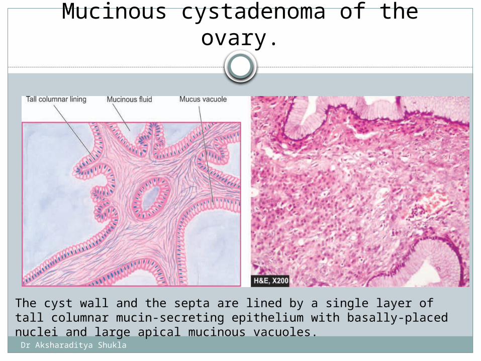
Mucinous cystadenoma of the ovary.
The cyst wall and the septa are lined by a single layer of tall columnar mucin-secreting epithelium with basally-placed nuclei and large apical mucinous vacuoles.
Dr Aksharaditya Shukla
Intestinal type (80%)
Endocervical type (20%)
Borderline mucinous tumors
Dr Aksharaditya Shukla
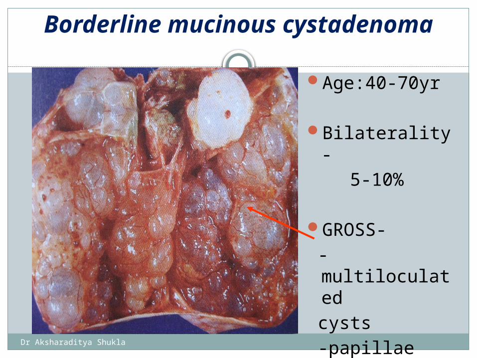
Borderline mucinous cystadenoma
Age:40-70yr
Bilaterality- 5-10%
GROSS- -multiloculated cysts -papillae
Dr Aksharaditya Shukla
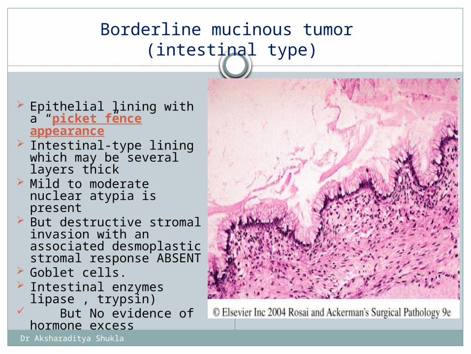
Borderline mucinous tumor (intestinal type)
Epithelial lining with a “picket fence appearance”
Intestinal-type lining which may be several layers thick
Mild to moderate nuclear atypia is present
But destructive stromal invasion with an associated desmoplastic stromal response ABSENT
Goblet cells. Intestinal enzymes
lipase , trypsin) But No evidence of
hormone excess
Lining of mucinous cystadenoma
Dr Aksharaditya Shukla
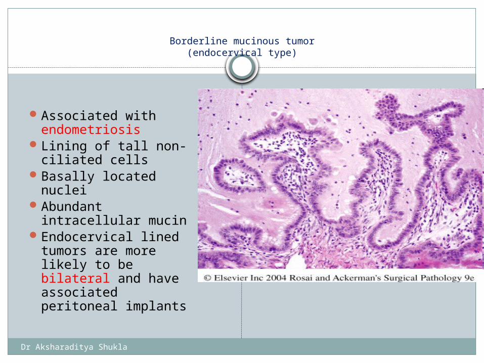
Borderline mucinous tumor(endocervical type)
Associated with endometriosis
Lining of tall non-ciliated cells
Basally located nuclei
Abundant intracellular mucin
Endocervical lined tumors are more likely to be bilateral and have associated peritoneal implants
Dr Aksharaditya Shukla
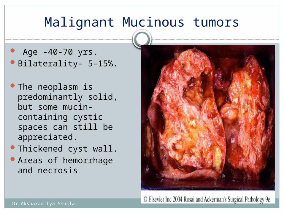
Malignant Mucinous tumors
Age -40-70 yrs.Bilaterality- 5-15%.
The neoplasm is predominantly solid, but some mucin-containing cystic spaces can still be appreciated.
Thickened cyst wall.Areas of hemorrhage
and necrosis
Dr Aksharaditya Shukla
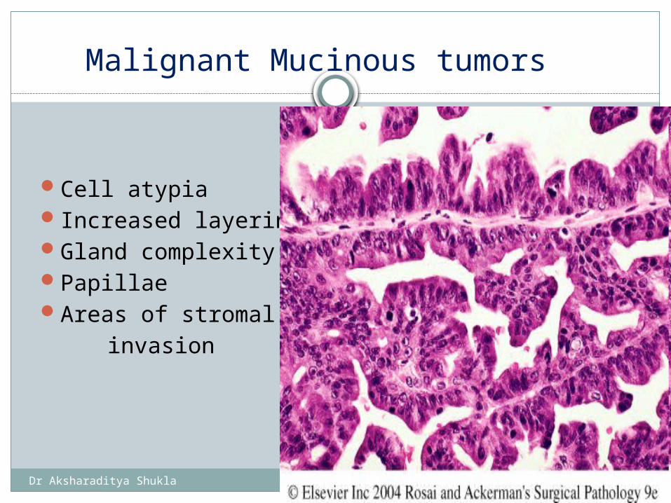
Malignant Mucinous tumors
Cell atypiaIncreased layeringGland complexityPapillaeAreas of stromal invasion
Complex architecture and obvious nuclear atypia in mucinous cystadenoma
Dr Aksharaditya Shukla
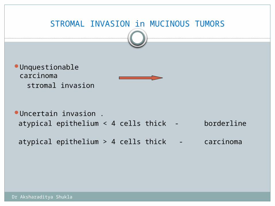
STROMAL INVASION in MUCINOUS TUMORS
Unquestionable carcinoma stromal invasion
Uncertain invasion . atypical epithelium < 4 cells thick - borderline
atypical epithelium > 4 cells thick - carcinoma
Dr Aksharaditya Shukla
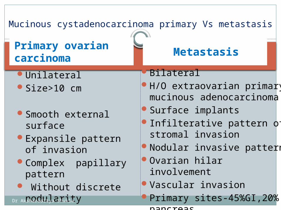
Primary ovarian carcinoma
Metastasis
UnilateralSize>10 cm
Smooth external surface
Expansile pattern of invasion
Complex papillary pattern
Without discrete nodularity
BilateralH/O extraovarian primary
mucinous adenocarcinomaSurface implantsInfilterative pattern of
stromal invasionNodular invasive patternOvarian hilar involvementVascular invasionPrimary sites-45%GI,20%
pancreas,
Mucinous cystadenocarcinoma primary Vs metastasis
Dr Aksharaditya Shukla
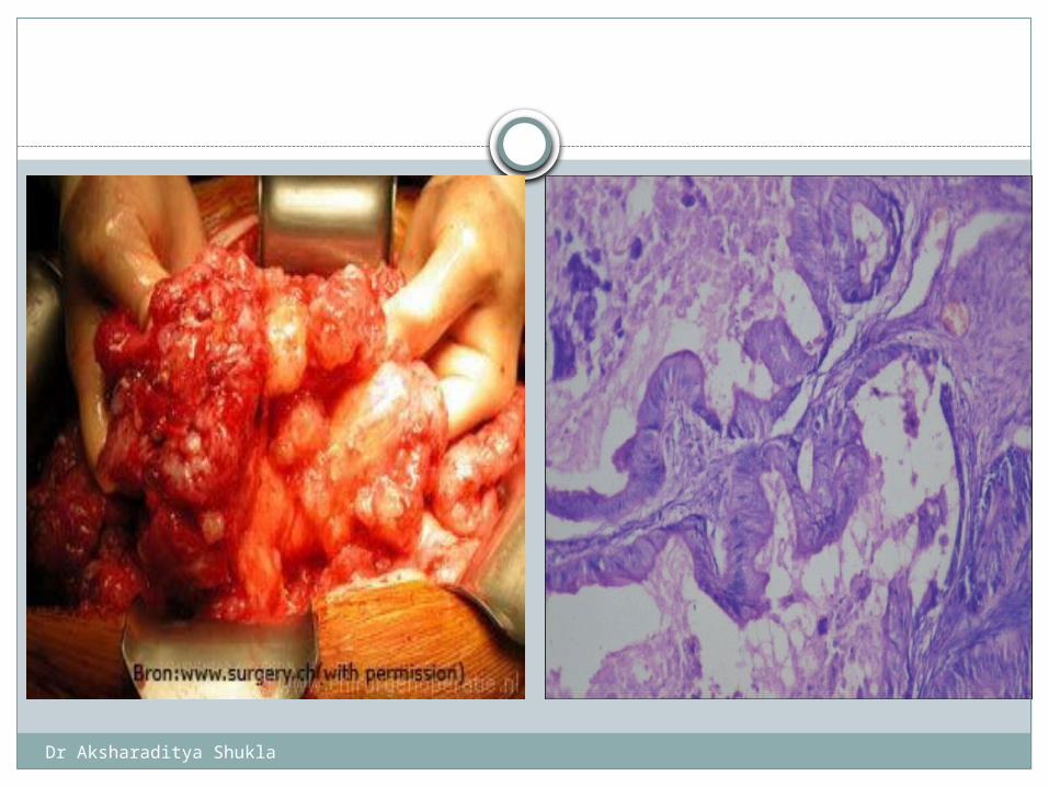
Pseudomyxoma peritonei
Mucinous tumors (like serous tumors) may involve the peritoneal surface with collection of extensive mucinous material resembling cystic contents within the peritoneal cavity.
Is a rare condition Seen with primarily borderline or malignant
neoplasms. Major complication:
Extensive interadherence and adhesion of the viscera, producing a matting together of the abdominal contents and intestinal obstruction
Dr Aksharaditya Shukla
Dr Aksharaditya Shukla
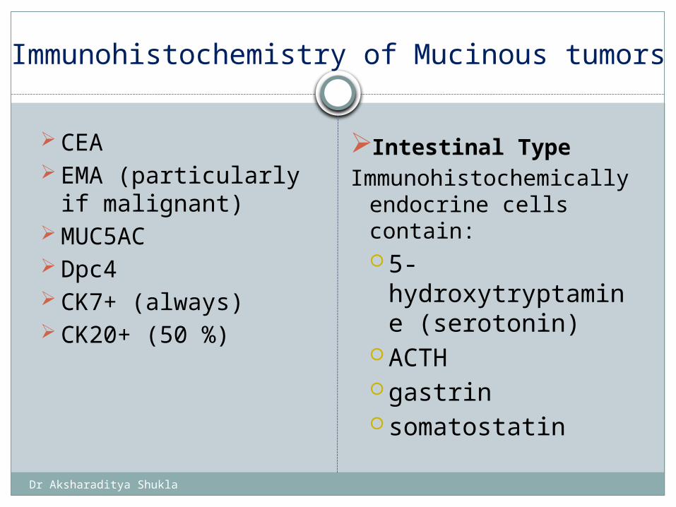
Immunohistochemistry of Mucinous tumors
CEA EMA (particularly if
malignant) MUC5AC Dpc4 CK7+ (always) CK20+ (50 %)
Intestinal TypeImmunohistochemically
endocrine cells contain: 5-
hydroxytryptamine (serotonin)
ACTH gastrin somatostatin
Dr Aksharaditya Shukla
1. Serous tumours2. Mucinous tumours Endometroid tumours including
variants of squamous differentiation3. Clear cell tumours 4. Transitional tumours5. Squamous cell tumours 6. Mixed epithelial tumours 7. Undifferentiated and unclassified tumours
SURFACE EPITHELIAL TUMOURS
Dr Aksharaditya Shukla
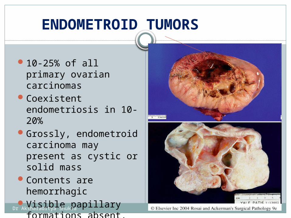
ENDOMETROID TUMORS
10-25% of all primary ovarian carcinomas
Coexistent endometriosis in 10-20%
Grossly, endometroid carcinoma may present as cystic or solid mass
Contents are hemorrhagic
Visible papillary formations absent.
Good prognosis.
Dr Aksharaditya Shukla
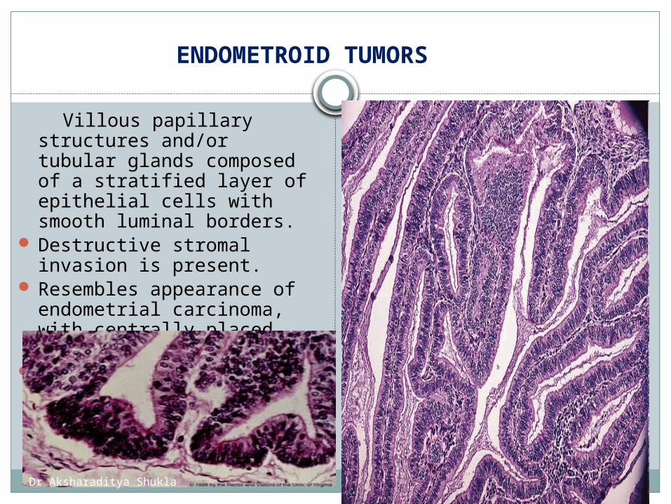
ENDOMETROID TUMORS
Villous papillary structures and/or tubular glands composed of a stratified layer of epithelial cells with smooth luminal borders.
Destructive stromal invasion is present.
Resembles appearance of endometrial carcinoma, with centrally placed nuclei.
Dr Aksharaditya Shukla
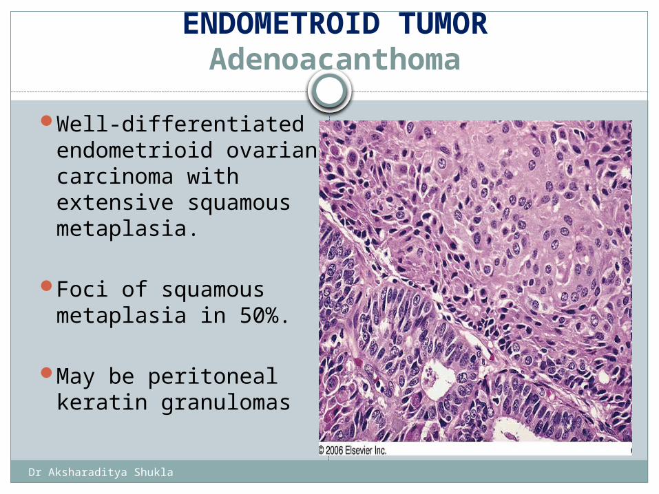
ENDOMETROID TUMORAdenoacanthoma
Well-differentiated endometrioid ovarian carcinoma with extensive squamous metaplasia.
Foci of squamous metaplasia in 50%.
May be peritoneal keratin granulomas
Well-differentiated endometrioid ovarian carcinoma with extensive squamous metaplasia
Dr Aksharaditya Shukla
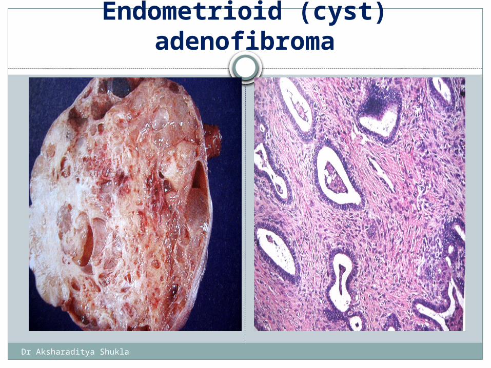
Endometrioid (cyst) adenofibroma
Dr Aksharaditya Shukla
Immunohistochemistry of endometroid carcinoma
Keratin
EMA
Vimentin
CEA usually negative or weak
Dr Aksharaditya Shukla
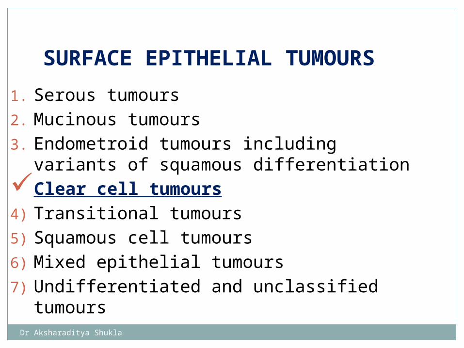
1. Serous tumours2. Mucinous tumours 3. Endometroid tumours including variants
of squamous differentiationClear cell tumours 4) Transitional tumours5) Squamous cell tumours 6) Mixed epithelial tumours 7) Undifferentiated and unclassified tumours
SURFACE EPITHELIAL TUMOURS
Dr Aksharaditya Shukla
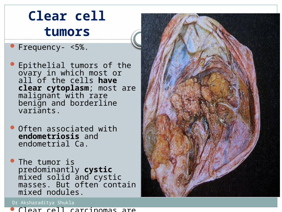
Clear cell tumors Frequency- <5%.
Epithelial tumors of the ovary in which most or all of the cells have clear cytoplasm; most are malignant with rare benign and borderline variants.
Often associated with endometriosis and endometrial Ca.
The tumor is predominantly cystic mixed solid and cystic masses. But often contain mixed nodules.
Clear cell carcinomas are always high grade. Poor prognosis,
Dr Aksharaditya Shukla
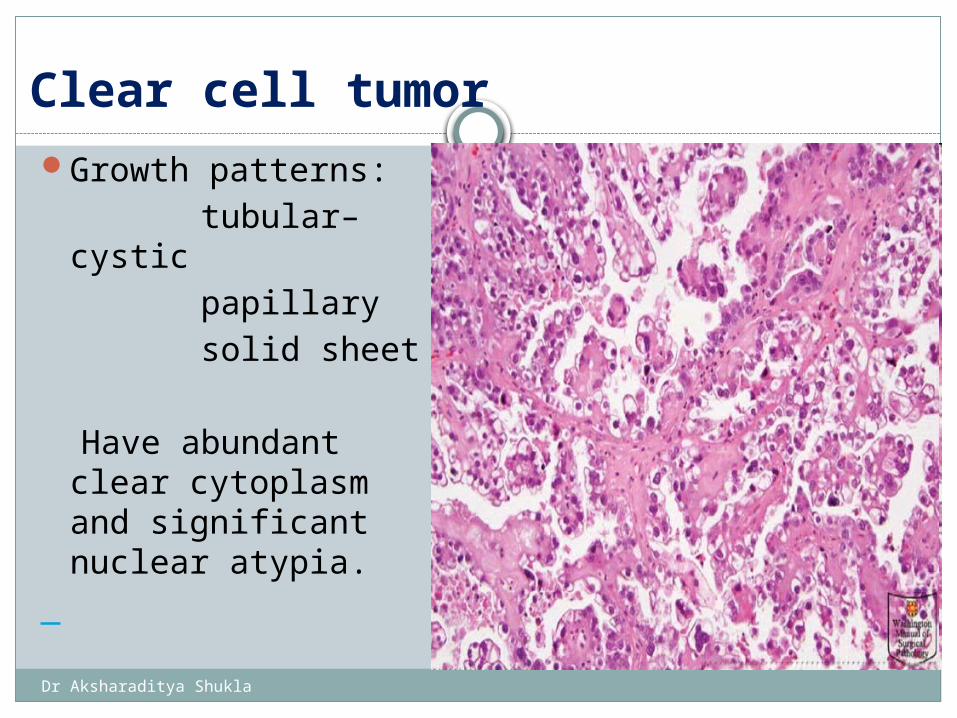
Growth patterns: tubular–cystic papillary solid sheet
Have abundant clear cytoplasm and significant nuclear atypia.
Clear cell tumor
Dr Aksharaditya Shukla
Clear cell tumors
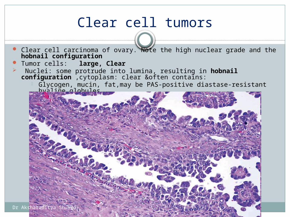
Clear cell carcinoma of ovary. Note the high nuclear grade and the hobnail configuration
Tumor cells: large, Clear Nuclei: some protrude into lumina, resulting in hobnail
configuration ,cytoplasm: clear &often contains: Glycogen, mucin, fat,may be PAS-positive diastase-resistant hyaline
globules
Dr Aksharaditya Shukla
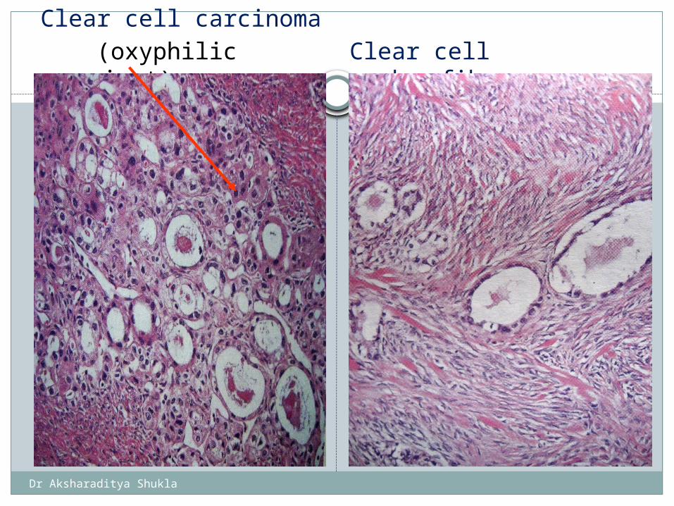
Clear cell carcinoma
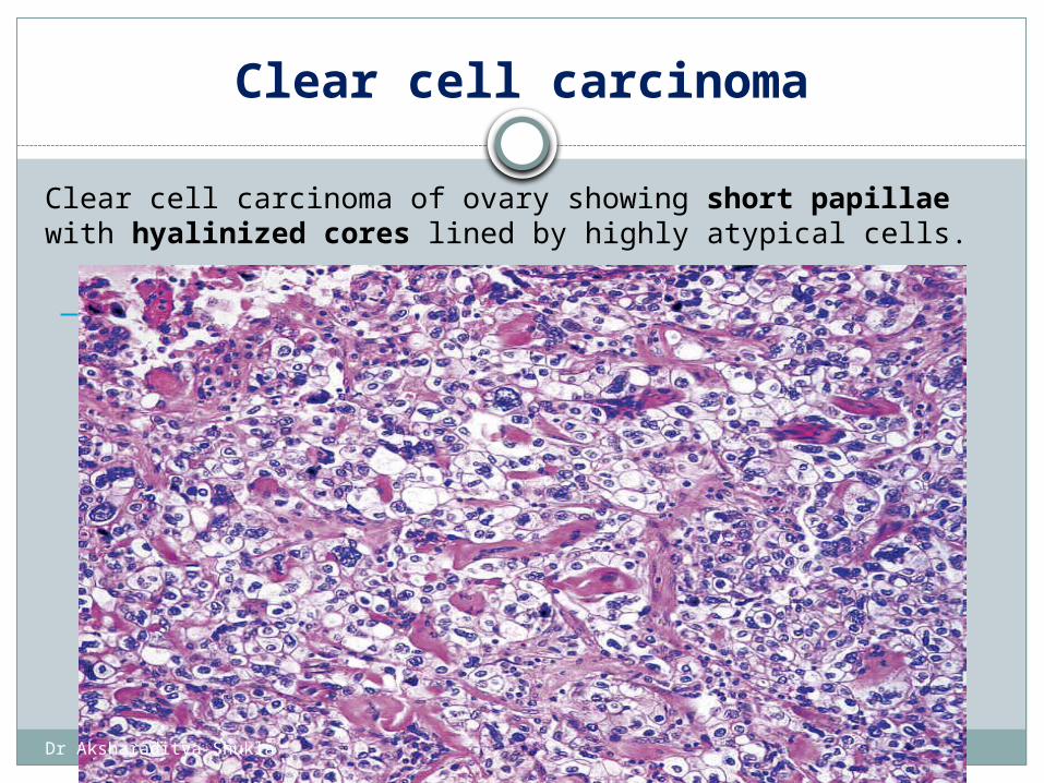
Clear cell carcinoma of ovary showing short papillae with hyalinized cores lined by highly atypical cells.
Dr Aksharaditya Shukla
Clear cell carcinoma (oxyphilic variant) Clear cell adenofibroma
Dr Aksharaditya Shukla
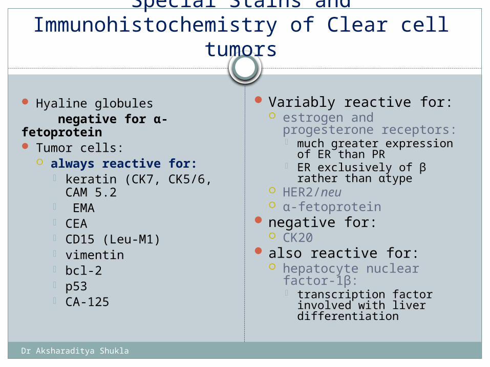
Special Stains and Immunohistochemistry of Clear cell
tumors
Hyaline globules negative for α-fetoprotein Tumor cells:
always reactive for: keratin (CK7, CK5/6, CAM
5.2 EMA CEA CD15 (Leu-M1) vimentin bcl-2 p53 CA-125
Variably reactive for: estrogen and
progesterone receptors: much greater expression of
ER than PR ER exclusively of β rather
than αtype HER2/neu α-fetoprotein
negative for: CK20
also reactive for: hepatocyte nuclear factor-
1β: transcription factor
involved with liver differentiation
Dr Aksharaditya Shukla
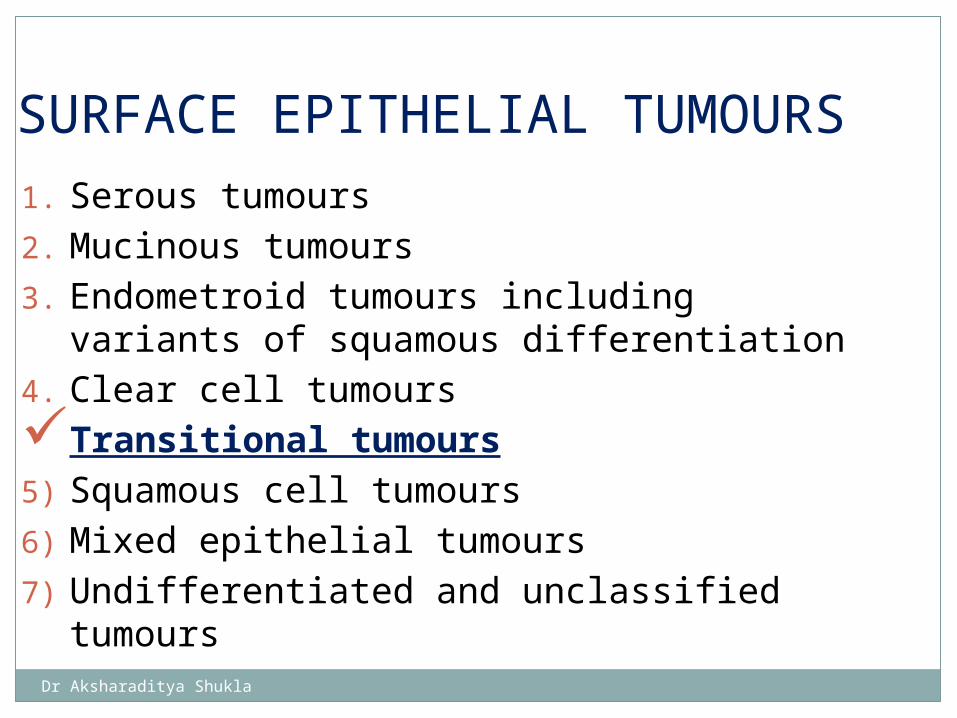
1. Serous tumours2. Mucinous tumours 3. Endometroid tumours including variants
of squamous differentiation4. Clear cell tumours Transitional tumours5) Squamous cell tumours 6) Mixed epithelial tumours 7) Undifferentiated and unclassified tumours
SURFACE EPITHELIAL TUMOURS
Dr Aksharaditya Shukla
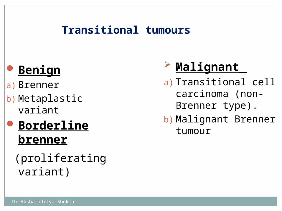
Benigna) Brennerb) Metaplastic variantBorderline
brenner
(proliferating variant)
Malignant a) Transitional cell
carcinoma (non-Brenner type).
b) Malignant Brenner tumour
Transitional tumours
Dr Aksharaditya Shukla
Brenner Tumor and Transitional Cell Carcinoma
Resemble those of transitional cell neoplasms of the urinary tract.
1–2% of all ovarian neoplasms.
Average age at presentation ≈50 years:
Sometimes signs of hyperestronism, such as postmenopausal uterine bleeding from endometrial hyperplasia.
* Slow rate of growth * Rarely ascites
Dr Aksharaditya Shukla
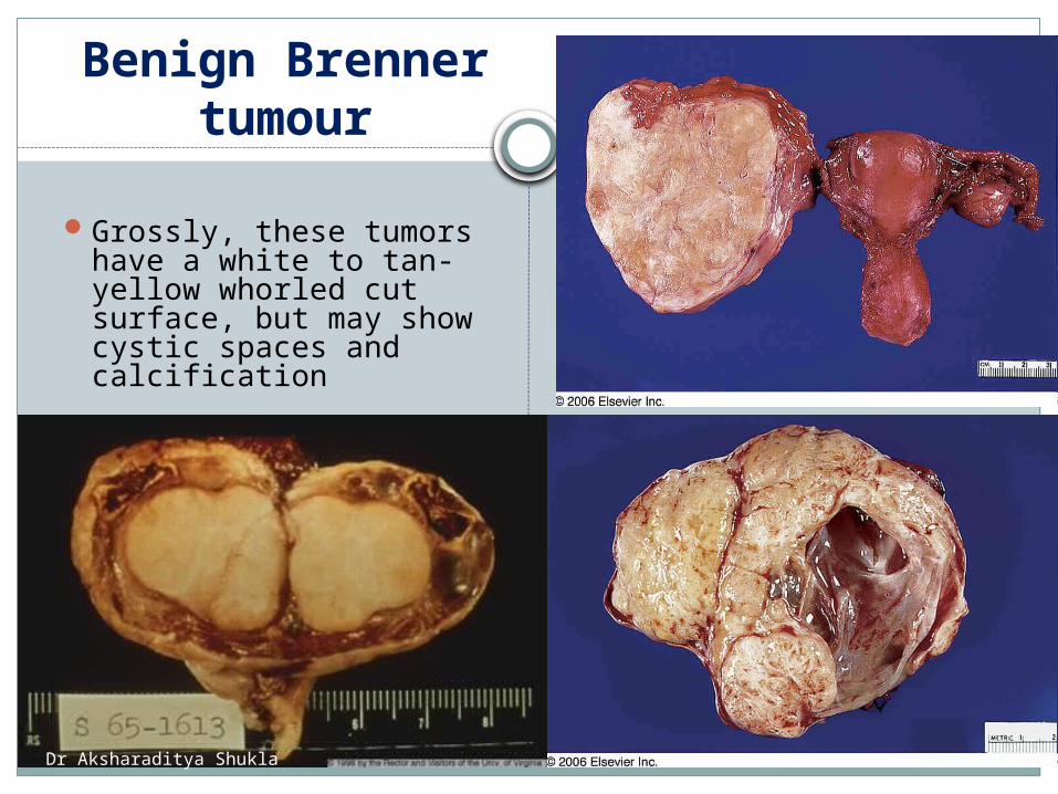
Benign Brenner tumour
Grossly, these tumors have a white to tan-yellow whorled cut surface, but may show cystic spaces and calcification
unilateral firm May be associated with: mucinous cystadenoma exceptionally struma ovarii also transitional cell
tumors of urinary bladder
Dr Aksharaditya Shukla
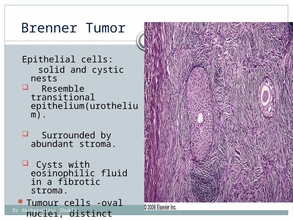
Brenner Tumor
Epithelial cells: solid and cystic nests Resemble transitional
epithelium(urothelium).
Surrounded by abundant stroma.
Cysts with eosinophilic fluid in a fibrotic stroma.
Tumour cells -oval nuclei, distinct nucleolus, longitudinal groove.
Brenner tumor of ovary showing solid and cystic epithelial cells embedded within fibrous tissue.
Dr Aksharaditya Shukla
Metaplastic Brenner tumor
* Unduly prominent cystic formations.
* Florid mucinous changes.
* Papillary fronds and nuclear atypia absent.
Dr Aksharaditya Shukla
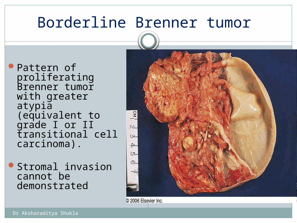
Borderline Brenner tumor
Pattern of proliferating Brenner tumor with greater atypia (equivalent to grade I or II transitional cell carcinoma).
Stromal invasion cannot be demonstrated
Borderline Brenner tumor showing solid area with papillary formations, associated with a large cystic space
Dr Aksharaditya Shukla
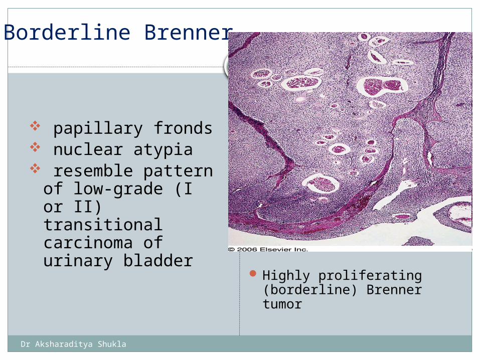
Borderline Brenner
papillary fronds nuclear atypia resemble pattern of
low-grade (I or II) transitional carcinoma of urinary bladder
Highly proliferating (borderline) Brenner tumor
Dr Aksharaditya Shukla
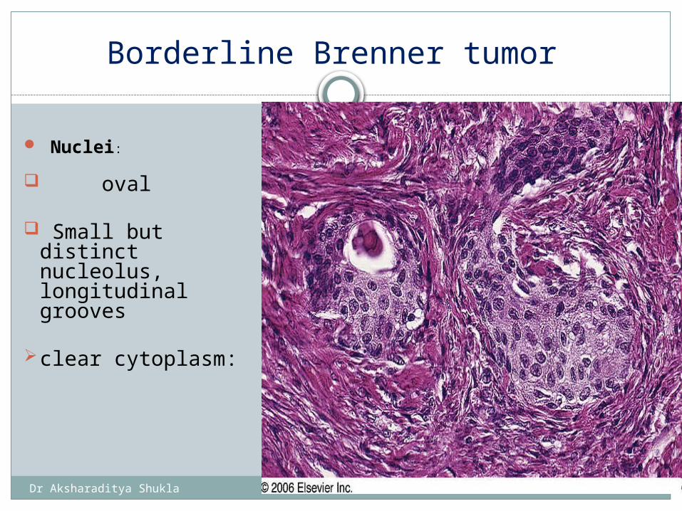
Borderline Brenner tumor
Nuclei:
oval
Small but distinct nucleolus, longitudinal grooves
clear cytoplasm:
The epithelial nests of Brenner tumor are composed of cells with oval nuclei, many of which exhibit longitudinal grooves
Dr Aksharaditya Shukla
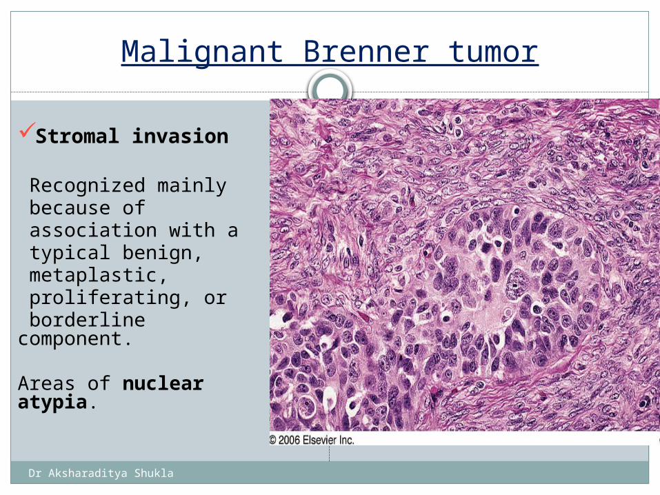
Malignant Brenner tumor
Stromal invasion
Recognized mainly because of association with a typical benign, metaplastic, proliferating, or borderline component.
Areas of nuclear atypia.
Dr Aksharaditya Shukla
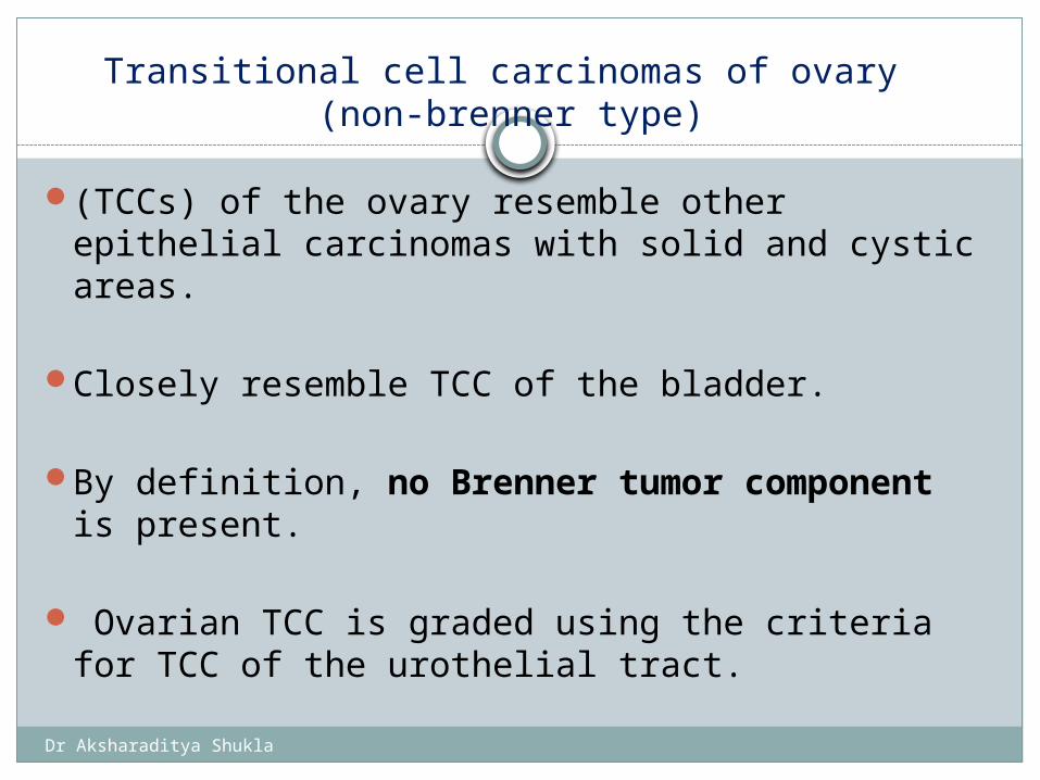
Transitional cell carcinomas of ovary (non-brenner type)
(TCCs) of the ovary resemble other epithelial carcinomas with solid and cystic areas.
Closely resemble TCC of the bladder.
By definition, no Brenner tumor component is present.
Ovarian TCC is graded using the criteria for TCC of the urothelial tract.
Dr Aksharaditya Shukla
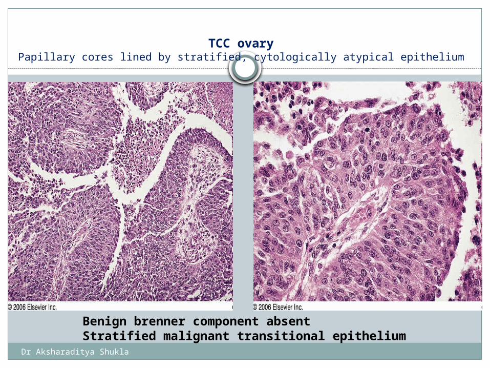
TCC ovaryPapillary cores lined by stratified, cytologically atypical epithelium
Benign brenner component absentStratified malignant transitional epithelium
Dr Aksharaditya Shukla
Special Stains and Immunohistochemistry of Brenner tumors and TCC
Cytoplasm of tumor cells: immunoreactive for: - keratin - EMA - CEA: + also in lumen of cysts * May contain: glycogen, mucin, lipid
Steroidogenic enzymes usually absent
Dr Aksharaditya Shukla
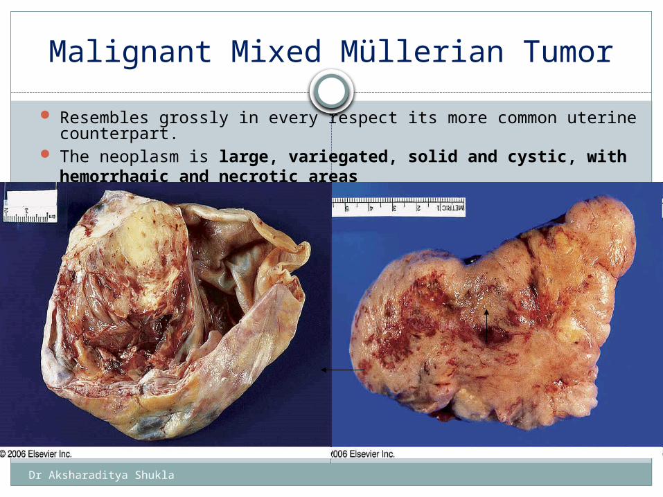
Malignant Mixed Müllerian Tumor
Resembles grossly in every respect its more common uterine counterpart.
The neoplasm is large, variegated, solid and cystic, with hemorrhagic and necrotic areas
Gross appearance of malignant mixed müllerian tumor of ovary. The neoplasm is large, variegated, solid and cystic, with hemorrhagic and necrotic areas
Dr Aksharaditya Shukla
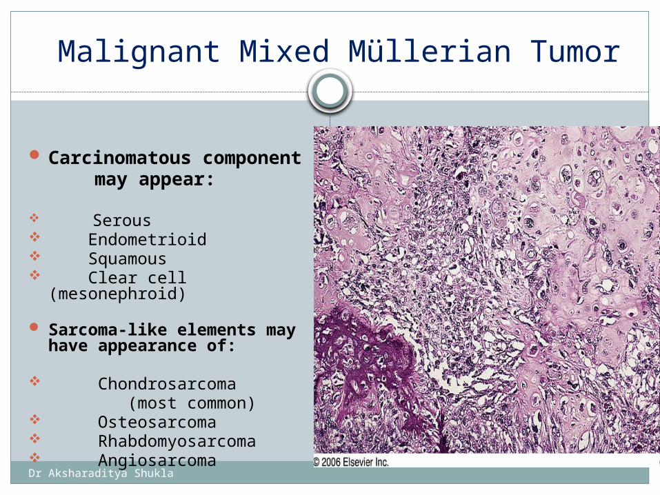
Malignant Mixed Müllerian Tumor
Carcinomatous component
may appear:
Serous Endometrioid Squamous Clear cell (mesonephroid)
Sarcoma-like elements may have appearance of:
Chondrosarcoma (most common) Osteosarcoma Rhabdomyosarcoma Angiosarcoma
Malignant mixed müllerian tumor of ovary exhibiting heterologous foci in the form of bone and cartilage
Dr Aksharaditya Shukla
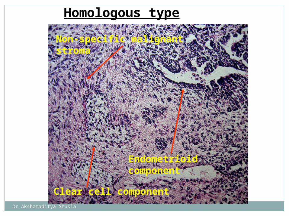
Non-specific malignant stroma
Endometrioid component
Clear cell component
Homologous type
Dr Aksharaditya Shukla
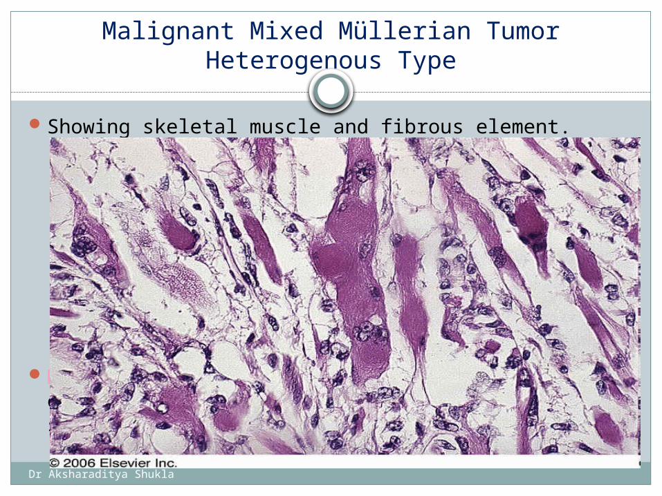
Malignant Mixed Müllerian TumorHeterogenous Type
Showing skeletal muscle and fibrous element.
Malignant mixed müllerian tumor of ovary exhibiting heterologous foci in the form of skeletal muscle
Dr Aksharaditya Shukla
Special Stains and Immunohistochemistryof MMMT
Often hyaline droplets containing α1-
antitrypsin in cytoplasm of tumor cells.
Prognosis: Extremely poor.
Most reliable prognostic criterion is initial tumor stage Most tumors have already extended outside ovary at
surgery.

Dr Aksharaditya Shukla
To be continue…
Presented By: Dr Aksharaditya ShuklaResident, Department Of Patholgy
MGM Medical College & M.Y. Hospital, Indore