Sustainable harvest of Finnish moose population? Esa Ranta, Anne Luoma, Veijo Kaitala &FGFRI.
ABCDEFG
UNIVERS ITY OF OULU P.O.B . 7500 F I -90014 UNIVERS ITY OF OULU F INLAND
A C T A U N I V E R S I T A T I S O U L U E N S I S
S E R I E S E D I T O R S
SCIENTIAE RERUM NATURALIUM
HUMANIORA
TECHNICA
MEDICA
SCIENTIAE RERUM SOCIALIUM
SCRIPTA ACADEMICA
OECONOMICA
EDITOR IN CHIEF
PUBLICATIONS EDITOR
Senior Assistant Jorma Arhippainen
Lecturer Santeri Palviainen
Professor Hannu Heusala
Professor Olli Vuolteenaho
Senior Researcher Eila Estola
Director Sinikka Eskelinen
Professor Jari Juga
Professor Olli Vuolteenaho
Publications Editor Kirsti Nurkkala
ISBN 978-951-42-9905-6 (Paperback)ISBN 978-951-42-9906-3 (PDF)ISSN 0355-3191 (Print)ISSN 1796-220X (Online)
U N I V E R S I TAT I S O U L U E N S I SACTAA
SCIENTIAE RERUM NATURALIUM
U N I V E R S I TAT I S O U L U E N S I SACTAA
SCIENTIAE RERUM NATURALIUM
OULU 2012
A 597
Sirpa Niinimäki
RECONSTRUCTING PHYSICAL ACTIVITY FROM HUMAN SKELETAL REMAINSPOTENTIALS AND RESTRICTIONS IN THE USE OF MUSCULOSKELETAL STRESS MARKERS
UNIVERSITY OF OULU GRADUATE SCHOOL;UNIVERSITY OF OULU,FACULTY OF SCIENCE, DEPARTMENT OF BIOLOGY;FACULTY OF HUMANITIES, ARCHAEOLOGY;UNIVERSITY OF SHEFFIELD,FACULTY OF ARTS AND HUMANITIES, DEPARTMENT OF ARCHAEOLOGY
A 597
ACTA
Sirpa Niinim
äki

A C T A U N I V E R S I T A T I S O U L U E N S I SA S c i e n t i a e R e r u m N a t u r a l i u m 5 9 7
SIRPA NIINIMÄKI
RECONSTRUCTING PHYSICAL ACTIVITY FROM HUMAN SKELETAL REMAINSPotentials and restrictions in the use of musculoskeletal stress markers
Academic dissertation to be presented with the assent ofthe Doctoral Training Committee of Technology andNatural Sciences of the University of Oulu for publicdefence in Kuusamonsali (Auditorium YB210), Linnanmaa,on 13 October 2012, at 12 noon
UNIVERSITY OF OULU, OULU 2012
Copyright © 2012Acta Univ. Oul. A 597, 2012
Supervised byProfessor Arja KaitalaProfessor Andrew ChamberlainDoctor Markku Niskanen
Reviewed byAssistant Professor Cynthia WilczakAssociate Professor Elizabeth Weiss
ISBN 978-951-42-9905-6 (Paperback)ISBN 978-951-42-9906-3 (PDF)
ISSN 0355-3191 (Printed)ISSN 1796-220X (Online)
Cover DesignRaimo Ahonen
JUVENES PRINTTAMPERE 2012
Niinimäki, Sirpa, Reconstructing physical activity from human skeletal remains.Potentials and restrictions in the use of musculoskeletal stress markersUniversity of Oulu Graduate School; University of Oulu, Faculty of Science, Department ofBiology, P.O. Box 3000, FI-90014 University of Oulu, Finland; Faculty of Humanities,Archaeology, P.O. Box 1000, FI-90014 University of Oulu, Finland; University of Sheffield,Faculty of Arts and Humanities, Department of Archaeology, Northgate House, West Street,Sheffield, S1 4ET, United KingdomActa Univ. Oul. A 597, 2012Oulu, Finland
Abstract
The purpose of my thesis is to improve the reliability of physical activity reconstructions bygaining better understanding of the effects of physical activity on bone structural adaptations:musculoskeletal stress markers (MSM) at muscle attachment sites and bone biomechanicalproperties. Bone responds to changes in mechanical loading resulting from activity and bodyweight. Activity reconstructions are important as they are the only means with which activitypatterns of historic humans can be studied. However, MSMs have recently increased the debateabout their reliability as activity indicators due to many bias factors which affect their appearance:age, size, sex and pathological changes.
I studied the effects of physical activity on entheses from three perspectives. First, individualsperforming heavy labour should have higher MSM scores compared to the light labour group dueto elevated degree of mechanical loading at enthesis. This was studied on a population with knownoccupation and was among the first study designs of its kind. Second, a covariance between bonebiomechanical properties and MSM was studied to infer etiology of MSM. The affects of activityand body weight on bone biomechanical properties are well known due to studies in sportsmedicine, whereas the causal mechanisms behind MSMs are not as clear. In theory, both shouldrespond to stress with similar mechanisms. This is a novel approach to investigate the etiologybehind MSMs. Third, if there is a possibility of site-specific adaptation of cortical bone, MSMs,which are local adaptations, can also result from site-specific stress.
I found that while individuals performing heavy labour had higher scores, age-related changesin MSM override activity effects after biological maturity around 40 to 50 years. Also, MSMs andbone biomechanical properties are likely to remodel under same causal mechanisms as wherethere is an increase in one there is likely to be an increase in the other. Furthermore, bone has apossibility of site-specific response, as cortical thickness was increased at muscle pull sitescompared to sites of no muscle pull. I propose that while MSM can be used to study the intensityof physical activity in individuals before they reach biological maturity, it is important to designstudies where biasing factors, such as age, are considered.
Keywords: aging, biological anthropology, bones, entheseal changes, enthesis,musculoskeletal stress markers, osteology, physical activity

Niinimäki, Sirpa, Lihasten kiinnittymiskohtien mahdollisuudet ja rajoituksetfyysisen aktiviteetin rekonstruktioissa. Oulun yliopiston tutkijakoulu; Oulun yliopisto, Luonnontieteellinen tiedekunta, Biologian laitos,PL 3000, 90014 Oulun yliopisto; Humanistinen tiedekunta, Arkeologia, PL 1000, 90014 Oulunyliopisto; University of Sheffield, Faculty of Arts and Humanities, Department of Archaeology,Northgate House, West Street, Sheffield, S1 4ET, United KingdomActa Univ. Oul. A 597, 2012Oulu
Tiivistelmä
Väitöskirjani tarkoitus on tutkia lihasten kiinnittymiskohtien mahdollisuuksia ja rajoituksia fyy-sisen aktiviteetin rekonstruktioissa ja näin parantaa rekonstruktioiden luotettavuutta. Aktiviteet-tihistoriaa voidaan tutkia lihasten kiinnittymiskohdista luun pinnalla tai luun poikkileikkauksenominaisuuksista, koska luu reagoi muutoksiin mekaanisen rasituksen määrässä. Mekaaniseenrasitukseen vaikuttaa aktiviteetin lisäksi ruumiin koko. Aktiviteetin rekonstruktiot mahdollista-vat ammatin ja harrastusten selvittämisen pelkän luustomorfologian perusteella. Ruumiin koonja aktiviteetin lisäksi myös ikä, sukupuoli, patologiset muutokset sekä ruokavalio vaikuttavatlihasten kiinnittymiskohtiin. Tästä syystä tämän menetelmän rajoitusten selvittäminen on oleel-lista luotettavien rekonstruktioiden aikaansaamiseksi.
Jos aktiviteetti heijastuu lihasten kiinnittymiskohtiin, raskasta ja kevyttä työtä tekevillä ihmi-sillä tulee olla erilainen luustomorfologia. Lisäksi, lihasten kiinnittymiskohtien morfologian sekäluun poikkileikkausten ominaisuuksien tulee muunnella yhdessä koska molemmat heijastavataktiviteettia. Luun poikkileikkausten ominaisuuksien aktiviteettisidonnaisuus tunnetaan parem-min liikuntalääketieteellisten tutkimusten ansiosta. Kolmanneksi, jos luu voi vastata rasitukseenpaikallisesti kasvattamalla luun paksuutta lihaksen vetosuuntaan nähden, myös luun pinnassapaikallisesti tapahtuvat muutokset ovat mahdollisia. Nämä ovat uusia lähestymistapoja aktivi-teettia heijastavien syntymekanismien selvittämisessä.
Tutkimustulosteni perusteella raskasta ja kevyttä työtä tekevillä ihmisillä on erilainen luusto-morfologia lihaksen kiinnittymiskohdassa. Nämä muutokset ovat alttiita myös ikäsidonnaisillemuutoksille, joten noin 40–50 ikävuoden jälkeen fyysisen aktiviteetin intensiteettiä ei voida enääluotettavasti rekonstruoida. Aktiviteetin aiheuttamien muutosten syntymekanismi lihaksen kiin-nittymiskohdissa on todennäköisesti sama kuin luun poikkileikkausten ominaisuuksilla, koskamolemmat muuntelevat yhdessä. Lisäksi huomasin, että luu voi reagoida rasitukseen myös pai-kallisesti, koska luun seinämät olivat paksumpia lihaksen vetosuunnassa verrattuna kohtaan,johon ei liittynyt suoraa lihaksen vetosuuntaa. Ehdotan, että lihasten kiinnittymiskohtia voidaankäyttää aktiviteetin rekonstruktioissa, kunhan tutkimuksessa otetaan huomioon muut vaikuttavattekijät, kuten ikä.
Asiasanat: biologinen antropologia, fyysinen aktiviteetti, historia, ikä, ikämuutokset,osteologia


To Joska

8
9
Acknowledgements
I thank my supervisors Arja Kaitala, Andrew Chamberlain and Markku Niskanen.
First of all, I am indebted to Arja Kaitala for her belief in me. Her experience in
science and practical matters has eased my journey into doctorate. I owe a great
deal to Andrew Chamberlain for his kindness and availability and not only in
helping with the material but also for discussions both on and off scientific topic.
Thanks are also due to Markku Niskanen, who was the very first person to
introduce and get me excited about osteology as well as being among the first to
see the potential I had to give to osteological science.
As my topic is both interdisciplinary as well as international I owe thanks to
people coming from many different institutions. I thank the students and staff
from the Department of Archaeology at Oulu University, where I started my
scientific career as an archaeologist. I especially wish to give thanks to Pentti
Koivunen, and to Milton Nuñéz who was the first along with Markku to get me
excited about osteology. Thanks are also due to my fellow osteologists, Juho-
Antti Junno and Anna-Kaisa Salmi, with whom I have collaborated on many
projects. People from the Department of Biology in Oulu University have been
very helpful, and I especially owe thanks to Eija Hurme, who has gone beyond
the call of duty in giving me invaluable comments which helped in organising
ideas in my papers, Sami Kivelä, who gave me advice on statistical analyses,
Laura Härkönen, with whom I made the last stretch towards defending our
prospective theses, and other members of the ‘Evolution and behaviour research
group’. In addition, people from the Department of Archaeology in Sheffield
University made me feel welcome there, first as a student and then as a
researcher. I thank Ari Karttunen, Jaakko Niinimäki, Juha Tuukkanen and Mikko
Finnilä from Oulu University Hospital, who were excited about collaboration and
kind enough to allow access to material. I also thank my colleagues at Johns
Hopkins University, Christopher Ruff and Evan Garofalo for their help and
making me feel a part of a greater scientific community. Furthermore, I thank
Cynthia Wilczak and Elizabeth Weiss for taking the time to review my thesis.
In addition, I am grateful to Timo Jussila, who was the very first person to
introduce me to archaeology. I thank Karen Niskanen and Andrew Chamberlain
for language check and Hanna Heikkinen for statistical advice. I would also like
to thank my students, especially Tiina Väre, Rosa Vilkama and Sonja Söderling,
who also participated in the data collection. Furthermore, I thank the members in

10
‘Tea and theory’ group for lively discussions in archaeological theory as well as
for peer support during my journey to doctorate.
Thanks are due to my family: my parents Anita and Sakari Niinimäki who
taught me the meaning of hard work and ambition, and my aunts Ulla Apell, Virpi
Haataja and Seija Hänninen and their families for giving me board and company
during my research trips. My greatest thanks belong to my husband, Joska Hieta,
to whom I dedicate my work.
My research was funded by the Finnish Cultural Foundation, the National
Science Foundation (grant number 0642297) and the Academy of Finland (grant
number 127241).
11
List of original papers
This thesis is based on the following papers, which are referred to in the text by
their Roman numerals:
I Niinimäki S (2011) What do muscle marker ruggedness scores actually tell us? International Journal of Osteoarchaeology 21(3): 292–299, first published online 2009 DOI10.1002/oa.1134.
II Niinimäki S (2012) The relationship between musculoskeletal stress markers and biomechanical properties of the humeral diaphysis. American Journal of Physical Anthropology 147(4): 618–628.
III Niinimäki S, Söderling S, Junno J-A, Finnilä M & Niskanen M (2012) Age and activity related trends in cross-sectional distribution of bone in the humerus. Manuscript.
IV Junno J-A, Niinimäki S, Niskanen M, Nunez M & Tuukkanen J (2011) Cross sectional properties of the human radial tuberosity. HOMO – Journal of Comparative Human Biology 62: 659–465.

12
13
Contents
Acknowledgements 9 List of original papers 11 Contents 13 1 Introduction 15
1.1 Bone cross-sectional geometry in activity reconstructions ..................... 16 1.2 Musculoskeletal stress markers (MSM) in activity
reconstructions ........................................................................................ 17 1.3 Sources of bias in activity reconstructions .............................................. 17 1.4 Objectives ............................................................................................... 19
2 Materials 23 2.1 Helsinki (I, II, III, IV) ............................................................................. 23 2.2 Blackgate (II, III) .................................................................................... 24 2.3 York (II, III) ............................................................................................ 24
3 Methods 25 3.1 Anatomical basis for biomechanical investigations ................................ 25 3.2 Bone cross-sectional properties (II, III, IV) ............................................ 26 3.3 Controlling for bias factors ..................................................................... 28 3.4 MSM scoring (I, II, III) ........................................................................... 29
4 Results and discussion 31 4.1 Interpreting activity from skeletal remains (I, III, IV) ............................ 31 4.2 Possible causal mechanisms behind MSM development (I, II, III) ........ 32 4.3 Bone response to stress: site-specific versus overall structural
adaptation (II, III, IV) ............................................................................. 33 4.4 Effects of biasing factors on results (I, II, III, IV) ................................... 35 4.5 Recommendations for application of MSM in activity
reconstruction (I, II, III, IV) .................................................................... 36 5 Conclusions 39 References 41 Original papers 47

14
15
1 Introduction
Activity reconstructions based on human skeletal remains are important as they
are the only means with which activity patterns and intensity of activity of
historic human populations or extinct species can be studied. However, these
reconstructions have recently been the focus of a debate about their reliability as
activity indicators (e.g. Jurmain & Roberts 2008). In this thesis, I aim for a better
understanding of the effects of physical activity on bone structural adaptations by
studying the relationship between musculoskeletal stress markers1 (MSM), bone
biomechanical properties and physical activity in the upper limb with respect to
possible bias factors, such as age, size, sex and pathological changes.
Activity reconstructions are based on bone biomechanical properties and
MSMs2 (Jurmain et al. 2012). Analyses of the biomechanical loading of skeletal
elements have been used to study the history of bipedal locomotion and
locomotion patterns (Ruff 2008), patterns of mobility in prehistoric and historic
human populations, such as marine-based versus terrestrial-based mobility (Stock
& Pfeiffer 2001, Weiss 2003a), effects of unilateral versus bilateral activity (Shaw
& Stock 2009), or even combatant weapon preference (Rhodes & Knüsel 2005).
In addition, biomechanical properties have been used to study sexual division of
labour (Wescott & Cunningham 2006). It is important to study biomechanical
loading with respect to past activity, as there may be unexpected patterns, such as
less marked sexual division of labour during the Early Upper Paleolithic
compared to present-day hunter-gatherer sexual division of labour, or changes in
behavioural patterns to a more sedentary lifestyle occurring before the onset of
agriculture during the Late Upper Paleolithic and Mesolithic (Holt 2003). Another
method used to reconstruct physical activity is to use MSMs. This method has
been applied in studies of sexual division of labour (Wilczak 1998, Molnar 2006,
Weiss 2007) as well as differences between populations in subsistence strategies
(Churchill & Morris 1998) or differences within populations over time (Lieverse
et al. 2009) or between socio-economic groups (Havelková et al. 2011), before
and after the onset of agriculture (Eshed et al. 2004) or, in case of the Pueblo
people, differences in labour intensity and labour patterns during pre-colonial and
colonial times (Spielmann et al. 2009).
1 Sometimes called entheseal changes (abbreviated EC) as some researchers want to set these changes apart more clearly from the aetiology originally associate with MSM formation (Jurmain et al. 2012). 2 Occupational stress has also been studied using presence and location of specific pathologies, such as osteoarthritis (Jurmain et al. 2012).
16
Activity reconstructions are based on bone’s ability to respond to stress. Bone
reacts to its mechanical environment - lean body mass, physical activity, body
size and muscle moment arm - by changing its shape and structure (Ruff et al. 1994, Trinkaus et al. 1994, Seeman et al. 1996, Ferretti et al. 1998, Schiessl et al. 1998, Schoenau et al. 2000, Ruff 2000b, Schoenau & Frost 2002, Ruff 2003,
Parfitt 2004, Petit et al. 2004). Bone mass can be increased or decreased with
mechanical stimulus according to mechanostat theory: bone is adapted to normal
levels of stress, whereas a response occurs at higher or lower levels of stress than
the equilibrium range (Frost 1987). Bone mass is increased if the stimulus is high
enough while decreased loading leads to removal of bone in a process called
modeling, and if bone mass stays constant after resorption and formation the
process is called remodeling (Schoenau & Frost 2002, Martin 2007). Peak bone
mass is achieved during the first thirty years of life based on individuals’ loading
history (Johnston & Slemenda 1993, Parfitt 2004). Loading history (Forwood &
Burr 1993) and cumulative effects of activity are important for bone mass
maintenance and distribution (Ruff et al. 2006), but exercise can add bone mass
even post-menopause (Kannus et al. 1995). However, bone remains adaptive in
adulthood (Forwood & Burr 1993, Kannus et al. 1995, Seeman et al. 1996,
Ferretti et al. 1998, Anderson 2001). Bone is strengthened towards the direction
of stress by changes in overall shape.
1.1 Bone cross-sectional geometry in activity reconstructions
The usual direction of stress can be deduced from the shape of the bone shafts and
thus bone cross-sectional properties have been utilised in activity reconstructions
(e.g. Stock & Pfeiffer 2001, Holt 2003, Weiss 2003a, Wescott & Cunningham
2006, Stock & Shaw 2007, Shaw & Stock 2009). Bone biomechanical properties,
such as the overall shape of a cross-section and the distribution of bone mass with
respect to bone bending and torsional strength and axial compression strength,
can be calculated from bone cross-sectional geometry (O’Neill & Ruff 2004).
While mechanostat theory explains how and why there are changes in bone mass,
the theoretical principles of beam theory have been applied to study bone
mechanics to understand changes in bone cross-sectional shape and structure
(Lieberman et al. 2004, Ruff et al. 2006, Stock & Shaw 2007). According to
beam theory, the periosteal contour of bone is most relevant for resisting bending
and torsion forces as it is located furthest from the neutral bending axis
(Lieberman et al. 2004, Ruff et al. 2006, Stock & Shaw 2007). Thus, the overall
17
shape of a bone shaft contributes significantly to the bending and torsion
resistance (Lieberman et al. 2004, Ruff et al. 2006). The endosteal contour has
relatively minimal impact on bone strength, but external architecture is strongly
correlated with internal architecture (Stock & Shaw 2007).
1.2 Musculoskeletal stress markers (MSM) in activity
reconstructions
The principal function of an attachment site is to provide surface area for the
attachment of a muscle or a tendon. Thus muscle attachment sites, also called
entheses, are sites where stress from muscle contraction force takes effect.
Entheses are divided into two categories according to the type of muscle-bone
attachments: fibrous and fibrocartilaginous. Fibrous attachments are further
divided into bony and periosteal attachments. Usually fibrous entheses are
associated with diaphyses, whereas fibrocartilaginous entheses are associated
with epiphyseal and apophyseal attachment sites (Benjamin et al. 1986, Benjamin
et al. 2002).
Entheses exhibit MSM as localised modifications of the bone surface. Several
methods have been developed to quantify these changes (Hawkey & Merbs 1995,
Robb 1998, Mariotti et al. 2004, Zumwalt 2005, Galtés et al. 2006, Villotte 2006).
Scoring methods record the development of osteophytes (also called tubercles or
ruggedness) and osteolytic defects (also called pits or stress lesions). Formation of
a pit and/or a tubercle at an enthesis has been explained theoretically by
Hirschberg (2005) under the standard biomechanical principles of bone
remodeling. Flat bone surfaces model in response to stress according to whether
deposition or resorption thresholds are reached, and a tubercle or a pit is formed
accordingly. Furthermore, the strain of a muscle pull directs the formed tubercle
or pit along the primary axis of stress (Hirschberg 2005).
1.3 Sources of bias in activity reconstructions
Both cross-sectional properties and MSM have limitations in their use and there
are a considerable number of bias factors when applying these methods for
activity reconstructions. Not all scientists agree on the extent to which loading
patterns can be reconstructed from dry bones (Lieberman et al. 2003, Lieberman
et al. 2004, Pearson & Lieberman 2004). Also, some authors emphasise the role
18
of pathological conditions in MSM which may seriously compromise the
accuracy of physical activity reconstructions (Jurmain et al. 2012).
Recording MSM for activity reconstructions may be problematic. A
multifactorial aetiology of MSMs has been acknowledged by several authors as,
in addition to physical activity, also age, body size, sex, genes, obesity, diet and
pathological conditions have been noted to affect their appearance (Zumwalt et al. 2000, Mariotti et al. 2004, Chen et al. 2007, Slobodin et al. 2007, Jurmain &
Roberts 2008, Villotte et al. 2010, Weiss et al. 2010, Jurmain et al. 2012). Body
size, age and sex also have profound effects on bone cross-sectional properties.
As body size is the baseline for the bone mechanical environment it affects bone
cross-sectional properties significantly (Ruff 2003, Ruff 2005). Thus, before any
attempts are made to reconstruct physical activity, bone cross-sectional properties
must be standardised to this baseline. Overall body size affects also MSMs
(Churchill & Morris 1998, Wilczak 1998, Zumwalt et al. 2000, Weiss 2003a,
Weiss 2003b, Weiss 2007). Large individuals produce slightly higher scores on
average, which may result from greater required effort for movement compared to
smaller individuals.
Internal factors, such as age and sex hormones, affect both cross-sectional
properties and MSM. In general, bone is deposited at the periosteal envelope prior
to mid-adolescence and at the endosteal envelope after that (Ruff et al. 1994,
Trinkaus et al. 1994, Seeman 2001, Daly 2007). Age-related changes include
expanded medullary cavities, thinner cortices, and the net loss of trabecular bone
(Trinkaus et al. 1994, Schiessl et al. 1998, Schoenau et al. 2000, Martin 2007).
Age affects also bending and torsion rigidity, which increases with age in young
adults due to expansion of the cortical and medullary areas (Petit et al. 2004) and
the increase may continue through middle age and well into old age (Ruff and
Hayes 1983, Feik et al. 1996, Sigurdsson et al. 2006). With respect to MSM,
older individuals are more likely to exhibit an elevated degree of modifications
(Kennedy 1989, Churchill & Morris 1998, Nagy 1998, Robb 1998, Weiss 2003b;
Mariotti et al. 2004, Galtés et al. 2006, Molnar 2006, Weiss 2007, Villotte et al. 2010). Possible reasons for elevated scores in elderly individuals have been
related to cumulative long-term activity patterns. Repetitions of activities alter
MSM appearance, and in old age cumulative effects of stress are more likely to
appear (Nagy 1998, Wilczak 1998). Rougher external bone may result from
reduced osteoblast activity in older individuals resulting in thinner cortical bone
with greater diameter and rougher external bone surfaces (Weiss 2007). This
likely applies mostly to MSMs at diaphyses, namely fibrous attachments. Age
19
effects on fibrocartilaginous attachments are likely due to changes in tissue
properties of tendons by increasing tendon stiffness and thus mechanical loading
of entheses (Jurmain et al. 2012).
There are systematic differences between the sexes in the degree of
modifications at entheses, but the causal factors behind this observation are not
known. Sex differences in bone cross-sectional properties and MSM may be due
to hormones (Seeman 2001, Daly 2007, Weiss et al. 2010) and size (Churchill &
Morris 1998, Wilczak 1998, Zumwalt et al. 2000, Weiss 2003a, Weiss 2003b,
Weiss 2007). Prior to puberty, exercise appears to increase periosteal apposition
in both sexes, whereas, during or late in puberty, exercise appears to result in
periosteal expansion in boys but endocortical contraction in girls (Seeman 2001,
Daly 2007). Sex differences may also result from possible gender-specific
patterns in habitual activities (Ruff et al. 2006) or differences in force production
capability between males and females (Miller et al. 1993, Kanehisa et al. 1994).
Male scores are usually higher than female scores, but also reverse sex
differences have been documented (Weiss et al. 2012 and references therein).
According to Weiss et al. (2010), reverse sex differences in the case of specific
cultures or populations are likely to be activity related, but when the same reverse
sex difference occurs in many different cultures or populations, it may be
hormonal or methodological.
1.4 Objectives
The scope of my research is to aim for a better understanding of the effects of
physical activity on bone adaptation to stress; in particular MSM and bone
biomechanical properties. This scope is divided into three perspectives (for
summary of research questions, hypotheses and results, see Table 1). First, I
evaluate whether MSM can be used to infer intensity of activity while considering
the effects of biasing factors (I). Second, I study the possible causal mechanisms
behind MSM development (II). Third, I explore the possibility of site-specific
versus overall structural adaptation of bone to stress (II, III, IV). These results
will be applicable for understanding how, why and to what extent bone is able to
respond to stress site-specifically as manifested in changes in MSM scores, cross-
sectional shape and cortical thickness. Thus, my thesis is a contribution to the
current discussion on the reliability of physical activity reconstructions.
My first objective is to test whether individuals performing heavy and light
labour exhibit differences in MSMs (I). The scores should be higher among the
20
heavy labour group compared to the light labour group, as individuals performing
heavy labour should have an elevated degree of mechanical loading at entheses.
To explore this, I use a population with recorded information about occupation,
age and sex (I). If differences in activity intensity are not evident as differences in
MSM, any inferences on type of activity are not possible either. Further, the
biasing effects of age and size on interpretation of activity intensity will also be
considered.
My second objective is to study the possible causal mechanisms behind MSM
development (II). The effects of activity on bone cross-sectional properties are
well documented (e.g. Trinkaus et al. 1994, Kannus et al. 1995, Shaw & Stock
2009, Shaw 2010). Mechanisms behind alterations of these properties are
explained through mechanostat theory (Frost 1987) and beam theory (Lieberman
et al. 2004, Ruff et al. 2006, Stock & Shaw 2007). Changes in MSM should be
based on the same mechanisms of bone remodeling (Hirschberg 2005). And, as
both bone torsion rigidity and MSM are considered to reflect physical activity,
they should covary. I use multiple cross-sectional locations of the humeral shaft to
explore site-specific as well as general trends in the relationship between MSMs
and biomechanical properties. This includes both those cross-sectional locations
housing the attachment of the muscles of interest as well as those that do not.
Previously, only the 35% cross-section location has been utilised in studies on the
relationship between MSM and bone cross-sectional properties (Bridges 1997,
Weiss 2003b).
My third objective is to study the effects of bone adaptation to stress as site-
specific and overall changes affecting the shape and distribution of cortical bone
in the humeral shaft and at the radial tuberosity (II, III, IV). Bone adapts to stress
by altering the distribution of bone mass as well as MSM development. Physical
activity loads the bone mechanically, which should, according to mechanostat
theory (Frost 1987), increase bone mass. There may be a possibility to increase or
maintain bone at muscle attachment sites against age-related bone loss (Forwood
& Burr 1993, Kannus et al. 1995, Nguyen et al. 1998, Sigurdsson et al. 2006).
Response to activity also varies between the proximal and distal humeral shaft
where the activity increases the width of the proximal shaft more than the distal
shaft (Haapasalo et al. 1996). This indicates that cortical bone mass and
distribution can vary between the proximal and distal shaft with age and activity.
However, cortical thickness and torsional rigidity may be governed by the overall
stress regime acting on the diaphysis, rather than localised stresses at attachment
sites. Furthermore, adaptation with shape might be an adequate stress response
21
(Rhodes & Knüsel 2005). I use multiple cross-sectional locations of the humeral
shaft to study site-specific versus overall structural adaptation of bone mass. For
the radius, I examine the mid-radial tuberosity site. In previous studies the focus
has usually been on the 35% or 50% cross-section of the humeral shaft (except
Rhodes & Knüsel 2005), and no studies exist on the cross-sectional properties of
the mid-radial tuberosity.
To sum up, in this research I aim for a better understanding of the possible
causal factors behind MSM development to improve the reliability of physical
activity reconstructions. Furthermore, studying site-specific versus overall bone
functional adaptation to stress will aid in understanding the processes resulting in
changes in bone mass distribution and shape and changes in MSM to physical
activity.
22
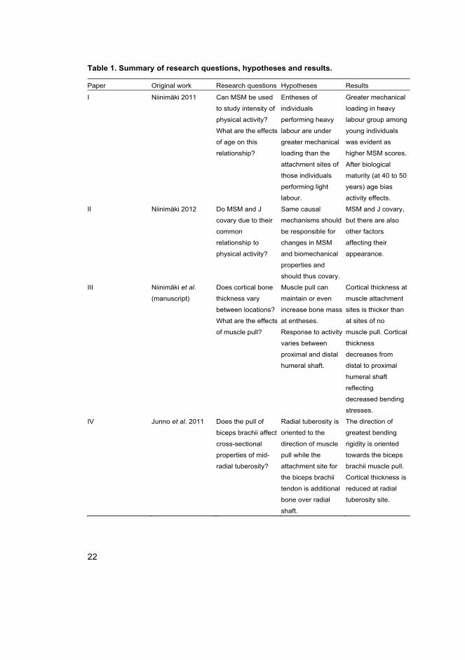
Table 1. Summary of research questions, hypotheses and results.
Paper Original work Research questions Hypotheses Results
I Niinimäki 2011 Can MSM be used
to study intensity of
physical activity?
What are the effects
of age on this
relationship?
Entheses of
individuals
performing heavy
labour are under
greater mechanical
loading than the
attachment sites of
those individuals
performing light
labour.
Greater mechanical
loading in heavy
labour group among
young individuals
was evident as
higher MSM scores.
After biological
maturity (at 40 to 50
years) age bias
activity effects.
II Niinimäki 2012 Do MSM and J
covary due to their
common
relationship to
physical activity?
Same causal
mechanisms should
be responsible for
changes in MSM
and biomechanical
properties and
should thus covary.
MSM and J covary,
but there are also
other factors
affecting their
appearance.
III Niinimäki et al.
(manuscript)
Does cortical bone
thickness vary
between locations?
What are the effects
of muscle pull?
Muscle pull can
maintain or even
increase bone mass
at entheses.
Response to activity
varies between
proximal and distal
humeral shaft.
Cortical thickness at
muscle attachment
sites is thicker than
at sites of no
muscle pull. Cortical
thickness
decreases from
distal to proximal
humeral shaft
reflecting
decreased bending
stresses.
IV Junno et al. 2011
Does the pull of
biceps brachii affect
cross-sectional
properties of mid-
radial tuberosity?
Radial tuberosity is
oriented to the
direction of muscle
pull while the
attachment site for
the biceps brachii
tendon is additional
bone over radial
shaft.
The direction of
greatest bending
rigidity is oriented
towards the biceps
brachii muscle pull.
Cortical thickness is
reduced at radial
tuberosity site.
23
2 Materials
I had three main requirements for the skeletal materials. First, I needed a large
number of relatively intact and well-preserved humeri available for pQCT
(peripheral quantitative computed tomography) scanning (II, III, IV) and
recording MSM at entheses (I, II, III). Second, material of known occupation was
essential to study the effects of heavy and light physical labour as well as age on
MSM (I). Third, radii of a population with known occupation were required to
study the cross-sectional properties of mid-radial tuberosity (IV).
I chose three skeletal populations: Helsinki, York (Barbican) and Newcastle
(Blackgate) as they are large, well preserved skeletal collections representing
populations with similar climatic adaptations. The Helsinki material consists of
skeletons obtained from cadavers and the material is housed in the Natural
History Museum, Helsinki. The York and Blackgate collections are archaeological
and are housed at the University of Sheffield. Only skeletons of adults (18 years
or older) were included in the samples. Age was known for the Helsinki material.
For the archaeological material, I assessed age from the changes in the pubic
symphysis, endocranial suture closure, and dental wear and sex from
morphological differences in the pelvis and cranium (Bass 1987).
My selection criteria for humeri were that maximum length for size control
and biomechanical length for determining cross-sectional locations for pQCT
scanning could be measured accurately. For radii selection the criterion was an
intact radial tuberosity. The observed radii were in excellent condition, as I
studied radii only from the Helsinki sample. I followed the criteria of Mariotti et al. (2004) for preservation of entheses, where at least 50% of an enthesis is
required for accurate scoring of the attachment. I rejected any pathological radii
and humeri. Relevant numbers of individuals available for analyses are stated
case-specifically in papers.
2.1 Helsinki (I, II, III, IV)
The Helsinki sample represents modern urban Finns of Southern Finland and
consists of approximately 300 adults. As this skeletal material was obtained from
cadavers, they were mostly in excellent condition with minimal taphonomic
interference, although not all bones of an individual were present. Age, sex and
occupation were known for most of these individuals (Kivalo 1957, I). The stated
occupations are, for example, workman, seaman, office worker and shoemaker for
24
males and housemaid, servant, and seamstress for females (I). Some ambiguous
titles such as prisoner, senior citizen and wife are also given (I). Heavy physical
labour was defined as tasks requiring lifting, moving heavy loads or getting short
of breath involved in the profession (Chaffin 1981). I evaluated the intensity of
activity as heavy or light where it could be assessed from the stated occupation.
There were very few indications of trauma and a relatively low prevalence of
degenerative joint disease among this sample (T Heikkilä, personal
communication July 11 2011).
2.2 Blackgate (II, III)
The Blackgate cemetery is located in Newcastle upon Tyne, adjacent to the site of
a former Roman fort. Radiocarbon dating of the skeletons shows that the
cemetery was in use from the late Anglo-Saxon to early Norman periods (late 7th
to the early 12th centuries AD). The cemetery was excavated between 1978 and
1992 with skeletal remains from a total of over 800 individuals being recovered.
There is no contemporary settlement evidence associated with the cemetery, thus
the origins and affiliations of the individuals buried in the cemetery are unknown
(Nolan et al. 2010). Approximately one-third of the individuals were immature at
time of death, and amongst the adults there were approximately equal proportions
of females and males. There is a relatively low prevalence of infection and trauma
among the skeletal remains, but there is a high prevalence of degenerative joint
disease (A Chamberlain, personal communication July 15 2011).
2.3 York (II, III)
The cemetery at the Barbican site in York, excavated between June 2007 and
February 2008, belonged to the former medieval Church of All Saints, Fishergate,
York. Of the number of excavated articulated human skeletons, 547 individuals
dated to the medieval period, and 113 individuals to the post-medieval period
buried in mass graves. Two radiocarbon dates on the skeletal remains show that
the medieval phase of burials at All Saints, Fishergate continued until about AD
1500. The post-medieval burials are likely to date to the time of the Siege of York
in AD 1644 (McIntyre & Bruce 2010, McIntyre et al. in preparation). The
medieval burials are from a population with normal attritional mortality with
approximately one quarter of the individuals being immature at time of death.
There is a high prevalence of degenerative joint disease in the sample (McIntyre
& Bruce 2010, McIntyre et al. in preparation).
25
3 Methods
Humeri and radii were scanned with pQCT and scored for MSM. Maximum
humeral length (Martin & Saller 1957, measurement M-1) was measured with an
osteometric board with 1mm accuracy to be used as a size-standard. Humeral
biomechanical length (Ruff 2000b) was measured with large sliding callipers with
1mm accuracy to determine cross-sectional locations defined as percentage of
length measured from the distal end of the humeral shaft. Radial tuberosity
(length and width) was measured with small sliding callipers with 0.1mm
accuracy for determining cross-sectional location on mid-radial tuberosity.
3.1 Anatomical basis for biomechanical investigations
The shape of a whole humeral cross-section as well as the direction of muscle
attachment sites at an enthesis should be oriented to the principal axis of stress,
directed by muscle pull (Ruff 2003, Ruff 2005, see also Table 2). The cross-
section at 80% from the distal humeral shaft houses the attachments for pectoralis
major, teres major, latissimus dorsi, and triceps brachii (Table 2). Pectoralis major
attaches on the proximal third of the anterolateral humeral shaft, teres major
attaches on the proximal third of the anteromedial humeral shaft, latissimus dorsi
attaches on the proximal third of the anterior humeral shaft, and triceps brachii on
the posterior aspect of the humeral shaft (Table 2). The cross-section at 65%
houses the attachments for deltoid and medial and lateral heads of triceps brachii.
Deltoid attaches on the lateral aspect of the mid-shaft of the humerus. While the
humeral flexion results in anterior and lateral deltoid pull, pectoralis major and
latissimus dorsi pull anteriorly. Humeral extension results in medial teres major
pull, but the overall resultant is an anteriorly directed force vector at the 80% and
65% cross-sectional locations. However, the pull of pectoralis major, teres major
and deltoid is no longer discernible at 65% (see III: Fig. 1). Triceps brachii pulls
posteriorly at 80% and 65%, and medially at 65%. The cross-section at 50%
houses the attachments for deltoid, brachialis, coracobrachialis and triceps brachii
where deltoid pulls laterally, brachialis pulls anteriorly, coracobrachialis pulls
medially and triceps brachii pulls posteriorly (Table 2, see also III: Fig. 1). The
shape of the 35% cross-section of the humeral shaft is affected by brachialis,
brachioradialis and triceps brachii where brachialis pulls anteriorly,
brachioradialis pulls laterally, and triceps brachii pulls posteriorly (Table 2, see
also III: Fig. 1).
26
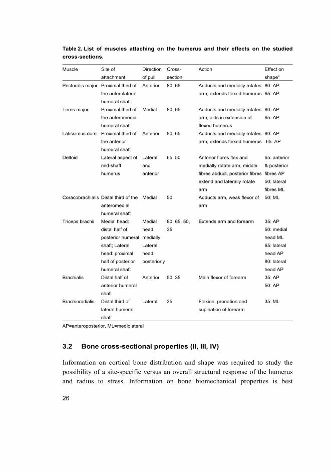
Table 2. List of muscles attaching on the humerus and their effects on the studied
cross-sections.
Muscle Site of
attachment
Direction
of pull
Cross-
section
Action Effect on
shape*
Pectoralis major Proximal third of
the anterolateral
humeral shaft
Anterior 80, 65 Adducts and medially rotates
arm; extends flexed humerus
80: AP
65: AP
Teres major Proximal third of
the anteromedial
humeral shaft
Medial 80, 65 Adducts and medially rotates
arm; aids in extension of
flexed humerus
80: AP
65: AP
Latissimus dorsi Proximal third of
the anterior
humeral shaft
Anterior 80, 65 Adducts and medially rotates
arm; extends flexed humerus
80: AP
65: AP
Deltoid Lateral aspect of
mid-shaft
humerus
Lateral
and
anterior
65, 50 Anterior fibres flex and
medially rotate arm, middle
fibres abduct, posterior fibres
extend and laterally rotate
arm
65: anterior
& posterior
fibres AP
50: lateral
fibres ML
Coracobrachialis Distal third of the
anteromedial
humeral shaft
Medial 50 Adducts arm, weak flexor of
arm
50: ML
Triceps brachii Medial head:
distal half of
posterior humeral
shaft; Lateral
head: proximal
half of posterior
humeral shaft
Medial
head:
medially;
Lateral
head:
posteriorly
80, 65, 50,
35
Extends arm and forearm 35: AP
50: medial
head ML
65: lateral
head AP
80: lateral
head AP
Brachialis Distal half of
anterior humeral
shaft
Anterior 50, 35 Main flexor of forearm 35: AP
50: AP
Brachioradialis Distal third of
lateral humeral
shaft
Lateral 35 Flexion, pronation and
supination of forearm
35: ML
AP=anteroposterior, ML=mediolateral
3.2 Bone cross-sectional properties (II, III, IV)
Information on cortical bone distribution and shape was required to study the
possibility of a site-specific versus an overall structural response of the humerus
and radius to stress. Information on bone biomechanical properties is best
27
obtained from CT scans (O’Neill & Ruff 2004, Stock & Shaw 2007). Thus,
humeri from the Helsinki, York and Blackgate materials and radii from the
Helsinki material were scanned using a pQCT scanner (XCT-960A with software
version 5.20, Norland Stratec Medizintechnik GmbH, Birkenfeld, Germany).
Humeri were aligned for pQCT scanning according to the protocol presented in
Ruff (2000b). Four cross-sectional locations at 80%, 65%, 50%, and 35% of
biomechanical length from the distal humeral end were scanned (II, III). Radii
were placed orthogonally and vertically to the axis of slice, where the radial
tuberosity was upwards and orienting the radial tuberosity surface horizontally
(IV). Radii were scanned at mid-radial tuberosity measured from the length of the
tuberosity (IV). Bones were attached securely to a rigid plastic platform with steel
wires 50 mm apart representing a parallel surface and to provide a control scale
for the pQCT measurements. The slice thickness was 1.25 mm and the voxel size
was 0.69*0.69 mm2. A default threshold of 560 mg/mm3 was used to separate
bone from air.
I obtained information on cortical area, total area, and second moments of
area and bone torsion rigidity. Cortical area (CA) represents the bone's resistance
to axial tension or compression loads. Total area (TA) represents the total size of a
cross-section including medullary and cortical areas. Second moment of area (I),
also called moment of inertia represents bending rigidity around the mediolateral
axis in the anteroposterior plane (Ix) and around the anteroposterior axis in the
mediolateral plane (Iy). Torsional rigidity is described by J which measures
torsional and average (twice) bending strength (Ruff 2003, O’Neill & Ruff 2004).
It is derived from summing two perpendicular Is. Therefore J= Ix+Iy or Imax+Imin
(Ruff 2003, O’Neill & Ruff 2004).
The effects of muscle pull were studied by measuring cortical bone thickness
at the middle of sites of muscle pull, as well as at sites with no evident direction
of muscle pull at the mid-lateral wall of the humeral shaft using Dataviewer.
Measurements were taken perpendicular to the periosteal surface at each site.
Mid-sites of muscle pull were measured for pectoralis major, teres major, and
triceps brachii at 80% cross-section; deltoid and triceps brachii medial and lateral
heads at 65% cross-section; deltoid, brachialis, coracobrachialis, and triceps
brachii at 50% cross-section; and brachialis, brachioradialis and triceps brachii at
35% cross-section (see III: Fig. 1). In addition, from each cross-section a site of
no muscle pull was measured.
28
3.3 Controlling for bias factors
I analysed males and females separately to account for hormonal influences on
bone modeling after puberty (Ruff et al. 1994, Trinkaus et al. 1994, Seeman 2001,
Daly 2007). For studies of activity intensity using the Helsinki material (I, IV) the
sexes were pooled because of the relatively modest sample size of females with
known activity (I, IV). I performed analyses both sex-specifically as well as for
pooled-sex samples to compare trends. As there was no significant effect of
including females in the data (I, IV; Niinimäki et al. 2012), only pooled sample
data are shown. I analysed left and right sides separately as handedness can alter
loads applied side-specifically.
The Helsinki sample was accompanied with reliable information about age
and thus I could use the material to study the effects of age on activity markers (I,
IV). However, because age in archaeological material can only be estimated as
age ranges, I divided my material into two age categories using a section-point of
40 years. This section-point was chosen as bone loss and osteoporotic changes
increase after biological maturity at 40 to 50 years (Forbes 1987, Johnston &
Slemenda 1993). Due to the large number of individuals in the old age category in
the Helsinki sample, the section-point was readjusted to 50 years to have more
equal numbers of individuals across the age groups in the study of radial
tuberosity cross-sectional properties (IV). Results remained similar regardless of
the section-point used; thus I presented only those results using a section-point of
50 years (IV).
Bone cross-sectional properties must be standardised for baseline mechanical
environment in order to study relative rather than absolute variation. Baseline
environment is represented by a product of body weight and moment arm length
(Ruff 2003, Ruff 2005). However, bones required to reconstruct body mass, such
as the femur head (Ruff et al. 1991) or pelvic breadth and the bones required to
reconstruct stature (Ruff 2000a), were present in only a few individuals. Thus, the
use of reconstructed body mass as a size variable would have decreased the
sample size. Adjusted humeral length can be used to reflect the baseline
mechanical environment (Ruff et al. 1993, Trinkaus et al. 1994). I used humeral
length raised to the power of 3 as a scaling factor for CA and cortical thickness
measurements, and the bone length raised to a power of 5.33 for J measurement
(Ruff et al. 1993, Trinkaus et al. 1994, see also Ruff 2000b, Stock & Pfeiffer
2001). These scaling factors describe the power relationship of J (torsional
strength resists beam length and volume) and CA (axial compression resists
29
volume) relative to humeral length. The scaling factor for J was theoretically
derived and empirically tested for femoral and humeral cross-sections (Ruff et al. 1993, Trinkaus et al. 1994). It should be borne in mind that this size scaling
method is problematic. The difficulty comes in terms of finding the appropriate
scaling factor to describe the power relationship between bone length and cross-
sectional properties (Ruff 2000b). Further, there are differences in the relationship
between body proportions and body size between individuals and populations
(Ruff 2000a, Ruff 2000b).
3.4 MSM scoring (I, II, III)
There are several available enthesis scoring systems (Hawkey & Merbs 1995,
Robb 1998, Mariotti et al. 2004, Galtés et al. 2006, Villotte 2006) but only Galtés
et al. (2006) and Villotte (2006) have considered the current anatomical
understanding of entheses, namely division of entheses according to their muscle-
bone –attachment: fibrous and fibrocartilaginous. In Villotte et al. (2010) this
method was further modified, where observed variance in fibrocartilaginous
attachments was reduced into a binary variable for statistical analyses. However,
although scoring fibrocartilaginous entheses is fairly accurate using the Villotte
(2006) guidelines, it is less accurate for fibrous entheses (Havelková & Villotte
2007). Therefore, I chose the Hawkey and Merbs (1995) method for recording
MSMs. Three fibrous (pectoralis major, teres major, deltoid) attachments (I, II,
III) and one fibrocartilaginous (biceps brachii) attachment (I) were used in the
analyses. An enthesis was scored for ruggedness and stress lesions (pits) on a 0–3
ordinal scale. Ossification was not included as it is considered to result from
trauma (Hawkey & Merbs 1995, Molnar 2006). I summed individual MSM scores
for ruggedness and stress lesions to represent overall muscle use. Some
researchers have used a continuum from ruggedness to stress lesions (e.g. Weiss
2003b; Eshed et al. 2004, Molnar 2006, Weiss 2007), but this over-emphasises
the relevance of stress lesions and pre-supposes that they only develop from a
pre-existing rugged lesion. For the teres major, insertion stress lesions are the
most prominent feature; for the pectoralis major insertion, stress lesions rarely
form as distinctive fossae, but severe pitting may exist; for the deltoid insertion,
stress lesions are rarely present.

30

31
4 Results and discussion
My main goal was to understand the relationship between physical activity and
bone structural adaptations to stress in order to explore the reliability of MSM as
an indicator of physical activity. I found that MSM and bone torsion rigidity
covaried, thus they are likely to remodel under the same casual mechanisms (II).
Changes in MSM were accompanied by changes in cross-sectional shape (II, III)
but not in the distribution of bone mass (III). However, cortical bone thicknesses
at muscle attachment sites were different compared to sites where there was no
evident direction of muscle pull (III, IV). Further, according to my results MSM
can be used to represent intensity of physical activity (I). However, this should be
applied with caution as there are also bias factors affecting their appearance, of
which the most prominent is age (I, II, III, IV).
4.1 Interpreting activity from skeletal remains (I, III, IV)
If MSMs reflect physical activity, individuals performing heavier tasks should
have higher degree of modifications at entheses than individuals performing
lighter tasks. My results on individuals with known occupation and age indicated
that the intensity of physical activity cannot be separated after biological maturity
at 40–50 years using fibrous muscle attachments in the humeral shaft (I)3. After
this age any interpretations of intensity of activity are biased by age-related
changes. Subsequently also Villotte et al. (2010) and Alves-Cardoso and
Henderson (2010) have studied individuals with documented occupation. Villotte
et al. (2010) found that fibrocartilaginous muscle attachments of the upper limb
can separate young individuals performing manual and non-manual labour.
Similar to my results, they also noted that old age biased interpretations of
physical activity (Villotte et al. 2010). Alves-Cardoso and Henderson (2010)
found that individuals performing manual labour tended to have higher scores
than non-manual labour individuals, but their results were significant only after
controlling for age.
The direction of greatest bending strength can be used to ascertain habitual
differences in muscle use. According to my results, the direction of greatest
bending strength was most variable at the 50% humeral cross-sectional location
3 Also biceps brachii attachment at radius, which is a fibrocartilaginous attachment, was included in the analysis for this paper. However, in a subsequent analysis where this attachment was dropped the results on age-related trends remained similar (Niinimäki et al. 2012).
32
(III). This cross-sectional location can be used to study differences in activity
patterns between groups on the preference of the elbow (AP) compared to the
shoulder (ML). The direction of greatest bending strength was constant at the
80% and 35% cross-sections. Bending strength at the 35% cross-section was
oriented towards the AP axis and bending strength at the 80% cross-section was
oriented towards the ML axis (III). These trends likely reflect flexion of the elbow
at 35% and fixing of the humerus close to the torso in the ML direction at 80%.
The direction of greatest bending strength was different between the sexes at the
65% cross-section, which in males was more directed towards the AP direction,
while in females there was mostly no preferential direction (III). These shape
differences between males and females may result from greater loading of the
shoulder among males or from muscle lever differences between sexes, as males
have broader shoulders compared to females (Holliday 1997). At the radial
tuberosity, the pull of the biceps brachii tendon provides the principal direction of
stress (IV).
4.2 Possible causal mechanisms behind MSM development (I, II, III)
As both bone biomechanical properties and MSM are considered to reflect
physical activity, both are considered to remodel under the same mechanisms and
thus they should covary. There was a covariance between bone biomechanical
properties and MSM, where higher MSM scores occurred with higher values of J
(II). Thus the same mechanisms of bone remodeling are likely responsible for
both the appearance of MSMs and the altering of diaphyseal dimensions as
predicted by mechanostat theory and beam theory, and as theoretically explained
by Hirschberg (2005). Hirschberg (2005) studied entheseal modifications in the
light of bone remodeling theory where remodeling thresholds explain whether
bone deposition is evident as ruggedness or whether bone resorption is evident as
pits develop. Furthermore, age seems to have a similar effect on both MSM and J,
where both increase with age (I, II; Ruff & Hayes 1983, Feik et al. 1996, Galtés et al. 2006, Sigurdsson et al. 2006, Villotte et al. 2010).
My interest in the relationship between biomechanical properties and MSM
was to study the overall versus the site-specific relationships, as I included both
those cross-sectional locations housing the attachment for the muscles of interest
as well as those that do not (II). The relationship between MSM and bone
biomechanical properties has been previously studied by Weiss (2003b) and
Bridges (1997) but with a slightly different focus. I utilised a total of four cross-
33
sectional locations along the humeral shaft to study site-specific as well as overall
covariance between MSM and biomechanical properties whereas Weiss (2003b)
and Bridges (1997) only included the 35% cross-section in their studies. I did not
find a correlation between MSM and CA (III), but did find one between MSM
and J (II). The correlation between MSM and J was mostly positive, with high
MSM scores occurring with high J (II), a trend also found by Weiss (2003b).
Some single muscle MSM and J correlated in my samples, but there was no such
correlation between MSM and cross-sectional properties in Weiss’ sample
(2003b). It seems that the relationship between MSM and J reflects also the
overall loads applied to the humeral shaft rather than a specific relationship.
Bridges (1997) found only a weak inverse correlation between pectoralis major
MSM and bone cross-sectional properties on the left side in males. She suggests
that this may be due to her sample composition and possible pathological cases in
her sample (Bridges 1997). To sum up, although a true causal relationship
between MSM and J could not be studied, where there is an increase in one there
is likely to be an increase in the other (II). This common increase is likely a result
of bone response to physical activity at a young age, whereas in old age the
increase can also be due to age-related changes in bone remodeling.
4.3 Bone response to stress: site-specific versus overall structural adaptation (II, III, IV)
My results on variation on cortical bone thickness indicate that there is a
possibility of both site-specific as well as an overall structural adaptation to stress
in the humeral shaft and radial tuberosity. As the main functions of MSM are to
provide surface area for the attachment of a muscle or a tendon, they can be
considered as site-specific adaptations to stress. In general, bone adapts to stress
with changes in shape which are correlated with changes in MSM (II) rather than
adaptation with bone mass, although there is some variation in cortical bone
thickness between the proximal and distal humeral shaft and within cross-
sectional locations between sites of muscle pull and no pull (III). This may be due
to preservation of bone at muscle pull sites compared to sites of no muscle pull.
Thus, muscle pull seems to have an effect on cortical bone thickness (III).
However, muscle activity evident as MSM did not have a significant relationship
with cortical bone mass (III).
The increase in cortical thickness found at muscle pull sites on the humeral
shaft was reversed at the radial tuberosity, where cortical bone was thinner at
34
muscle pull site than in sites of no muscle pull (IV). This difference is likely due
to reduced bending stresses at epiphyses and apophyses where fibrocartilaginous
attachments are found. Reduced bending stresses reduce the need for greater
cortical thickness at these sites. Thus, it is likely that other locations of
fibrocartilaginous attachments are associated with thinner cortical bone as well.
In addition to site-specific variation in cortical bone thickness, CA was
reduced from the distal to the proximal humeral shaft (III). However, only CA at
the 80% cross-section was statistically significantly smaller than in other cross-
sectional locations (III). This is in agreement with results by Rhodes and Knüsel
(2005) where CA was smallest at the 80% cross-section. Differences between the
distal and proximal humeral shaft were also found by Haapasalo et al. (1996)
where changes due to activity were greater in the proximal shaft than in the distal
shaft. This is likely due to reduced bending stresses towards the ends of the
diaphyses, which is evident as pit formation at MSM sites near the ends of the
diaphysis.
Differences in CA between the proximal and distal humeral shaft may further
illustrate the significance of adaptation with shape rather than bone mass. Thus, it
seems that adaptation to stress occurs mostly by changes in shape (II, III, IV) and
the geometrical properties of the bone are adapted to accommodate the principal
directions of stress. Modeling towards the direction of stress occurs by increasing
bone mass locally. Periosteal deposition is compensated by higher endosteal
resorption rates as suggested by the results of papers III and IV. With this
deposition-resorption mechanism as explained by mechanostat theory cross-
sectional shafts can accommodate bone cross-sectional shape towards the
direction of muscle pull without resulting in thick cortices at muscle attachment
sites. This was most evident among older individuals where in some cases the
original, more circular endosteal wall was still visible as denser trabecular bone
(III, IV). Regardless of site-specific variation in cortical bone thickness where
cortical thickness was thicker at muscle pull sites in the humeral shaft and thinner
at the radial tuberosity, there was a very close approximation between the internal
and external contours of cortical bone (III, IV). This is in agreement with Stock
and Shaw (2007), who noted that external architecture is strongly correlated with
internal architecture.
Although bone mass can be increased or at least preserved locally against
age-related bone loss at sites of direct muscle pull it is not likely that cortical
thickness and MSM reflect similar aspects of bone functional adaptation (III).
While CA represents resistance to axial compression, MSM and J are functional
35
responses to stress. Thus, there is no need to maintain or develop a thick cortical
bone wall if stress can be accommodated with bone shape as also suggested by
Rhodes and Knüsel (2005), and by surface structure as MSM. As MSMs are
associated with J rather than CA, it suggests that the aetiology behind MSMs is
related to muscle activity where surface development can aid in the orientation of
cross-sections towards the direction of stress.
4.4 Effects of biasing factors on results (I, II, III, IV)
A variety of biasing factors affect MSM and bone biomechanical properties as
well as interpretations of physical activity based on them. While a covariance
between MSM and J was found, there were also other factors affecting their
variance (II). Age and sex are known to affect the mechanisms involved in bone
modelling, and these factors also affect the variance in activity markers: MSM
and biomechanical properties.
Age effects are profound on MSM after biological maturity (Galtés et al. 2006), where age overrides activity effects in both fibrous and fibrocartilaginous
muscle attachments (I; Villotte et al. 2010). Robb (1998) has suggested that after
biological maturity, from 40–50 years onwards, the mechanisms affecting MSM
formation may level off. Biological maturity also affects bone bending strength,
where J increases and continues to increase through middle age and well into old
age in both sexes (Ruff & Hayes 1983, Feik et al. 1996, Sigurdsson et al. 2006).
In my data, cortical bone decreased with age, both as CA and as cortical thickness
measured at sites where there was not direct muscle pull (III, IV). This is in
accordance with earlier studies on aging effects on CA (Trinkaus et al. 1994,
Schiessl et al. 1998, Schoenau et al. 2000, Russoa et al. 2006, Martin 2007). It
may be that a bone's structural response to mechanical loading is similar to its
aging response, thus affecting physical activity reconstructions based on MSM
and J.
Differences in activity patterns between the sexes are mostly studied from the
upper limb (Wilczak 1998, Lieverse et al. 2009, Spielmann et al. 2009, Villotte et al. 2010b, Weiss et al. 2010), but this is also where there is most sexual
dimorphism due to hormones. Bone adaptation to stress differs between males
and females due to sex hormones. It seems that among females bone response to
stress may be under greater hormonal influence than activity influence: while
there were significant trends in the male sample in the relationship between MSM
and J (II), trends among females usually remained statistically insignificant.
36
Estrogen is responsible for greater endosteal apposition in females during puberty
(Schiessl et al. 1998, Schoenau et al. 2000, Petit et al. 2004) and decrease during
menopause (Schiessl et al. 1998, Schoenau et al. 2000). Also muscle mass is
influenced by hormones (Gallagher et al. 1997, Ferretti et al. 1998, Janssen et al. 2000, Schoenau et al. 2000, Seeman 2001, Ruff 2003, Schoenau et al. 2002, Petit
et al. 2004, Sumnik et al. 2006, Daly 2007). Hormonal influences are more
marked in the upper limb where males have more muscle mass (Gallagher et al. 1997, Janssen et al. 2000, Ruff 2003, Sumnik et al. 2006) and as a result males
develop relatively stronger humeri (Ruff 2003). Males have larger muscles both
in absolute terms and relative to body mass starting from puberty, probably due to
high testosterone levels (Schoenau et al. 2000, Ruff 2003) and greater force
production capability (Miller et al. 1993, Kanehisa et al. 1994). As males usually
have higher scores than females, reverse cross-cultural sex patterns are most
likely to reflect hormonal influence or methodological problems (Weiss et al. 2010). Thus, hormonal influences on MSM and bone biomechanical properties in
relation to physical activity should be considered.
Sexual dimorphism in size may also contribute to some sex differences.
Males are on average larger than females and, as size is known to influence
MSMs higher scores in males, may reflect their larger size (Weiss 2007).
However, for a large body to affect MSM, some activity is needed. Sex
differences may also result from more environmentally sensitive growth in males
(Wilczak 1998).
4.5 Recommendations for application of MSM in activity
reconstruction (I, II, III, IV)
My results indicate that MSM can be used to infer intensity of muscle activity
among individuals before or after reaching biological maturity (I), as there is a
possibility of site-specific response to stress (III, IV) and because the causal
mechanisms behind MSM appearance are likely those which reflect physical
activity and age (II). However, care should be taken when applying this method,
as other factors also affect their appearance (I, II).
A distinction should be borne in mind between activity type and intensity of
activity. When the interest is in intensity of activity, it is advisable to account for
the total variation of the studied MSM sites (II), and in order to obtain a measure
less sensitive to anomalies, to combine the MSM scores. A composite variable of
several ordinal variables can be created if variables are measures of the same
37
thing (in this case, intensity of activity affecting MSM sites) and then treated as a
continuous variable (Rowe 2006). This enables considering the continuous nature
of at least some fibrous MSMs in statistical analyses. Furthermore, Weiss (2003b;
2007) suggests using a composite variable (which she calls an aggregated MSM)
as it is a measure less sensitive to anomalies. Havelková et al. (2011) combined
information from one individual using percentage prevalence of enthesopathies.
They argued for the use of parametric testing to detect significant differences
between populations (Havelková et al. 2011).
Interpretations of type of activity may be more difficult. Discerning
movements of the upper limb are difficult due to great mobility and instability of
the shoulder joint. Many muscles around the shoulder joint are active during
upper limb movement either as prime movers, antagonists or stabilisers (Gosling
et al. 2002). Also, there are multiple muscles attaching to the same location - as
seen with flexor and extensor origins, and on the linea aspera in the femur
(Gosling et al. 2002).
Difficulties in interpreting activity type also arise from the variability in the
degree of modifications in MSMs between different entheses. In addition to
differences between fibrous and fibrocartilaginous muscle attachment sites
(Villotte 2006), there is variance within fibrous muscle attachments. Most notable
is the presence or absence of pits (II). Pits usually form towards the ends of the
diaphyses, where bending stresses on the bone shaft are minimised. For the
insertion of teres major, stress lesions are the most prominent feature; the
pectoralis major insertion exhibits severe pitting, but pitting is rarely evident as
distinctive fossae; for the insertion of deltoid, pits are rarely present (II). In
addition, not all attachment sites exhibit similar scale of ruggedness. There have
been some attempts to solve the problem of variation in MSM scores between
muscle attachments. Standardisation of MSMs into z-scores has been used (Weiss
2003a, Weiss 2003b, Weiss 2007). However, as MSM are recorded as ordinal
variables, the use of z-scores has been criticised (Stefanović & Porčić 2011).
Havelková et al. (2011) used parametric testing on percentage prevalence of
enthesopathies to compare differences between populations but non-parametric
testing to compare differences in individual insertions. Robb (1998) and Mariotti
et al. (2004) have suggested creating separate scales for each muscle attachment.
This may be the best approach to consider the differences in the degree of surface
development. However, large inter-population studies are needed for creating
universal recording standards for each muscle attachment.
38
To sum up, due to differences in variation between attachment sites in MSM
scores, difficulties interpreting specific tasks, the unknown effect of sex, and
difficulties in methodology, I suggest that at the moment inferences concerning
activity intensity or type should be made at a more general level, between
populations or groups, within same sex- and age-groups (before or after biological
maturity) and comparing between similar muscle attachment sites.
39
5 Conclusions
My main result was that bone has a possibility to respond to stress site-
specifically, and, thus, MSM can be used to reconstruct physical activity if
biasing factors are taken into account. For inferences about labour intensity,
MSM is a useful method when young individuals are studied, as age-effects after
biological maturity can override the effects of intense physical activity (I). A
covariance between MSM and J suggests a possibility that they reflect similar
properties, physical activity and age, under similar causal mechanisms (II).
However, there was a considerable amount of variation not explained by their
common covariance (II). Thus, care should be taken when applying this method,
as other factors are also likely to affect their appearance. According to my results
bone can adapt to stress site-specifically as well as through overall adaptation
with bone cross-sectional shape and distribution (II, III, IV). In the humeral shaft,
cortical bone was thicker at muscle pull sites than at sites of no pull (III). At the
radial tuberosity, cortical thickness at the muscle pull site of the biceps brachii
tendon was thinner (IV). This difference between cortical thicknesses at muscle
pull sites of fibrous attachments on the humeral shaft compared to the
fibrocartilaginous attachment at the radial tuberosity may reflect differences in
mechanical loading between the two types of muscle-bone -attachment. In both
types of attachments, bone mass was increased at the periosteal envelope which
results in cross-sectional shapes oriented towards the direction of stress. However,
in the case of the radial tuberosity, even a substantial increase of bone mass at the
stressed sites was compensated for by endosteal resorption (IV). There was also a
reduction in CA from the distal to the proximal shaft which was likely due to
reduced bending stress towards the proximal humeral shaft (III). Both CA and
cortical thickness at sites of no muscle pull decreased with age (III). This suggests
that bone mass is maintained at muscle attachment sites against age related bone
loss. Finally, MSM was associated with J rather than CA, where the former
represents bone adaptation to torsional stress resulting in shape changes towards
the direction of stress and the latter represents the withstanding of axial stress,
where overall changes in CA in adulthood are related to aging. To conclude, I
propose that MSM can be used to study the intensity of physical activity in
individuals before they reach biological maturity. However, it is important to
design studies where age, sex, population and muscle attachment type are
considered.

40
41
References
Alves-Cardoso F & Henderson CY (2010) Enthesopathy formation in the humerus: Data from known age-at-death and known occupation skeletal collections. Am J Phys Anthropol 141(4): 550–60.
Anderson JJB (2001) Calcium requirements during adolescence to maximize bone health. J Am Coll Nutr 20(2): 186S–191S.
Bass WM (1987) Human Osteology: A Laboratory and Field Manual. 3rd edition. Columbia, MA: Missouri Archaeological Society.
Benjamin M, Evans EJ & Copp L (1986) The histology of tendon attachment to bone in man. J Anat 149: 89–100.
Benjamin M, Kumai T, Milz S, Boszczyk BM, Boszczyk AA & Ralphs JR (2002) The skeletal attachment of tendon – tendon ‘enthesis’. Comp Biochem Physiol A Mol Integr Physiol 133: 931–945.
Bridges PS (1997) The relationship between muscle markings and diaphyseal strength in prehistoric remains from West-Central Illinois. Am J Phys Anthropol 24:82 [abstract].
Chaffin DB (1981) Functional assessment for heavy physical labour. Occupational Health and Safety 50(1): 24–32, 64.
Chen X, Macica C, Nasiri A, Judex S & Broadus AE (2007) Mechanical regulation of PTHrP expression in entheses. Bone 41(5): 752–759.
Churchill SE & Morris AG (1998) Muscle marking morphology and labour intensity in prehistoric Khoisan foragers. Int J Osteoarchaeol 8: 390–411.
Daly R (2007) The Effect of Exercise on Bone Mass and Structural Geometry during Growth. In: Daly R & Petit M (eds.) Optimizing Bone Mass and Strength. The Role of Physical Activity and Nutrition during Growth. Med Sport Sci 51, Basel, Karger: 33–49.
Eshed V, Gopher A & Galili E (2004) Musculoskeletal stress markers in Natufian hunter-gatherers and Neolithic farmers in the Levant: The upper limb. Am J Phys Anthropol 123(4): 303–315.
Feik SA, Thomas CDL & Clement JG (1996) Age trends in remodeling of the femoral midshaft differ between the sexes. J Orthopaed Res 14: 590–597.
Ferretti JL, Capozza RF, Cointry GR, Garcia SL, Plotkin H, Alvarez Filgueira ML & Zanchetta JR (1998) Gender-related differences in the relationship between densitometric values of whole-body bone mineral content and lean body mass in humans between 2 and 87 years of age. Bone 22(6): 683–690.
Forbes GB (1987) Human body composition: growth, aging, nutrition and activity. New York, Springer-Verlag.
Forwood MR & Burr DB (1993) Physical activity and bone mass: exercises in futility? Bone Miner 21(2): 89–112.
Frost HM (1987) Bone “mass” and the “mechanostat”. A proposal. Anat Rec 219(1): 1–9. Gallagher D, Visser M, de Meersman RE, Sepúlveda D, Baumgartner RN, Pierson RN,
Harris T & Heymsfield SB (1997) Appendicular skeletal muscle mass: effects of age, gender, and ethnicity. J Appl Physiol 83(1): 229–239.
42
Galtés I, Rodríguez-Baeza A & Malgosa A (2006) Mechanical morphogenesis: a concept applied to the surface of the radius. Anat Rec 288A: 794–805.
Gosling JA, Harris PF, Whitmore I & Willan PLT (2002) Human anatomy color atlas and text. 4th edition. Mosby, Elsevier Science Limited.
Haapasalo H, Sievänen H, Kannus P, Heinonen A, Oja P & Vuori I (1996) Dimensions and estimated mechanical characteristics of the humerus after long-term tennis loading. J Bone Miner Res 11: 864–872.
Havelková P & Villotte S (2007) Enthesopathies: test of the reproducibility of the new scoring system based on current medical data. Slovenská antropológia 10: 51–57.
Havelková P, Villotte S, Velemínský P, Poláček L & Dobisíková M (2011) Enthesopathies and activity patterns in the early Medieval Great Moravian population: evidence of division of labour. Int J Osteoarchaeol 21: 487–504.
Hawkey D & Merbs C (1995) Activity-induced musculoskeletal stress markers (MSM) and subsistence strategy changes among ancient Hudson Bay Eskimos. Int J Osteoarchaeol 5(4): 324–338.
Hirschberg J (2005) Simulations of mechanical adaptation and their relationship to stress bearing in skeletal tissue. Ph.D. Thesis, University of Western Australia.
Holliday TW (1997) Body proportions in Late Pleistocene Europe and modern human origins. J Human Evol 32: 423–447.
Holt BM (2003) Mobility in Upper Paleolithic and Mesolithic Europe: evidence from the lower limb. Am J Phys Anthropol 122: 200–215.
Janssen I, Heymsfield SB, Wang Z & Ross R (2000) Skeletal muscle mass and distribution in 468 men and women aged 18–88 yr. J Appl Physiol 89: 81–88.
Johnston CC & Slemenda CW (1993) Determinants of peak bone mass. Osteoporosis Int Suppl 1: S54–55.
Jurmain R & Roberts C (2008) Juggling the evidence: the purported “acrobat” from Tell Brak. Antiquity 28(318).
Jurmain R, Alves Cardoso F, Henderson C & Villotte S (2012) Bioarchaeology’s Holy Grail: the reconstruction of activity. In: Grauer A (ed). Companion to Paleopathology. Wiley/Blackwell: 531–552.
Kanehisa H, Ikegawa S & Fukunaga T (1994) Comparison of muscle cross-sectional area and strength between untrained women and men. Eur J Appl Physiol O 68(2): 148–154.
Kannus P, Haapasalo H, Sankelo M, Sievänen H, Pasanen M, Heinonen A, Oja P & Vuori I (1995) Effect of starting age of physical activity on bone mass in the dominant arm of tennis and squash players. Ann Intern Med 123(1): 27–31.
Kennedy KAR (1989) Skeletal markers of occupational stress. In: Iscan M, Kennedy K (eds.) Reconstruction of Life from the Skeleton. New York, Alan R. Liss: 129–160.
Kivalo E (1957) On weight of Finnish brains. Annales Academiae Scientiarum Fennicae series A. V. Medica-Anthropologica 66: 3–6.
Lieberman DE, Pearson OM, Polk JD, Demes B & Crompton AW (2003) Optimization of bone growth and remodeling in response to loading in tapered mammalian limbs. J Exp Biol 206: 3125–3138.
43
Lieberman DE, Polk JD & Demes B (2004) Predicting long bone loading from cross-sectional geometry. Am J Phys Anthropol 123: 156–171.
Lieverse AR, Bazaliiskii VI, Goriunova OI & Weber AW (2009) Upper limb musculoskeletal stress markers among middle Holocene foragers of Siberia's Cis-Baikal region. Am J Phys Anthropol 138(4): 458–72.
Mariotti V, Facchini F & Belcastro MG (2004) Enthesopathies – proposal of a standardised scoring method and applications. Coll Anthropol 1: 145–159.
Martin RB (2007) The importance of mechanical loading in bone biology and medicine. J Musculoskelet Neuronal Interact 7(1): 48–53.
Martin R & Saller K (1957) Lehrbuch der Anthropologie, Band I. Stuttgart, Fischer. McIntyre LM & Bruce G (2010) Excavating All Saint's: a medieval church rediscovered.
Current Archaeology 245: 30–37. McIntyre LM, Bruce G & Chamberlain AT. The Cemetery of All Saints, Fishergate, York.
Archaeological Investigations at the Barbican Site. On-Site Archaeology Monograph Number 3. On-Site Archaeology Ltd, York (in preparation).
Miller AEJ, MacDougall JD, Tarnopolsky MA & Sale DG (1993) Gender differences in strength and muscle fiber characteristics. Eur J Appl Physiol O 66(3): 254–262.
Molnar P (2006) Tracing prehistoric activities: musculoskeletal stress marker analysis of a Stone-Age population on the island of Gotland in the Baltic sea. Am J Phys Anthropol 129(1): 12–23.
Nagy BLB (1998) Age, activity, and musculoskeletal stress markers. Am J Phys Anthropol 26: 168–169 [abstract].
Nagy BLB (1999) Bioarchaeological evidence for atl-atl use in prehistory. Am J Phys Anthropol 28: 208 [abstract].
Nguyen TV, Sambrook PN & Eisman JA (1998) Bone loss, physical activity, and weight change in elderly women: the Dubbo osteoporosis epidemiology study. J Bone Miner Res 3(9): 1458–1467.
Niinimäki S, Niskanen M, Niinimäki J, Nieminen M, Tuukkanen J & Junno J-A (2012) Modeling skeletal traits and functions of the upper body: comparing archaeological and anthropological material. J Anthropol Archaeol DOI 10.1016/j.jaa.2012.01.004.
Nolan J, Harbottle B & Vaughan J (2010) The early medieval cemetery at the Castle, Newcastle upon Tyne. Arch Aeliana 5th Series 39: 147–287.
O’Neill MC & Ruff CB (2004) Estimating human long bone cross-sectional geometric properties. A comparison of noninvasive methods. J Human Evol 47: 221–233.
Parfitt AM (2004) The attainment of peak bone mass: what is the relationship between muscle growth and bone growth? Bone 34: 767–770.
Pearson OM & Lieberman DE (2004) The aging of Wolff's “Law”: Ontogeny and responses to mechanical loading in cortical bone. Yearb Phys Anthropol 47: 63–99.
Petit MA, Beck TJ, Lin H-M, Bentley C, Legro RS & Lloyd T (2004) Femoral bone structural geometry adapts to mechanical loading and is influenced by sex steroids: the Penn State young women’s health study. Bone 35: 750–759.
Rhodes JA & Knüsel CJ (2005) Activity-related skeletal change in medieval humeri: cross-sectional and architectural alterations. Am J Phys Anthropol 128: 536–546.
44
Robb J (1998) The interpretation of skeletal muscle sites: a statistical approach. Int J Osteoarchaeol 8: 363–377.
Rowe KR (2006) The measurement of composite variables from multiple indicators: applications in quality assurance and accreditation systems – childcare. Australian Council for Educational Research. Background paper prepared for the National Childcare Accreditation Council, Australia.
Ruff CB (2000a) Body mass prediction from skeletal frame size in elite athletes. Am J Phys Anthropol 113: 507–517.
Ruff CB (2000b) Body size, body shape, and long bone strength in modern humans. J Human Evol 38: 269–290.
Ruff CB (2003) Growth in bone strength, body size, and muscle size in a juvenile longitudinal sample. Bone 33: 317–329
Ruff CB (2005) Mechanical determinants of bone form: insights from skeletal remains. J Musculoskelet Neuronal Interact 5(3): 202–212.
Ruff CB (2008) Femoral/humeral strength in early African Homo erectus. J Hum Evol 54: 383–390.
Ruff CB & Hayes W (1983) Cross-sectional geometry of Pecos Pueblo femora and tibiae – a biomechanical investigation: II. Sex, age, and side differences. Am J Phys Anthropol 60(3): 383–400.
Ruff CB, Scott WW & Liu A Y-C (1991) Articular and diaphyseal remodeling of the proximal femur with changes in body mass in adults. Am J Phys Anthropol 86: 397–413.
Ruff CB, Trinkaus E, Walker A & Larsen CS (1993) Postcranial robusticity in Homo. I: temporal trends and mechanical interpretation. Am J Phys Anthropol 91: 21–53.
Ruff CB, Walker A & Trinkaus E (1994) Postcranial robusticity in Homo. III: Ontogeny. Am J Phys Anthropol 93: 35–54.
Ruff C, Holt B & Trinkaus E (2006) Who’s afraid of the Big Bad Wolff? “Wolff’s law” and bone functional adaptation. Am J Phys Anthropol 129: 484–498.
Schiessl H, Frost HM & Jee WSS (1998) Estrogen and bone-muscle strength and mass relationships. Bone 22(1): 1–6.
Schoenau E, Neu CM, Mokov E, Wassmer G & Manz F (2000) Influence of puberty on muscle area and cortical area of the forearm in boys and girls. J Clin Endocrinol Metab 85(3): 1095–1098.
Schoenau E & Frost HM (2002) The “muscle-bone unit” in children and adolescents. Calcif Tissue Int 70: 405–407.
Schoenau E, Neu CM, Beck B, Manz F & Rauch F (2002) Bone mineral content per muscle cross-sectional area as an index of the functional muscle-bone unit. J Bone Miner Res 17(6): 1095–1101.
Seeman E (2001) Clinical review 137: Sexual dimorphism in skeletal size, density, and strength. J Clin Endocrinol Metab 86(10): 4576–84.
Seeman E, Hopper JL, Young NR, Formica C, Goss P & Tsalamandris C (1996) Do genetic factors explain associations between muscle strength, lean mass, and bone density? A twin study. Am J Physiol Endocrinol Metab 270: E320–E327.
45
Shaw C (2010) “Putting flesh back onto the bones?” Can we predict soft tissue properties from skeletal and fossil remains? J Hum Evol 59: 484–492.
Shaw CN & Stock JT (2009) Habitual throwing and swimming corresponds with upper limb diaphyseal strength and shape in modern human athletes. Am J Phys Anthropol 140: 160–172.
Sigurdsson G, Aspelund T, Chang M, Jonsdottir B, Sigurdsson S, Eiriksdottir G, Gudmundsson A, Harris TB, Gudnason V & Lang TF (2006) Increasing sex difference in bone strength in old age: the age, gene/environment susceptibility-Reykjavik study (AGES-REYKJAVIK). Bone 39(3): 644–651.
Slobodin G, Rozenbaum M, Boulman N & Rosner I (2007) Varied Presentations of Enthesopathy. Semin Arthritis Rheu 37(2): 119–126.
Spielmann KA, Clark T, Hawkey D, Rainey K & Fish SK (2009) “…being weary, they had rebelled”: Pueblo subsistence and labour under Spanish colonialism. J Anthropol Archaeol 28(1): 102–125.
Stefanović S & Porčić M (2011) Between-group differences in the patterning of musculo-skeletal stress markers: avoiding confounding factors by focusing on qualitative aspects of physical activity. Int J Osteoarchaeol DOI: 10.1002/oa.1243.
Stock JT & Pfeiffer S (2001) Linking structural variability in long bone diaphyses to habitual behaviors: foragers from the Southern African Later Stone Age and the Andaman Islands. Am J Phys Anthropol 115: 337–348.
Stock JT & Shaw CN (2007) Which measures of diaphyseal robusticity are robust? A comparison of external methods of quantifying the strength of long bone diaphyses to cross-sectional geometric properties. Am J Phys Anthropol 134: 412–423.
Sumnik Z, Land C, Coburger S, Neu C, Manz F, Hrach K & Schoenau E (2006) The muscle-bone unit in adulthood: influence of sex, height, age and gynecological history on the bone mineral content and muscle cross-sectional area. J Musculoskelet Neuronatal Interact 6(2): 195–200.
Trinkaus E, Churchill SE & Ruff C (1994) Postcranial robusticity in Homo. II: Humeral bilateral asymmetry and bone plasticity. Am J Phys Anthropol 93: 1–34.
Villotte S (2006) Connaissances médicales actuelles, cotation des enthésopathies: nouvelle méthode. Bull Mém Soc Anthropol Paris 18: 65–85.
Villotte S, Castex D, Couallier V, Dutour O, Knüsel C & Henry-Gambier D (2010) Enthesopathies as occupational stress markers: evidence from the upper limb Am J Phys Anthropol142: 224–234.
Weiss E (2003a) Effects of rowing on humeral strength. Am J Phys Anthropol 121: 293–302.
Weiss E (2003b) Understanding muscle markers: aggregation and construct validity. Am J Phys Anthropol 121: 230–240.
Weiss E (2007) Muscle markers revisited: activity pattern reconstruction with controls in a Central California Amerind population. Am J Phys Anthropol 133: 931–940.
Weiss E, Corona L & Schultz B (2010) Sex differences in musculoskeletal stress markers: problems with activity pattern reconstructions. Int J Osteoarchaeol 22(1): 70–80.
46
Wescott DJ & Cunningham DL (2006) Temporal changes in Arikara humeral and femoral cross-sectional geometry associated with horticultural intensification. J Archaeol Sci 33(7): 1022–1036.
Wilczak C (1998) Consideration of sexual dimorphism, age, and asymmetry in quantitative measurements of muscle insertion sites. Int J Osteoarchaeol 8: 311–325.
Zumwalt A (2005) A new method for quantifying the complexity of muscle attachment sites. Anat Rec 286B: 21–28.
Zumwalt A, Ruff C & Wilczak C (2000) Primate muscle insertions: what does size tell you? Am J Phys Anthropol 30: 331 [abstract].
47
Original papers
I Niinimäki S (2011) What do muscle marker ruggedness scores actually tell us? International Journal of Osteoarchaeology 21(3): 292–299, first published online 2009 DOI10.1002/oa.1134.
II Niinimäki S (2012) The relationship between musculoskeletal stress markers and biomechanical properties of the humeral diaphysis. American Journal of Physical Anthropology 147(4): 618–628.
III Niinimäki S, Söderling S, Junno J-A, Finnilä M & Niskanen M (2012): Age and activity related trends in cross-sectional distribution of bone in the humerus. Manuscript.
IV Junno J-A, Niinimäki S, Niskanen M, Nunez M & Tuukkanen J (2011) Cross sectional properties of the human radial tuberosity. HOMO – Journal of Comparative Human Biology 62: 659–465.
Reprinted with permission from Wiley-Liss (I, II) and Elsevier (III).
Original publications are not included in the electronic version of the dissertation.

48
A C T A U N I V E R S I T A T I S O U L U E N S I S
Book orders:Granum: Virtual book storehttp://granum.uta.fi/granum/
S E R I E S A S C I E N T I A E R E R U M N A T U R A L I U M
581. Nykänen, Kari (2011) Tietoturvakoulutuksen vaikuttavuuden arviointi yksilön jaorganisaation tietoturvakäyttäytymiseen
582. Haataja, Tatu (2011) Peroxisomal multifunctional enzyme type 2 (MFE-2) : thecatalytic domains work as independent units
583. Pakanen, Veli-Matti (2011) Linking demography with dispersal and habitatselection for species conservation
584. Hautala, Johanna (2011) Academic knowledge creation as a spatio-temporalprocess : The case of international research groups in Finland
585. Saarikettu-Känsälä, Mari (2011) Coevolution of male signals and femalepreferences in Drosophila montana and D. virilis
586. Leinonen, Päivi (2011) Local adaptation and its genetic basis in Arabidopsis lyrata
587. Rajanen, Mikko (2011) Applying usability cost-benefit analysis — explorations incommercial and open source software development contexts
588. Tervo, Heli (2011) Information technology incidents in the present informationsociety : Viewpoints of service providers, users, and the mass media
589. Riipinen, Katja-Anneli (2011) Genetic variation and evolution among industriallyimportant Lactobacillus bacteriophages
590. Lampila, Petri (2011) Populations and communities in human modified forestlandscapes
591. Liukkunen, Kari (2011) Change process towards ICT supported teaching andlearning
592. Segerståhl, Katarina (2011) Cross-platform functionality in practice : Exploringthe influence of system composition on user experiences of personal exercisemonitoring
593. Tiikkaja, Marjo (2012) Value creation in collaboration between software suppliersand customers: suppliers’ perspective
594. Rousu, Timo (2012) Liquid chromatography–mass spectrometry in drugmetabolism studies
595. Kangas, Teija (2012) Theoretical study of the oxidation of a pure and alloyedcopper surface
596. Härkönen, Laura (2012) Seasonal variation in the life histories of a viviparousectoparasite, the deer ked
ABCDEFG
UNIVERS ITY OF OULU P.O.B . 7500 F I -90014 UNIVERS ITY OF OULU F INLAND
A C T A U N I V E R S I T A T I S O U L U E N S I S
S E R I E S E D I T O R S
SCIENTIAE RERUM NATURALIUM
HUMANIORA
TECHNICA
MEDICA
SCIENTIAE RERUM SOCIALIUM
SCRIPTA ACADEMICA
OECONOMICA
EDITOR IN CHIEF
PUBLICATIONS EDITOR
Senior Assistant Jorma Arhippainen
Lecturer Santeri Palviainen
Professor Hannu Heusala
Professor Olli Vuolteenaho
Senior Researcher Eila Estola
Director Sinikka Eskelinen
Professor Jari Juga
Professor Olli Vuolteenaho
Publications Editor Kirsti Nurkkala
ISBN 978-951-42-9905-6 (Paperback)ISBN 978-951-42-9906-3 (PDF)ISSN 0355-3191 (Print)ISSN 1796-220X (Online)
U N I V E R S I TAT I S O U L U E N S I SACTAA
SCIENTIAE RERUM NATURALIUM
U N I V E R S I TAT I S O U L U E N S I SACTAA
SCIENTIAE RERUM NATURALIUM
OULU 2012
A 597
Sirpa Niinimäki
RECONSTRUCTING PHYSICAL ACTIVITY FROM HUMAN SKELETAL REMAINSPOTENTIALS AND RESTRICTIONS IN THE USE OF MUSCULOSKELETAL STRESS MARKERS
UNIVERSITY OF OULU GRADUATE SCHOOL;UNIVERSITY OF OULU,FACULTY OF SCIENCE, DEPARTMENT OF BIOLOGY;FACULTY OF HUMANITIES, ARCHAEOLOGY;UNIVERSITY OF SHEFFIELD,FACULTY OF ARTS AND HUMANITIES, DEPARTMENT OF ARCHAEOLOGY
A 597
ACTA
Sirpa Niinim
äki