Otto Friedrich Karl Deiters...
25
Otto Friedrich Karl Deiters (1834–1863) Vera S. Deiters, 1 and R.W. Guillery 2 * 1 Am Krausberg 8, 52351, D€ uren, Germany 2 MRC Anatomical Neuropharmacology Unit, Mansfield Road, Oxford, OX1 3TH, United Kingdom ABSTRACT Otto Deiters, for whom the lateral vestibular nucleus and the supporting cells of the outer auditory hair cells were named, died in 1863 aged 29. He taught in the Bonn Anatomy Department, had an appointment in the University Clinic, and ran a small private practice. He published articles on the cell theory, the structure and development of muscle fibers, the inner ear, leukaemia, and scarlet fever. He was the second of five surviving children in an academic family whose private corre- spondence revealed him to be a young man with lim- ited social skills and high ambitions to complete a deeply original study of the brainstem and spinal cord. However, first his father and then his younger brother died, leaving him and his older brother responsible for a suddenly impecunious family as he failed to gain aca- demic promotion. Otto died of typhus two years after his younger brother’s death, leaving his greatest scien- tific achievement to be published posthumously. He showed that most nerve cells have a single axon and several dendrites; he recognized the possibility that nerve cells might be functionally polarized and pro- duced the first illustrations of synaptic inputs to den- drites from what he termed a second system of nerve fibers. J. Comp. Neurol. 521:1929–1953, 2013. V C 2013 Wiley Periodicals, Inc. INDEXING TERMS: history; Deiters cells; Deiters nucleus; axons; dendrites; synapses Otto Deiters died on December 5, 1863, 150 years ago. When he died, he was 29, unmarried, and had been working intensely on what might have become an early textbook of neuroanatomy and neurohistology. He had been working under adverse conditions; he had a recently widowed mother and two teenage sisters in need of financial support, and his younger brother had died shortly after his father’s death. His efforts to es- tablish a career as a neuroanatomist at Bonn University, with the strong support of his professor, Max Schultze, had been unsuccessful in spite of his significant pub- lished scholarly contributions. The unpublished material he left behind reveals a microscopist and illustrator of unusual skill with deep insights into the problems that faced our predecessors. It is difficult not to speculate what he might have achieved had he lived into the 20th century, as did his older brother, Hermann. 1 In this review of his life and accomplishments, the first three sections describe his personal life and the later sections his published results. We present an account of a somewhat lost young man moving from medical school to a year in Berlin, where he is greatly stimulated by the great men at the recently opened Charit e (Fig. 1). He returns home and during the last few years of his short life turns himself into a gifted neuroanatomist, struggling to understand the puzzles of the brain while his family faces serious problems and his personal life is neglected. The present authors, both descendants of Hermann Deiters (Fig. 2), met recently 2 through their shared in- terest in Otto’s life and work. Both had wanted to learn more when, as medical students, they encountered the family name linked to the lateral vestibular nucleus and the supporting cells of the inner ear. VSD has written a *Correspondence to: R.W. Guillery, MRC Anatomical Neuropharmacology Unit, Mansfield Road, Oxford, OX1 3TH, UK. E-mail:[email protected]. Received December 5, 2012; Revised January 22, 2013; Accepted for publication February 1, 2013. DOI 10.1002/cne.23316 Published online February 22, 2013 in Wiley Online Library (wileyonlinelibrary.com) V C 2013 Wiley Periodicals, Inc. 1 He was 17 years younger than K€ olliker, 18 years older than Ram on y Cajal, and 9 years older than Golgi. 2 RWG has written, “Our acquaintance grew out of a conversation with a medical student in Istanbul, where I was teaching. The student said that a visiting German Erasmus student wanted to meet me. The German student was from Bonn. I spoke of my interest in Bonn as a place where early publications by Otto Deiters might be available and mentioned my rela- tionship to Otto. The German student had to leave unexpectedly and I never learnt his name. Later, a medical historian at Bonn, PD Dr. Walter Bruchhausen, wrote to me, saying that a doctor at Bonn had recently writ- ten a thesis on Otto Deiters. He added that she was also called Deiters, was related to Otto, and must also be related to me. He added her address. This footnote is written as a formal thanks to Dr. Bruchhausen and the anonymous student for putting the two authors in touch, thus generating this article.“ The Journal of Comparative Neurology | Research in Systems Neuroscience 521:1929–1953 (2013) 1929 REVIEW
Transcript of Otto Friedrich Karl Deiters...
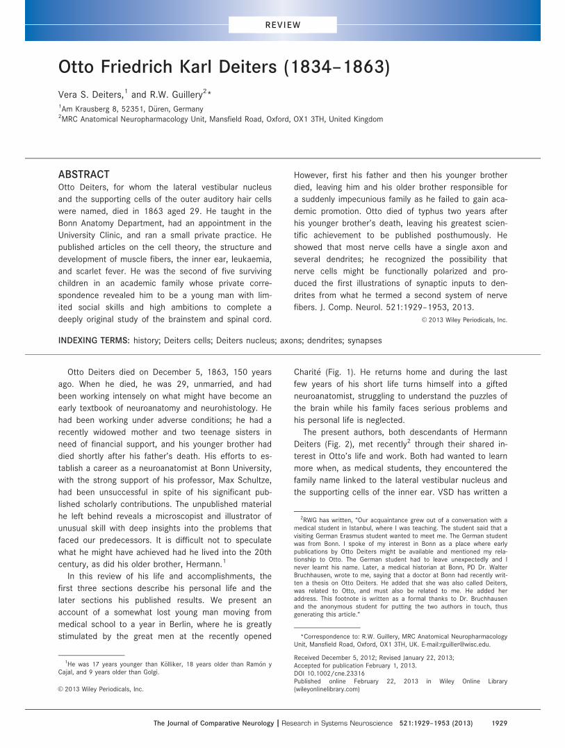
Otto Friedrich Karl Deiters (1834–1863)
Vera S. Deiters,1 and R.W. Guillery2*1Am Krausberg 8, 52351, D€uren, Germany2MRC Anatomical Neuropharmacology Unit, Mansfield Road, Oxford, OX1 3TH, United Kingdom
ABSTRACTOtto Deiters, for whom the lateral vestibular nucleus
and the supporting cells of the outer auditory hair cells
were named, died in 1863 aged 29. He taught in the
Bonn Anatomy Department, had an appointment in the
University Clinic, and ran a small private practice. He
published articles on the cell theory, the structure and
development of muscle fibers, the inner ear, leukaemia,
and scarlet fever. He was the second of five surviving
children in an academic family whose private corre-
spondence revealed him to be a young man with lim-
ited social skills and high ambitions to complete a
deeply original study of the brainstem and spinal cord.
However, first his father and then his younger brother
died, leaving him and his older brother responsible for
a suddenly impecunious family as he failed to gain aca-
demic promotion. Otto died of typhus two years after
his younger brother’s death, leaving his greatest scien-
tific achievement to be published posthumously. He
showed that most nerve cells have a single axon and
several dendrites; he recognized the possibility that
nerve cells might be functionally polarized and pro-
duced the first illustrations of synaptic inputs to den-
drites from what he termed a second system of nerve
fibers. J. Comp. Neurol. 521:1929–1953, 2013.
VC 2013 Wiley Periodicals, Inc.
INDEXING TERMS: history; Deiters cells; Deiters nucleus; axons; dendrites; synapses
Otto Deiters died on December 5, 1863, 150 years
ago. When he died, he was 29, unmarried, and had
been working intensely on what might have become an
early textbook of neuroanatomy and neurohistology. He
had been working under adverse conditions; he had a
recently widowed mother and two teenage sisters in
need of financial support, and his younger brother had
died shortly after his father’s death. His efforts to es-
tablish a career as a neuroanatomist at Bonn University,
with the strong support of his professor, Max Schultze,
had been unsuccessful in spite of his significant pub-
lished scholarly contributions. The unpublished material
he left behind reveals a microscopist and illustrator of
unusual skill with deep insights into the problems that
faced our predecessors. It is difficult not to speculate
what he might have achieved had he lived into the 20th
century, as did his older brother, Hermann.1
In this review of his life and accomplishments, the
first three sections describe his personal life and the
later sections his published results. We present an
account of a somewhat lost young man moving from
medical school to a year in Berlin, where he is greatly
stimulated by the great men at the recently opened
Charit�e (Fig. 1). He returns home and during the last
few years of his short life turns himself into a gifted
neuroanatomist, struggling to understand the puzzles of
the brain while his family faces serious problems and
his personal life is neglected.
The present authors, both descendants of Hermann
Deiters (Fig. 2), met recently2 through their shared in-
terest in Otto’s life and work. Both had wanted to learn
more when, as medical students, they encountered the
family name linked to the lateral vestibular nucleus and
the supporting cells of the inner ear. VSD has written a
*Correspondence to: R.W. Guillery, MRC Anatomical NeuropharmacologyUnit, Mansfield Road, Oxford, OX1 3TH, UK. E-mail:[email protected].
Received December 5, 2012; Revised January 22, 2013;Accepted for publication February 1, 2013.DOI 10.1002/cne.23316Published online February 22, 2013 in Wiley Online Library(wileyonlinelibrary.com)VC 2013 Wiley Periodicals, Inc.
1He was 17 years younger than K€olliker, 18 years older than Ram�on yCajal, and 9 years older than Golgi.
2RWG has written, “Our acquaintance grew out of a conversation with amedical student in Istanbul, where I was teaching. The student said that avisiting German Erasmus student wanted to meet me. The German studentwas from Bonn. I spoke of my interest in Bonn as a place where earlypublications by Otto Deiters might be available and mentioned my rela-tionship to Otto. The German student had to leave unexpectedly and Inever learnt his name. Later, a medical historian at Bonn, PD Dr. WalterBruchhausen, wrote to me, saying that a doctor at Bonn had recently writ-ten a thesis on Otto Deiters. He added that she was also called Deiters,was related to Otto, and must also be related to me. He added heraddress. This footnote is written as a formal thanks to Dr. Bruchhausenand the anonymous student for putting the two authors in touch, thusgenerating this article.“
The Journal of Comparative Neurology | Research in Systems Neuroscience 521:1929–1953 (2013) 1929
REVIEW

doctoral thesis (Deiters, 2006) based largely on perso-
nal records kept in her family, and RWG wrote briefly
about Otto’s work and life in three earlier papers. One
marked the 100th anniversary of Otto’s death (Deiters
and Guillery, 1964) and was written with his great aunt,
who was a psychiatrist in D€usseldorf and the daughter
of Otto’s Brother Hermann. The other two were about
the neuron doctrine that included comments on the
relevance of Otto’s work (Guillery, 2005, 2007). We
combine and extend these earlier studies now to mark
the 150th anniversary of Otto’s death. He died showing
significant promise of becoming one of the great neuro-
histologists of his generation. Most of the material pre-
sented in the early sections is based on VSD’s thesis,
and the rest is largely based on Otto’s published work.
EARLY LIFE AND EDUCATION
ChildhoodOtto Deiters was born on November 15, 1834, the
second son of Franz Peter Ignaz Deiters and Emilie
Eleonore Henriette Deiters (born Bausch). The family
tree in Figure 2 includes his siblings, two of which died
in their infancy; Otto and his younger brother Max died
before they were 30. The father was a professor in the
Law Faculty of Bonn University. He served five terms as
Dean of the Faculty and two (1845–1846 and 1856–
1857) as Rector. The University, founded in 1818 and
small by today’s standards, had many eminent faculty
members including von Helmholtz, Johannes M€uller, and
Eduard Pfl€uger, and students including Theodor
Schwann (who followed M€uller to Berlin), Heinrich
Heine, Prince Albert, and Karl Marx. Bonn was a small
town of 35,0003 inhabitants, first reached by the rail-
way in 1848. In that year Franz Peter Deiters served as
an elected representative from Bonn to the Frankfurt
National Assembly, which attempted to establish a par-
liamentary democracy in Germany.4 He served on the
constitutional committee of that parliament as a liberal
member. He died, aged 57, in 1861, leaving a finan-
cially strapped widow and five children still needing fi-
nancial support: Hermann just 27, Otto at 26 struggling
to establish his professional status, a younger brother
Max, at 23 starting a career in mining, and the two
youngest daughters, aged 15 and 19.5
The family was, like most others in the Rhineland,
Catholic. It was a respectable and serious academic
family, the children learning Latin and French at an
early age.6 When he was 10, Otto wrote in German to
his parents: “Today on the first day of the New Year, I
find myself particularly impelled (besonders angetrie-
ben) to say to you in writing, how much I love you and
my heart thanks you for the manifest blessings
(erzeigten Wohltaten). May the Almighty hear my prayer
and grant you the proper blessings, for your great care
and the many sacrifices you have made to give me
pleasure and through a good upbringing make me
happy. . ..”7 This was a family dedicated to high aca-
demic achievement.
While the father was at the Frankfurt parliament, he
wrote to 15-year-old Hermann about some political
issues Hermann had raised, advising him not to get
involved with politics as they might keep him from his
present duties (presumably school), and then adding
that he would like to hear more about what he and
Otto did during the holidays, “. . .and at your next op-
portunity to communicate this also to Otto. If it is not
overly burdensome for Otto, with a definite topic in
mind letter writing would not be as difficult as he
Figure 1. Portrait of Otto Deiters by August Bausch, 1858.
3https://commons.wikimedia.org/wiki/File:Bonn_population.svg. Thiscompares to Berlin’s 400,000 at the same time (Namier, 1992).
4This “revolutionary“ parliament included 49 university professors andlecturers and 57 schoolmasters among its more than 800 elected mem-bers, according to Namier (1992), who described the 1848 revolution asthe revolution of the intellectuals.
5We have no information about the reasons for the family’s financialproblems. They added to the pressures on Otto before his own death twoand a half years after his father’s and two years after that of his youngerbrother, Max.
6Hermann wrote accounts of his holidays in Latin for his father.7Quotation marks here and subsequently show passages translated
from the German. Parentheses enclose original German words having pos-sible alternative meanings.
V.S. Deiters and R.W. Guillery
1930 The Journal of Comparative Neurology |Research in Systems Neuroscience

seems to believe. This would please me especially,
although you can assure Otto that I have not taken his
past silence amiss, but have understood it.” The father
was aware of Otto’s withdrawn and unsociable nature,
which figures significantly in the family correspondence,
and one wonders how much of the father’s message
Hermann passed on to Otto.
The family was musical. Otto played violin and viola.
He and Hermann organized a musical circle in 1852 that
met every week and played classical music, mostly quar-
tets, whose rules still survive (Deiters, 2006). The last
rule states that each member has the duty to practice
with others and if necessary on their own. Hermann was
much more sociable and outgoing than Otto and had a
lifelong interest in music; much of the correspondence
between the brothers was about music or drama, com-
plete with details of performances and critical com-
ments, e.g., “Mr. Frank from Cologne played Beethoven’s
C minor piano concerto, then a Bach fugue, which I have
never heard you play, and then a little charming compo-
sition of his own. . .and then Frau Nissen Salomon sang
a few charming Swedish songs and a quite abominable
aria by Verdi tolerably, but a wonderful aria from Ales-
sandro Stradella very badly. She trilled and warbled
frightfully.”8 This group of musicians continued to meet
while Otto was in medical school and included a fellow
medical student, Carl Liebermeister (see below).
Medical school at Bonn (1852–1857)In 1852 Otto graduated from his school with positive
comments from his teachers about his moral leader-
ship, his diligence and his knowledge of the subjects
that included, German, Latin, Greek, French, Hebrew,
Religious studies, Mathematics, History and Geography,
Physics and Philosophical Propaedeutics. He started in
medical school with no Biology or Chemistry, which
would play a major role in his later studies. Although
his father and older brother studied humanities, he was
not the only medical student in the family. A maternal
uncle, Friedrich Bausch, had studied medicine at Bonn
in 1832 and left a bound 400 page volume of his neatly
handwritten notes of the lectures that Johannes Mueller
delivered to the students that year9.
In reaction to Hermann’s suggestion, that Otto was
not establishing enough friendships, Otto wrote: “You
seem to think that I am one of those who invest all of
their needed learning abilities in the courses and who
have a who knows what sort of a conscience if they
just once, horribile dictum, cut classes for an hour.
There you are mightily in error. . . as a medic one is
closer to the professors, and if in addition to that one
is the son of one’s father and the father happens to be
a colleague of the professor, if, further, one is sitting
with a maximum of 3 to 6 colleagues, then you will
understand that one can’t always do what one wants.”
We have relatively little information about Otto’s years
in medical school. As the above letter shows, he felt iso-
lated. His later correspondence (see the next section)
indicates that he still had to learn to focus his studies
on the essentials. His musical activities continued, but
he worked through his studies rather mechanically, and
he wrote to Hermann in 1854: “I go to the college, then
sit again in my room, go again to the college and then
sit once more in my room, and that’s how it goes, from
one day to the next. There are no people that I am close
to, I have no friends. That’s bleak (trostlos), isn’t it?” We
have very little information about this period of his life.
He was living at home, was insecure about his studies,
and left limited correspondence.
Otto’s doctoral thesis, “De Incremento Musculorum”
(Deiters, 1856), in Latin, was about the growth of
muscles, documenting the sizes of many different
muscles in many different species (human, ox, rabbit,
dormouse, dove, several species of frog, salamander
and carp), comparing ages, and on the basis of the
means and what would today be recorded as variances
of the measurements concludes that muscle growth is
due to an increase of the volume of the individual mus-
cle fibers not to an increase in their numbers10. His
thesis advisor was Julius Budge.
Figure 2. Family tree of the immediate Deiters family.
8A letter from Otto to Hermann, November 17, 1854.
9Now held by Bonn University.10For non-Latin speakers Otto summarizes this study in Deiters (1861a),
with regrets that these results are not well known.
Otto Friedrich Karl Deiters (1834–1863)
The Journal of Comparative Neurology | Research in Systems Neuroscience 1931

BERLIN: LIVING AWAY FROM HOME,MILITARY DUTY, AND LECTURES AT THECHARIT�E (1857–1858)
Otto’s one year in Berlin was an important period in
his short life, significantly igniting his interest in medical
studies. Although this was primarily his year as a volun-
teer in the army, it included attendance at lectures by
Virchow, Johannes M€uller, and Romberg, and was the
only extended period of his life away from his family. It
stimulated him to start thinking seriously about his ca-
reer goals, and woke him to a wider view for his future.
Otto served as a volunteer in the artillery of the 2nd
Guards Regiment in Berlin. He had a low view of the
military and for him this was primarily an opportunity to
visit Berlin and attend Virchow’s lectures at the recently
established Charit�e. Writing to his younger brother Max,
he evaluated his choices, basing them on how close to
the University the barracks were, how well educated
the officers were, and the severity of the drill exercises.
Of the artillery he wrote: “. . .when the regiment is on
the move the soldiers dawdle without a heavy pack and
in no strict order behind the cannon,” contrasting this
to the soldiers of the infantry with heavy packs and in
strict order.11
Otto went to Berlin two months ahead of his army
service so he could attend Virchow’s lectures. Virchow
had written: “Only a few have advanced enough to have
learnt really to think in microscopical terms. . .. For
most older doctors, microscopy is like a foreign lan-
guage, where one can freely use foreign words but
thinks in one’s own language.”12 Otto’s brief period
with the students who were learning the “new
language” from Virchow would be strongly reinforced af-
ter his return to Bonn in 1858 by Max Schultze, who
was appointed to the Bonn Anatomy chair in 1859.
Virchow had also written: “The natural scientist knows
the body and the properties of the body, what is
beyond he calls transcendent and regards the tran-
scendent as an error of the human mind.” Otto cited
this at the head of his dissertation.
During this period in Berlin, Otto also attended the
comparative anatomy lectures of Johannes M€uller, and
wrote to Hermann, “. . .everything that M says is signifi-
cant and ingenious to the highest degree but in one
summer semester there is not time to go into the spe-
cial points, and this also applies to Virchow’s pathologi-
cal anatomy. In both [fields] private studies are
essential. Here microscopic anatomy is only taught by
younger staff (Privatdocenten). For my own studies one
thing is absolutely necessary. Requirement one
microscope.”13
The father had previously advised Otto to borrow a
microscope, and Otto had told him this was impossible.
The crucial issue at the time was the achromatic lens,
which was produced by only a few companies, and pro-
vided much clearer views because these lenses cor-
rected the chromatic aberration characteristic of earlier
lenses, which had brought each wavelength to a differ-
ent focal point and introduced significant blurring of the
image, especially at higher magnifications. In July 1857
the father advised Otto that Leitz had begun to produce
microscopes that were greatly in demand and told Otto
not to delay in placing his order. It appears that the fa-
ther found funds, but did not live long enough to see
the extraordinary results Otto would produce.
Here we encounter the sad financial position of the
Deiters family. Otto’s father had previously written to a
colleague requesting help to find a position for Otto, his
talented but impecunious son. The father complained
about his own financial position, and added “if you find
him [Otto] somewhat enclosed, restrained, reserved
(verschwiegen), then I can tell you that I hear and have
grounds for believing that if this ring can be broken, a
good core will be found” (Deiters, 2006). Not surpris-
ingly, the letter produced nothing.
The letters from Otto in Berlin to his brothers (cited
more fully in Deiters, 2006) are entertaining and include
critiques of (good and bad) musical performances in
Berlin, showing that he was more aware of others than
many of his later actions and his views of himself might
suggest. He writes about the Tiergarten, implicitly com-
paring city promenades with the real walks possible in
the Rhineland. The Tiergarten walks are spoilt by the
many typical Berliner family outings: “. . .maids with
their cousins and children, old women in Amazon hats
(Grisetten in Amazonh€uten),14 careful parents with their
marriageable daughters. . .arrive at one of the many
inns, they all sit, the whole Berliner hoi-polloi (Philiste-
rium), each family at one table in the middle of which a
towering glass of Weissbier (eine Weisse) is enthroned,
and then with dignified demeanour the honoured family
father takes a draught whereupon the others follow in
order (secundum ordinensis). Such a thing is only possi-
ble in Berlin. . ..”
11Otto to Max Deiters, May 18, 1858. This was before the reorganiza-tion of the Prussian Army (see Grenville, 1976).
12Cited by Deiters (2006).
13Otto to Hermann, May 26, 1857.14A stylish hat. A poem appeared in 1857 entitled “ Der Amazonenhut—
Mit achtzehn Jahren wohlgetan—mit zweiundzwanzig geht’s noch an—Mitdreißig Jahr’ bewahr’ uns Gott!—Mit sechsunddreißig—Kinderspott!“ See“Eisenbergisches Nachrichtsblatt 1857 at http://zs.thulb.uni-jena.de/receive/jportal_jparticle_00081264. In English: The Amazon hat: At 18 itis well done—at 22 just possible—at 30, ’God preserve us’—and at 36 thechildren mock.
V.S. Deiters and R.W. Guillery
1932 The Journal of Comparative Neurology |Research in Systems Neuroscience
Otto’s Berlin letters also tell us more about family
relationships: “Papa’s letter has arrived safely. The
somewhat unworthy (unw€urdige) title, junior doctor, can
be left out of the address.” Another letter, in which
Otto replies to Hermann’s request that Otto look in Ber-
lin for a suitable silver wedding gift for the parents, pro-
vides a close domestic view: “We must respect the
principle of usefulness so valued in our home.” Such
items as pictures or valuable rings are not to be consid-
ered. He discusses a clock for the hall or some com-
fortable chairs, which may be needed, but advises that
they can be purchased in Bonn or Cologne as well as
Berlin. We do not know whether the family gained the
comfortable armchairs they lacked or the clock that
would keep them all on time.
In 1857 Otto also began to act as informal medical
adviser to Hermann, sounding authoritative, dogmatic,
and uncompromising. On the basis of Hermann’s
account of his indisposition, Otto had diagnosed a gas-
tric and intestinal problem, “perhaps augmented, so far
as I can see, by a gallbladder problem. . .you think that
in spite of Albers’s15 energetic treatment you are not
completely cured. I can believe that. These conditions
usually go away on their own without any energetic
treatment. . .above all you must not be thinking about
yourself as having an unhealthy nature. Our family has
a healthy nature.” Otto goes on to advise Hermann to
live a less sedentary life, take more walks, exercise,
and drink less tea and coffee. He sounds stern, not like
a younger brother. Hermann must have replied by
reporting that he was better, and complaining that he
was not ready to follow Otto’s rather rigorous rules,
because Otto’s next letter expresses pleasure at the
improvement and hopes it will last: “You consider the
correct dietary regime to be of no consequence, and I
could hardly expect anything different, but that must
not be. . ..” Another harangue about healthy diet and
exercise follows complete with a statement about
patients who fail to follow the doctor’s advice. But he
ends on a peace-making note, saying, “I did not mean,
as you seem to have assumed. . .that you should at this
point give up your social life and follow my bad
example.”
Although Otto was a private, self-enclosed person
and was recognized as such by family and colleagues,
he had some close friends, including his fellow musi-
cians, particularly Carl Liebermeister,16 with whom he
corresponded after they graduated. The letters from
Liebermeister contain advice about examiners at other
universities. From Greifswald, where he was studying,
he wrote: “. . .he who knows his subject reasonably well
is better off going to Greifswald, but he who knows
nothing could fake his way in Berlin. . ..You could, with
a clear conscience, advise people to go to Greifswald.”
He invited Otto to visit him in Greifswald, and a later
letter to Otto from N. Simrock, school friend and fellow
medical student, includes a statement that he was glad
to hear that the two had met: “You both always fol-
lowed the same academic tendencies.” Later, Otto vis-
ited T€ubingen while Liebermeister was there as
prosector in Pathological Anatomy.
While he was in Berlin, Otto began to think seriously
about his future. He wrote a thoughtful letter to Her-
mann: “I have always worked hard, though not as hard
as people said. I had to, because quite apart from the
amount of material that had to be mastered, which was
overpowering, the time that was available to me because
of my reserved nature had to be occupied. There was a
lack of distractions. So at the beginning this work was
not of the right sort.” He now sees more specialist areas
that he had no time to review properly as a student. “I
don’t think that I judge my strengths too highly but I
don’t want to estimate them too lowly. . .. The position at
present is as follows: I no longer doubt that I would pre-
fer an academic career to one in a practice. . .this fits
best with my strengths. So I will be a docent, no matter
what.” He then asks about the long-term possibilities of
an academic career and about his choices. He is clear
about these: on the one hand, anatomy, particularly mi-
croscopic and comparative anatomy, and on the other,
pathological anatomy. He considers an assistant position
with Geheimrat Naumann in Bonn as a docent in patho-
logical anatomy, and finding a medical practice that
would later provide extra income (Sp€ater die n€othigen
Bed€urfnisse deckte). However, he worries about income,
and sees a Professor of Pathological Anatomy as a “fifth
wheel,” not making much money, while a serious clinical
commitment would limit his plans for academic work. He
reflects negatively about the position of a doctor in a
German practice: “If I don’t settle in the first best village
and don’t go to America, then I would risk as little by
planning on a simple academic career as I would by
expecting a remunerative practice: particularly so as the
field to which I always return has many income possibil-
ities. And that field. . .is anatomy. . .and I have decided to
dedicate myself to it.” He lists the income opportunities:
publications generated income then, as did private tutori-
als, which were more in demand as exams increased in
difficulty.
He decided nonetheless to maintain his clinical skills,
and in Berlin attended the clinical sessions presented
by Romberg and Langenbeck.
15Another Bonn faculty member in the pathology department.16Later Professor of Pathology and Therapy in Basel from 1865 and in
T€ubingen from 1871 (Deiters, 2006).
Otto Friedrich Karl Deiters (1834–1863)
The Journal of Comparative Neurology | Research in Systems Neuroscience 1933
N. Simrock, who had moved to Cologne, compared
Otto’s enthusiasm about academic opportunities with
his own lack of academic ambition. He was glad to be
in Cologne, he wrote, where “. . .a zealous academic
engagement is impossible. . .I know for you it is differ-
ent, and even if your well-known unassuming modesty
stops you from recognizing it, it will later be a benefit
for the faculty that has the good luck to count you
among its young chosen ones.”
In another letter, before Otto moved back to Bonn,
Simrock wrote about Otto’s planned appointment with
Naumann: “That you have accepted the position with
Naumann not only does not surprise me, but, on the
contrary, I think it totally understandable, since in such
a position you can soonest reach your goals. Only I
would. . .in relation to him put myself in an independent
position, which is something he may assume.” Here
Simrock anticipates later problems for Otto’s career,
based on the differing approaches to medicine of Otto
and Naumann: the former scientific, the latter philo-
sophical and romantic.
In 1858 Otto completed his military service and
returned to Bonn as first assistant to Geheimrat Nau-
mann, spending the next three years with him.
MEDICAL CAREER IN BONN (1858–1863)
Clinical work and publicationsIn Bonn Otto took on clinical responsibilities in Nau-
mann’s clinic and also started private clinical work as a
general practitioner, visiting patients in Bonn and sur-
rounding areas17 to boost his income. He also began to
work on his habilitation, which would qualify him as a
lecturer at the University. He must also at an early
stage have begun detailed investigations of the inner
ear in several different species, which led to four publi-
cations by 1862 (see the section, The organ of Corti
and Deiters cells, below).
In 1859 he published a brief account of two cases of
scarlet fever (Deiters, 1859a) and later a longer article
about leukemia (Deiters, 1861a). The former stressed
that current treatments were generally ineffective and
showed that incidence of the infection could not be
related to particular home conditions of the patients;
the latter presented a detailed account of the physical
(wasted) condition of a terminally ill young man who
died in the hospital. Otto described the postmortem
findings, the major features being lymphatic swelling of
the peritoneum, in which he noted hypertrophic cells
and abnormal blood cells. He stressed how little was
known about the disease. From our point of view, his
rigorous demand for convincing evidence before coming
to any conclusions about a disease is the most note-
worthy part of both accounts.
HabilitationOtto wrote an essay on the present position of the
cell theory (Deiters, 1859b). It has a strange beginning:
“Half a century has passed since the sharp eyes of a
genial French investigator succeeded in winning an
insight for academic anatomy that until then was com-
pletely strange and for which hardly anyone else was
suited. . ..” The long, complex sentence continues, but
Xavier Bichat, who is often regarded as the father of
histology (although he never used a microscope), is not
identified as the sharp-eyed French investigator until
Deiters had introduced many others (Leeuwenhoek,
Malpighi, Linnaeus, and Cuvier) and compared their
efforts at categorizations in biology with the successes
of chemists, Lavoisier in particular. In this long,
“scholarly” review, Otto covered much of the relevant
literature, although the version that was published as a
lecture lacked a list of references and looks as though
it was prepared in a hurry, probably because Otto
needed the qualification to start giving lectures that
would bring in some fees. Although it is now considered
that the cell theory was established earlier by Schleiden
and Schwann, when Otto wrote, there was significant
disagreement about exactly what the cell theory
claimed and how far Schwann’s account was an accept-
able view of how tissues are organized and develop (for
fuller historical accounts, see Baker, 1948, 1949; Har-
ris, 1999). Otto had started to look at the inner ear and
had noticed some cells that had a large hole, which
had pushed the cytoplasm, nucleus, and nucleolus to a
small border region. For reasons not made clear, Otto
found it difficult to conceive of these fenestrated cells
as bound by a membrane. He introduced these cells as
presenting one of several difficulties faced by the cell
theory at that time. Other difficulties included cells
lacking a nucleus and cells lacking a nucleolus. The
lack of a membrane long remained a difficulty until the
electron microscope allowed clear identification of the
thin, approximately 5-nm cell membrane.18 Otto is look-
ing for a strong theory based on the physics and chem-
istry of the cell, one that covers not only the anatomy
of the cell but also the physiology. Even the strongest,
most basic part of the cell theory, that cells only arise
from other cells, he considers not yet sufficiently well
17He advised the patients on the local farms to not drink fresh milk, buthis advice went largely unheeded.
18The 1948 edition of Maximow and Bloom’s A Textbook of Histology,predating the use of electron microscopes in biology, relied on the semi-permeable nature of the membrane and its resistant and elastic propertiesdemonstrable by microdissection as the major evidence for the existenceof a cell membrane.
V.S. Deiters and R.W. Guillery
1934 The Journal of Comparative Neurology |Research in Systems Neuroscience

established, in spite of mounting evidence, which he
summarizes. The problem of exactly what the cell
theory does include was not resolved at the time and
may even today not be clearly agreed. This is an issue
that plays an important role in Otto’s later work and is
revisited in the section on Otto’s book, below.
EARLY PUBLICATIONS IN ANATOMYThe organ of Corti and Deiters cells
In 1859, soon after returning to Bonn, Otto published
an important study of the organ of Corti (Deiters,
1859c) that included a detailed account of the cells
that, at Max Schultze’s suggestion, were later named
Deiters cells. These cells support the outer hair cells
and provide a link between these hair cells and the
basilar membrane. Between 1859 and 1862, Otto also
published a book (Deiters, 1860a) and two other papers
on the inner ear (Deiters, 1860b, 1862). These are
based partially on sectioned material, but primarily rely
on preparations that were teased out under the micro-
scope. Otto compared the structures, based on their
flexibility or fragility, watching how they reacted to vari-
ous chemicals serving as fixatives (dichromates, chro-
mates, or acetic acid) or dyes (iodine, carmine). These
methods were widely used by early microscopists. The
practice of first fixing a piece of tissue and then
embedding and sectioning it before ever looking at it
developed later. The method that Otto used is relevant
for all of Otto’s microscopic studies. He was watching
the reactions of tissues to his chemical and physical
manipulations under the microscope in order to reach
his conclusions about their characteristics. We know no
details about the microscope or the lenses that Otto
used, and have no information about the “tools” that he
used (probably needles, fine pipettes, delicately shaped
glass rods, perhaps even fine, hand-sharpened knives,
etc.).
It is important to recognize that for this work and for
his later microscopic studies of nerve and glial cells,
Otto’s tissues were generally in an aqueous solution as
he was manipulating them. They would not have been
cleared in a medium of appropriate refractive index nor
would he have been able to manipulate the tissue had
they been covered by a coverslip.19 Water immersion
lenses were available in the late 1850s and early
1860s,20 but we have no information about the lenses
that Otto used. It is possible that Otto did some of his
fine dissections with a water immersion lens, but the
difficulties of manipulating small objects in conditions
where surface tension effects can make the manipula-
tions difficult (or impossible) have to be recognized. For
these studies of the inner ear (see Figs. 3 and 4) and
for his later studies of neurons and glia (e.g., Plate
ODU II, Fig. 10), he produced extraordinary details of
very small cells, often unmatched at the time, but the
special tools, methods, or skills that he used are not
known to us.
Otto’s first study of the organ of Corti (Deiters,
1859a) was based on material from dog, cat, and calf.
It is extensively illustrated by Otto’s own drawings. Otto
criticizes K€olliker’s earlier account of nerve fibers end-
ing in the large “cells of Claudius,” describing this as a
physiological impossibility21 but focuses on the struc-
tures that link the hair cells to the reticular lamina and
the basilar membrane. Figure 3 shows the structure of
a Deiters cell as seen in a current electron microscope
study for comparison with Otto’s figure (Fig. 4), which
Figure 3. A Deiters cell shown schematically. The figure is based
on a scanning electron micrograph (Fig. 6B of Parsa et al., 2012).
The cell stretches between the reticular membrane (above) and
the basilar membrane (below), and the stereocilia of the hair cells
(not shown) project above the reticular membrane. Note that in
the original figure there are fine nerve fibers that run horizontally
across the Deiters cells. These nerve fibers are in part often
enclosed within the cytoplasm of the Deiters cell and are not
shown, nor are the rich terminals that lie deep within the region
where the Deiters cell encloses the base of the hair cell. Scale
bar5 20 lm.
19However, Schultze in the introduction to Otto’s posthumous bookmentions many microscopic preparations that Otto left behind, preservedand cleared in balsam and covered.
20See http://www.smecc.org/history_of_oil_immersion_lenses.htm.
21Presumably because the body of these nerves is in the spiral ganglioncells. Here Otto is possibly suggesting, but not exploring, an objection toa fusion of two cells. K€olliker in the 1863 edition of his textbook withdrewthis suggestion on the basis of newer developmental studies but withoutmentioning Deiters.
Otto Friedrich Karl Deiters (1834–1863)
The Journal of Comparative Neurology | Research in Systems Neuroscience 1935
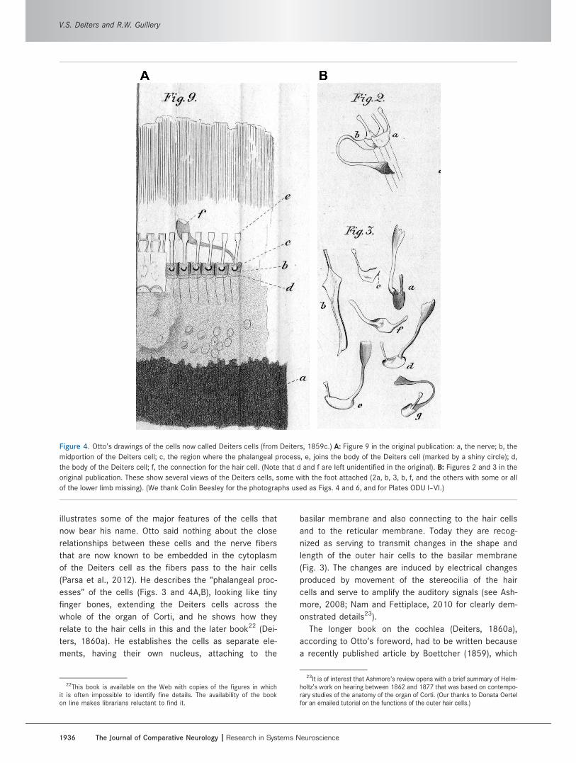
illustrates some of the major features of the cells that
now bear his name. Otto said nothing about the close
relationships between these cells and the nerve fibers
that are now known to be embedded in the cytoplasm
of the Deiters cell as the fibers pass to the hair cells
(Parsa et al., 2012). He describes the “phalangeal proc-
esses” of the cells (Figs. 3 and 4A,B), looking like tiny
finger bones, extending the Deiters cells across the
whole of the organ of Corti, and he shows how they
relate to the hair cells in this and the later book22 (Dei-
ters, 1860a). He establishes the cells as separate ele-
ments, having their own nucleus, attaching to the
basilar membrane and also connecting to the hair cells
and to the reticular membrane. Today they are recog-
nized as serving to transmit changes in the shape and
length of the outer hair cells to the basilar membrane
(Fig. 3). The changes are induced by electrical changes
produced by movement of the stereocilia of the hair
cells and serve to amplify the auditory signals (see Ash-
more, 2008; Nam and Fettiplace, 2010 for clearly dem-
onstrated details23).
The longer book on the cochlea (Deiters, 1860a),
according to Otto’s foreword, had to be written because
a recently published article by Boettcher (1859), which
Figure 4. Otto’s drawings of the cells now called Deiters cells (from Deiters, 1859c.) A: Figure 9 in the original publication: a, the nerve; b, the
midportion of the Deiters cell; c, the region where the phalangeal process, e, joins the body of the Deiters cell (marked by a shiny circle); d,
the body of the Deiters cell; f, the connection for the hair cell. (Note that d and f are left unidentified in the original). B: Figures 2 and 3 in the
original publication. These show several views of the Deiters cells, some with the foot attached (2a, b, 3, b, f, and the others with some or all
of the lower limb missing). (We thank Colin Beesley for the photographs used as Figs. 4 and 6, and for Plates ODU I–VI.)
22This book is available on the Web with copies of the figures in whichit is often impossible to identify fine details. The availability of the bookon line makes librarians reluctant to find it.
23It is of interest that Ashmore’s review opens with a brief summary of Helm-holtz’s work on hearing between 1862 and 1877 that was based on contempo-rary studies of the anatomy of the organ of Corti. (Our thanks to Donata Oertelfor an emailed tutorial on the functions of the outer hair cells.)
V.S. Deiters and R.W. Guillery
1936 The Journal of Comparative Neurology |Research in Systems Neuroscience
contained “next to some correct results, a number of
incomplete and mistaken results. . .made it my duty not
to wait any longer with my own results.”24 The book is
based on studies of rabbit, mouse, cat, dog, calf, horse,
human, and guinea pig. Two further studies (Deiters,
1860b, 1862) extended his material to birds (starling,
yellow hammer, hen, pigeon, crow, magpie, two species
of lizard, slow-worms, grass-snakes, and probably more
than one species of frog.) Reading these studies today,
or K€olliker’s 1863 account, and comparing them with
what is now known, shows how much our knowledge
has advanced and how difficult it was in the 1860s to
trace nerve fibers when they lie very close to or are
enclosed in the cytoplasm of the supporting cells.
Studies of striated muscles (Deiters,1861b)25
This was a continuation of his thesis on muscles,
using developmental studies of muscle growth in the
regenerating tip of a tadpole tail. The surviving muscle
fibers grow but do not contribute to the regenerated
tail. New cells form from stellate or bipolar connective
tissue cells, and these develop a striated fiber along
one border. He draws these finely striated fibers lying
next to the cells, and parts of the fibers appearing to
separate from the cells, with the fibers then growing
beyond the cells. He regards the striated fibers pro-
duced in this way as an intercellular component that is
subsequently incorporated together with the cell in a
common membrane, to make a new multinucleate mus-
cle fiber. He mentions that J. Budge disagreed with
Otto’s conclusions and has agreed that Otto should
include a discussion of their different views, which he
does. Here Otto is more than usually deferential to his
former thesis adviser, who later became his friend.
TEACHING, LEAVING CLINICAL WORK,AND STARTING TO STUDY THE CENTRALNERVOUS SYSTEM
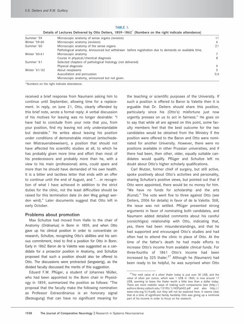
TeachingIn April 1859 Otto was granted the position of Privat-
docent. In addition to continuing with his clinical
responsibilities in the hospital, he worked with his pri-
vate patients and started to offer lectures in the medi-
cal school (Table 1). The lectures on auscultation were
a novelty in Bonn at the time. Although Otto was popu-
lar among the students, his occasional disrespectful
comments about colleagues, whose approach to medi-
cine he considered less modern and objective than his
own, damaged his relationships with some faculty col-
leagues and would cost him dearly before long.
A family crisis; leaving clinical workIn March of 1861, the death of Otto’s father follow-
ing a stroke added a significant extra burden to Otto’s
life, as did the death of his younger brother Max, 8
months later, under circumstances that leave suicide a
possibility (Deiters, 2006). This left Hermann and Otto
with the sole responsibility for supporting the family,
which had to move into cheaper accommodations. The
youngest daughter, Paula, started training as a teacher
in order to generate her own income, with Otto writing
the health certificate that supported her application. At
this time Otto also started to add microscopic reports
for colleagues to his own earnings and time
commitments.
On May 28, 1861, he wrote a letter to a professor,
unidentified on the surviving draft26 and unknown to us,
calling on the goodwill the professor had often shown
Otto in the past. He reported the father’s death and
the family’s limited financial circumstances. He men-
tioned his present position (Assistant in the Medical
Clinic and Privatdocent in Anatomy) as holding no
promise for the future and leaving only limited opportu-
nities for scientific work. If he were to continue in
Bonn, he would be forced by financial considerations to
sacrifice his scientific work for practical medical activ-
ities. He asked for help in finding a suitable position
and recalled that in the past the professor had indi-
cated an inclination to find a position for Otto in
Heidelberg.
We have no record of any answer from Heidelberg.
Otto’s views about his future in Bonn represented a
correct assessment of relationships there. In spite of
the family’s needs, in June Otto gave notice to Nau-
mann that in view of his changed family circumstances
he would be leaving his Clinical Assistant position in
the summer in order to concentrate on research. We
do not know the reasons for this decision, but subse-
quent events suggest that he knew his microscopic
studies were promising a rich harvest and that he had
to concentrate on them. Otto and his chief had differ-
ent approaches to medicine—modern and empirical ver-
sus older and more intuitive. The two did not relate
well, as anticipated in Simrock’s earlier letter. Otto
24Boettcher’s angry reaction (Boettcher, 1860) and Otto’s four-pageresponse (Deiters, 1860c) will not be explored here, but they representthe reactions that Otto’s lack of tact could generate. We have not beenable to address issues of priorities that arise from this interchange.
25In the same year Max Schultze (1961) published a paper on muscle,arguing that each nucleus of the multinucleate muscle cell represents adistinct cell not separated from the other nuclei (cells) by any membrane.Schultze’s account would have caused Otto some further problems had helived, and should be seen in relation to Otto’s interpretation in this study.
26Possibly Helmholtz, who had been a faculty colleague of Otto’s fatheruntil 1858, and who was in Heidelberg in 1861.
Otto Friedrich Karl Deiters (1834–1863)
The Journal of Comparative Neurology | Research in Systems Neuroscience 1937
received a brief response from Naumann asking him to
continue until September, allowing time for a replace-
ment. In reply, on June 21, Otto, clearly offended by
this brief note, wrote a formal reply: A verbal discussion
of his motives for leaving was no longer desirable: “I
have had to conclude from your note that you, from
your position, find my leaving not only understandable
but desirable.” He writes about leaving his position
under conditions of demonstrable mistrust (entschiede-
nen Mistrauensbeweisen), a position that should not
have affected his scientific studies at all, to which he
has probably given more time and effort than most of
his predecessors and probably more than he, with a
view to his main (professional) aims, could spare and
more than he should have demanded of his own health.
It is a bitter and tactless letter that ends with an offer
to continue until the end of August, and “. . .in recogni-
tion of what I have achieved in addition to the strict
duties for the clinic, not the least difficulties should be
raised for this termination date (in den Weg gelegt wer-
den wird).” Later documents suggest that Otto left in
early October.
Problems about promotionMax Schultze had moved from Halle to the chair of
Anatomy (Ordinarius) in Bonn in 1859, and when Otto
gave up his clinical position in order to concentrate on
research, Schultze, recognizing Otto’s abilities and his seri-
ous commitment, tried to find a position for Otto in Bonn.
Early in 1862 Baron de la Valette was suggested as a can-
didate for a prosector position in Anatomy, and Schultze
proposed that such a position should also be offered to
Otto. The discussions were protracted (langwierig), as the
divided faculty discussed the merits of this suggestion.
Eduard F.W. Pfl€uger, a student of Johannes M€uller,
who had been appointed to the Bonn chair in Physiol-
ogy in 1859, summarized the position as follows: “The
proposal that the faculty make the following nomination
as Professor Extraordinarius is an honorary signal
(Bezeugung) that can have no significant meaning for
the teaching or scientific purposes of the University. If
such a position is offered to Baron la Valette then it is
arguable that Dr. Deiters should share this position,
particularly since his (Otto’s) misfortune just now
urgently presses on us to act in fairness.” He goes on
to say that while all are agreed on this point, some fac-
ulty members feel that the best outcome for the two
candidates would be obtained from the Ministry if the
position were offered to the Baron and Otto were nomi-
nated for another University. However, there were no
positions available in other Prussian universities, and if
there had been, then other, older, equally suitable can-
didates would qualify. Pfl€uger and Schultze left no
doubt about Otto’s higher scholarly qualifications.
Carl Wutzer, former chief of surgery, but still active,
spoke positively about Otto’s activities and personality,
sharing Schultze’s positive views, but pointed out that if
Otto were appointed, there would be no money for him.
“We have no funds for scholarship and the arts
(Kunst).” The vote went five to three against Otto (see
Deiters, 2006 for details) in favor of de la Valette. Still,
the issue was not settled. Pfl€uger presented strong
arguments in favor of nominating both candidates, and
Naumann added detailed comments about his careful
(vorsichtiges) relationship with Otto, indicating that,
yes, there had been misunderstandings, and that he
had supported and encouraged Otto’s studies and had
often had to attend the clinic in place of Otto. At the
time of the father’s death he had made efforts to
increase Otto’s income from available clinical funds. For
three-fourths of 1861 Otto’s income had been
increased by 325 thaler.27 Although he (Naumann) had
been ready to be helpful, he was surprised when Otto
TABLE 1.
Details of Lectures Delivered by Otto Deiters, 1859–1862* (Numbers on the right indicate attendance)
Summer ’59 Microscopic anatomy of sense organs (revision) 11Winter ’59-60 Microscopic anatomy (revision) 6Summer ’60 Microscopic anatomy of the sense organs 21
Pathological anatomy. Announced but withdrawn before registration due to demands on available time.Winter ’60-61 Microscopic anatomy 7
Course in physical/chemical diagnosis 8Summer ’61 Selected chapters of pathological histology (not delivered)
Physical diagnosis 9Winter ’61-’62 About neoplasms 5
Auscultation and percussion 0.6Microscopic anatomy, announced but not given.
*Numbers on the right indicate attendance.
27The melt value of a silver thaler today is just over 30 US$, and thevalue of silver per ounce, which was 1 US$ in 1860, is now around 17US$, seeming to leave the thaler worth a little less than a dollar today.There are more realistic ways of making such comparisons (see (http://authors.library.caltech.edu/13155/1/HOFjeh02.pdf and also http://www.roiw.org/5/4.pdf), but they will not be explored here. It seems clearthat at a time of significant family hardship Otto was giving up a nontrivialpart of his income in order to focus on his research.
V.S. Deiters and R.W. Guillery
1938 The Journal of Comparative Neurology |Research in Systems Neuroscience
suddenly announced that in “consideration of his family
and his partiality (Vorliebe) for microscopy,” he could
not stay with the clinic; Naumann had pointed out to
Otto that his continuation in the clinic would bring an
annual income of 250 thaler. “I had to let him go. I was
sorry because I had got used to his friendly face and
modest ways.”
After an unusually long debate,28 it was decided to
send both names to the Minister, with strong recom-
mendations written for both by Schultze. The Minister
accepted the nomination of Baron de la Valette, so far
as we can tell without a salary, and rejected that of Dr.
Otto Deiters. Otto himself was and would remain bitter
about this decision during the brief and busy last two
years of his life. In 1874, Baron de la Valette suc-
ceeded Max Schultze as head of the Anatomy Depart-
ment, retiring in 1906. Today the Anatomy Department
has a bust of the Baron in the entrance hall but nothing
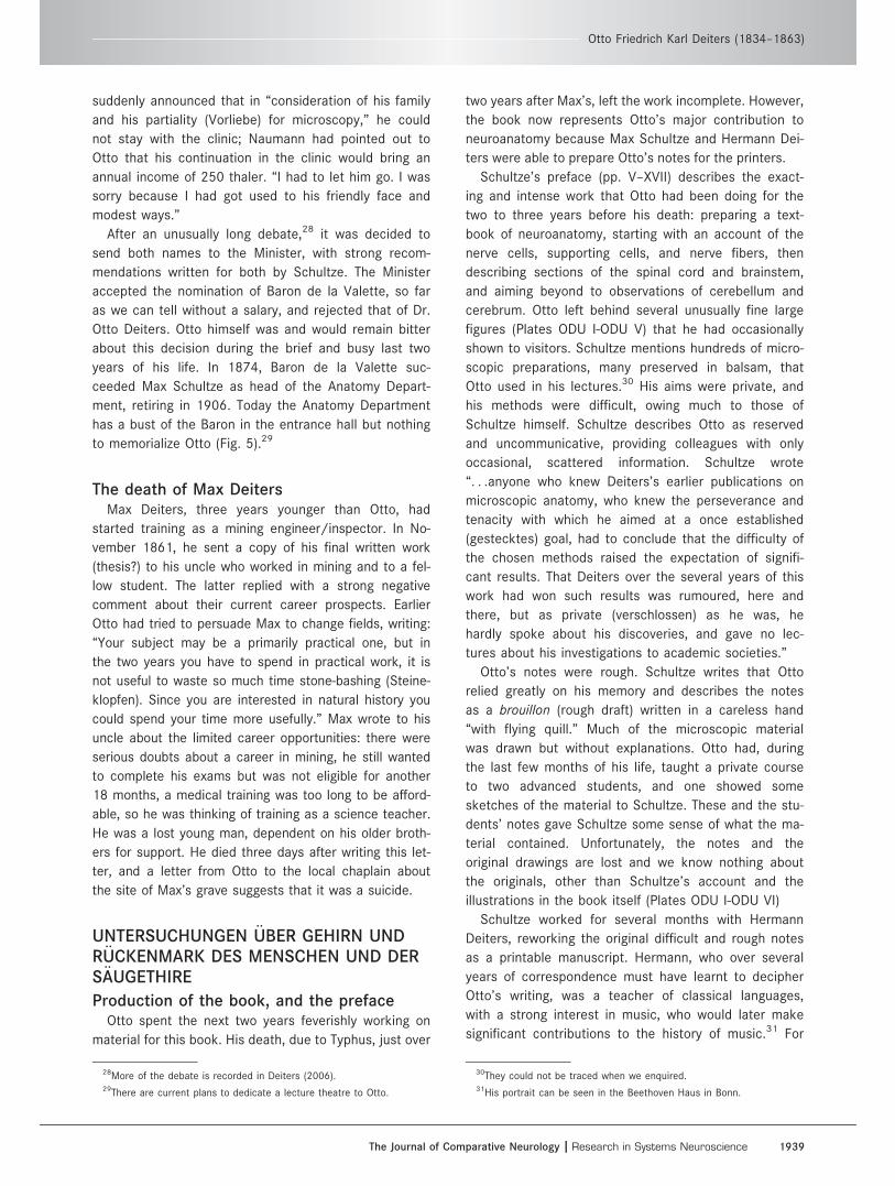
to memorialize Otto (Fig. 5).29
The death of Max DeitersMax Deiters, three years younger than Otto, had
started training as a mining engineer/inspector. In No-
vember 1861, he sent a copy of his final written work
(thesis?) to his uncle who worked in mining and to a fel-
low student. The latter replied with a strong negative
comment about their current career prospects. Earlier
Otto had tried to persuade Max to change fields, writing:
“Your subject may be a primarily practical one, but in
the two years you have to spend in practical work, it is
not useful to waste so much time stone-bashing (Steine-
klopfen). Since you are interested in natural history you
could spend your time more usefully.” Max wrote to his
uncle about the limited career opportunities: there were
serious doubts about a career in mining, he still wanted
to complete his exams but was not eligible for another
18 months, a medical training was too long to be afford-
able, so he was thinking of training as a science teacher.
He was a lost young man, dependent on his older broth-
ers for support. He died three days after writing this let-
ter, and a letter from Otto to the local chaplain about
the site of Max’s grave suggests that it was a suicide.
UNTERSUCHUNGEN €UBER GEHIRN UNDR€UCKENMARK DES MENSCHEN UND DERS€AUGETHIRE
Production of the book, and the prefaceOtto spent the next two years feverishly working on
material for this book. His death, due to Typhus, just over
two years after Max’s, left the work incomplete. However,
the book now represents Otto’s major contribution to
neuroanatomy because Max Schultze and Hermann Dei-
ters were able to prepare Otto’s notes for the printers.
Schultze’s preface (pp. V–XVII) describes the exact-
ing and intense work that Otto had been doing for the
two to three years before his death: preparing a text-
book of neuroanatomy, starting with an account of the
nerve cells, supporting cells, and nerve fibers, then
describing sections of the spinal cord and brainstem,
and aiming beyond to observations of cerebellum and
cerebrum. Otto left behind several unusually fine large
figures (Plates ODU I-ODU V) that he had occasionally
shown to visitors. Schultze mentions hundreds of micro-
scopic preparations, many preserved in balsam, that
Otto used in his lectures.30 His aims were private, and
his methods were difficult, owing much to those of
Schultze himself. Schultze describes Otto as reserved
and uncommunicative, providing colleagues with only
occasional, scattered information. Schultze wrote
“. . .anyone who knew Deiters’s earlier publications on
microscopic anatomy, who knew the perseverance and
tenacity with which he aimed at a once established
(gestecktes) goal, had to conclude that the difficulty of
the chosen methods raised the expectation of signifi-
cant results. That Deiters over the several years of this
work had won such results was rumoured, here and
there, but as private (verschlossen) as he was, he
hardly spoke about his discoveries, and gave no lec-
tures about his investigations to academic societies.”
Otto’s notes were rough. Schultze writes that Otto
relied greatly on his memory and describes the notes
as a brouillon (rough draft) written in a careless hand
“with flying quill.” Much of the microscopic material
was drawn but without explanations. Otto had, during
the last few months of his life, taught a private course
to two advanced students, and one showed some
sketches of the material to Schultze. These and the stu-
dents’ notes gave Schultze some sense of what the ma-
terial contained. Unfortunately, the notes and the
original drawings are lost and we know nothing about
the originals, other than Schultze’s account and the
illustrations in the book itself (Plates ODU I-ODU VI)
Schultze worked for several months with Hermann
Deiters, reworking the original difficult and rough notes
as a printable manuscript. Hermann, who over several
years of correspondence must have learnt to decipher
Otto’s writing, was a teacher of classical languages,
with a strong interest in music, who would later make
significant contributions to the history of music.31 For
28More of the debate is recorded in Deiters (2006).29There are current plans to dedicate a lecture theatre to Otto.
30They could not be traced when we enquired.31His portrait can be seen in the Beethoven Haus in Bonn.
Otto Friedrich Karl Deiters (1834–1863)
The Journal of Comparative Neurology | Research in Systems Neuroscience 1939
Hermann the technical neuroterminology must have
been foreign, but one has the impression that, although
evidence of the brouillon remains, the man of letters
and the teacher may have introduced a degree of order,
less obvious in Otto’s earlier work.
Without this significant effort of Max Schultze and
Hermann Deiters, the book would not exist. We would
then still hold Deiters in high regard for his earlier pub-
lications, but the view provided by the book of a deeply
thoughtful and skilful young scientist arguing (with him-
self) about conclusions to draw from his carefully pre-
pared and studied material would be lost to us, as
would the details of his new observations.
For us the book provides a vivid view of the uncer-
tainties that prevailed 150 years ago, upon which many
of our current “certainties” have been built in innumera-
ble, mostly small, steps. We lack the space and the his-
torical sense to present the material in relation to what
was known at the time. Otto was remarkably bad at cit-
ing others; when he did, it was usually cursory, mention
of a name, or occasionally a journal title.32
A sense of the then contemporary relevant knowl-
edge is best obtained from the 3rd, 4th, and 5th edi-
tions of K€olliker’s Handbuch (K€olliker, 1859, 1863,
1867). Van Der Loos (1967) and Shepherd (1971) pro-
vide many relevant details concerning the nerve cells
and Meyer (1971), Clarke and O’Malley (1996), and Fin-
ger (1994) about the brain more generally.
Purkinje (1838; cited by Finger, 1994) had described
the cerebellar cells that bear his name and illustrated
the basal parts of their dendrites; Wagner (1845, 1847)
had illustrated the dendrites extending some greater dis-
tance from the cell body of sympathetic cells and cells
in the electric organ of fish (Torpedo); Remak (1855) had
described the several processes (dendrites) that leave
the large neuronal cell body of the ventral horn cells,
and had traced one single, distinctive process (the axon)
into the ventral root. Although Brown S�equard (see Fin-
ger, 1994) had demonstrated the crossing of the pyrami-
dal tract and of some of the sensory spinal pathways, it
had not yet been possible to trace the sensory pathways
through their relay stations in the brainstem. Tracing
nerve fibers from their cells of origin into their peripheral
cranial nerves was still extremely difficult, and the
results were often conflicting. Although the demonstra-
tion by Bell and Magendie (Finger, 1994) that the spinal
dorsal roots are sensory and the ventral roots motor
was widely accepted, the connections that might provide
a pathway for the spinal reflex remained undefined. The
cell theory was still the subject of significant debate and
misunderstanding (Baker, 1949; Harris, 1999).
We present Otto’s book under several different head-
ings, not in order of the chapters, but summarizing
what appear to us as the most interesting points for
readers of The Journal of Comparative Neurology.
On page XI of the preface, Schultze lists novel obser-
vations included in the book: conspicuously the identifi-
cation of the superior olive in the human brain,
previously only reported in animals since it is visible in
many mammals, but is hidden in the human brain by
the large pons; also the identification of a large-celled
gray nucleus between the origin of the inferior cerebel-
lar peduncles that Schultze suggested be called Deiters
nucleus (Plate ODU V, Fig. 14). Schultze draws atten-
tion to Otto’s account of a lateral (intermediate) motor
component of the cranial nerves, lying between those
corresponding to the spinal dorsal and ventral roots,
specifically including the accessory, seventh, and fifth
cranial nerves. Schultze describes as a major contribu-
tion Deiters’s account of nerve cells having a single
axon33 and multiple “protoplasmic processes” (den-
drites), with an additional second system of fibers
attaching to the dendrites. However, Schultze then
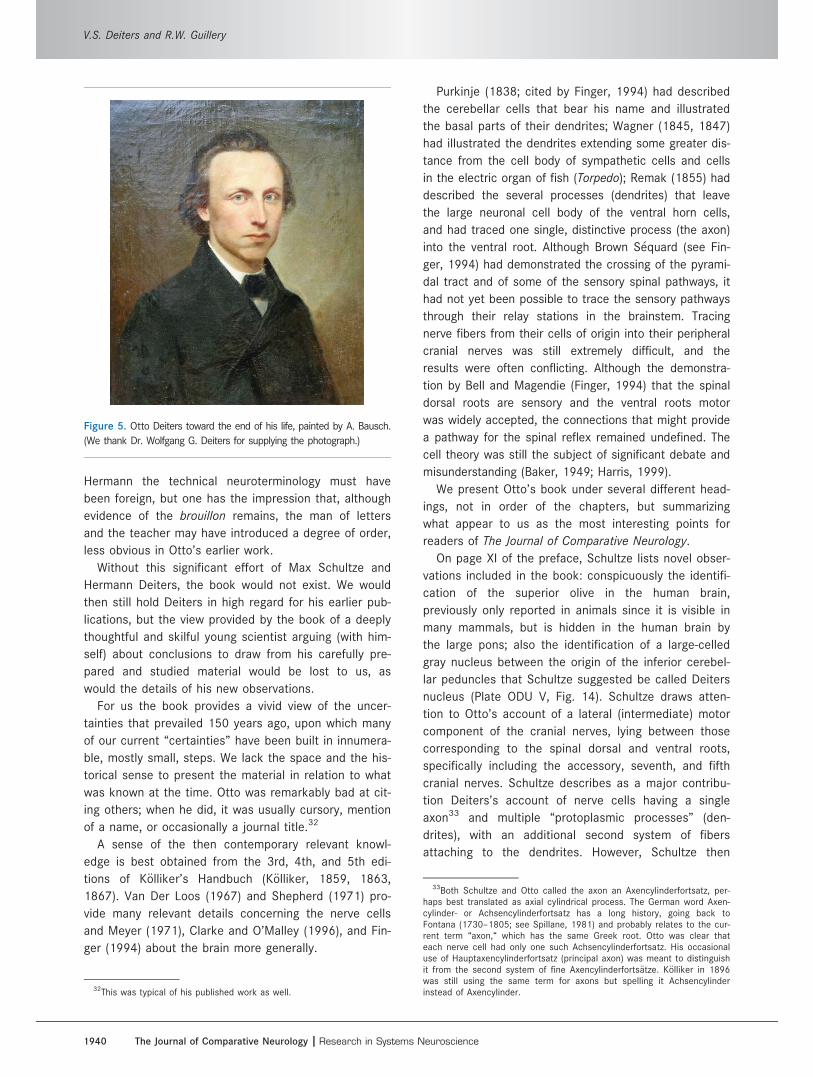
Figure 5. Otto Deiters toward the end of his life, painted by A. Bausch.
(We thank Dr. Wolfgang G. Deiters for supplying the photograph.)
32This was typical of his published work as well.
33Both Schultze and Otto called the axon an Axencylinderfortsatz, per-haps best translated as axial cylindrical process. The German word Axen-cylinder- or Achsencylinderfortsatz has a long history, going back toFontana (1730–1805; see Spillane, 1981) and probably relates to the cur-rent term “axon,“ which has the same Greek root. Otto was clear thateach nerve cell had only one such Achsencylinderfortsatz. His occasionaluse of Hauptaxencylinderfortsatz (principal axon) was meant to distinguishit from the second system of fine Axencylinderforts€atze. K€olliker in 1896was still using the same term for axons but spelling it Achsencylinderinstead of Axencylinder.
V.S. Deiters and R.W. Guillery
1940 The Journal of Comparative Neurology |Research in Systems Neuroscience

argues at length that Otto misnamed the protoplasmic
processes because he (Schultze) considered that they are
not an extension of the nerve cell’s protoplasmic contents.
Finally, Schultze draws attention to the fact that Otto
reports never having been able to trace a nerve process
from one nerve cell into continuity with another nerve cell.
This is relevant to the view held by several commentators
that Otto was a reticularist, or a precursor of this theory
(Shepherd, 1971; Swanson and Swanson, 1995; Albright
et al., 2000) and is of interest since Schultze himself had
described several olfactory fibers fusing to form a single
axon and another member of his department (G. Walter34)
had published an account of rich fusions of neural proc-
esses in the leech, fusions today readily recognized as
artefacts (Walter, 1863).35 Otto mentions this study in the
text and reports that he succeeded in persuading Walter
to take another look. Unfortunately, Otto died soon after
the Walter paper was published and we don’t know
whether Walter ever did look again.
The methods used (mostly from Chapter I)We know very little about the microscope Otto used
except that it had achromatic lenses (see section on Otto’s
life in Berlin, above). He worked with tissues from humans,
calves, dogs, cats, rabbits, and goats, comparing one spe-
cies with another, and recognizing that human tissue is
never well preserved. He cut his sections in conventional
cross sections (see Plates ODU III to ODU VI) but also in
planes suitable for following particular pathways. He
depended heavily on teasing and manipulating fresh pieces
of tissue under the microscope, using different chemicals
at different concentrations and different temperatures. He
watched the extent to which he could preserve the tissues
with some solutions but not others, the rates at which dif-
ferent tissue elements disintegrated (depending on species
or solutions), and the extent to which different parts were
firm and resistant to manipulation or could be moved, dis-
torted, or separated from each other. He (rightly) distrusted
the use of sections for judging the continuity of two proc-
esses; he preferred to tease out the finer processes. The
figures (especially the first two plates), drawn by Otto him-
self, illustrate how closely related were his skills at manipu-
lating the tissues under the microscope and his
considerable ability as an illustrator. The figures in Plates
ODU I and II show nerve cells as a three-dimensional tac-
tile experience with potentially mobile parts, a vision that
even the Golgi method does not provide.
Figures 1–15 from the book appear here on six plates
numbered ODU I to ODU VI. We refer to them here by the
plate and original figure numbers, e.g., Plate ODU I, Fig-
ure 2. Plates ODU I and ODU II show dissections of nerve
cells and two glial cells. These two plates represent the
most original and striking part of the book. Plates ODU
III–VI show sections through different levels of the brain-
stem: In caudorostral order, they are III, IV, VI, and V.
Whereas III is identified as being from a human brain, IV
and V are not identified; from the appearance of the pyra-
mids, they may well be from a sheep or a calf. VI, judging
from the appearance of the pyramids, is human. The
legends are translations of the original legends.
Otto tells how to prepare the carmine stain and the im-
portance of freshness, filtering, and other factors. He notes
that the intensity of the reaction varies from one tissue ele-
ment to another and from one region to another. He argues
that different chemical compositions of the parts imbibe the
dye differentially, but concludes that strict chemical inter-
pretations are not yet possible. He was impressed by the
beauty of the carmine preparations, but warns that carmine
can give false views of fibers fusing with each other and
states that “The aesthetic gratification (Befriedigung) that
some find in such preparations has led, I believe, to the
overestimation of their scientific significance” (p. 5). He
tried double staining with carmine and indigo blue, hoping
that the nerve cells might be red and the connective tissues
blue, but found that one dye drove out the other (p. 26).
Otto considers the “elementary parts” of the nervous
system (p. 5). In view of his earlier publication on the cell
theory, this can be seen as an unsuccessful search for
the units that were later to be defined by Waldeyer and
fought for by Ram�on y Cajal. He cites the relevance of
K€olliker’s and K€uhne’s observations of nerve endings on
muscle fibers but notes that their methods did not serve
to separate/distinguish the neural elements (p. 6).
The nerve cell (mostly from Chapter III)The “typical” nerve cellOtto stresses how little we know about even the large,
well-recognized ventral horn cells: “If one thinks of the
ganglion cell of the ventral horn as being the connec-
tion/link between the ventral roots entering the spinal
cord and the ventral columns that are leading to the
brain, one has the simplest function that can be
assigned to a ganglion cell” (p. 54; italics added36).
34Many years later, after Hermann Deiters’s daughter Maria Deiters hadmarried Hippolyte Guillery, their children could claim two grand-uncleswho had worked in the Bonn Anatomy Department in the early 1860s.Otto Deiters was Maria’s uncle, and Georg Walter was Hippolyte’s uncle.
35It is puzzling that Ram�on y Cajal (1954) describes Schultze as a fatherof the neuron doctrine and treats Deiters as a reticularist (see also Guil-lery, 2005).
36This description of the ventral roots as “entering the cord“ is puzzling.Otto wrote in German: (Man denke sich hier also z.B. zwischen die in dasR€uckenmark eingetretenen Vorderwurzeln und die Vorderstr€ange als dieleitenden Bahnen zum [sic] Gehirn eine Verbindung durch die Ganglien-zellen der Vorderh€orner, so hat man doch die simpelste Function, die maneiner Ganglionzelle zuschreiben kann.) Here he is thinking of the connec-tions as he traced them, but not of the direction of the signal, which wasthen known to travel from the brain through the ventral horn cells to themuscle.
Otto Friedrich Karl Deiters (1834–1863)
The Journal of Comparative Neurology | Research in Systems Neuroscience 1941
However, he concludes that there is no answer to the
question of how the connections from the brain to the
muscles are established through the ventral horn cells:
“If cells as conspicuous as the ventral horn cells have
frustrated us, one can expect much less from the other,
smaller cells.” He then concludes that our knowledge
of nerve cells lacks a solid basis (is “bodenlos”).
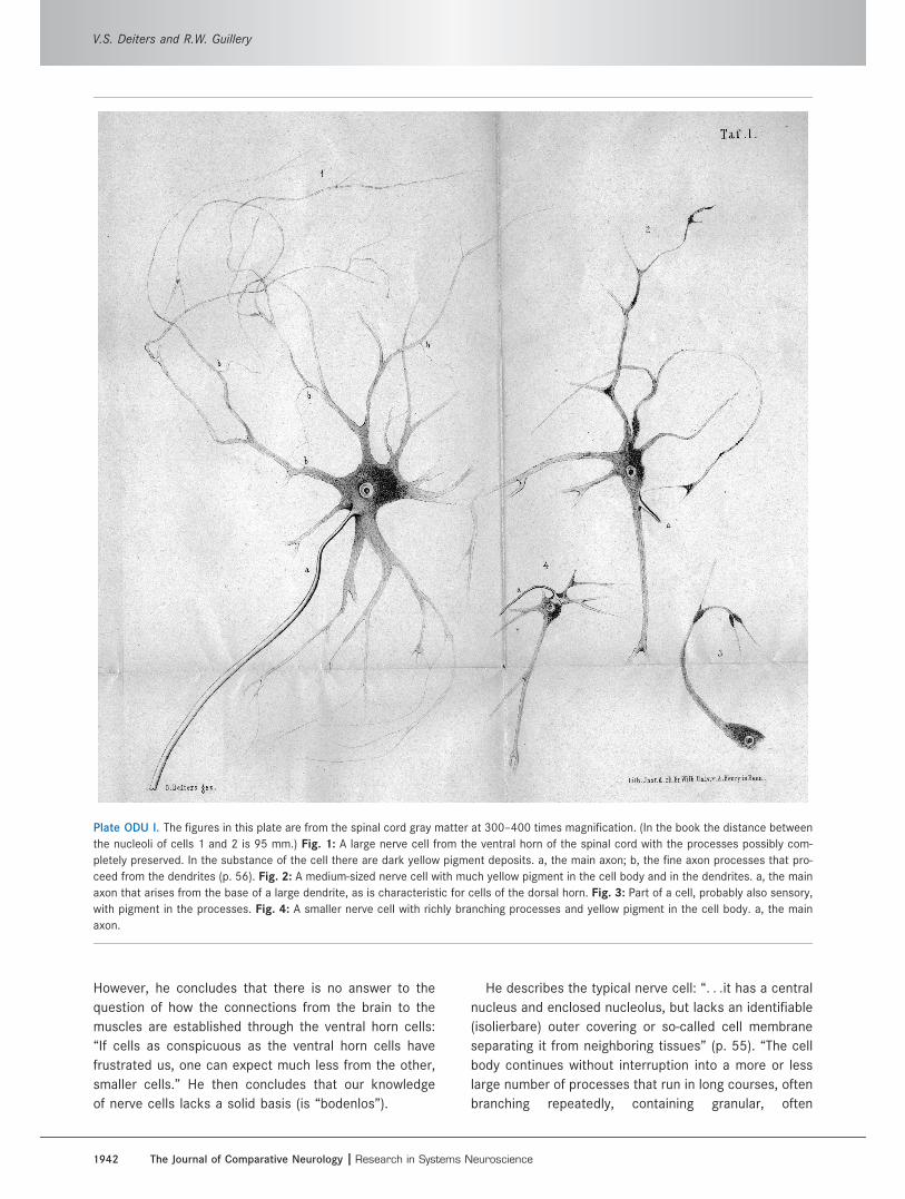
He describes the typical nerve cell: “. . .it has a central
nucleus and enclosed nucleolus, but lacks an identifiable
(isolierbare) outer covering or so-called cell membrane
separating it from neighboring tissues” (p. 55). “The cell
body continues without interruption into a more or less
large number of processes that run in long courses, often
branching repeatedly, containing granular, often
Plate ODU I. The figures in this plate are from the spinal cord gray matter at 300–400 times magnification. (In the book the distance between
the nucleoli of cells 1 and 2 is 95 mm.) Fig. 1: A large nerve cell from the ventral horn of the spinal cord with the processes possibly com-
pletely preserved. In the substance of the cell there are dark yellow pigment deposits. a, the main axon; b, the fine axon processes that pro-
ceed from the dendrites (p. 56). Fig. 2: A medium-sized nerve cell with much yellow pigment in the cell body and in the dendrites. a, the main
axon that arises from the base of a large dendrite, as is characteristic for cells of the dorsal horn. Fig. 3: Part of a cell, probably also sensory,
with pigment in the processes. Fig. 4: A smaller nerve cell with richly branching processes and yellow pigment in the cell body. a, the main
axon.
V.S. Deiters and R.W. Guillery
1942 The Journal of Comparative Neurology |Research in Systems Neuroscience
pigmented protoplasm continuous [with that of the cell
body] and extending finally into immeasurably fine proc-
esses. . ..For convenience I will call them protoplasmic
processes” (the dendrites; p. 56). These are immediately
distinguishable from “. . .an outstanding single process
that arises from the cell body or, as can occur, from the
root of one of the larger protoplasmic processes (Figs. 1,
2, 4A). This single process, ’Nervenfaser-oder Axencylin-
derfortsatz’ (see footnote 33) at its origin can be seen to
contain the granular protoplasm. . .but as soon as it
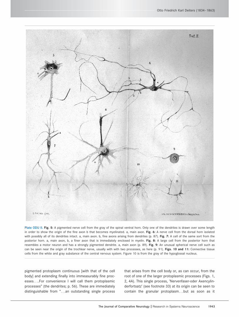
Plate ODU II. Fig. 5: A pigmented nerve cell from the gray of the spinal ventral horn. Only one of the dendrites is drawn over some length
in order to show the origin of the fine axon b that becomes myelinated. a, main axon. Fig. 6: A nerve cell from the dorsal horn isolated
with possibly all of its dendrites intact. a, main axon. b, fine axons arising from dendrites (p. 87). Fig. 7: A cell of the same sort from the
posterior horn. a, main axon, b, a finer axon that is immediately enclosed in myelin. Fig. 8: A large cell from the posterior horn that
resembles a motor neuron and has a strongly pigmented dendrite. a, main axon (p. 89). Fig. 9: An unusual spherical nerve cell such as
can be seen near the origin of the trochlear nerve, usually with with two processes, as here (p. 91). Figs. 10 and 11: Connective tissue
cells from the white and gray substance of the central nervous system. Figure 10 is from the gray of the hypoglossal nucleus.
Otto Friedrich Karl Deiters (1834–1863)
The Journal of Comparative Neurology | Research in Systems Neuroscience 1943

Plate ODU III. Fig. 12: Transverse section through half of the lower part of a human spinal cord. Probably near the beginning of the conus
medullaris. R.a., bundle of the ventral root arising from the ventral horn within which one can see nerve fibers running among the large
cells; R.p., dorsal root leaving the dorsal horn; here the cells are paler and with very few exceptions smaller than those in the ventral
horn; R.i.p., inner part of the dorsal root; C.c., central canal with its lining of ciliated epithelium surrounded by connective tissue; C.p., dor-
sal gray commissure; C.a.a., ventral white commissure. Around the gray horns are myelinated fibers and axons that vary in size in the dif-
ferent regions.
V.S. Deiters and R.W. Guillery
1944 The Journal of Comparative Neurology |Research in Systems Neuroscience
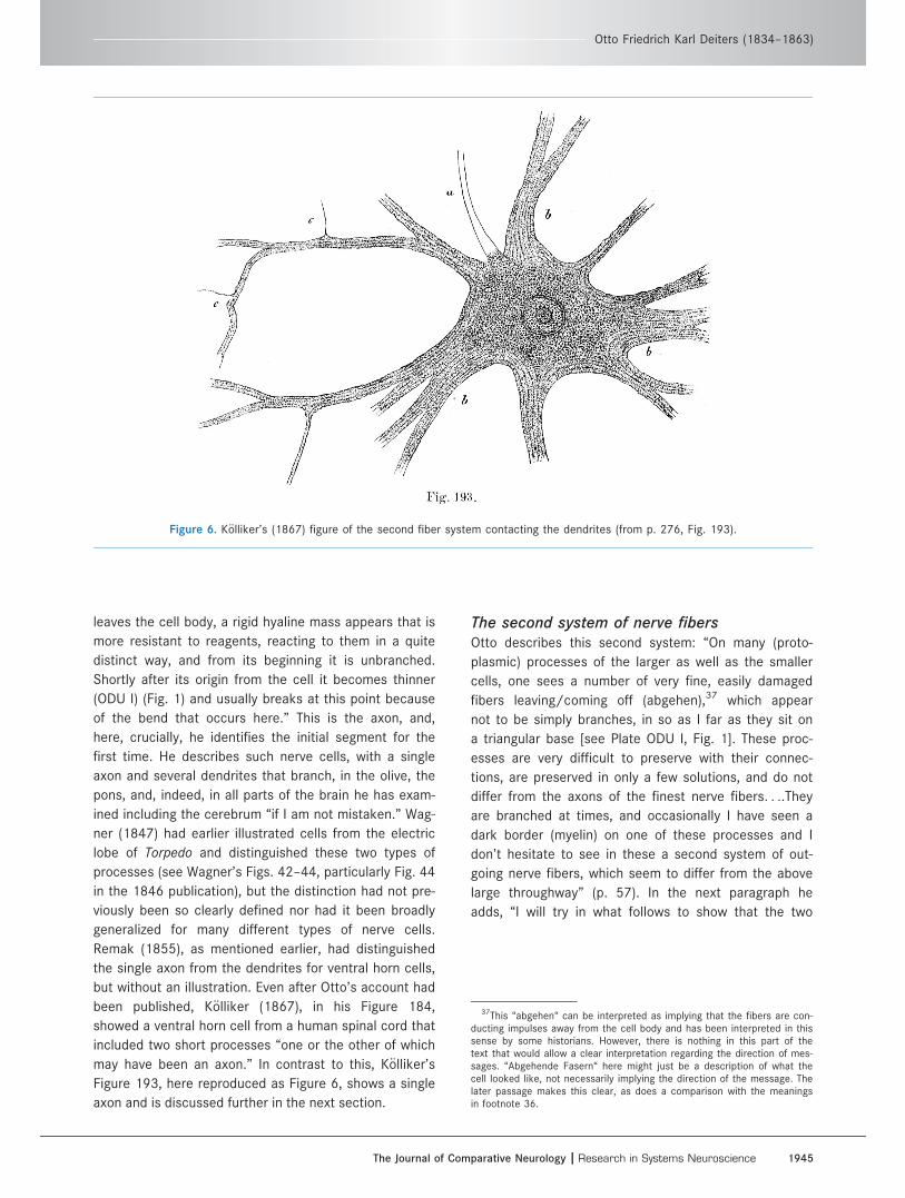
leaves the cell body, a rigid hyaline mass appears that is
more resistant to reagents, reacting to them in a quite
distinct way, and from its beginning it is unbranched.
Shortly after its origin from the cell it becomes thinner
(ODU I) (Fig. 1) and usually breaks at this point because
of the bend that occurs here.” This is the axon, and,
here, crucially, he identifies the initial segment for the
first time. He describes such nerve cells, with a single
axon and several dendrites that branch, in the olive, the
pons, and, indeed, in all parts of the brain he has exam-
ined including the cerebrum “if I am not mistaken.” Wag-
ner (1847) had earlier illustrated cells from the electric
lobe of Torpedo and distinguished these two types of
processes (see Wagner’s Figs. 42–44, particularly Fig. 44
in the 1846 publication), but the distinction had not pre-
viously been so clearly defined nor had it been broadly
generalized for many different types of nerve cells.
Remak (1855), as mentioned earlier, had distinguished
the single axon from the dendrites for ventral horn cells,
but without an illustration. Even after Otto’s account had
been published, K€olliker (1867), in his Figure 184,
showed a ventral horn cell from a human spinal cord that
included two short processes “one or the other of which
may have been an axon.” In contrast to this, K€olliker’s
Figure 193, here reproduced as Figure 6, shows a single
axon and is discussed further in the next section.
The second system of nerve fibersOtto describes this second system: “On many (proto-
plasmic) processes of the larger as well as the smaller
cells, one sees a number of very fine, easily damaged
fibers leaving/coming off (abgehen),37 which appear
not to be simply branches, in so as I far as they sit on
a triangular base [see Plate ODU I, Fig. 1]. These proc-
esses are very difficult to preserve with their connec-
tions, are preserved in only a few solutions, and do not
differ from the axons of the finest nerve fibers. . ..They
are branched at times, and occasionally I have seen a
dark border (myelin) on one of these processes and I
don’t hesitate to see in these a second system of out-
going nerve fibers, which seem to differ from the above
large throughway” (p. 57). In the next paragraph he
adds, “I will try in what follows to show that the two
Figure 6. K€olliker’s (1867) figure of the second fiber system contacting the dendrites (from p. 276, Fig. 193).
37This “abgehen“ can be interpreted as implying that the fibers are con-ducting impulses away from the cell body and has been interpreted in thissense by some historians. However, there is nothing in this part of thetext that would allow a clear interpretation regarding the direction of mes-sages. “Abgehende Fasern“ here might just be a description of what thecell looked like, not necessarily implying the direction of the message. Thelater passage makes this clear, as does a comparison with the meaningsin footnote 36.
Otto Friedrich Karl Deiters (1834–1863)
The Journal of Comparative Neurology | Research in Systems Neuroscience 1945

systems of axons serve two different directions” (das
diese beiden Systeme verschiedenen Richtungen
angeh€oren).
He returns to the second axonal system, and dis-
cusses the possibility that they might contact the cell
body as well as the dendrites, stresses how easily they
are destroyed, and gives details of how they can be
preserved. He says that they are not artefacts;
occasionally they can be identified as the unmyelinated
end of a myelinated fiber (pp. 64–65; Plate ODU I,
Fig.7b). “I am convinced that with careful treatment, as
described, the appearances in the figure and my
description can be confirmed” (p. 65). K€olliker (1867),
who clearly recognized the necessity for some commu-
nication involving the dendrites, subsequently illustrated
something similar to the fine fibers of the second
Plate ODU IV. Fig. 13: Transverse section through half of the beginning of the medulla, with nerve roots of the accessory and hypoglossal
nerves. J.a., ventral fissure, partially filled by the beginning of the pyramidal decussation; C.p., posterior gray commissure; C.c., central
canal; F.r., reticular formation, the reticular connective tissue that arises from (is continuous with) the ventral horns and increases to
occupy the greater part of the medulla; C.p., posterior horn, at its base also continuous with the framework of the reticular formation; A.,
accessory nerve; H., hypoglossal nerve; A0., transverse section of the accessory nerve; Hyp., cells of the hypoglossal nucleus.
V.S. Deiters and R.W. Guillery
1946 The Journal of Comparative Neurology |Research in Systems Neuroscience

system (Fig. 6). He pointed out that he had described
them earlier and that he could not distinguish them
from the other processes given off by the dendrites. He
could not trace any of these into a myelinated stretch,
as Otto had shown in Plate ODU I, Figures 5 and 7.
K€olliker concluded that the existence of the second sys-
tem of fibers was “not a decided matter” (Eine nicht
abgeschlossene Sache).
Plate ODU V. Fig. 14: Transverse section through half of the medulla close to its junction with the pons, with nerve roots of abducens, fa-
cial, and acoustic nerves. E., fibers of the pyramids38; R., raphe—to its right the framework of the reticular formation within whose dorsal
portion (part b, smaller) and ventral portion (part a, larger) nerve fibers are found. Ol. (Ol.s.), section through the superior olive; C.tr., fibers
of the trapezoid body; Cr.c., inferior cerebellar peduncle with many large ganglion cells that appear to belong to the origin of the acoustic
nerve, Ac.; Fac., facial nerve; F., the same cut in transverse section at the facial genu; Abd., abducens nerve. The drawing is incomplete;
the transverse sections of the many thick fibers (at J [sic]) should be shown.
38This is an error. There is no E in the figure and probably the P wasintended.
Otto Friedrich Karl Deiters (1834–1863)
The Journal of Comparative Neurology | Research in Systems Neuroscience 1947
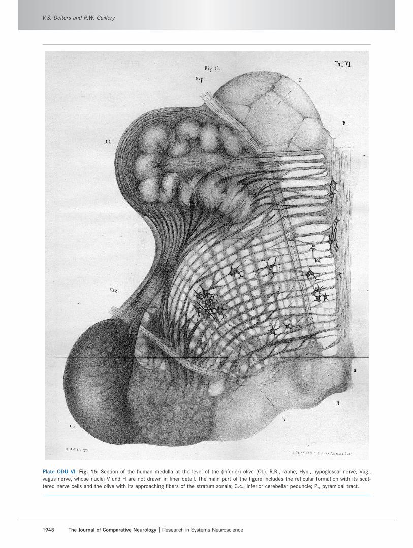
Plate ODU VI. Fig. 15: Section of the human medulla at the level of the (inferior) olive (Ol.). R.R., raphe; Hyp., hypoglossal nerve, Vag.,
vagus nerve, whose nuclei V and H are not drawn in finer detail. The main part of the figure includes the reticular formation with its scat-
tered nerve cells and the olive with its approaching fibers of the stratum zonale; C.c., inferior cerebellar peduncle; P., pyramidal tract.
V.S. Deiters and R.W. Guillery
1948 The Journal of Comparative Neurology |Research in Systems Neuroscience

The fact that Otto had described incoming axons
making what we can now recognize as synaptic con-
tacts with the dendrites was not appreciated by later
workers and was altogether lost sight of by many. In
part this was because the adhesive properties of synap-
ses were not recognized, and in part because when
finally, some decades later, synaptic contacts were
described light microscopically, they appeared to show
a light microscopically visible synaptic gap, which Otto’s
figures did not show. The fact that the actual synaptic
gap was too narrow to be visible with the light micro-
scope was not revealed until it could be defined by
electron micrographs as only about 20 nm in width.39
This now makes Otto’s drawing entirely believable, and
allows us to see these fine fibers of the second system
as ending on the dendrites in synaptic contacts (see
also Van Der Loos, 1967). Much later (on p. 145), Otto
returns to this second system. He points out that the
second system of fibers characterizes sensory as well
as motor neurons, and raises the possibility that sen-
sory fibers may open into (eim€unden) a motor cell. He
recognizes that such a connection has not been demon-
strated and that there is (thus) no evidence for an ana-
tomical basis of reflex connections. However, he then
suggests: “If there should be specific differences
between sensory and motor nerve cells, then they must
have a physiological and an anatomical significance
from which an effect on the spread and the direction of
the current would result, so that, for example, the
motor roots [would] connect directly with the axon and
the sensory, in contrast, [would] connect to the second
fiber system.” That is, the dendrites on which the sec-
ond system forms its triangular contacts would be
receiving inputs from the second system.
Here he was postulating a functionally polarized
nerve cell, with the second fiber system bringing the
afferents to the dendrites, and the axon providing the
output. This is a stunning, but fleeting anticipation of
the dynamically polarized neuron identified later by
Ram�on y Cajal and Van Gehuchten. Unfortunately, Otto
does not have the actual evidence for such connections
and does not return to it. We are left with this tantaliz-
ing thought: that he recognized the importance of func-
tionally polarized neuron and saw the second system of
fibers as a likely input to the dendrites, with the axon
as the output. He would surely have followed this up
had he lived.
Classifying nerve cellsOn page 86, Otto says that the basic structure of cells
in the dorsal horn is the same as in the ventral horn.
He does not plan to describe differences in form, size,
number, and length of processes and their branching. . .
The cells of the dorsal horn are more difficult to dissect
and survive less well but they are basically the same. He
refers to the dorsal horn cell that can be seen in Plate
ODU I, Figure 6: “On the branches one sees a fair num-
ber of the little branches (Reiserchen) [Plate ODU I, Fig.
6b,b] leaving from a triangular basis (the second system
of fibers)”40 (p. 87). That is, nerve cells are generally the
same everywhere. On page 90, he says that in the me-
dulla the cells of the accessory, hypoglossal, and vagus
nuclei show the same characteristics, with some special
features (but they are harder to stain than spinal cells).
On pages 94–95, the same basic cell structure is seen in
the nerve cells of the inferior olive, pons, and dentate nu-
cleus, in the substantia nigra, and for some cells in the
granule cell layer of the cerebellum. The large cells of the
medulla only differ from the small cells in their size. Ear-
lier, in the chapter on connective tissues, he considers
the “so-called granules in the second layer of the cerebel-
lum. I will show that these so-called free nuclei have
structures surrounding the nucleus that form a narrow
border of protoplasm that continues into enormously long
fibers”41 (p. 37). He concludes that these are nerve cells,
although he says nothing about their dendrites, and he
does not return to this issue later in the book.
Only for the cells of the superior olive has he failed
to arrive at a clear view of their structure. On page 95,
he says that “Finally there is a third cell type scattered
in the cerebellar cortex that can continue on both sides
as an axon42. . ..This strange small cell is one of the
few examples of a cell that I have not yet been able to
fit into a general schema of nerve cells.” In addition, on
pages 91–92 he describes cells near the origin of the
trochlear nucleus as another exception (ODU II, Fig. 9).
He likens them to dorsal root or trigeminal ganglion
38This is an error. There is no E in the figure and probably the P wasintended.
39The reasons for this apparent discrepancy between some of the lightmicroscopic images of a large synaptic gap and the electron microscopicimages of a much narrower gap are discussed by Gray and Guillery (1961;and see Guillery, 2006). Essentially, the views of the wider gap werebased on reduced silver stains of the neurofibrillary skeleton that left sig-nificant parts of the nerve cell unstained.
40On page 84 he describes dorsal horn cells whose axons enter the dor-sal root, but does not mention the species.
41Presumably he traced the axon into a parallel fiber. There is a prob-lem about this statement since in other notes (pp. XI–XII of the Preface),he treats the granule cells as connective tissue elements (glia). Althoughthis account of the granule cells as neurons is definitive in denying thatthey are glial, and the observations must have been much more difficultthan those leading him to see them as glia, we have no way of knowingwhich observations he would have included in his final account. Certainly,Schultze, citing these notes in the Preface, not knowing what granule cellsare really like, took the glial categorization seriously and ignored the otherpossibility.
42Cerebellar fusiform cell?
Otto Friedrich Karl Deiters (1834–1863)
The Journal of Comparative Neurology | Research in Systems Neuroscience 1949
cells, having hardly any protoplasmic processes. He is
puzzled by these,43 is uncertain about having obtained
a complete dissection, and recommends further study
by others.
The lack of nerve cell fusionsOtto notes how readily the beauty of carmine-stained
preparations can mislead one into interpreting every pro-
cess of a nerve cell as an axon, and interpreting the com-
plex interconnections as supporting the doctrine of
anastomoses between ganglion cells (p. 54). He is
entirely clear that such anastomoses are not to be found.
Some authors see “the presence of such connections as
a physiological necessity44. . ..Those who hold these
views about the easy and frequent opportunities for
observing such anastomoses must have been startled
that K€olliker, whose experience and observational skills
no one would wish to deny, had never asserted that he
had seen one. . ..Every reasonable author working with-
out preconceptions must come to the same conclusion”
(p. 67). “I have. . .never seen an anastomosis. . .in prepa-
rations whose processes could be followed to their finest
ramifications, as in (ODU I) Figure 1” (p. 69).
The nerve fibersOtto quotes Bidder regarding the absence of Schwann
cells in the central nervous system (pp. 101–102).
“Many authors opposed this on theoretical grounds,
others like K€olliker were doubtful, and others like M.
Schultze agreed with Bidder. . ..I must admit that from
my studies I have learnt nothing that proved to me the
existence of such a sheath, and the ways [for finding a
sheath] given by the authors led in the opposite
direction.” Otto describes that where the axon leaves
the nerve cell as an uncovered structure, and where
the myelin begins there is no evidence for the begin-
ning of a cover. And, similarly at the nerve ending,
where a cover should stop as the myelin stops, there is
no evidence of a separate cover (p. 103).
Some generalizationsOn page 98, Otto says that “The only thing that I can
contribute in relation to this question [classification of
cells] is something very insignificant [sic], namely, the
fact that the size of the cell body is proportional to the
thickness of the axon that leaves the cell body.” He
notes further that “The arrangement and quantity of
nerve cells in a part [of the central nervous system] is
in a certain (gewissen) proportion to the nerve fibers
with which it is connected.” He cites the brachial and
lumbar enlargements, the size of the pontine nuclei in
relation to the middle cerebellar peduncle, and the tra-
pezoid body in relation to the superior olive.
The connective tissues (mainly Chapter II)Although Virchow had identified glia earlier, Otto
writes about connective tissues and argues (on pp. 29–
30) that recent accounts have provided no agreement
concerning the differences between neural and support-
ing elements. At the beginning of Chapter II, Otto is
critical: “The central organs of the nervous system have
a connective tissue framework (Ger€ust), which more or
less follows the general form of all parts, is nowhere
missing and within whose meshwork the neural appara-
tus is embedded. When this general statement, which
in this form in itself can raise no doubts, is first
announced, one might well regard it as an ingenious
guess (geistreiche Divination) rather than one supported
by stringent observations” (p. 27). He then asks: What
can we see to support such generalizations? How can
we distinguish between neural and non-neural ele-
ments? If we follow Reissner, that “only a connection
with an indubitable nerve fiber can identify a nerve cell,
then this is an aid that must have the first priority” (p.
28). However, Otto notes, this leads to misinterpreta-
tions wherever such continuity is lost.
Most usefully, on page 38 he turns to Max Schultze’s
study of the retina and compares the non-neural ele-
ments of the central nervous system with Mueller
fibers. On page 33, he then contrasts white matter cells
(which are stellate with many branching processes
[Plate ODU I, Fig. 10], presumably fibrous astrocytes)
with gray matter cells having sponge-like processes,
presumably protoplasmic astrocytes. On pages 44–45,
it can be seen that wherever he can trace the cyto-
plasm of a cell into a neural process this must be neu-
ral. Other cells in the white matter have delicate
processes, with sharp borders, and a bright appear-
ance. They are easily moved, coil around isolated cells,
are not brittle, and branch richly at forked branch
points (Plate ODU I, Fig. 10).45
Otto argues that many small cells seen in the tis-
sues linking the central canal and pia had been
incompletely drawn by K€olliker: “I have no doubt that
43Cells of the mesencephalic root of the trigeminal nerve look like thisand have been interpreted as displaced trigeminal ganglion cells.
44K€olliker (1863) considered fusions between dendrites of ventral anddorsal nerve cells as necessary for producing spinal reflexes, but admittednever having seen such fusions. Otto’s comment: “As concerns the argu-ment that physiology demands connections of this sort, I hold that suchan assumption does not give us the right to assume particular formula-tions of anatomical facts, particularly in areas where the unknowndominates.“
45There can be debate about the identification of this cell as an astro-cyte, but we are inclined to accept it as such on the basis of the observa-tions that Otto made as he dissected it.
V.S. Deiters and R.W. Guillery
1950 The Journal of Comparative Neurology |Research in Systems Neuroscience

here one is dealing with genuine nerve cells. . .and
would ask K€olliker to look at this point once more”
(p. 33). The possibility of confusion between connec-
tive tissues and neural elements seems like a
“phantom (Gespenst) of which investigators have
allowed themselves to be frightened more than is nec-
essary.” Later, on page 36, he notes that “If one
starts with the pia to win a secure basis for the parts
that can indubitably be described as not neural, then
there is a straight path open.” Next, he stresses the
differences between neural processes and the connec-
tive tissues (p. 37).
The spinal cord and medulla (mainlyChapter VI)
In terms of understanding the organization of the me-
dulla, Otto wrote: “here one must deal with concepts
about cells and pathways that can hardly be conceived
as sufficiently complex. . ..He who concludes that here
one is dealing with a thankless task is certainly not
wrong” (p. 149). Then he asks: “what is the point of a
whole lot of pictures of cross sections if they leave the
inner architectonics unexplained?” (p. 150). This is a
valid summary of contemporary accounts, which were
largely descriptive and offered few organizational princi-
ples. He continues that the human medulla is far too
difficult and that other mammals provide a much read-
ier view; on the basis of the comparative anatomy, he
then provides a useful schematic view of the medulla
as first of all a continuation of the basic structure of
the cord (pp. 152–156), with the central canal opened
as the fourth ventricle, the dorsal columns shifted later-
ally, and the last 10 cranial nerves continuing the pat-
tern of the spinal nerves. He relates this analysis to the
segmental origins of the skull up to the end of the
notochord. Second, there is the addition of a third, in-
termediate column that includes the motor trigeminal,
facial, and cranial accessory nerves between the contin-
uations of the dorsal and the ventral columns (later
categorized by others as “special visceral efferent”),
and, third (on pp. 166–167), the important major modi-
fications that are added to accommodate the connec-
tions of the cerebellum. This creates a novel
interpretative schema. He traces axons from the inferior
olives into the inferior cerebellar peduncle (p. 167) and
fibers from the pontine nuclei into the middle cerebellar
peduncle.
This general scheme has a surprisingly modern ring
to it, and readers of early issues of The Journal of Com-
parative Neurology will recognize indications of an orga-
nization for the cranial nerves to which the Journal’s
earlier volumes made significant additions.46 We have
not found a comparable schematic view in other early
accounts (Stilling, 1843, 1846; Wagner, 1845, 1847;
Clarke, 1858; Schroeder Van der Kolk, 1859; Reichert,
1859; Henle, 1871), and K€olliker’s 3rd edition of his
Handbuch (K€olliker, 1859) does not provide a signifi-
cant source for something comparable, other than rec-
ognizing the continuation of the dorsal horn into the
medulla and seeing the central gray of the cord as con-
tinuing into the floor of the fourth ventricle. The early
accounts all provide a fascinating and wonderfully illus-
trated view of how difficult it was to sort out the sepa-
rate pathways and components of the brainstem before
experimental methods for tracing the pathways became
available. However, they do not present a proposed
schema of the sort that Otto suggests, beyond Wagn-
er’s (1845) recognition of the close relation between
the motor trigeminal and the facial nerves.
It is important to note that Otto did not separate the
several pathways that cross the midline in the medulla.
He treated them as a single “framework” (Balken-
ger€ust), within which the cranial nerve nuclei and other
gray masses were embedded. This framework includes
what we now recognize as the crossings of the medial
lemniscus and the pyramidal tract and all of the other
fibers crossing the midline in the medulla and pons
including those that link to the inferior olives and the
reticular zones of the medulla and pons (p. 226).
“It is wrong to separate the medulla from the pons”
(p. 168). Otto points out that the pons varies greatly in
size between species so that the ponto-medullary bor-
der leaves some pontine structures of the human brain
(e.g., the superior olive and the facial nerve) in the me-
dulla of other mammals.
On pages 180–181, he discusses the role of cell
groups that interrupt fiber pathways of the
“Balkenger€ust.” These are often seen where pathways
turn. “It is possible to consider that the cells only repre-
sent a junction (Knotenpunkt) that does not modify the
transmission in any way.” But his own observations
contradict this view: the cell groups “appear all to be
the central point of a complicated system of different,
complicated fibers, so that there is never a simple
through passage or turning point of a fiber” (p. 181).
He proposes “a change in the total pattern of currents,”
one that suggests a synthetic function for the Knoten-
punkt. He has in mind “mechanisms (Apparate) that
serve as voluntary impulses on moving body parts, on
the one hand, or work as perceptual mechanisms for
sensory appearances, on the other, and these would
thus have sensory functions, and others that can con-
vey stimuli and modify them without serving as direct46See, for example, references in Johnston (1907).
Otto Friedrich Karl Deiters (1834–1863)
The Journal of Comparative Neurology | Research in Systems Neuroscience 1951
sensory centers.” Thus he is suggesting a hierarchy of
functions, some simple relays and some with more
complex functions relating to the motor and sensory
pathways. He is not altogether clear, but he is wrestling
with the very real and functionally significant problems
of pathways linking the brain to the periphery.
He traces each spinal column into the medulla (p.
185 and onward). This is not altogether in accordance
with current views. He includes the large-celled compo-
nent medial to the inferior cerebellar peduncle (Deiters’
nucleus; Plate ODU V, Fig. 14) in the lateral columns
and on page 206 connects the dorsal columns, not the
lateral columns, to the cerebellum in the inferior cere-
bellar peduncle, recognizing, however, the enormous
contribution that the inferior olive makes to the inferior
cerebellar peduncle (p. 167).
OverviewWe have condensed 300 pages, written hurriedly and
often somewhat confusingly, into a much smaller sum-
mary that may, nonetheless, provide a view of Otto’s
methods of working and thinking. Although his name is
still widely recognized in terms of the lateral vestibular
nucleus and the supporting cells of the outer hair cells
in the cochlea, both named after him, our summary
should show that his account of nerve cells as charac-
teristically having a single axon and several dendrites
was a far more significant contribution. Individual cells
having a single unbranched process (axon) near the cell
body and multiple (dendrites) that branch close to the
cell body had been described earlier by Wagner (1845,
1847) and Remak (1855), and Otto’s generalization of
this picture for most nerve cells (with only a few excep-
tions) was a singular step forward, as was his recogni-
tion of the axon’s initial segment. The importance of
Otto’s view of the nerve cell comes into better focus
when we appreciate the significance of the second sys-
tem of nerve fibers with their triangular contacts on the
dendrites. These must now be seen as strong candi-
dates for being synapses and arguably the earliest
description and drawing of a synapse. His brief refer-
ence to the second system as the input to a nerve cell
and the axon as the output must serve to demonstrate
how carefully Otto had thought about the functional
issues, how difficult it often is to formulate new con-
cepts, and how elusive they can be even when they
have been formulated a first time. This view of the
polarized nerve cell was not raised again until Ram�on y
Cajal formulated a clear view of the polarized neuron
many years later after studying the olfactory pathway
and the cerebellar Purkinje cells, where the newer Golgi
method revealed the inputs and outputs much more
clearly than Otto could see them in the spinal cord.
Otto’s account of the changes that occur as the spi-
nal cord broadens out into the medulla and pons and of
the hints that comparative anatomy and the segmental
anatomy of the skull provide for understanding how the
anatomy of the brainstem relates to that of the spinal
cord, and how the columns of the cranial nerve nuclei
relate to the spinal nerves, represents another area in
which Otto sounds remarkably modern.
We have attempted to show Otto’s unusual scientific
achievements in relation to his difficult family and per-
sonal life. It is a sad story that will, we hope, speak to
the reader without further comment from us.
The illustrationsIt is worth adding a brief extra note on the illustrations.
This is not to stress how skilled an illustrator Otto was,
which should be obvious, but to draw attention to the
extent to which the illustrations themselves, with their
three-dimensional shading and their curved processes
occasionally crossing over each other, reflect Otto’s
methods. He had moved these processes about, had
judged how flexible, how friable, how tough or delicate
they were. These were dead cells, but they differed from
the cells that many of us have studied over more recent
years: cells that were passive, preserved in the sections
for us to view. Otto had actively explored these cells, and
the drawings give some indication of this. The Deiters
cell portrayed in ODU I Figure 2 shows the scale on
which he worked, as do his published drawings (Plates
ODU I and I, Figs. 1–15), in which the large ventral horn
cell (Plate ODU I, Fig. 1) was probably about 50 lm in di-
ameter and the finer dendritic branches 1–2 lm in diam-
eter. These methods were also used by some of his
contemporaries, although most relied on sectioned mate-
rial more than Otto did, and he seems to have taken
these methods further than most others.
CONFLICT OF INTEREST STATEMENT
There is no conflict of interest related to this article
for either of the authors.
ROLE OF AUTHORS
VSD summarized the family correspondence, the uni-
versity documents relating to Otto’s departmental
appointment, and some of the published research (see
Deiters, 2006). RWG summarized some of this material in
English, added details about the research, and worked
with VSD on difficult parts of the original German texts.
V.S. Deiters and R.W. Guillery
1952 The Journal of Comparative Neurology |Research in Systems Neuroscience
LITERATURE CITEDAlbright TD, Jessel, TM, Kandel ER, Posner MI. 2000. Neural
science: a century of progress and the mysteries thatremain. Neuron 25:S1–S55.
Ashmore J. 2008. Cochlear outer hair cell mobility. PhysiolRev 88:173–210.
Baker JR. 1949. The cell-theory; a restatement, history, andcritique. Q J Microsc Sci 90:87–108.
Boettcher A. 1859. Weitere Beitrage zur Anatomie derSchnecke. Arch Pathol Anat 17:243–281.
Boettcher A. 1860. Herr Doctor Otto Deiters und dieLamina Spiralis membranacea. Arch Pathol Anat XIX:224–225.
Clarke E, O’Malley CD. 1996. The human brain and spinalcord, 2nd ed. Berkely, CA: University of California Press.
Clarke JL. 1858. Researches on the intimate structure of the brain,human and comparative. First Series. On the structure of themedulla oblongata. Philos Trans R Soc 148:231–259.
Deiters E, Guillery RW. 1964. Otto Deiters, 1834–1863. ExpNeurol 9:iii–vi.
Deiters O. 1856. De incremento musculorum. Observationesanatomico-physiologicae. Inaugural dissertation, Bonn.
Deiters O. 1859a. Merkw€urdige Scharlachf€alle. Deutsche Klin12:119; 31:312; 34:342.
Deiters O. 1859b. }Uber den heutigen Stand der Lehre von derZelle. Deutsche Klin 18:175–179.
Deiters O. 1859c. Beitr€age zur Kenntniss der Lamina spiralismembranacea der Schnecke. Z Wissenschaft Zool 1–12.
Deiters O. 1860a. Untersuchungen €uber die Lamina spiralismembranacea. Bonn: Henry and Cohen.
Deiters O. 1860b. Untersuchungen €uber die Schnecke derV€ogel. Arch Anat Physiol Wissenschaft Med 409–465.
Deiters O. 1860c. Erkl€arung, die Lamina spiralis membranaceabetreffend. Arch Pathol Anat XIX:445–449.
Deiters O. 1861a. }Uber einen Fall von Leuch€amie. DeutscheKlin 15:142–145; 18:179–180; 19:186–187.
Deiters O. 1861b. Beitrag der Histologie der quergestreiftenMuskeln. Arch Anat Physiol Wissenschafl Med 393–424.
Deiters O. 1862. }Uber das innere Geh€ororgan der Amphibien.Arch Anat Physiol 272–310.
Deiters O. 1865. Untersuchungen €uber Gehirn undR€uckenmark des Menschen und der S€augethiere. Editedby Max Schultze after the death of the author. Braunsch-weig: Vieweg und Sohn.
Deiters VS. 2006. Otto Friedrich Karl Deiters Leben undWerk. Inaugural-Dissertation zur Erlangung des Doktor-grades der Hohen Medizinischen Fakult€at der Rheini-schen Friedrich-Wilhelms Universit€at, Bonn.
Finger S. 1994. Origins of neuroscience. A history of explorationsinto brain function. New York: Oxford University Press.
Gray EG, Guillery RW. 1961. A basis for staining of synapsesof the mammalian spinal cord: a light and electronmicroscope study. J Physiol 157:581–588.
Grenville JAS. 1976. Europe reshaped, 1848–1878. London:Fontana Press.
Guillery RW. 2005. Observations of synaptic structures: ori-gins of the neuron doctrine and its current status. PhilosTrans R Soc B 360:1281–1307.
Guillery RW. 2007. Relating the neuron doctrine to the celltheory. Should contemporary knowledge change our viewof the neuron doctrine? Brain Res Rev 55:411–421.
Harris H. 1999. The birth of a cell. New Haven, CT: Yale Uni-versity Press.
Henle J. 1871. Handbuch der systematischen Anatomie desMenschen, vol 3. Part 2. Nervenlehre. Braunschweig: FVieweg und Sohn.
Johnston JB. 1907. The nervous system of vertebrates. Lon-don: John Murray.
K€olliker A von. 1859. Handbuch der Gewebelehre des Men-schen, 3rd ed. Leipzig: W Engelmann.
K€olliker A von. 1863. Handbuch der Gewebelehre des Menschenf€ur Aerzte und studierende, 4th ed. Leipzig: W Engelmann.
K€olliker A von. 1867 Handbuch der Gewebelehre, 5th ed.Leipzig: W Engelmann.
Maximow AA, Bloom W. 1948. A textbook of histology, 5thed. Philadelphia: WB Saunders.
Meyer A. 1996. Historical aspect of cerebral anatomy. Oxford,UK: Oxford University Press.
Nam JH, Fettiplace R. 2010. Force transmission in the organof Corti micromachine. Biophys J 98:2813–2821.
Namier L. 1992. 1848: the revolution of the intellectuals.Oxford, UK: Oxford University Press.
Parsa A, Webster P, Kalinec F. 2012. Deiters cells tread a nar-row path—the Deiters cells–basilar membrane junction.Hearing Res 290:13–20.
Ram�on y Cajal S. 1954. Neuron theory of reticular theory?Translated by Purkiss MU, Fox CA. Madrid: ConsejoSuperior de Investigaciones Cientificas, Instituto Ram�ony Cajal.
Remak R. 1855. €Uber den Bau der grauen S€aulen derS€augethiere. Deutsche Klin 7:295.
Reichert CB. 1859. Der Bau des menschlichen Gehirns durchAbbildungen mit erl€auternden Texte. Erste Abteilung,Leipzig: W Englemann.
Shepherd G. 1971. Foundations of the neuron doctrine.Oxford, UK: Oxford University Press.
Van der Kolk S. 1859. On the minute structure and functionof the spinal cord and medulla. Translated by WD Moore.London: New Sydenham Society.
Schultze M. 1861. €Uber Muskelk€orperchen und das, was maneine Zelle zu nennen habe. Arch Anat Physiol Wissen-schaft Med 1–27.
Spillane JD. 1981. The doctrine of the nerves. Chapters in thehistory of neurology. Oxford: Oxford University Press.
Stilling B. 1843. Textur und Function der Medulla oblongata.Erlangen: Ferdinand Enke.
Stilling B. 1846. Untersuchungen €uber den Bau und die Ver-ichtungen des Gehirns. I. }Uber den Bau des Hirnkno-tens oder der Varoli’schen Br€ucke. Jena: FriedrichMauke.
Swanson N, Swanson LW. 1995. Preface. In: English transla-tion of Ram�on y Cajal S, Histology of the nervous systemof man and vertebrates, vol 1. Oxford: Oxford UniversityPress, p xxi–xxxiv.
Van Der Loos H. 1967. The history of the neuron. In: HydenH, editor. The neuron. Amsterdam: Elsevier, p 1–47.
Wagner R. 1845. Elements of the comparative anatomy of thevertebrate animals: designed especially for the use ofstudents. Translated from the German by A Tulk. London:Longman, Brown, Green and Longmans.
Wagner R. 1847. Sympathischer Nerven mit besondererR€ucksicht auf die Herzbewegung. Handw€orterbuch derPhysiologie mit R€ucksicht auf physiologische Pathologie.Band 3, Abtheilung 1:360–407.
Walter G. 1863. Microscopische Studien €uber das CentralNervensystem wirbelloser Thiere. Bonn: A Henry.
Otto Friedrich Karl Deiters (1834–1863)
The Journal of Comparative Neurology | Research in Systems Neuroscience 1953