Otopathologic Abnormalities - Mass Eye and Ear
12
continued on page 2 Winter 2020–21 Vol. 28, No. 1 MISSION STATEMENT The NIDCD Naonal Temporal Bone, Hearing and Balance Pathology Resource Registry was established in 1992 by the Naonal Instute on Deafness and Other Communicaon Disorders (NIDCD) of the Naonal Instutes of Health (NIH) to connue and expand upon the former Naonal Temporal Bone Banks (NTBB) Program. The Registry promotes research on hearing and balance disorders and serves as a resource for the public and scienfic communies about research on the pathology of the human auditory and vesbular systems. Featured Research Otopathologic Abnormalies in CHARGE syndrome.............................1 Associaon of Internal Auditory Canal Divercula and Otosclerosis: A Histopathological and Radiological Study.............................6 Registry News Otopathology Mini-Travel Fellowship .......................................11 Order Form for Temporal Bone Donaon Brochures........................12 THE Newsletter of the NIDCD National Temporal Bone, Hearing and Balance Pathology Resource Registry CONTENTS Otopathologic Abnormalities in CHARGE Syndrome C HARGE syndrome is a genetic disorder of autosomal dominant inheritance, with an estimated incidence at 0.1 per 10,000 live births. 1 Over 70% of the patients surpass the age of 5 years old, requiring rehabilitation for the associated disabilities. 2 Most CHARGE patients have severe or profound sensorineural hearing loss and may benefit from cochlear or auditory brainstem implants. However, the surgical approach may be hindered by middle and inner-ear abnormalities, and cochlear implant (CI) outcomes are less predictable in CHARGE patients. 3 It is critical that all aspects related with hearing rehabilitation are studied in-depth, and correct identification of anatomic abnormalities might potentially reduce the risks of intraoperative and postoperative complications. 4 e aim of this study was to perform an otopathological analysis of human temporal bones (HTBs) with CHARGE syndrome. Our study included 12 HTBs (6 donors) with CHARGE syndrome (mean age of death: 11.83 months). We evaluated morphologic abnormalities affecting the external ear canal (EAC) and tympanic membrane (TM), ossicular chain, facial nerve (FN), round window (RW), and mastoid. Inner-ear malformations were analyzed using the methodology proposed by Sennaroglu and Bajin. 5 We measured the width of the facial recess, diameter and length of the internal auditory canal (IAC), and diameter of the bony cochlear nerve canal (BCNC). e number of spiral (SGN) and Scarpa (ScGN) ganglion neurons were assessed using validated methods 6,7 , and counts were normalized by historical age-matched controls. 6,7 Rafael da Costa Monsanto, MD, PhD 1 ; Renata Malimpensa Knoll, MD 2 ; Felipe Santos, MD 2 ; Michael Mauro Paparella, MD 1 ; Sebahattin Cureoglu, MD 1 1 Department of Otolaryngology, Head & Neck Surgery – University of Minnesota, Minneapolis, MN, USA 2 Department of Otolaryngology, Head & Neck Surgery – Massachuses Eye and Ear/ Harvard Medical School, Boston, MA, USA
Transcript of Otopathologic Abnormalities - Mass Eye and Ear
continued on page 2
Winter 2020–21Vol. 28, No. 1
MISSION STATEMENT
The NIDCD National Temporal Bone, Hearing and Balance Pathology Resource Registry was established in 1992 by the National Institute on Deafness and Other Communication Disorders (NIDCD) of the National Institutes of Health (NIH) to continue and expand upon the former National Temporal Bone Banks (NTBB) Program. The Registry promotes research on hearing and balance disorders and serves as a resource for the public and scientific communities about research on the pathology of the human auditory and vestibular systems.
Featured Research Otopathologic Abnormalities in CHARGE syndrome.............................1
Association of Internal Auditory Canal Diverticula and Otosclerosis: A Histopathological and Radiological Study.............................6
Registry NewsOtopathology Mini-Travel Fellowship .......................................11
Order Form for Temporal Bone Donation Brochures........................12
THE
Newsletter of the NIDCD National Temporal Bone, Hearing and Balance Pathology Resource Registry
CONTENTS
Otopathologic Abnormalities in CHARGE Syndrome
CHARGE syndrome is a genetic disorder of autosomal dominant inheritance, with an estimated incidence at 0.1 per 10,000 live births.1 Over 70% of the patients surpass the age of 5 years old, requiring rehabilitation for the associated disabilities.2
Most CHARGE patients have severe or profound sensorineural hearing loss and may benefit from cochlear or auditory brainstem implants. However, the surgical approach may be hindered by middle and inner-ear abnormalities, and cochlear implant (CI) outcomes are less predictable in CHARGE patients.3 It is critical that all aspects related with hearing rehabilitation are studied in-depth, and correct identification of anatomic abnormalities might potentially reduce the risks of intraoperative and postoperative complications.4 The aim of this study was to perform an otopathological analysis of human temporal bones (HTBs) with CHARGE syndrome.
Our study included 12 HTBs (6 donors) with CHARGE syndrome (mean age of death: 11.83 months). We evaluated morphologic abnormalities affecting the external ear canal (EAC) and tympanic membrane (TM), ossicular chain, facial nerve (FN), round window (RW), and mastoid. Inner-ear malformations were analyzed using the methodology proposed by Sennaroglu and Bajin.5 We measured the width of the facial recess, diameter and length of the internal auditory canal (IAC), and diameter of the bony cochlear nerve canal (BCNC). The number of spiral (SGN) and Scarpa (ScGN) ganglion neurons were assessed using validated methods6,7, and counts were normalized by historical age-matched controls.6,7
Rafael da Costa Monsanto, MD, PhD1; Renata Malimpensa Knoll, MD2; Felipe Santos, MD2;
Michael Mauro Paparella, MD1; Sebahattin Cureoglu, MD1 1Department of Otolaryngology, Head & Neck Surgery – University of Minnesota, Minneapolis, MN, USA
2Department of Otolaryngology, Head & Neck Surgery – Massachusetts Eye and Ear/ Harvard Medical School, Boston, MA, USA

Vol. 28.1 | Winter 2020-21THE
The medical records revealed profound hearing loss in all 4 donors from which results of hearing evaluation were available. The EAC was atretic in 2 (16.6%) specimens. The most frequent middle-ear abnormalities were the presence of otitis media (66.6%) and stapes dysplasia (75%). The RW niche was obliterated by bone, mesenchymal tissue, or the FN in 11 (91.6%).
The FN traveled through an aberrant course in all cases (Fig. 1): the labyrinthine segment was more inferior, and the tympanic segment was more lateral than normal, and the angle of the first genu was obtuse. The mean facial recess width was 3.18mm (range = 1.80–5.09mm), being narrow (<2.5mm) in 37.5%.
All specimens had cochlear hypoplasia (CH), and the modiolus was dysplastic in 8 (66.6%) (Fig. 2). None of the specimens presented modiolar base defects. In 3 cases, we observed different types of CH between the ears of the same donor. The vestibule was hypoplastic in 10 (83.3%) TBs, and semicircular canals were aplastic in all 10 specimens with hypoplastic vestibules (Fig. 2).
The IAC architecture varied greatly among specimens. Most HTBs (83.3%) had a “short” (length <5mm) IAC, and in 2 (16.6%) cases the IAC was narrow (diameter <2mm). The BCNC was aplastic or narrow (<1.5mm) in 8 (66.6%). The cochlear nerve (CN) was normal in 4 (33.4%) specimens, hypoplastic in 6 (50.0%) and aplastic in 2 (16.6%). In 4 (66.6%) cases, the development of the CNs was different between ears from the same donor. The vestibular nerves were abnormal in 91.6%.
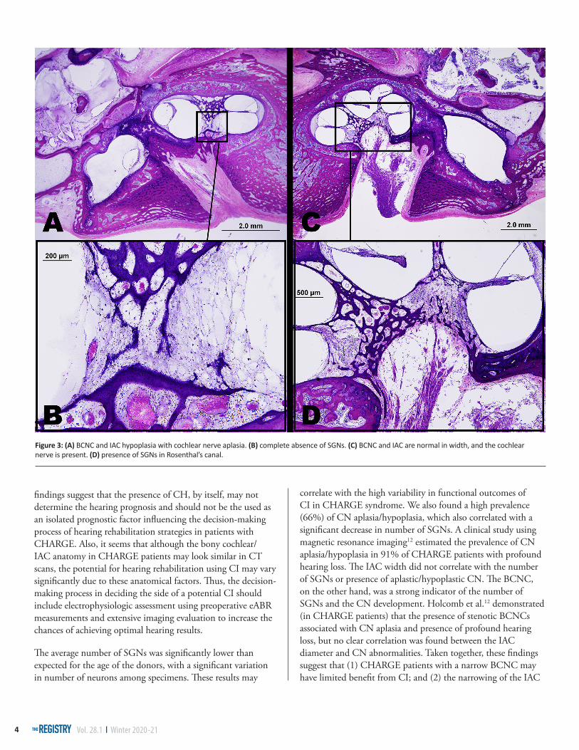
The mean SGN number was 68% lower than expected for age. There was a significant positive correlation between the number of SGNs and the BCNC diameter (P = 0.013) (Fig. 3), but no correlations were observed between number of SGNs and IAC length or diameter (P >0.05). As expected, no neuron bodies were found in the Rosenthal’s canal of specimens with aplastic CN. There was no difference in the number of SGNs between TBs with hypoplastic or normal CNs (p = 0.60). Mean ScGN counts were 80% lower than expected for their age. There were no significant correlations between ScGN counts and IAC diameter/length (P >0.05) or BCNC diameter (P = 0.17).
In the peripheral vestibular system, the saccule and utricle were markedly small in the majority (83.3%) of cases. The presence of either aplasia (33.3%) or hypoplasia (50%) of the utricle (pars superior) was more frequent than aplasia or hypoplasia of the saccule (pars inferior) (58.3%). In 4 (33.3%) TBs there was a large confluence between the saccule and the utricle. The hair cells in the otolithic organs seemed decreased due to the hypoplastic maculae.
Our findings provide insights regarding developmental abnormalities affecting the ears of patients with CHARGE syndrome. The understanding of structural and cellular TB abnormalities is critical for defining the best hearing rehabilitation strategy.4,8 We found in these 12 specimens several external, middle and inner-ear abnormalities that can lead to hearing impairment. These findings are consistent with clinical studies, which revealed a high prevalence of hearing loss (>60%) in this specific population.
We found a high prevalence of EAC malformations in the medical records and in the otopathological analysis, which is consistent with the literature (80-100%). The middle-ear analysis also revealed small middle-ear clefts and underdeveloped, sclerotic mastoids. Furthermore, the stapes were dysplastic in most TBs. Considering these abnormalities, these donors would certainly have a conductive hearing deficit – in this population, conductive or mixed-type hearing loss may be caused by stapes dysplasia, eustachian tube dysfunction, otitis media, and external ear malformations.9 These conductive deficits
DIRECTORSJoseph B. Nadol, Jr., MDM. Charles Liberman, PhDAlicia M. Quesnel, MDFelipe Santos, MD
COORDINATORCsilla Haburcakova, PhD
ADMINISTRATIVE STAFFKristen Kirk-Paladino
EDITORMedical: Felipe Santos, MD General: Michael Kotsopoulos
DESIGNERGaryfallia Pagonis
NIDCD National Temporal Bone, Hearing and Balance Pathology Resource Registry
Massachusetts Eye and Ear243 Charles StreetBoston, MA 02114
(800) 822-1327 Toll-Free Voice(617) 573-3711 Voice(617) 573-3838 Fax
Email: [email protected]: www.tbregistry.org
The reports in the Registry Newsletter are not peer reviewed.
2
THE

Vol. 28.1 | Winter 2020-21THE 3
continued on page 4
may be addressed with a hearing aid (in cases with no EAC malformation), a stapedectomy (in cases with dysplastic stapes), or a bone-anchored hearing device.5
The FN was abnormal in all TBs with CHARGE syndrome, which was consistent with the findings of clinical studies.10 The narrowing of the facial recess observed in some of the specimens could hinder access to the RW through traditional posterior tympanotomy approach for CI; therefore, a comprehensive preoperative imaging routine is warranted to identify and prepare the surgeon for this potential adversity.4,8 Additionally, other anatomical abnormalities may constitute additional drawbacks for CI surgery: the sclerotic and small mastoids, RW niche obliteration, and the hypoplastic cochlea and semicircular canals. These challenges could be circumvented by using
an alternative route (such as middle fosse, suprameatal, or RAMBO transcanal approaches) or a subtotal petrosectomy.8,11
In our study, all TBs had CH, with significant differences in the cochlear architecture among specimens, findings that are congruent with those of clinical studies: a retrospective review of 379 patients with CHARGE showed a 91% prevalence of CH.2 Although the CH may be responsible for the profound hearing loss, the hearing in CHARGE patients may range from normal hearing to complete deafness, suggesting multifactorial causes. Our results corroborate these assumptions: the cochlear architecture, number of SGNs, and development of the CN varied substantially among specimens, even between ears from the same donors. These
Figure 1: (A) Facial nerve (first genu) in abnormal course (obtuse angle). (B) Facial nerve (black arrow) abnormally positioned at the level of the round window, narrowing the facial recess (white arrow = chorda tympani nerve).
Figure 2: Cochlear hypoplasia and modiolar defects; vestibules and semicircular canals are hypoplastic. The oval windows (A and B) are aplastic.
Vol. 28.1 | Winter 2020-21THE4
findings suggest that the presence of CH, by itself, may not determine the hearing prognosis and should not be the used as an isolated prognostic factor influencing the decision-making process of hearing rehabilitation strategies in patients with CHARGE. Also, it seems that although the bony cochlear/IAC anatomy in CHARGE patients may look similar in CT scans, the potential for hearing rehabilitation using CI may vary significantly due to these anatomical factors. Thus, the decision-making process in deciding the side of a potential CI should include electrophysiologic assessment using preoperative eABR measurements and extensive imaging evaluation to increase the chances of achieving optimal hearing results.
The average number of SGNs was significantly lower than expected for the age of the donors, with a significant variation in number of neurons among specimens. These results may
correlate with the high variability in functional outcomes of CI in CHARGE syndrome. We also found a high prevalence (66%) of CN aplasia/hypoplasia, which also correlated with a significant decrease in number of SGNs. A clinical study using magnetic resonance imaging12 estimated the prevalence of CN aplasia/hypoplasia in 91% of CHARGE patients with profound hearing loss. The IAC width did not correlate with the number of SGNs or presence of aplastic/hypoplastic CN. The BCNC, on the other hand, was a strong indicator of the number of SGNs and the CN development. Holcomb et al.12 demonstrated (in CHARGE patients) that the presence of stenotic BCNCs associated with CN aplasia and presence of profound hearing loss, but no clear correlation was found between the IAC diameter and CN abnormalities. Taken together, these findings suggest that (1) CHARGE patients with a narrow BCNC may have limited benefit from CI; and (2) the narrowing of the IAC
Figure 3: (A) BCNC and IAC hypoplasia with cochlear nerve aplasia. (B) complete absence of SGNs. (C) BCNC and IAC are normal in width, and the cochlear nerve is present. (D) presence of SGNs in Rosenthal’s canal.

Vol. 28.1 | Winter 2020-21THE 5
should not be used as a single prognostic factor in the decision-making process of CIs versus auditory brainstem implants.
We found a high prevalence of peripheral vestibular system abnormalities secondary to CHARGE syndrome, including vestibular hypoplasia, semicircular canal aplasia, and severe decrease of ScGNs. Previous studies estimated the prevalence of semicircular canal aplasia in CHARGE at 77%-99%.1 It was also demonstrated that CHARGE patients have no responses detected by bi-thermal caloric testing.13 The testing of otolithic organs frequently yields normal results;13 however, most CHARGE patients have delayed development of gross motor skills and balance dysfunction.14 Although these balance issues are multifactorial, the semicircular canal aplasia and malformation of the otolithic organs and vestibular nerves may play a role.13 l
Keywords: CHARGE syndrome; otopathology; temporal bone pathology; Inner ear; middle ear; congenital abnormalities; hearing loss; cochlear implantation; deafness; vestibular diseases.
This article was presented at the AAO-HNSF 2020 Virtual Annual Meeting and OTO Experience, September 13-October 25, 2020 (Presentation number: 002209)
Conflict of Interest and financial disclosures: None
REFERENCES
1. Lalani SR, Hefner MA, Belmont JW, Davenport SL. CHARGE Syndrome. In: Adam MP, Ardinger HH, Pagon RA, et al., eds. GeneReviews®. University of Washington, Seattle; 1993. Accessed August 28, 2020. http://www.ncbi.nlm.nih.gov/books/NBK1117/
2. Zentner GE, Layman WS, Martin DM, Scacheri PC. Molecular and phenotypic aspects of CHD7 mutation in CHARGE syndrome. Am J Med Genet A. 2010;152A:674-686. doi:10.1002/ajmg.a.33323
3. Vincenti V, Di Lella F, Falcioni M, Negri M, Zanetti D. Cochlear implantation in children with CHARGE syndrome: a report of eight cases. Eur Arch Otorhinolaryngol. 2018;275:1987-1993. doi:10.1007/s00405-018-5053-x
4. Sennaroglu L. Cochlear implantation in inner ear malformations–a review article. Cochlear Implants Int. 2010;11:4-41. doi:10.1002/cii.416
5. Sennaroğlu L, Demir Bajin M. Classification and Current Management of Inner Ear Malformations. Balk Med J. 2017;34:397-411. doi:10.4274/balkanmedj.2017.0367
6. Otte J, Schunknecht HF, Kerr AG. Ganglion cell populations in normal and pathological human cochleae. Implications for cochlear implantation. Laryngoscope. 1978;88:1231-1246. doi:10.1288/00005537-197808000-00004
7. Velázquez-Villaseñor L, Merchant SN, Tsuji K, Glynn RJ, Wall C, Rauch SD. Temporal bone studies of the human peripheral vestibular system. Normative Scarpa’s ganglion cell data. Ann Otol Rhinol Laryngol. 2000;181:14-19. doi:10.1177/00034894001090s503
8. Monsanto R da C, Sennaroglu L, Uchiyama M, Sancak IG, Paparella MM, Cureoglu S. Histopathology of Inner Ear Malformations: Potential Pitfalls for Cochlear Implantation. Otol Neurotol 2019;40:e839-e846. doi:10.1097/MAO.0000000000002356
9. Davenport SL, Hefner MA, Thelin JW. CHARGE syndrome. Part I. External ear anomalies. Int J Pediatr Otorhinolaryngol. 1986;12:137-143. doi:10.1016/s0165-5876(86)80071-4
10. Wineland A, Menezes MD, Shimony JS, Shinawi MS, Hullar TE, Hirose K. Prevalence of Semicircular Canal Hypoplasia in Patients With CHARGE Syndrome: 3C Syndrome. JAMA Otolaryngol Neck Surg. 2017;143:168-177. doi:10.1001/jamaoto.2016.3175
11. Chen JX, Nourmahnad A, O’Malley J, Reinshagen K, Nadol JB, Quesnel AM. Otopathology in CHARGE syndrome. Laryngoscope Investig Otolaryngol. 2020;5:157-162. doi:10.1002/lio2.347
12. Holcomb MA, Rumboldt Z, White DR. Cochlear nerve deficiency in children with CHARGE syndrome. Laryngoscope. 2013;123:793-796. doi:10.1002/lary.23682
13. Wiener-Vacher SR, Amanou L, Denise P, Narcy P, Manach Y. Vestibular Function in Children With the CHARGE Association. Arch Otolaryngol Neck Surg. 1999;125:342-347. doi:10.1001/archotol.125.3.342
14. Green GE, Huq FS, Emery SB, Mukherji SK, Martin DM. CHD7 mutations and CHARGE syndrome in semicircular canal dysplasia. Otol Neurotol. 2014;35:1466-1470. doi:10.1097/MAO.0000000000000260
CORRESPONDING AUTHORRafael da Costa Monsanto, MD, PhDR Alexandre Francisco Dall’ava 50 – Jd Res Tivoli Park – Sorocaba/SP, Brasil. CEP 1848-210Email: [email protected]
Vol. 28.1 | Winter 2020-21THE6
Association of Internal Auditory Canal Diverticula and Otosclerosis:
A Histopathological and Radiological StudyRenata M. Knoll, MD* 1,2, Elliot Kozin, MD* 1,2, Dawson Wells* 1,2, Jenny X. Chen, MD1,2, Katherine Reinshagen, MD3, Hinrich Staecker, MD, PhD4, Michael J. McKenna, MD1,2,
Joseph Nadol, Jr, MD1,2, Alicia Quesnel, MD1,2
1Department of Otolaryngology, Harvard Medical School, Boston, MA 2Department of Otolaryngology, Massachusetts Eye and Ear, Boston, MA
3Department of Radiology, Massachusetts Eye and Ear Infirmary, Boston, MA4Department of Otolaryngology-Head and Neck Surgery, University of Kansas School of Medicine, Kansas City, KS
Otosclerosis is a progressive osteodystrophy disease of the otic capsule that results in acquired hearing loss, with a peak onset in the third to fifth decades of life.1-3 While the estimated incidence of clinical otosclerosis in the Caucasian population is about
0.1%, postmortem histological evidence of otosclerosis in otherwise asymptomatic individuals can reach 11%.4 Even though the majority of these histological changes involve the area anterior to the oval window, otosclerosis has also been reported in other locations of the inner ear, such as the anterior wall of the internal auditory canal (IAC).1 In fact, several small histologic and imaging case series have referred to this finding, also known as IAC diverticula, as a form of “cavitary otosclerosis,”5-7 and suggested that its prevalence may be related to the severity of the disease.8 However, IAC diverticula have also been described in high resolution temporal bone (TB) computed tomography (CT) of patients without radiographic evidence of fenestral or retrofenestral otosclerosis.9
While these lesions have been identified in both otopathology specimens and CT scans, their etiology, prevalence, and clinical implications are not completely understood. Additionally, sensitivity and specificity of IAC diverticula as a radiologic marker of otosclerosis has never been specifically explored. Herein, this study aimed to (1) determine the significance of diverticula in cases with otosclerosis and (2) compare TB histologic sections and CT scans to evaluate the sensitivity and specificity of CT scans in identifying diverticula.
To approach these aims, consecutive TB specimens from the Massachusetts Eye and Ear Otopathology Laboratory collection with available histologic slides and CT scans were identified. We excluded specimens with severe postmortem changes or tumoral lesions causing bony destruction of the IAC. Slides were examined using light microscopy for otosclerosis and/or IAC diverticula, which was defined as any cavitary lesion – regardless of size – on the anterolateral side of the IAC.
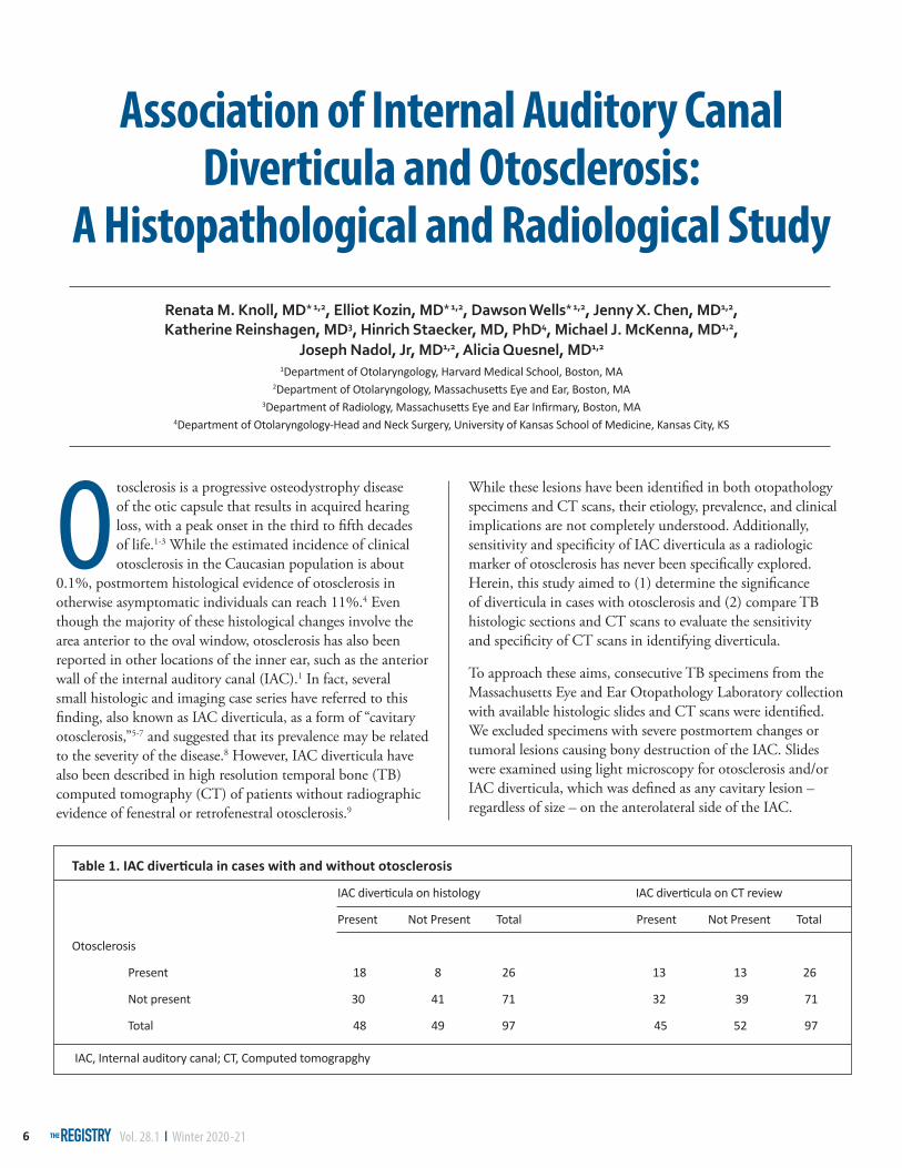
Table 1. IAC diverticula in cases with and without otosclerosis
IAC diverticula on histology IAC diverticula on CT review
Present Not Present Total Present Not Present Total
Otosclerosis
Present 18 8 26 13 13 26
Not present 30 41 71 32 39 71
Total 48 49 97 45 52 97
IAC, Internal auditory canal; CT, Computed tomograpghy
Vol. 28.1 | Winter 2020-21THE 7
continued on page 8
CT scans were also reviewed retrospectively by a neuro-radiologist who was blinded to the histologic evaluations. Radiographically, diverticula were defined as any low-density outpouching on the anterolateral wall of the IAC, regardless of size. The area of the IAC diverticula was measured on histology, and sensitivity and specificity of CT scan to detect diverticula were determined.
Ninety-seven TB specimens (42% male) were included, with average age of death of 77 years old (SD=18). IAC diverticula were found on histology in 48 specimens (49%), with a mean
area of 0.34mm2 (SD: ±0.60, range: 0.02-3.07mm2; Table 1; Fig. 1-4). Histological evidence of otosclerosis was found in 26 out of the 97 specimens (27%). In patients with histologically identified IAC diverticula, the rate of otosclerosis was 37.5%, which was significantly higher than the rate of 16% for those without diverticula (p=0.019). Of all patients with IAC diverticula identified histologically, those with otosclerosis typically had larger diverticula (p=0.001). The diverticula mean area for cases with otosclerosis was 0.69 mm2 (SD ±0.88 mm2), whereas non-otosclerosis cases had a mean area of 0.14 mm2 (SD ±0.13 mm2; Figure 4).
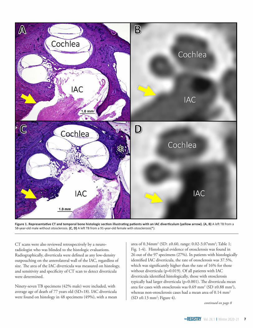
Figure 1. Representative CT and temporal bone histologic section illustrating patients with an IAC diverticulum (yellow arrow). (A, B) A left TB from a 58-year-old male without otosclerosis. (C, D) A left TB from a 91-year-old female with otosclerosis(*).
Vol. 28.1 | Winter 2020-21THE8
CT review identified 45 patients (46%) with diverticula, of which 70% had evidence of IAC diverticula on histology. The sensitivity and specificity to detect IAC diverticula based on CT was 77% and 63%, respectively (Table 2). Thirteen out of the 45 cases with a diverticula identified on CT review had histological evidence of otosclerosis (Table 1). The sensitivity and speci- ficity of CT in identifying IAC diverticula that were associated with otosclerosis was 50% and 55%, respectively. The mean histological diverticula area for those patients with otosclerosis that had an IAC diverticula identified in the CT was 0.65 mm2
(SD ±0.85 mm2), which was significantly greater than those without otosclerosis (mean= 0.13 mm2, SD ±0.15 mm2; p<0.001).
Previous radiological studies have already linked these cavitary lesions on the anterior wall of the IAC to otosclerosis.7, 9-11 In a retrospective study by Puac et al.,10 35% of cases with otosclerotic lesions were also reported to have a cavitary lesion, which is consistent with our findings. However, our study also showed that the presence of IAC diverticula on imaging is not
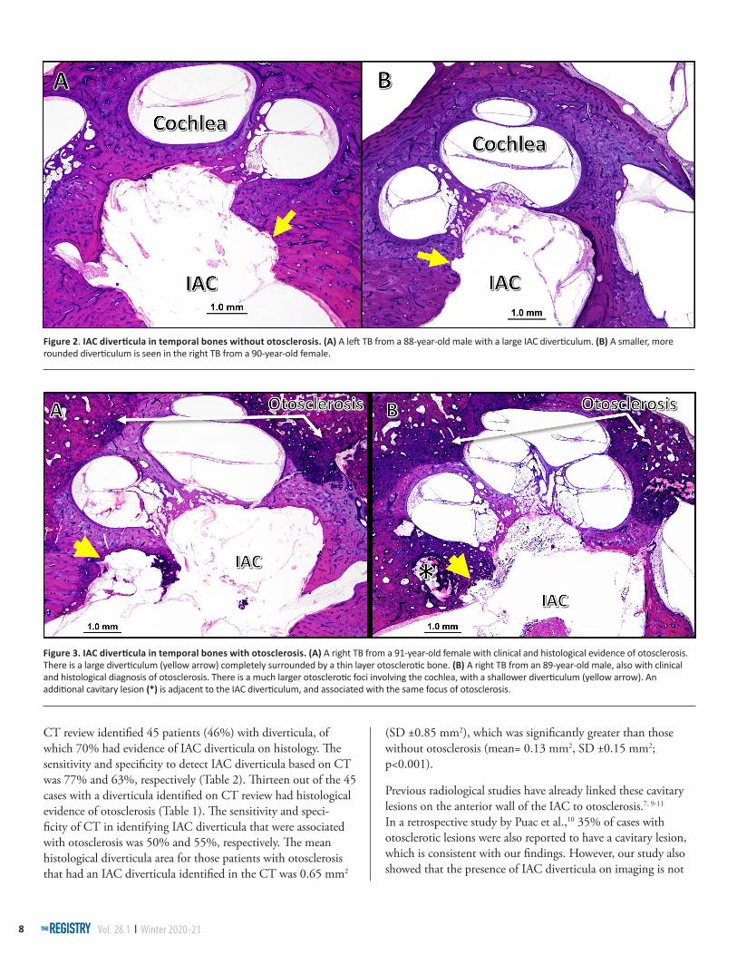
Figure 2. IAC diverticula in temporal bones without otosclerosis. (A) A left TB from a 88-year-old male with a large IAC diverticulum. (B) A smaller, more rounded diverticulum is seen in the right TB from a 90-year-old female.
Figure 3. IAC diverticula in temporal bones with otosclerosis. (A) A right TB from a 91-year-old female with clinical and histological evidence of otosclerosis. There is a large diverticulum (yellow arrow) completely surrounded by a thin layer otosclerotic bone. (B) A right TB from an 89-year-old male, also with clinical and histological diagnosis of otosclerosis. There is a much larger otosclerotic foci involving the cochlea, with a shallower diverticulum (yellow arrow). An additional cavitary lesion (*) is adjacent to the IAC diverticulum, and associated with the same focus of otosclerosis.

Vol. 28.1 | Winter 2020-21THE 9
Figure 4. Scatter plot with diverticula area in otosclerosis and non-otosclerosis cases. Cases with otosclerosis had a greater mean diverticula area compared to non-otosclerosis cases (p = 0.001). The horizontal lines and error bars represent the mean and standard error of the mean, respectively.
Table 2. Diagnostic Efficacy of CT Scanning
Statistic Value 95% CI
Sensitivity 77.08 62.69 - 87.97
Specificity 63.27 48.29 - 76.58
sensitive nor specific enough to diagnose cases with otosclerosis, as they may be still present in normal ears or associated with other pathologies. Additionally, our findings suggest that the presence of small IAC diverticula on imaging are less likely to be associated with otosclerosis.
In fact, other studies have suggested that these cavitary lesions most likely represent a normal anatomic variant.12,13 In another recent study by Pippin et al.9, IAC diverticula were usually described as an isolated CT scan finding, and rarely occurred alongside otosclerosis, which conflicts with our findings. One possible explanation for this discrepancy is that their study was entirely based on CT scans rather than histologic slides to identify diverticula,9 and small diverticula were probably missed.
Although there was variability in IAC diverticula shape and size in our study, we provide evidence of an association between large IAC diverticula and otosclerosis.
Taken together, while IAC diverticula may be more commonly associated with otosclerosis, they are not pathognomonic. CT scan was neither sensitive nor specific in identifying IAC diverticula. The presence of a diverticulum on CT, particularly when large, may be an indicator, but not a definitive marker, of otosclerosis. Nevertheless, we suggest that TB CT scan evaluations include a survey for IAC diverticula, particularly when ordered in the setting of clinical suspicion of otosclerosis. l
REFERENCES
1. Schuknecht HF. Pathology of the ear. Philadelphia: Lea & Febiger, 1993.
2. Quesnel A, McKenna M. Otosclerosis. In: Sataloff RT, Lalwani AK, eds. Sataloff’s Comprehensive Textbook of Otolaryngology: Head & Neck Surgery: Otology/Neurotology/Skull Base Surgery: JP Medical Ltd, 2015:231-242.
3. Cureoglu S, Schachern PA, Ferlito A, Rinaldo A, Tsuprun V, Paparella MM. Otosclerosis: etiopathogenesis and histopathology. Am J Otolaryngol 2006; 27:334-340.
4. Declau F, Van Spaendonck M, Timmermans JPet al. Prevalence of otosclerosis in an unselected series of temporal bones. Otol Neurotol 2001; 22:596-602.
5. Makarem AO, Linthicum FH. Cavitating otosclerosis. Otol Neurotol 2008; 29:730-731.
6. Makarem AO, Hoang TA, Lo WW, Linthicum FH, Jr., Fayad JN. Cavitating otosclerosis: clinical, radiologic, and histopathologic correlations. Otol Neurotol 2010; 31:381-384.
7. Bou-Assaly W, Mukherji S, Srinivasan A. Bilateral cavitary otosclerosis: a rare presentation of otosclerosis and cause of hearing loss. Clin Imaging 2013; 37:1116-1118.
8. Hoeberigs M, Postma A, Waterval J, Stokroos R, Stadler A. Prevalence of anterior internal auditory canal ‘‘diverticulum’’ on high resolution CT in patients with otosclerosis Proceedings of the Radiological Society of North America 2012 Scientific Assembly and Annual Meeting. Chicago, Illinois, 2012.
9. Pippin KJ, Muelleman TJ, Hill J, Leever J, Staecker H, Ledbetter LN. Prevalence of Internal Auditory Canal Diverticulum and Its Association with Hearing Loss and Otosclerosis. AJNR Am J Neuroradiol 2017; 38:2167-2171.
10. Puac P, Rodriguez A, Lin HCet al. Cavitary Plaques in Otospongiosis: CT Findings and Clinical Implications. AJNR Am J Neuroradiol 2018; 39:1135-1139.
11. Shim YJ, Bae YJ, An GSet al. Involvement of the Internal Auditory Canal in Subjects With Cochlear Otosclerosis: A Less Acknowledged Third Window That Affects Surgical Outcome. Otol Neurotol 2019; 40:e186-e190.
12. Mihal DC, Feng Y, Kodet ML, Lohse CM, Carlson ML, Lane JI. Isolated Internal Auditory Canal Diverticula: A Normal Anatomic Variant Not Associated with Sensorineural Hearing Loss. AJNR Am J Neuroradiol 2018; 39:2340-2344.
13. Gulya AJ, Gulya AJ. Gulya and Schuknecht’s anatomy of the temporal bone with surgical implications. New York: Informa Healthcare, 2007.
CORRESPONDING AUTHOR Alicia M. Quesnel, MD Massachusetts Eye and Ear 243 Charles Street Boston, MA 02114 Email: [email protected]

Vol. 28.1 | Winter 2020-21THE10
Temporal Bone Removal Technicians Needed Nationwide!
Seeking trained autopsy technicians for the removal of temporal bones on an on-call basis.
Technicians must be in the U.S. and are paid by case.
Interested? Email us at [email protected]
Explore Past Issues!
The editors of The Registry have created an online archive where you can access every issue.
The archive can be found on our website at: https://masseyeandear.org/tbregistry/research-resources
Interested in receiving digital newsletter copies?Email Csilla Haburcakova, PhD, Registry Coordinator, at Mass Eye and Ear

Vol. 28.1 | Winter 2020-21THE 11
Otopathology Mini-Travel Fellowship Program
The NIDCD National Temporal Bone Registry’s mini-travel fellowships provide funds for research technicians and young investigators to visit a temporal bone laboratory for a brief educational visit, lasting approximately one week. The emphasis is on the training of research assistants, technicians, and junior faculty.
These fellowships are available to:
• U.S. hospital departments who aspire to start a new temporal bone laboratory
• Inactive U.S. temporal bone laboratories who wish to reactivate their collections
• Active U.S. temporal bone laboratories who wish to learn new research techniques
Up to two fellowship awards will be made each year ($1,000 per fellowship). The funds may be used to defray travel and lodging expenses. Applications will be decided on merit.
Interested applicants should submit the following:
• An outline of the educational or training aspect of the proposed fellowship (1–2 pages)
• Applicant’s curriculum vitae
• Letter of support from a temporal bone laboratory director or department chairman
• Letter from the host temporal bone laboratory indicating willingness to receive the traveling fellow
Applications should be submitted to:
Felipe Santos, MDNIDCD Temporal Bone RegistryMassachusetts Eye and Ear243 Charles Street, Boston, MA [email protected]
12
NIDCD National Temporal Bone, Hearing and BalancePathology Resource Registry Massachusetts Eye and Ear 243 Charles Street Boston, MA 02114-3096
Free Brochures for your Office or Clinic about Temporal Bone Research and DonationThe Gift of Hearing and Balance: Learning About Temporal Bone Donation is a 16-page, full-color booklet that describes in detail the benefits of temporal bone research. It also answers commonly asked questions regarding the temporal bone donation process. Dimensions: 7”x10”
If you would like to display this brochure, please complete the form below and return it to The Registry by mail or fax. The brochures will be sent to you free of charge. Please circle the amount requested for each brochure or write in the amount if not listed.
The Gift of Hearing and Balance __________ 25 50 100
Name: ________________________________________________________________________________________________
Address: ______________________________________________________________________________________________
City, State, Zip: _________________________________________________________________________________________
Telephone: ____________________________________________________________________________________________
Mail or fax this form to the Registry at: NIDCD National Temporal Bone, Hearing and Balance Pathology Resource Registry Massachusetts Eye and Ear, 243 Charles Street, Boston, MA 02114 Toll-free phone: (800) 822-1327, Fax: (617) 573-3838 Email: [email protected]
NON-PROFIT ORGU.S. POSTAGE PAID
BOSTON, MAPERMIT NO. 53825