Osteopathic approach to treatment of developmental hip ... di 9 aprile/Pediatria/1... · The aim of...
32
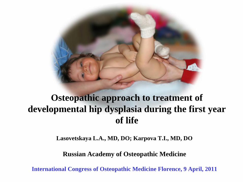
Lasovetskaya L.A., MD, DO; Karpova T.I., MD, DO Russian Academy of Osteopathic Medicine International Congress of Osteopathic Medicine Florence, 9 April, 2011 Osteopathic approach to treatment of developmental hip dysplasia during the first year of life
Transcript of Osteopathic approach to treatment of developmental hip ... di 9 aprile/Pediatria/1... · The aim of...
Lasovetskaya L.A., MD, DO; Karpova T.I., MD, DO
Russian Academy of Osteopathic Medicine
International Congress of Osteopathic Medicine Florence, 9 April, 2011
Osteopathic approach to treatment of
developmental hip dysplasia during the first year
of life
• The modern term “developmental hip dysplasia”, further DHD, is used to describe a broad spectrum of hip joint abnormalities from merely detectable to frankly visible, which are present at birth or in early infancy.
• It is our true belief that lately diagnosed or incompletely treated DHD independent of its severity has a negative influence over gait development, distribution of force lines in the body and posture on the whole, which among other things may cause predisposition to premature degenerative changes in the affected joint and painful arthritis

The aim of the presentation
• to demonstrate the perspectives of osteopathic
approach to diagnosis and treatment of different DHD
types during the first year of life.
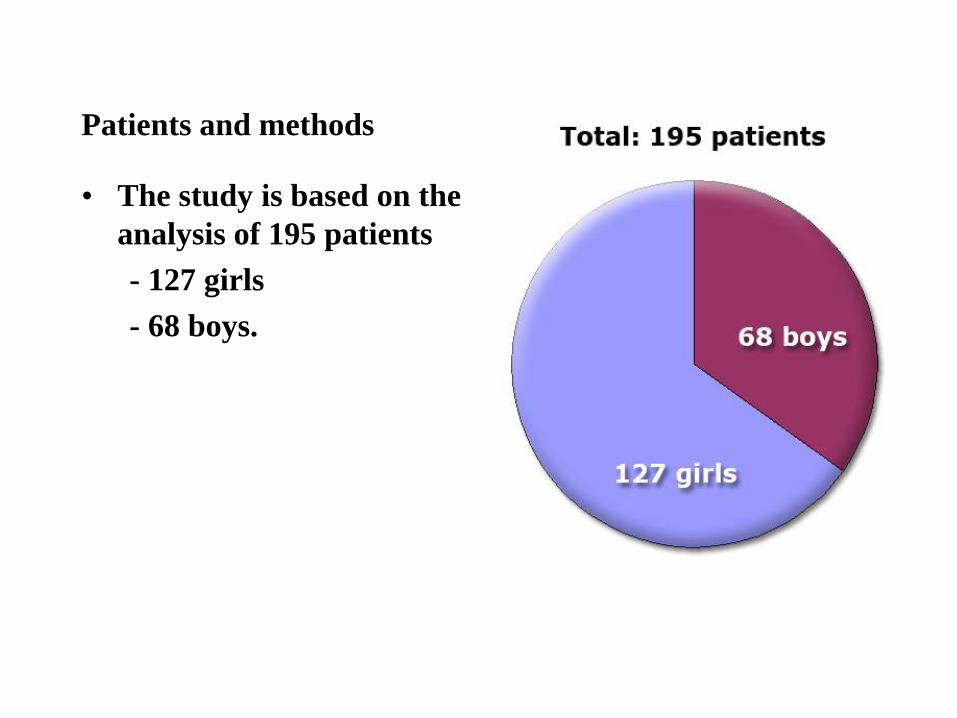
Patients and methods
• The study is based on the
analysis of 195 patients
- 127 girls
- 68 boys.

Inclusion criteria:
• diagnosed DHD based upon osteopathic, orthopedic
and ultrasound examinations;
• age from 0 to 6 months at the moment of DHD
diagnosis and treatment.

Exclusion criteria:
• prematurity;
• presence of genetic diseases, syndromes, neurological
pathology.

• At the whole - 195 infants
• 122 (study group) received
only osteopathic treatment
(6–8 procedures 10–21 days
apart),
• 73 (reference group) were
treated with bracing for
three months under the
supervision of an orthopedic
surgeon.

• All 195 infants had another ultrasound examination at
the age of 8 months.
At the age of 10–12 months all the patients were examined
by an osteopath and an orthopedist.
• All examinations were blinded.

Results and discussion

Hierarchic classification of DHD
• Formerly it was believed that the discussed condition
began prior to birth.
• But later on it has been realized that some hips may be
normal at birth and gradually become dysplastic.
• Basing on our findings we distinguish three DHD types:
primary, secondary and mixed.

• The primary type (38.5%) develops during the intrauterine period, and postnatally is characterized by presence of distinctly palpated lesions in the pelvis and hip joint or joints with intra-osseous strains of the innominate bones, sacrum and coccyx.
• The secondary type (15.6%) forms during the first weeks of the infant’s life, and is caused by
- (1) severe non-physiological sphenobasilar strains,
- (2) vertebral lesions,
- (3) fascial tensions including those coming from the viscera or the thoracic diaphragm
• The mixed DHD type appears to be the most frequent (45.9%);
- here both variants (primary and secondary) are present.
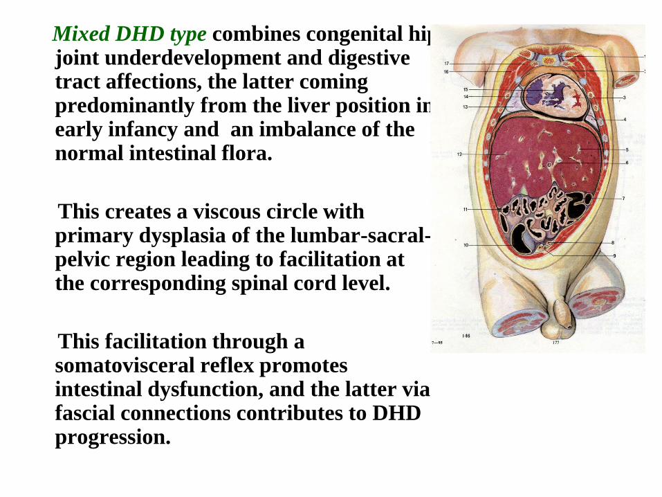
Mixed DHD type combines congenital hip joint underdevelopment and digestive tract affections, the latter coming predominantly from the liver position in early infancy and an imbalance of the normal intestinal flora.
This creates a viscous circle with primary dysplasia of the lumbar-sacral-pelvic region leading to facilitation at the corresponding spinal cord level.
This facilitation through a somatovisceral reflex promotes intestinal dysfunction, and the latter via fascial connections contributes to DHD progression.
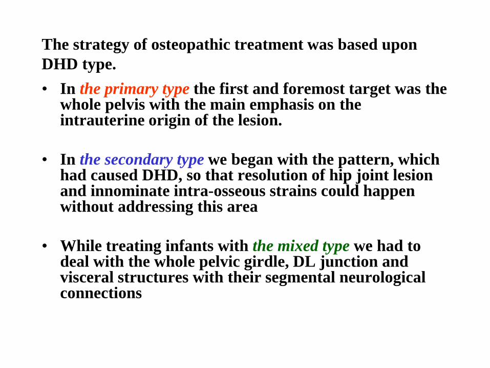
The strategy of osteopathic treatment was based upon
DHD type.
• In the primary type the first and foremost target was the whole pelvis with the main emphasis on the intrauterine origin of the lesion.
• In the secondary type we began with the pattern, which had caused DHD, so that resolution of hip joint lesion and innominate intra-osseous strains could happen without addressing this area
• While treating infants with the mixed type we had to deal with the whole pelvic girdle, DL junction and visceral structures with their segmental neurological connections

• Although ultrasound
examination conducted at
the age of 8 months showed
normalization of all the
affected hip joints,
orthopedic evaluation of
the patients at the age of
10–12 months
demonstrated differences
between the study group
and the reference group
A «healthy» child from reference group

• Clinical orthopedic signs in infants of both groups at the age of 10–12 months
• Clinical signs Groups of patientsχ2 Р-level reference group (n=73)study group (n=122)Varus position of one or both feet45 (61.6%)8 (6.6%)70.02< 0,0001 Scoliotic spinal pattern 32 (43.8%)-60.82< 0,0001Restriction of hip abduction 18 (24.7%)-30.26< 0,0001Asymmetry of gluteal and/or femoral skin folds19 (26.0%)3 (2.5%)12.45= 0,0004 Excessive external or internal leg rotation63 (86.3%)-151.62< 0,0001Leg length inequality10 (13.7%)-14.91= 0,0001
Table
Clinical orthopedic signs in infants of both groups at the
age of 10–12 months

• We have studied the whole complex of palpatory
changes in the infant’s body accompanying DHD.
• Five distinct palpatory patterns were found. It’s worth
mentioning that these patterns have never been
described in osteopathic and orthopedic literature.

Pattern 1 frontal-horizontal
• was registered in 45.9% of cases.
• with insufficient stability both in horizontal and frontal plane, predominantly at the expense of the pelvic component.
• mainly accompanied the mixed DHD type and demonstrated such features as
- pelvic rotation;
- ilium inflare on the affected side;
- umbilical ligament tension;
- intra-osseous strains of the coccyx, sacrum and innominate bones.

Pattern 2 frontal
• was diagnosed in 24.6% of infants.
• with insufficient hip stability in the frontal plane only.
• belongs to primary DHD type and has the following characteristics:
- intra-osseous strain of the coccyx and innominate bone;
- “deep location” of the acetabulum, the ilium on the affected side being oblique and flattened;
- absence of congruency between the size of the femoral head and that of the acetabulum, the former usually being larger;
- intra-osseous compression of the femur and the innominate bone.

Fig. 2. Pattern 2
Pattern 3 bilateral frontal
• was quite rare,
• present only in 5.7%.
• actually it was a bilateral version of the second
pattern manifesting in multi-vector pelvic
compression

Fig. 4. Pattern 3

Pattern 4 torsion • 8.2%
• was characterized by pelvic torsion around the horizontal axis,
• one innominate bone being rotated anteriorly, and the other one – posteriorly.
• affected the whole of the pelvis with intra-osseous strains of the sacrum (predominantly of S1 and S2 segments)
• torsion existed between the primary (embryological) sacral segments.
• was regarded as a manifestation of the primary DHD type.
• If it represented a part of a general fascial pattern without crude involvement of the sacrum and the lumbar spine it was interpreted as a variant of the secondary DHD type.

Fig. Pattern 4

Pattern 5 lateral
• was found in 15.6% of infants.
• in analogy with SBS lesions we called it a lateral pelvic
pattern.
• in more than half of cases the SBS strain was primary,
and the pelvic pattern – secondary.
• the whole pelvic girdle was deformed, one ilium being
anterior and in outflare, and the other one – posterior
and in inflare.

Fig. Pattern 5

The frequency of different patterns
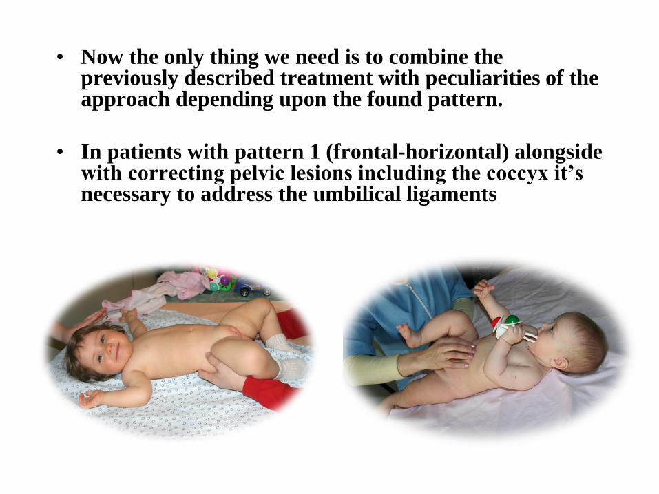
• Now the only thing we need is to combine the previously described treatment with peculiarities of the approach depending upon the found pattern.
• In patients with pattern 1 (frontal-horizontal) alongside with correcting pelvic lesions including the coccyx it’s necessary to address the umbilical ligaments

• The main emphasis in
infants with the second
(frontal) and third (bilateral
frontal) patterns should be
made on resolution of
innominate bone
compression to allow it
expressing its inherent
primary respiration.

• Treatment of the fourth
(torsional) pattern in
primary DHD should begin
with resolution of sacral
intra-osseous strains, and in
secondary DHD should
predominantly deal with
pelvic correction in context
of the whole body without
forgetting about the
sacroiliac joints.
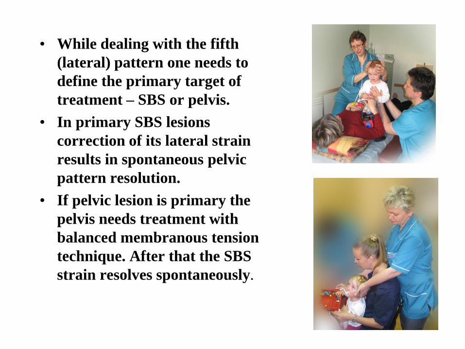
• While dealing with the fifth
(lateral) pattern one needs to
define the primary target of
treatment – SBS or pelvis.
• In primary SBS lesions
correction of its lateral strain
results in spontaneous pelvic
pattern resolution.
• If pelvic lesion is primary the
pelvis needs treatment with
balanced membranous tension
technique. After that the SBS
strain resolves spontaneously.

• Thus, defining the strategy and tactics of osteopathic treatment it’s necessary to take into account the type of developmental hip dysplasia in accordance with the suggested classification (primary, secondary and mixed) and the found lesion pattern (frontal-horizontal, frontal, bilateral frontal, torsional, lateral).
• There are all reasons to believe that this complete, pathogenically based approach to DHD treatment will lead to resolution of all components of the problem and in the long run – to optimal joint development.

T
H
A
N
K
Y
O
U
F
O
R
Y
O
U
R
A
T
T
E
N
T
I
O
N