ORTHOPEDICS NOW - Connecticut Children's
10
Summer 2017 Inside this Issue ... From the Director’s Desk ...................... 2 Dancer Rebounds from Cervical Bone Tumor .............................................. 3 New Surgical Technique for ACL Reconstruction ............................... 4 Meniscus Tear Doesn’t Sideline Shortstop .................................................. 4 Novel Techniques Put Gymnast Back on Top ............................................. 5 Scoliosis Corrected with a Little “MAGEC” ....................................... 6 Research Focuses on Concussions ... 7 Your Support Counts ............................. 8 Special Insert: Researchers Shine Light on Charcot-Marie-Tooth Disease Welcome to Orthopedics Now, an annual publication of Connecticut Children’s Medical Center Foundation, prepared especially for friends and patient families of Connecticut Children’s Division of Orthopedics. Read on to learn more about the many programs and services the hospital provides and about our generous friends and donors who help make it happen. Geno Auriemma’s Charity Golf Tournament . . Fundraiser Scores Imaging Equipment “Fore the Kids” Continued on page 2 Coach Geno Auriemma joined 160 golfers on the links in late June to raise the remaining funds needed to purchase specialized imaging equipment for Connecticut Children’s Orthopedics Division. The 2017 Geno Auriemma’s “Fore the Kids” Charity Golf Tournament, which celebrated its 15th year of play June 26th, raised approximately $130,000 toward the purchase of an ultra-low-dose imaging system to benefit patient care. Funds raised at the past three charity golf tournaments, hosted by Coach Auriemma at the Hartford Golf Club, will be used to purchase the technology, which will provide Connecticut Children’s orthopedic surgeons with state-of-the-art image quality. ORTHOPEDICS NOW From Connecticut Children’s Medical Center Foundation 17-419 “I want to thank Geno, the organizing committee and everyone who participated in making this such a great event,” said Orthopedics Division Chief Jeffrey Thomson, MD. “The money raised will be used to purchase specialized radiology equipment that will allow us to obtain more rapid and accurate X-rays while significantly decreasing the radiation exposure for our patients.” Specialized Imaging Equipment The new imaging equipment, called the EOS ® Imaging System, is a low-dose, three-dimensional imaging system that scans a child while standing. With an EOS scan, a child’s natural, weight-bearing posture is shown, so that physicians can view the interaction between the joints and A Football Player’s Comeback Meet Khyon…Page 4 Pictured with Coach Geno Auriemma (second from right) are Connecticut Children’s Medical Center Foundation President David Kinahan (left), and Tournament Co-Chairs Sal Giuliano, and Randy Holmeen. Lily Velez, daughter of Tony and Dawn Velez (pictured here), was born with severe scoliosis. Learn how the new MAGEC Rod technology has worked “magic” in her treatment. (See page 6 of this issue.)
Transcript of ORTHOPEDICS NOW - Connecticut Children's

Summer 2017
Inside this Issue ...From the Director’s Desk ......................2
Dancer Rebounds from Cervical Bone Tumor ..............................................3
New Surgical Technique for ACL Reconstruction ...............................4
Meniscus Tear Doesn’t Sideline Shortstop ..................................................4
Novel Techniques Put Gymnast Back on Top .............................................5
Scoliosis Corrected with a Little “MAGEC” .......................................6
Research Focuses on Concussions ...7
Your Support Counts .............................8
Special Insert: Researchers Shine Light on
Charcot-Marie-Tooth Disease
Welcome to Orthopedics Now, an annual publication of Connecticut Children’s Medical Center Foundation, prepared especially for friends and patient families of Connecticut Children’s Division of Orthopedics. Read on to learn more about the many programs and services the hospital provides and about our generous friends and donors who help make it happen.
Geno Auriemma’s Charity Golf Tournament . .
Fundraiser Scores Imaging Equipment “Fore the Kids”
Continued on page 2
Coach Geno Auriemma joined 160 golfers on the links in late June to raise the remaining funds needed to purchase specialized imaging equipment for Connecticut Children’s Orthopedics Division.
The 2017 Geno Auriemma’s “Fore the Kids” Charity Golf Tournament, which celebrated its 15th year of play June 26th, raised approximately $130,000 toward the purchase of an ultra-low-dose imaging system to benefit patient care.
Funds raised at the past three charity golf tournaments, hosted by Coach Auriemma at the Hartford Golf Club, will be used to purchase the technology, which will provide Connecticut Children’s orthopedic surgeons with state-of-the-art image quality.
ORTHOPEDICS NOWFrom Connecticut Children’s Medical Center Foundation
17-4
19
“I want to thank Geno, the organizing committee and everyone who participated in making this such a great event,” said Orthopedics Division Chief Jeffrey Thomson, MD. “The money raised will be used to purchase specialized radiology equipment that will allow us to obtain more rapid and accurate X-rays while significantly decreasing the radiation exposure for our patients.”
Specialized Imaging Equipment The new imaging equipment, called the EOS® Imaging System, is a low-dose, three-dimensional imaging system that scans a child while standing. With an EOS scan, a child’s natural, weight-bearing posture is shown, so that physicians can view the interaction between the joints and
A Football Player’s Comeback
Meet Khyon…Page 4
Pictured with Coach Geno Auriemma (second from right) are Connecticut Children’s Medical Center Foundation President David Kinahan (left), and Tournament Co-Chairs Sal Giuliano, and Randy Holmeen.
Lily Velez, daughter of Tony and Dawn Velez (pictured here), was born with severe scoliosis. Learn how the new MAGEC Rod technology has worked “magic” in her treatment. (See page 6 of this issue.)

Orthopedics, Sports Medicine (Elite Sports Medicine) and the Center for Motion Analysis compose Connecticut Children’s busy Orthopedics Division, where we adapt to seasonal increase demands for outpatient visits. Summer is a busy time, but in pediatric orthopedics, we are always available for same-day appointments.
Sports Medicine Section Growing Our Sports Medicine section is growing with the addition of a third orthopedic surgeon, Dr. Lee Pace, who relocated from Los Angeles Children’s Hospital. He has been in practice for about five years and is highly skilled in all aspects of pediatric sports medicine. (See Dr. Pace’s bio on this page.)
Our Sports Medicine section also has the ability to perform computerized baseball pitching analysis. In fact, our specialists, Matt Solomito, PhD, Carl Nissen, MD, Matt Milewski, MD, and Sylvia Õunpuu, MsC, designed the methods to evaluate baseball pitching using computerized motion analysis. Our motion analysis laboratory utilizes 12 infrared cameras to analyze a baseball pitcher’s motion. This level of analysis and expertise is not available anywhere else in the Northeast.
Center for Motion Analysis Our Center for Motion Analysis has also made exciting headway in evaluating and treating patients with Charcot-Marie-Tooth (CMT) disease. Sylvia Õunpuu, MsC, Kristan Pierz, MD, and Gyula Acsadi, MD, of Neurology, have teamed up and are now
2.
recognized by the Charcot-Marie-Tooth Association as a Center of Excellence.
Connecticut Children’s was also designated a Pediatric CMT Center of Excellence by the international Inherited Neuropathy Consortium. It is one of the few sites that collaborates with a pediatric gait laboratory for studying the natural history and surgical outcomes in the pediatric population.
Meet Our Patients In this issue of Orthopedics Now, you will not only learn about our CMT research activities and our busy Concussion Management Clinic, you will also meet some of our amazing patients whose lives have been touched – and transformed – by Connecticut Children’s caring professionals.
Among them, you will meet Bridget, who won first place in an Irish dance competition six weeks after undergoing cervical spine bone tumor surgery, as well as Khyon, who is back in the game after a torn ACL threatened his future in football and a new physeal-sparing technique saved it.
You will also meet Connor, who is back on the ballfield following a rare meniscus surgery, as well as Madelyn, a gymnast whose response to alternative treatment methods has helped her return to the sport she loves. And finally, you will meet Lily, whose severe scoliosis has responded well to “MAGEC.”
At Connecticut Children’s, our Orthopedics Division is committed to providing high-quality care for all of our pediatric patients and young athletes from across the region.
Division Welcomes Lee Pace, MD Connecticut Children’s Elite Sports Medicine is happy to welcome a new member to its team: James Lee Pace, MD, has signed on as a pediatric sports
medicine surgeon.
Dr. Pace comes to Connecticut Children’s Medical Center from Children’s Hospital Los Angeles, where he was a staff orthopedic surgeon and director of the Sports Medicine Program for the Children’s Orthopaedic Center. He also served as Assistant Team Physician for the Los Angeles Galaxy Soccer Team and as Venue Medical Director for the 2015 World Special Olympics.
Dr. Pace earned his medical degree at Boston University School of Medicine and completed an internship and residency at University of Washington Affiliated Hospitals in Seattle. He completed subsequent fellowships in Pediatric Orthopedic Surgery at Children’s Hospital Los Angeles and in Orthopedic Sports Medicine at Children’s Hospital Boston.
In addition to his new role at Elite Sports Medicine, Dr. Pace is Assistant Professor of Orthopedic Surgery at the University of Connecticut School of Medicine.
He assumed his new roles Aug. 1.
the rest of the musculoskeletal system, including the spine, hips and legs.
The application of EOS imaging is especially useful in relation to scoliosis and in treating vertical imbalances in the spine. Its use has also been helpful in the study of pelvic and lower-limb deformities and pathology in adult and pediatric patient populations. The new equipment is expected to be installed later this year.
In the past 15 years, Geno Auriemma’s “Fore the Kids” Charity Golf Tournament has raised more than $1.6 million for Connecticut Children’s.
Fundraiser Scores Imaging Equipment “Fore the Kids”, continued from page 1.
Members of the UConn Lady Huskies supported the Geno Auriemma “Fore the Kids” Charity Golf Tournament June 26. They are pictured here along with former Lady Husky Rebecca Lobo-Rushin (second from right), who co-emceed the event with ESPN sportscaster Kevin Negandhi.
From the Director’s Desk . . .
Busy Orthopedics Division Offers Array of Services By Jeffrey Thomson, MD, Division Chief, Division of Orthopedics

3
Following Cervical Spine Bone Tumor Surgery, Bridget Souriyamath Dances Her Way to the Top in Irish Dance Competition When it comes to Irish dance competitions, Irish eyes are smiling on 16-year-old Bridget Souriyamath.
Bridget, who has been taking Irish dance lessons for the past 12 years, has fared well at local and regional competitions. But it was her participation in the McInerney Championship Feis in Worcester, Mass., on Oct. 29, 2016, that was most notable.
“I was in a competition with 35 to 40 other dancers and danced really well,” said Bridget, who received a sash and a trophy for her first-class performance in her age group.
But even more important than winning first place in the competition – her first time ever – she danced her way to the top a mere six weeks after undergoing cervical spine bone tumor surgery at Connecticut Children’s Medical Center.
Pain Leads to Diagnosis Bridget, who takes dance classes five times a week at the Gray School of Irish Dance, began experiencing pain in her cervical spine in early 2016.
“It started becoming a problem about a year ago and affected the sports I played,” said Bridget, the daughter of Erin and Sith Souriyamath of Old Saybrook, who is also a sprinter on the high school track team. “The pain would run down my neck into my shoulders and down my arms. It felt like I was paralyzed. I couldn’t sleep and it started getting really bad.”
At first, doctors suspected a disc issue. Then she was referred to Jeffrey Thomson, MD, an orthopedic surgeon and Director of Orthopedics at Connecticut Children’s Medical Center, whom she met in August 2016. At the next appointment, Bridget met with both Dr. Thomson and Jonathan Martin, MD, a neurosurgeon at Connecticut Children’s.
“They knew right from the start she had a bone tumor, but they thought it was benign,” her mother, Erin, said. The pain, they would learn, was due to an osteoid osteoma located on the C6 vertebra at the base of Bridget’s neck.
“Our lives were turned upside down,” Erin said. “It was the scariest time of our lives.”
Benign Bone Tumor According to Dr. Thomson, about 11 percent of benign bone tumors are
osteoid osteomas, and about 50 percent occur in the hip region. “The tumor is benign, but it is classically painful and keeps the person up at night, so sleeping is difficult,” he said.
“Although primary bone tumors involving the spine are unusual, osteoid osteomas are among the more common benign bone tumors,” Dr. Martin added. “The joint Orthopedic/Neurosurgery Spine Program at Connecticut Children’s typically treats three to five patients annually for these lesions.”
“Surgery is necessary if it is near the spinal cord, which was the case for Bridget,” Dr. Thomson explained. “The other option is radiofrequency ablation, which involves inserting a radiofrequency electrode into the tumor, which heats the tumor and ablates it.”
Surgery and Recovery On Sept. 15, 2016, Bridget underwent surgery, which took less than two hours to complete. “They couldn’t pinpoint what caused it,” her mother said, “but Dr. Martin said it looked well contained.”
“Bridget’s tumor was straightforward to address,” Dr. Martin said. “Although our team is ready and able to address spinal instability created by neoplastic (benign or malignant) lesions, no such reconstruction was required in her case.”
Bridget, who missed seven days of school and couldn’t hold her head up at first, found out three-and-a-half weeks later that she could resume her normal activities at her own pace, which led her back to Irish dance.
“There were no restrictions and there was no more pain,” she said.
“She had a very quick recovery, which is due to Dr. Martin’s excellent technique,” Dr. Thomson said, “but it is also due to Bridget’s physical fitness prior to surgery.
People who are more physically fit prior to surgery will recover faster than those who are not. Her wonderful attitude also helped her recover quickly.”
“Bridget’s rapid recovery was remarkable,” Dr. Martin said. “She is a resilient and courageous young woman.”
Thankful for Care Bridget and her mom say they are grateful for the care she received at Connecticut Children’s. “Everyone was so good to us,” said Bridget.
“I think about the doctors every day and the miracle they were able to do,” Erin added. “They changed our lives, and we will never forget it.”
Bridget Souriyamath, who underwent surgery for a cervical spine bone tumor at Connecticut Children’s in 2016 – and went on to win an Irish Dance cham-pionship six weeks after surgery – demonstrates an Irish dance move for her physicians, Dr. Jonathan Martin (left), and Dr. Jeffrey Thomson. (Photo Credit: Erin Blinn-Curran).
Bridget took second place at a Fairfield County Championship Feis in January 2017.

4
New Technique for ACL Reconstruction Keeps Football Player in the GameIt can happen in almost any sport. For Khyon Gillespie, it was football. He was 12 years old and already an up-and-coming football prodigy. His whole life revolved around the sport, along with his plans for the future. But an instant of twisting the wrong way in a game had left him with a torn ACL, or anterior cruciate ligament, one of the central ligaments that holds the knee together. Khyon was devastated.
“He was having nightmares about not playing and crying,” his mother, Nahkia, said. He knew of professional football players who were permanently sidelined by a torn ACL, and he thought his dream of being a football player was over. Until recently, it would have been.
A torn ACL can’t simply be sewn back together again; it has to be replaced with other tendon or ligament tissue. The surgeon drills vertical holes into the ends of the patient’s femur and tibia, feeds the new tendon into those holes, and anchors each end with screws.
That’s the adult approach, but it cannot be used in young children, especially those in early adolescence, as their skeletons are still growing. Each young bone has a growth plate at its end that generates new bone tissue until it hardens into bone itself as the body stops growing.
Growth Plate Concerns According to Carl Nissen, MD, a sports medicine surgeon at Connecticut Children’s Elite Sports Medicine, athletes who have two or more years of growth left require a different approach.
“Damage to the growth plates can result in leg-length discrepancies or other deformities,” Dr. Nissen explained. “In
some cases, one side of the leg may stop growing while the other continues to grow, resulting in a crooked leg.”
Because of that risk, the traditional approach to younger patients with a torn ACL was to have them wear a brace and refrain from sports until their skeletons were finished growing and they could safely have the surgery. For Khyon, there would be no way to take three or four years off of football and expect a career.
Physeal-Sparing Technique But surgeons at Connecticut Children’s Elite Sports Medicine had an option for him: a relatively new surgical technique called physeal-sparing ACL reconstruction. There are a number of different versions of this technique, corresponding to different stages of bone growth. First, orthopedic surgeon Matthew Milewski, MD, removed the torn ligament and then harvested part of Khyon’s hamstring tendon to replace it. He wrapped the tendon around itself
several times, like braiding a rope for extra strength, then he inserted it into holes carefully drilled above Khyon’s growth plate, and the tendon was passed across the protected area and secured.
Physeal-sparing techniques are still relatively new, having developed over the past five or 10 years, and in some parts of the country are not widely used. That’s partly because it requires extra training and skills, and it involves more of an investment in following patients long term.
In Khyon’s case, following him is an exciting process. Now 17, he is entering his senior year at Capital Preparatory Magnet School in Hartford and is a running back for the school’s football team. Last season, he scored 25 touchdowns and had 1,750 yards rushing, more than 10 times the national average—an astonishing accomplishment for an appreciative young man.
“The operation didn’t just heal me,” Khyon said. “It made me better, stronger and faster.”
Khyon Gillespie carries the football against Bassick High School’s team, helping the Capital Preparatory School’s team win the game 46-6 on Nov. 5, 2016. Khyon rushed 18 times for 204 yards and scored four touchdowns. (Photo Credit: David Butler II. Copyright © 1764-2017. Hartford Courant. Used with Permission.)
Knee Surgery Keeps Pitcher-Shortstop from Stopping ShortConnor Naspo loves baseball. And he’s really good at it—pitcher and shortstop are his favorite positions.
But last year, when he was 10, he was experiencing problems with his knee that threatened to take him out of the game. He had significant pain all the time, and his knee would periodically lock up. His mother took him to the pediatrician, who referred him to Carl Nissen, MD, an orthopedic sports medicine surgeon at Connecticut Children’s. Dr. Nissen discovered that the problem was in Connor’s meniscus.
There are two menisci within the knee that are C-shaped pieces of cartilage between the thigh bone and the shin bone that protects the bones from rubbing against each other and safely transmits stress from one bone to the other. It works like memory foam, compressing under pressure and then restoring when the pressure has passed.
In Connor’s case, instead of being C-shaped, he was born with a meniscus that was almost square. And because of its shape, it had torn under stress and pieces of it had lodged in the joint, preventing the
knee from bending.
A Special Case Dr. Nissen planned to use arthroscopic surgery—miniaturized camera and tools inserted through tiny incisions—to remove the broken pieces, repair the tear and reshape the meniscus to prevent future problems.
It should have been fairly straightforward—Elite Sports Medicine typically sees about 100 patients a year with meniscus issues, and almost all can be repaired—but Connor’s knee was a special case.
Continued on page 5

5
Connor Naspo, an avid baseball player, continues to play following a rare meniscus surgery.
Achieving Results When All Else Fails . . . Call Dr. David Wang the “Un-Ortho-Doc” David Wang, MD, the Medical Director of Connecticut Children’s Elite Sports Medicine, does not have a secret laboratory in the basement of the Medical Center, but it almost seems like he should.
That’s because he gets results when results seem impossible. “I tend to see the patients who don’t get better with all the other treatments that are normally recommended,” he said. “My feeling is that just because the book says there’s nothing else you can do, it doesn’t mean there’s nothing else you can do. I use techniques that, in some cases, you would call novel, in some cases you would call experimental, and in other cases you would call alternative.”
Treatment for Madelyn Take, for example, the case of Madelyn Chamis, a recent graduate of Hartford’s Sport and Medicine Academy magnet school and a dedicated gymnast. She broke her foot while doing gymnastics in her sophomore year, which was followed by time in a boot to immobilize the foot while it healed. She broke her foot again in her junior year and required surgery.
When Madelyn got out of the boot the first time, she developed shin splints. After the second boot, the splints were worse, with severe pain. It was looking like she might not be able to pursue the thing that drove her life. “For a long time, I couldn’t really train, I could only do the bare minimum,” she said.
Her Connecticut Children’s orthopedic sports medicine surgeon told her she would need to take time off and avoid a variety of activities, and she tried that, but she says it didn’t help. So, she was referred to Dr. Wang.
“In Madelyn’s case,” Dr. Wang said, “I wanted to create an inflammatory response, which is basically tricking the body to heal itself. I create the inflammation with a couple of different mechanisms. One is dry needling: using a needle similar to an acupuncture needle to create lots of micro-injuries across the area. I typically use prolotherapy injections or platelet-rich plasma injections, as well as a meticulous needling technique to create an inflammatory reaction that promotes healing.”
The solution was injected directly into the surface of the shin bone, in four different places. “That made a huge difference,” Madelyn recalled. “I started feeling better, but then I started to get a different pain in the same area—it was more the tendon and the muscles along the shin bone. So, he started with the dry needling, and I felt immediate relief after he did it.”
Madelyn’s therapy is ongoing, with periodic visits when the pain returns, but it means she can now train and participate fully in gymnastics. And that participation is impressive: In February, she won the women’s Level 9 event at the Excalibur Cup in Virginia, competing against athletes from 10 countries. She’ll be carrying that passion forward to college next fall.
One of Many Successes Madelyn’s case is one of many successes Dr. Wang has achieved with his somewhat unorthodox methods. There was a girl in the Gastroenterology Division whose severe pain had defied all attempts to find a cause or a cure. Dr. Wang manipulated a spot on her back with a needle, and the pain immediately stopped.
Another girl suffered from a body that continuously bounced, like she was having a seizure. She had stopped going to school, and no one could help her.
“I identified a hyper-excitable muscle in her back,” Dr. Wang said, “so I hit the muscle with a needle and it went away. That stopped the bouncing on half of her body. Then I found a second spot on her abdomen that corresponded to it, and I needled that, and it went away. In literally 15 minutes she was permanently cured. She went back to school and got her life back.”
The key, Dr. Wang says, is being open-minded. “When I was a resident, I spent a lot of time with osteopaths, acupuncturists, and others,” he said. “I wanted to learn everything that everybody had to teach. I spent the last 20 or 30 years learning everything else that anybody had to teach, but my best educators are my patients. I usually find the answer by listening to them, more so than by examining them. You just have to listen to the details.”
Gymnast Madelyn Chamis, pictured here with David Wang, MD, was able to return to sport free from pain, thanks to Dr. Wang’s specialized treatment methods.
Knee Surgery Keeps Pitcher-Shortstop from Stopping Short, continued from page 4.
Because of the odd shape of Connor’s meniscus, there were unusual stresses on it, and the damage meant it had to come out altogether. Surprisingly, there is no replacement for a meniscus, at least not for an 11-year-old boy.
“There are artificial menisci out there,” Dr. Nissen said, “but they’re intended for people my age, who aren’t going to pound them and jump. It’s not really an option for young people, because it’s not biological. It’s not going to adapt and change.”
Still in the Game Happily, Connor can function without a meniscus, so long as he continues doing exercises to strengthen the muscles, tendons, and ligaments around the knee. “Most of the time, I don’t think about it,” said Connor, who plays on two baseball teams and one soccer team and plays basketball and golf as well.
Connor will continue to have follow-up therapy, but coming back every six months is just fine with him. “The practice has been wonderful,” his mother, Erin, said. “They’ve been very helpful in giving information and
support, and Connor really enjoys going to see them. He asks on the way over whether all the doctors will be there.”
Correcting Curved Spines Takes a Little MAGEC Tony and Dawn Velez had tried everything to have a child, including in vitro fertilization seven times, but nothing worked, so they finally gave up trying and accepted the reality of their situation. As Dawn puts it, “We said, ‘That’s it; it’s just going to be us and dogs, and we’re OK with that. We’ll just live our lives. It’s in God’s hands.’” And so it was.
One New Year’s Eve, Tony and Dawn were sitting at home when the phone rang. “It was my cousin in Pennsylvania,” said Tony, “and she said, ‘My daughter is pregnant and she already has two children and can’t keep this one, so we immediately thought of you and Dawn.’”
Tony and Dawn said “yes,” and after some legal paperwork and procedural efforts, they adopted Lily on the day she was born. “Lily dropped into our lives on my birthday like it was meant to be,” Tony said.
But there was a problem.
Severe Scoliosis When Lily was born, her spine was curved almost into the shape of the letter “C.” She had severe scoliosis, and there was concern it would interfere with her internal organs as she grew.
The conventional treatment for this kind of condition is to surgically
attach the ends of an adjustable rod to the child’s uppermost and lowest ribs and then periodically adjust the rod’s length by three millimeters or so, gradually pulling the spine into a more upright position over a period of several years. The rods work wonders, but each time the rod has to be adjusted, the doctor has to conduct surgery on the child’s back, with all the complexities that entails.
Instead of the conventional treatment, Lily is being treated with a new and much more benign tool, called the MAGEC Rod. She is one of the first five patients to be treated with this technology at Connecticut Children’s Medical Center.
MAGEC Rod Magic “With the MAGEC Rod, we do adjustments with magnets,” said Lily’s doctor, Connecticut Children’s orthopedist Mark Lee, MD. There are magnets in the rod itself, and there are more magnets in an external adjustment tool that looks a little like a hand vacuum.
When Lily went in for her rod adjustment in March, she lay on the table in Dr. Lee’s office, and Dr. Lee placed the adjustment tool on Lily’s back, turned a dial, and the magnets in the machine moved forward a few millimeters, pulling the rod in Lily’s back in the same direction. Within three
minutes, the whole procedure was done and Lily was ready to go home.
“With MAGEC, there will, hopefully, be only two surgeries: One to put it in and one to take it out,” Dr. Lee explained. There are other benefits as well: The MAGEC Rod adjustment can be done in the office in a few minutes, where the conventional rod requires days of prep, surgery and recovery each time it is adjusted.
“Patients with older rods are asking to switch to the MAGEC Rod,” Dr. Lee said.
One More Miracle With the less invasive approach of the MAGEC Rod, Lily has been able to do all the things a 4-year-old does without spending days recovering from frequent surgeries.
Once the whole process is complete, Lily – who will likely finish treatment in her teens – will live a perfectly ordinary life (or, given how bright and spirited she is, more likely an extraordinary life). She wants to be a firefighter when she grows up, an ambition that has replaced her earlier goal of becoming a flight attendant.
For Tony and Dawn, Lily’s remarkable transformation is one more miracle in an already miraculous story, and they are immensely grateful for it.
“The hospital was phenomenal,” Dawn said. “Everyone really cares. The first time we went for an MRI there, Lily was looking at the Beanie Babies, and she really wanted a tiger, but they couldn’t find a tiger. Then, when she was in the room waiting for her MRI, the security guard came in with two tigers. She brought them to school with her every day. We felt like, ‘This is too good to be true.’”Lily Velez, pictured here with her mom, Dawn, and her doctor, Mark Lee, MD, is one of the first five patients at
Connecticut Children’s to undergo treatment for scoliosis with the MAGEC Rod.
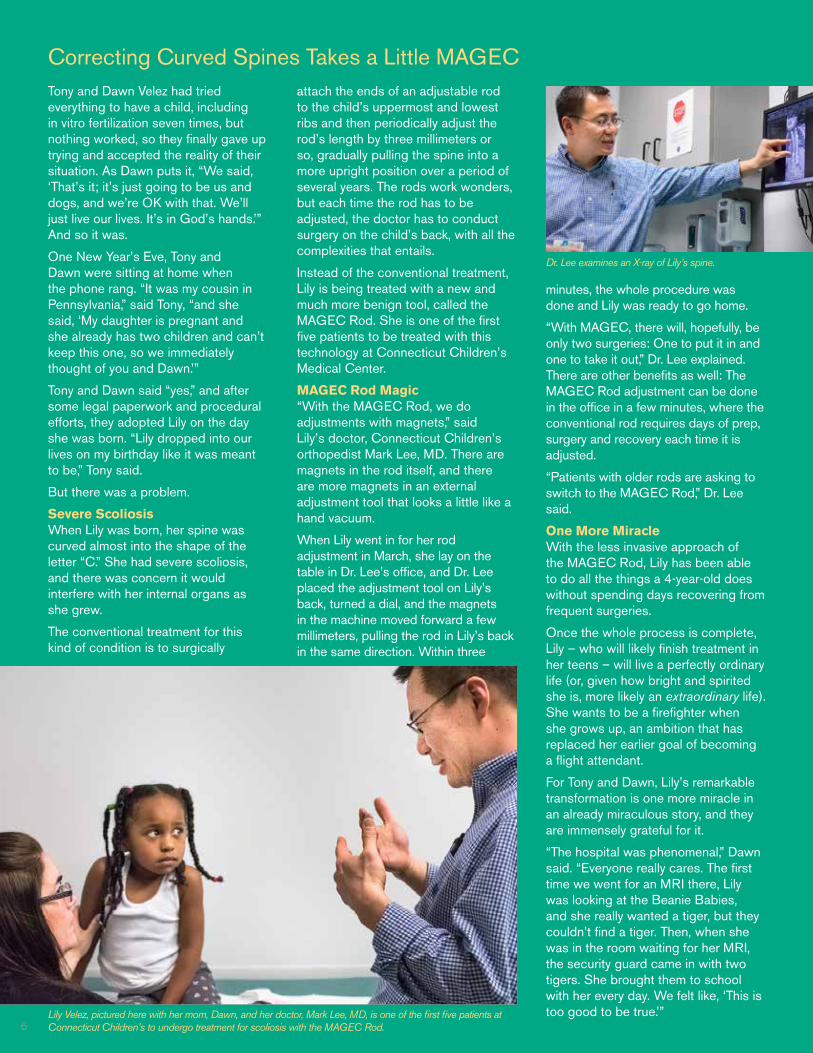
Dr. Lee examines an X-ray of Lily’s spine.
6

7
Busy Concussion Clinic Combines Management and Research Jacey Clarke of East Haddam knows all too well what a concussion feels like.
As a basketball player for Nathan Hale-Ray High School, Jacey, 16, was scrimmaging with her team last winter when she took a severe blow to the head.
“I was running down to get in front of the Center and she side-checked me,” Jacey explained. It was with just enough force to send the high school sophomore into the concrete wall, head first.
“I was on the ground for about 30 seconds and when I got up, I was instantly nauseous,” she said. “I felt completely unlike myself. My depth perception was off, and I couldn’t even catch the ball.”
“The coach recommended I go to the doctor because she knew it could be a concussion,” Jacey said.
And she was right.
Busy Concussion Clinic Each year, hundreds of teenage athletes, like Jacey, are referred to Connecticut Children’s Elite Sports Medicine in Farmington, which offers a comprehensive sports-related concussion program for adolescent and young adult athletes.
“Concussion alone makes up one-third of our patient population,” said Concussion Program Coordinator Regina Kostyun, MSEd, ATC. “We were seeing between 400 and 500 patients a year between 2013 and 2015. Our main demographic is athletes between 10 and 20 years of age.”
One of the first concussion clinics in the state, its services include concussion management, baseline and post-injury neurocognitive testing, physical therapy, vestibular therapy and more.
Concussion 101 A concussion is a traumatic brain injury, resulting from a blow to the head, face, neck or body. It commonly occurs in sports but can also result from whiplash or other injuries.
“On the outside, the patient may look normal,” Kostyun said. On the inside, other things are going on at the cellular level.
When an athlete takes a hard blow to the head, calcium floods into the cells, triggering an increased need for glucose from the brain to allow healing to take place. But while the brain needs glucose to heal, the cells are leaking potassium, which results in a surge of events that
lead to constriction of blood vessels and a decrease in the amount of blood delivered to the brain.
Recovery can take four to six weeks, or even longer.
Traditionally, concussions were seen most often in football, hockey and lacrosse, Kostyun said. Today, a higher concussion rate is being seen in female soccer players, but basketball players, swimmers and even cross-country runners are not exempt. “It can, unfortunately, happen in any sport,” she said.
Concussion Research At Connecticut Children’s Concussion Clinic, staff are not only treating concussion, they are studying it — from multiple angles.
“We have a number of different projects underway,” said Matthew Solomito, PhD, lead research engineer in the Division of Orthopedics. One of the current studies is looking at balance.
“Everybody’s balance is different,” Dr. Solomito said. But software he developed will help the researchers determine if a patient is still experiencing concussion symptoms at follow-up visits by mapping postural sway (horizontal and backwards and forward movement) and shift in weight to maintain postural balance after concussion.
Patients who agree to participate in the balance study begin by completing a survey on their first visit. Subsequent visits involve patients answering a series of questions with their eyes open and closed while using a specialized balance board.
“We currently have 30 concussed adolescents participating, along with 18 healthy adolescents who are not concussed,” Dr. Solomito said. Collegiate athletes have also been recruited for the study.
What the findings seem to suggest thus far is that patients who have lower scores on certain questions answered with their eyes closed are still experiencing concussion symptoms.
“Postural balance and dynamic balance (balance while in motion) both seem to be affected by concussion,” Dr. Solomito explained. “What we’ve learned is that many patients will try to mask their symptoms, saying they feel OK when, in fact, they are still experiencing symptoms.”
Much More to Learn While the balance board has been useful in helping determine whether a patient is physically ready to return to sport, the researchers are also collecting data on the psychological impact on athletes returning to sports after concussion recovery.
“The knowledge we have gained from studying concussions has exploded in the last decade,” Kostyun said. “But we are still going to be learning more about this condition 30 or 40 years from now. We’re still early in the spectrum.”
To learn more about concussion, view the online video, “Know the Signs: The Concussion Anthem,” written by David Wang, MD, Medical Director of Connecticut Children’s Elite Sports Medicine Division. To access the video, visit www.connecticutchildrensfoundation.org/concussion.
Jacey Clarke is part of a balance study conducted by the Concussion Clinic at Connecticut Children’s Elite Sports Medicine. She is pictured here with lead research engineer Matthew Solomito, PhD.

NONPROFIT U.S. POSTAGE
PAIDHARTFORD, CT
PERMIT NO. 3745
Your Support Counts! Your gift, large or small, makes a difference in the lives of children. Donations toward equipment, technology, research, people and programs bring hope and health to those who entrust their care to Connecticut Children’s. Won’t you please consider making a tax-deductible donation today? Your generosity helps us provide our region’s children with the best care possible – each and every day.
To learn more, please contact the Foundation at 860.837.5700, or visit our website at www.connecticutchildrensfoundation.org.
NEWSLETTER CONTRIBUTORS: Sharon Napolitano, Senior EditorEd Jalinskas, Creative ServicesMark Cherrington, Contributing Writer/Photographer
Orthopedics Now is an annual publication of Connecticut Children’s Medical Center Foundation, highlighting the programs and services of Connecticut Children’s. To be added to or removed from our mailing list, please contact [email protected].
Sports Medicine . . . At Connecticut Children’s Medical Center, our Elite Sports Medicine team works together with the ultimate goal of helping youth, adolescent and young adult athletes recover from sidelining injuries and return to their favorite sports and activities. Our sports medicine program is the only one in Connecticut that offers a full spectrum of care with experts focused exclusively on the health of young athletes.
While the Elite Sports Medicine program at Connecticut Children’s places a large emphasis on wellness and injury prevention, athletic injuries—minor or serious—are inevitable. We offer children of all ages and young athletes a state-of-the-art resource for sports injury care, sports injury prevention, performance training and education.
Here, Carl Nissen, MD, a sports medicine surgeon at Connecticut Children’s Elite Sports Medicine in Farmington, follows up with a patient who underwent surgery and rehabilitation for a torn ACL (anterior cruciate ligament).
Connecticut Children’s Medical Center282 Washington Street Hartford, CT 06106

Research on a century-old disease continues at Connecticut Children’s Medical Center, where researchers are working to unlock the mysteries of Charcot-Marie-Tooth (CMT) disease, a relatively rare condition but one of the most commonly inherited neurological disorders observed in children and youth.
First identified in 1886, CMT – also known as hereditary motor and sensory neuropathy – affects 1 in 2,500 people in the United States and comprises a group of disorders that damage the peripheral nerves that carry signals from the brain and spinal cord to the muscles. CMT causes muscle weakness and deterioration, as well as loss of sensation in the feet and lower legs, hands and forearms. While not fatal, the disease is variably progressive.
“Charcot-Marie-Tooth disease tends to run in families and tends to affect the feet more than hands,” said Kristan Pierz, MD, an orthopedic surgeon and Medical Director of Connecticut Children’s Center for Motion Analysis (CMA) in Farmington, where comprehensive assessments of gait abnormalities are carried out. “Progression of symptoms is gradual and can affect patients differently. Symptoms can range from subtle to severe,” Dr. Pierz said.
Onset of the disease may occur any time and may include weakness of the foot and lower leg muscles, resulting in foot drop and a steppage (high-stepping) gait leading to frequent tripping or falls. Foot deformities, such as high arches and hammertoes (a deformity of one or both joints of the toes), are also common characteristics and can affect a patient’s ability to walk.
Forefront of Research While knowledge about Charcot-Marie-Tooth disease is still considered to be in its infancy, Connecticut Children’s is at the forefront of CMT research.
“We started our local research efforts in motion analysis in 2011 and published our first paper on a retrospective review of gait function in 2013,” said Sylvia Õunpuu, a Connecticut Children’s kinesiologist and Director of Research at the CMA. “We have since published two other chapters on gait function and proposed treatment options based on gait patterns.”
“Our biggest contributions to date are classification of gait function with a focus on the ankle in CMT; documentation of preliminary treatment outcomes for orthopedic surgery; and documentation of bracing outcomes,” she said.
Two ongoing studies include a postoperative analysis in children with CMT who have had surgery, and a study that tracks the natural history of gait decline in patients who have not.
The Center for Motion Analysis has been collecting motion data on pediatric patients with CMT disease since 2004, allowing researchers to look at data retrospectively.
“The long-term goal is to provide evidence-based criteria for surgical intervention for kids with CMT,” Õunpuu said.
CMT Center of Excellence In addition to running a CMT Multispecialty Clinic for patients and families, Connecticut Children’s was named a Pediatric Charcot-Marie-Tooth Center of Excellence in 2016. Designated as a Clinical
Sylvia Õunpuu, Director of Research at Connecticut Children’s Center for Motion Analysis (left), reviews research findings with CMA Medical Director Kristan Pierz, MD. (Photo Credit: Erin Blinn-Curran)
Center of Excellence by the Inherited Neuropathy Consortium (INC), supported by the National Institutes of Health, the Center is led by neuromuscular specialist Gyula Acsadi, MD, PhD, Division Head of Neurology and Rehabilitation, who diagnoses pediatric patients with CMT.
Dr. Acsadi sees approximately 40 patients with CMT at Connecticut Children’s each year, while the Center for Motion Analysis sees about 10. The designation as a CMT Center of Excellence is significant because it reflects both high-level patient care and research.
According to Dr. Acsadi, there are 20 such Centers of Excellence in the worldwide consortium, including only five pediatric centers. Connecticut Children’s is one of only three gait labs involved in research by this Consortium.
“The INC is a worldwide group that is gathering a minimal data set, which is a standardized set of natural history measures, across sites and storing the information collected in a central database,” Õunpuu explained. “The database is then available for all members to access for research purposes. The members also collaborate on treatment protocols, which allows for greater patient numbers and, therefore, better statistical significance to prove research findings.”
“When you are part of a clinical trial, the patients know that they get access for the most advanced treatments.,” Dr. Acsadi noted. “We have patients from other states receiving care and participating in our research.”
What is unique is that Connecticut Children’s CMT Center of Excellence utilizes a state-of-the-art gait analysis to aid bracing and surgical treatments, if needed.
Spotlight on Research at Connecticut Children’s . . .
Researchers Shine Light on Charcot-Marie-Tooth Disease
Following Cervical Spine Bone Tumor Surgery, Bridget Souriyamath Dances Her Way to the Top in Irish Dance Competition
Research at the Center for Motion Analysis relies heavily on donor support for its research activities. To support research, including that for Charcot-Marie-Tooth disease, please contact Connecticut Children’s Medical Center Foundation at 860.837.5700.
Living with Charcot-Marie-Tooth Disease
Most of the patients seen at Connecticut Children’s Center for Motion Analysis (CMA) with Charcot-Marie-Tooth disease range in age from 4 to 21.
Emma Gonsalves, a patient of Kristan Pierz, MD, an orthopedic surgeon and Medical Director of the CMA, is a 24-year-old school teacher.
The Avon resident, who was born with the rare, inherited neurological disorder, is a longtime patient at the CMA.
“When I was a few months old, I started wearing casts on my legs,” said Gonsalves. “When I was about 7, my heel cords (Achilles tendons) were cut in both legs. When I was 13, my knees were growing inward, so I had both of my knees done. In 2009, when I was 17, I broke my hip. That’s when I met Dr. Pierz.”
After fixing her hip, Dr. Pierz began addressing the issues with her feet caused by CMT.
Foot Work CMT is a group of inherited disorders that affect the peripheral nerves, located outside the brain and spinal cord. According to the Charcot-Marie-Tooth Association, there are 90 kinds of CMT and each is caused by a different kind of mutation, with more causes being discovered every year.
“Patients can definitely have different presentations of CMT,” Dr. Pierz said. “Emma had foot deformities with painful high arches and claw toes that are often seen in patients with CMT.”
Gonsalves underwent whole-foot reconstruction to fix her bent toes and plantar fasciitis, one of the most common causes of heel pain.
“It was a long recovery, but my toes are now permanently straight,” Gonsalves said. “I walked barefoot for the first time this year.”
Importance of Follow-Up “CMT is a progressive disorder, so following patients over time is important,” Dr. Pierz said. “The Center for Motion Analysis offers objective data that can truly guide treatment decision-making. One treatment that isn’t appropriate at one point in time may become appropriate over a few years. Tracking patients with objective data will lead to better decision making. X-rays and physical exams in the office are not nearly as useful as full-motion data when evaluating function,” she explained.
While adults typically end up seeing foot and ankle specialists, Dr. Pierz said she looks forward to expanding care to older patients. “We have seen parents and their children with CMT in the Center for Motion Analysis, and the data is interesting,” she said.
Gonsalves, who says she can now wear regular shoes again, returns to the CMA at least once a year to meet with Dr. Pierz.
“She has completely made my life so much better,” Gonsalves said. “She has given me hope. I think she is helping me be the best person I can be.”
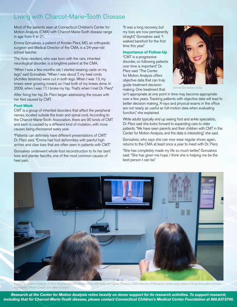
Center for Motion Analysis (CMA) Medical Director Kristan Pierz, MD (left), and Sylvia Õunpuu, CMA Director of Research, review early videos of long-time patient Emma Gonsalves. (Photo Credit: Erin Blinn-Curran)
Emma Gonsalves today.