Orphan Nuclear Bodies - cshperspectives.cshlp.org
13
Orphan Nuclear Bodies Maria Carmo-Fonseca 1 , Maria T. Berciano 2 , and Miguel Lafarga 2 1 Instituto de Medicina Molecular, Faculdade de Medicina, Universidade de Lisboa, 1649-028 Lisboa, Portugal 2 Department of Anatomyand Cell Biology, and “Centro de Investigacion Biomedica en Red sobre Enfermedades Neurodegenerativas (CIBERNED)”, University of Cantabria, Santander 39011, Spain Correspondence: [email protected] Orphan nuclear bodies are defined as nonchromatin nuclear compartments that have been less well studied compared with other well-characterized structures in the nucleus. Nuclear bodies have traditionally been thought of as uniform distinct entities depending on the protein “markers” they contain. However, it is becoming increasingly apparent that nuclear bodies enriched in different sets of transcriptional regulators share a link to the ubiq- uitin-proteasome and SUMO-conjugation pathways. An emerging concept is that some orphan nuclear bodies might act as sites of protein modification by SUMO and/or proteaso- mal degradation of ubiquitin-tagged proteins. By defining a specialized environment for protein modification and degradation, orphan nuclear bodies may increase the capacity of cells to survive under varying environmental conditions. A nuclear body is a region within the nucleus that is morphologically distinct from its surroundings when observed by transmission electron microscopy (reviewed in Spector 2006). This excludes structures that are only detected upon overexpression of tagged nuclear proteins and most likely result from nonphysio- logical aggregation of excess protein. On the basis of their structure observed at the electron microscopic level, nuclear bodies have been classified as either simple or complex (Bouteille et al. 1974). The simple nuclear bodies are small (0.2 – 0.5 mm), round, compact and finely fibrillar, whereas the complex nu- clear bodies are larger (0.2 –1.2 mm), hetero- geneous in shape and texture, and enveloped by a peripheral capsule, which gives them a doughnut-shaped appearance. Orphan nuclear bodies are primarily proteinaceous structures enriched in either transcriptional regulators or RNA-binding proteins (Table 1). Although it was initially thought that these structures might act as specialized compartments for gene ex- pression, most genes and RNA species targeted by the protein factors enriched in nuclear bodies show a widespread distribution through- out the nucleoplasm. Moreover, nuclear bodies contain only a fraction of the total pool of tran- scriptional regulators and RNA-binding pro- teins in the nucleus, and many of these factors are in constant flux in and out of the bodies. A breakthrough in this field was the recent discovery that transcription factors enriched in nuclear bodies are modified by SUMO and/or ubiquitin, and that SUMO-conjugation plays an important role in nuclear body assembly Editors: Tom Misteli and David Spector Additional Perspectives on The Nucleus available at www.cshperspectives.org Copyright # 2010 Cold Spring Harbor Laboratory Press; all rights reserved; doi: 10.1101/cshperspect.a000703 Cite this article as Cold Spring Harb Perspect Biol 2010;2:a000703 1 on November 16, 2021 - Published by Cold Spring Harbor Laboratory Press http://cshperspectives.cshlp.org/ Downloaded from
Transcript of Orphan Nuclear Bodies - cshperspectives.cshlp.org
Orphan Nuclear Bodies
Maria Carmo-Fonseca1, Maria T. Berciano2, and Miguel Lafarga2
1Instituto de Medicina Molecular, Faculdade de Medicina, Universidade de Lisboa, 1649-028 Lisboa, Portugal2Department of Anatomyand Cell Biology, and “Centro de Investigacion Biomedica en Red sobre EnfermedadesNeurodegenerativas (CIBERNED)”, University of Cantabria, Santander 39011, Spain
Correspondence: [email protected]
Orphan nuclear bodies are defined as nonchromatin nuclear compartments that have beenless well studied compared with other well-characterized structures in the nucleus. Nuclearbodies have traditionally been thought of as uniform distinct entities depending on theprotein “markers” they contain. However, it is becoming increasingly apparent thatnuclear bodies enriched in different sets of transcriptional regulators share a link to the ubiq-uitin-proteasome and SUMO-conjugation pathways. An emerging concept is that someorphan nuclear bodies might act as sites of protein modification by SUMO and/or proteaso-mal degradation of ubiquitin-tagged proteins. By defining a specialized environment forprotein modification and degradation, orphan nuclear bodies may increase the capacity ofcells to survive under varying environmental conditions.
Anuclear body is a region within the nucleusthat is morphologically distinct from its
surroundings when observed by transmissionelectron microscopy (reviewed in Spector2006). This excludes structures that are onlydetected upon overexpression of tagged nuclearproteins and most likely result from nonphysio-logical aggregation of excess protein.
On the basis of their structure observed atthe electron microscopic level, nuclear bodieshave been classified as either simple or complex(Bouteille et al. 1974). The simple nuclearbodies are small (0.2–0.5 mm), round, compactand finely fibrillar, whereas the complex nu-clear bodies are larger (0.2–1.2 mm), hetero-geneous in shape and texture, and envelopedby a peripheral capsule, which gives them adoughnut-shaped appearance. Orphan nuclear
bodies are primarily proteinaceous structuresenriched in either transcriptional regulators orRNA-binding proteins (Table 1). Although itwas initially thought that these structures mightact as specialized compartments for gene ex-pression, most genes and RNA species targetedby the protein factors enriched in nuclearbodies show a widespread distribution through-out the nucleoplasm. Moreover, nuclear bodiescontain only a fraction of the total pool of tran-scriptional regulators and RNA-binding pro-teins in the nucleus, and many of these factorsare in constant flux in and out of the bodies.
A breakthrough in this field was the recentdiscovery that transcription factors enriched innuclear bodies are modified by SUMO and/orubiquitin, and that SUMO-conjugation playsan important role in nuclear body assembly
Editors: Tom Misteli and David Spector
Additional Perspectives on The Nucleus available at www.cshperspectives.org
Copyright # 2010 Cold Spring Harbor Laboratory Press; all rights reserved; doi: 10.1101/cshperspect.a000703
Cite this article as Cold Spring Harb Perspect Biol 2010;2:a000703
1
on November 16, 2021 - Published by Cold Spring Harbor Laboratory Press http://cshperspectives.cshlp.org/Downloaded from
(reviewed in Heun 2007). It is therefore possiblethat some nuclear bodies act as protein modifi-cation and degradation centers.
THE CLASTOSOME: A NUCLEAR BODYLINKED TO THE UBIQUITIN-PROTEASOMESYSTEM
Proteins in cells and organisms exist in a dy-namic state with individual half-lives rangingfrom minutes to years. This implies that proteindegradation must be highly selective and tightly
controlled (reviewed in Ravid and Hochstrasser2008). Eukaryotic cells have two major mecha-nisms for protein degradation: lysosomes andproteasomes. Although exogenous particles andendogenous cellular organelles are targeted fordigestion in lysosomes, the majority of intracel-lular proteins are degraded by proteasomes(reviewed in Murata et al. 2009). Proteasomesdegrade short-lived regulatory proteins andthereby control cellular processes such as signaltransduction, cell cycle, and gene transcription.Proteasomes additionally clear misfolded and
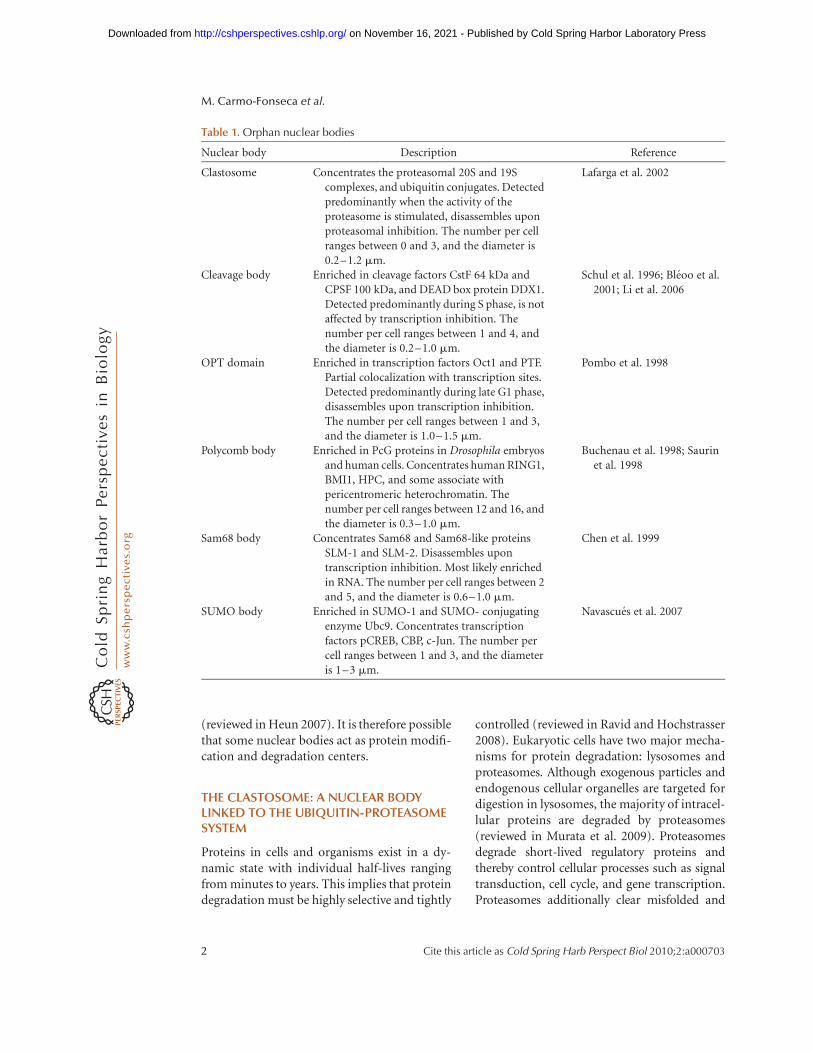
Table 1. Orphan nuclear bodies
Nuclear body Description Reference
Clastosome Concentrates the proteasomal 20S and 19Scomplexes, and ubiquitin conjugates. Detectedpredominantly when the activity of theproteasome is stimulated, disassembles uponproteasomal inhibition. The number per cellranges between 0 and 3, and the diameter is0.2–1.2 mm.
Lafarga et al. 2002
Cleavage body Enriched in cleavage factors CstF 64 kDa andCPSF 100 kDa, and DEAD box protein DDX1.Detected predominantly during S phase, is notaffected by transcription inhibition. Thenumber per cell ranges between 1 and 4, andthe diameter is 0.2–1.0 mm.
Schul et al. 1996; Bleoo et al.2001; Li et al. 2006
OPT domain Enriched in transcription factors Oct1 and PTF.Partial colocalization with transcription sites.Detected predominantly during late G1 phase,disassembles upon transcription inhibition.The number per cell ranges between 1 and 3,and the diameter is 1.0–1.5 mm.
Pombo et al. 1998
Polycomb body Enriched in PcG proteins in Drosophila embryosand human cells. Concentrates human RING1,BMI1, HPC, and some associate withpericentromeric heterochromatin. Thenumber per cell ranges between 12 and 16, andthe diameter is 0.3–1.0 mm.
Buchenau et al. 1998; Saurinet al. 1998
Sam68 body Concentrates Sam68 and Sam68-like proteinsSLM-1 and SLM-2. Disassembles upontranscription inhibition. Most likely enrichedin RNA. The number per cell ranges between 2and 5, and the diameter is 0.6–1.0 mm.
Chen et al. 1999
SUMO body Enriched in SUMO-1 and SUMO- conjugatingenzyme Ubc9. Concentrates transcriptionfactors pCREB, CBP, c-Jun. The number percell ranges between 1 and 3, and the diameteris 1–3 mm.
Navascues et al. 2007
M. Carmo-Fonseca et al.
2 Cite this article as Cold Spring Harb Perspect Biol 2010;2:a000703
on November 16, 2021 - Published by Cold Spring Harbor Laboratory Press http://cshperspectives.cshlp.org/Downloaded from
aggregated proteins from the cell and producesome of the peptides to be displayed at thecell surface as part of the adaptive immuneresponse.
A fundamental question about intracellu-lar proteolysis is how specific proteins are re-cognized by the proteolytic machinery. Ineukaryotic cells, most substrate proteins aretargeted to the proteasome by the covalent at-tachment of many copies of the small proteinubiquitin. Once a protein has been tagged bypolyubiquitination, it is recognized by the pro-teasome, unfolded and then degraded (Fig. 1).However, ubiquitination can function inde-pendently of the proteasome and proteasomescan degrade some proteins without their priormodification by ubiquitin (Ravid and Hoch-strasser 2008).
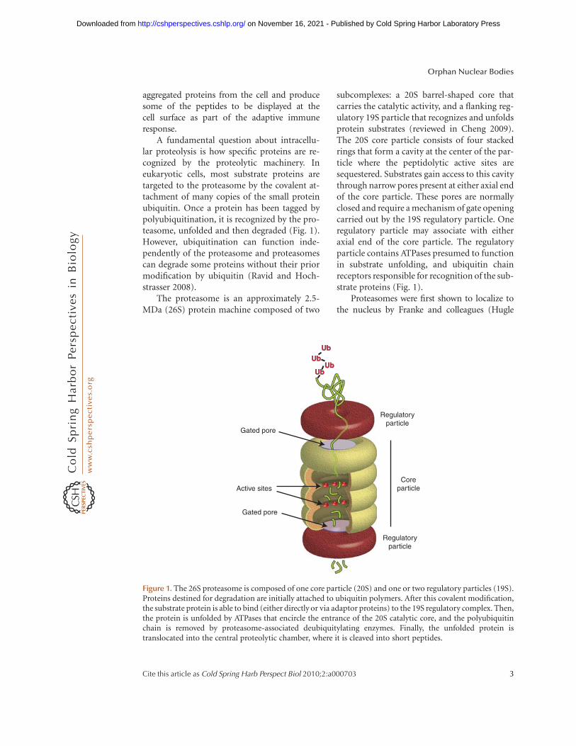
The proteasome is an approximately 2.5-MDa (26S) protein machine composed of two
subcomplexes: a 20S barrel-shaped core thatcarries the catalytic activity, and a flanking reg-ulatory 19S particle that recognizes and unfoldsprotein substrates (reviewed in Cheng 2009).The 20S core particle consists of four stackedrings that form a cavity at the center of the par-ticle where the peptidolytic active sites aresequestered. Substrates gain access to this cavitythrough narrow pores present at either axial endof the core particle. These pores are normallyclosed and require a mechanism of gate openingcarried out by the 19S regulatory particle. Oneregulatory particle may associate with eitheraxial end of the core particle. The regulatoryparticle contains ATPases presumed to functionin substrate unfolding, and ubiquitin chainreceptors responsible for recognition of the sub-strate proteins (Fig. 1).
Proteasomes were first shown to localize tothe nucleus by Franke and colleagues (Hugle
Regulatoryparticle
Regulatoryparticle
Gated pore
Gated pore
Active sitesCore
particle
UbUb
UbUbUbUb
UbUb
Figure 1. The 26S proteasome is composed of one core particle (20S) and one or two regulatory particles (19S).Proteins destined for degradation are initially attached to ubiquitin polymers. After this covalent modification,the substrate protein is able to bind (either directly or via adaptor proteins) to the 19S regulatory complex. Then,the protein is unfolded by ATPases that encircle the entrance of the 20S catalytic core, and the polyubiquitinchain is removed by proteasome-associated deubiquitylating enzymes. Finally, the unfolded protein istranslocated into the central proteolytic chamber, where it is cleaved into short peptides.
Orphan Nuclear Bodies
Cite this article as Cold Spring Harb Perspect Biol 2010;2:a000703 3
on November 16, 2021 - Published by Cold Spring Harbor Laboratory Press http://cshperspectives.cshlp.org/Downloaded from
et al. 1983; Kleinschmidt et al. 1983). Subse-quent studies confirmed that proteasomes,as well as components of the ubiquitin-pro-teasome system occur both in the nucleusand in the cytoplasm. Although some nuclearproteins, such as p53, are first exported fromthe nucleus and then degraded in the cytoplasm,several lines of evidence indicate that proteinscan be ubiquitylated and degraded by the pro-teasome within the nucleus (Tao et al. 2005;Rockel et al. 2005).
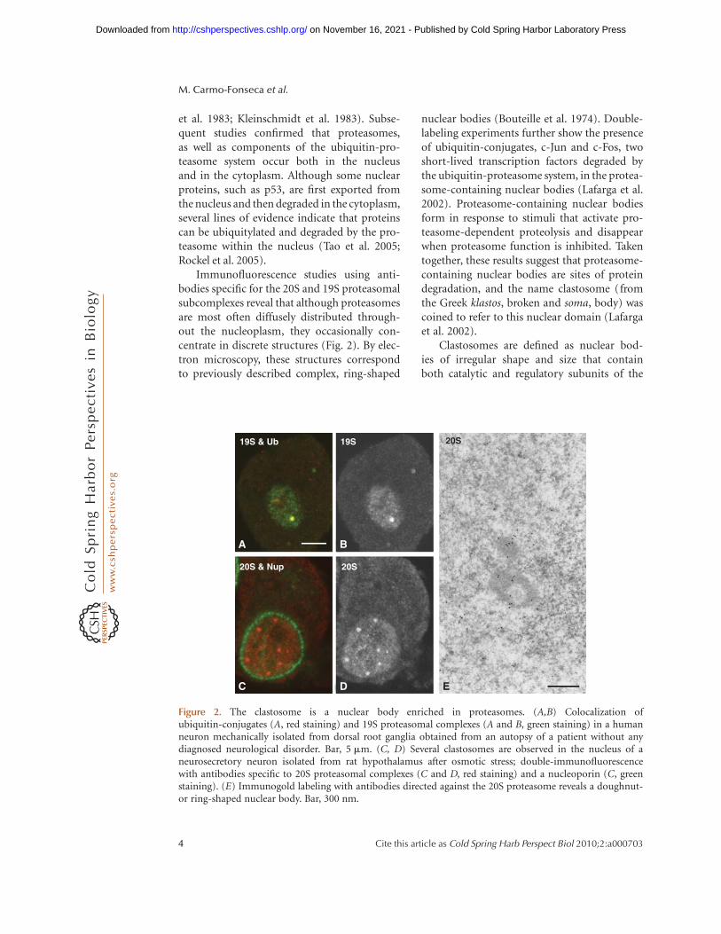
Immunofluorescence studies using anti-bodies specific for the 20S and 19S proteasomalsubcomplexes reveal that although proteasomesare most often diffusely distributed through-out the nucleoplasm, they occasionally con-centrate in discrete structures (Fig. 2). By elec-tron microscopy, these structures correspondto previously described complex, ring-shaped
nuclear bodies (Bouteille et al. 1974). Double-labeling experiments further show the presenceof ubiquitin-conjugates, c-Jun and c-Fos, twoshort-lived transcription factors degraded bythe ubiquitin-proteasome system, in the protea-some-containing nuclear bodies (Lafarga et al.2002). Proteasome-containing nuclear bodiesform in response to stimuli that activate pro-teasome-dependent proteolysis and disappearwhen proteasome function is inhibited. Takentogether, these results suggest that proteasome-containing nuclear bodies are sites of proteindegradation, and the name clastosome (fromthe Greek klastos, broken and soma, body) wascoined to refer to this nuclear domain (Lafargaet al. 2002).
Clastosomes are defined as nuclear bod-ies of irregular shape and size that containboth catalytic and regulatory subunits of the
19S & Ub
20S & Nup 20S
20S
A B
C ED
19S
Figure 2. The clastosome is a nuclear body enriched in proteasomes. (A,B) Colocalization ofubiquitin-conjugates (A, red staining) and 19S proteasomal complexes (A and B, green staining) in a humanneuron mechanically isolated from dorsal root ganglia obtained from an autopsy of a patient without anydiagnosed neurological disorder. Bar, 5 mm. (C, D) Several clastosomes are observed in the nucleus of aneurosecretory neuron isolated from rat hypothalamus after osmotic stress; double-immunofluorescencewith antibodies specific to 20S proteasomal complexes (C and D, red staining) and a nucleoporin (C, greenstaining). (E) Immunogold labeling with antibodies directed against the 20S proteasome reveals a doughnut-or ring-shaped nuclear body. Bar, 300 nm.
M. Carmo-Fonseca et al.
4 Cite this article as Cold Spring Harb Perspect Biol 2010;2:a000703
on November 16, 2021 - Published by Cold Spring Harbor Laboratory Press http://cshperspectives.cshlp.org/Downloaded from
proteasome and ubiquitylated proteasomesubstrates. Clastosomes are normally absent orscarce in most cell types, indicating that thesenuclear bodies are not essential for proteasomefunction. Most likely, protein substrates taggedfor proteolysis are rapidly encountered, recog-nized and degraded by the diffusely distributedproteasomes in the nucleoplasm. Clastosomesbecome prominent when cells are exposed tostimuli that suddenly increase the levels ofproteins targeted for degradation, for example,inducible expression of c-fos by serum, or stress.Clastosomes may therefore form as a result of anoverload of protein substrates. Clastosomesassemble transiently and revert after the stimu-lus, consistent with the view that they accu-mulate substrates queuing up for proteolysis.Moreover, clastosomes are not detected in cellstreated with specific proteasome inhibitors,arguing that clastosome assembly requires pro-teasomal activity. Clastosomes may also act bysequestering proteasomes, thereby controllingthe availability of proteolytic machines fornuclear protein degradation.
Recent studies suggest that regulated pro-tein degradation by the ubiquitin proteasomesystem is an important modulator of neuronalfunction (reviewed in Tai et al. 2008). In partic-ular, the localization of proteasomes to synapsesis controlled by synaptic stimulation, thus pro-viding a mechanism for local protein turnover(Bingol and Schuman 2006). The observationthat clastosomes are widespread in glial cellsand many types of brain neurons (Adori et al.2006; Baltrons et al. 2008) raises the possibilitythat these nuclear bodies participate in distinc-tive features of protein turnover in the nervoussystem. The importance of proteolysis in neu-rons is further underlined by the presence in awide spectrum of neurodegenerative diseasesof pathological protein aggregates that lead to asevere impairment of the ubiquitin-proteasomesystem (Bennett et al. 2005). Although the pres-ence of ubiquitin and proteasomes in inclusionbodies formed by disease-associated proteinaggregates suggests a link with dysfunction ofthe ubiquitin-proteasome system (Janer et al.2006), the mechanism by which accumula-tion of protein aggregates in inclusion bodies
contributes to disease remains unknown (seeOrr 2010).
THE PROTEASOME IS A COMPONENT OFBOTH CLASTOSOMES AND PML NUCLEARBODIES
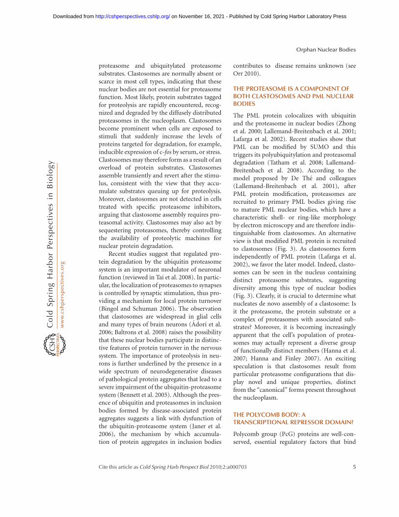
The PML protein colocalizes with ubiquitinand the proteasome in nuclear bodies (Zhonget al. 2000; Lallemand-Breitenbach et al. 2001;Lafarga et al. 2002). Recent studies show thatPML can be modified by SUMO and thistriggers its polyubiquitylation and proteasomaldegradation (Tatham et al. 2008; Lallemand-Breitenbach et al. 2008). According to themodel proposed by De The and colleagues(Lallemand-Breitenbach et al. 2001), afterPML protein modification, proteasomes arerecruited to primary PML bodies giving riseto mature PML nuclear bodies, which have acharacteristic shell- or ring-like morphologyby electron microscopy and are therefore indis-tinguishable from clastosomes. An alternativeview is that modified PML protein is recruitedto clastosomes (Fig. 3). As clastosomes formindependently of PML protein (Lafarga et al.2002), we favor the later model. Indeed, clasto-somes can be seen in the nucleus containingdistinct proteasome substrates, suggestingdiversity among this type of nuclear bodies(Fig. 3). Clearly, it is crucial to determine whatnucleates de novo assembly of a clastosome: Isit the proteasome, the protein substrate or acomplex of proteasomes with associated sub-strates? Moreover, it is becoming increasinglyapparent that the cell’s population of protea-somes may actually represent a diverse groupof functionally distinct members (Hanna et al.2007; Hanna and Finley 2007). An excitingspeculation is that clastosomes result fromparticular proteasome configurations that dis-play novel and unique properties, distinctfrom the “canonical” forms present throughoutthe nucleoplasm.
THE POLYCOMB BODY: ATRANSCRIPTIONAL REPRESSOR DOMAIN?
Polycomb group (PcG) proteins are well-con-served, essential regulatory factors that bind
Orphan Nuclear Bodies
Cite this article as Cold Spring Harb Perspect Biol 2010;2:a000703 5
on November 16, 2021 - Published by Cold Spring Harbor Laboratory Press http://cshperspectives.cshlp.org/Downloaded from
to specific DNA regions in target genes andrepress their transcription. PcG proteinsmaintain the silenced state of developmentalgenes, including homeotic genes and genesinvolved in mammalian embryonic stem celldifferentiation (Schuettengruber et al. 2007).PcG proteins were first described in Droso-phila as factors responsible for maintaining the
transcriptionally repressed state of Hox/home-otic genes throughout development. A growingnumber of vertebrate genes related to the Droso-phila PcG proteins have recently been identi-fied. PcG proteins exist in at least two distinctcomplexes termed Polycomb repressive com-plex 1 (PRC1) and Polycomb repressive com-plex 2 (PRC2). Members of PRC1, including
20S & PML
19S & c-fos
20S
A B
C D
Primary PML body
E
Clastosome
SUMOSUMO UbUb
19S
Figure 3. Clastosomes concentrate protein substrates for proteasomal degradation. (A, B) The panels depict ahuman neuron mechanically isolated from dorsal root ganglia obtained from an autopsy of a patient withoutany diagnosed neurological disorder, double-labeled with anti-PML (B, green staining) and anti-20Sproteasomal complexes (A and B, red staining). Note that only one of the multiple PML bodies concentratesproteasomes (arrowhead) and that one body enriched in proteasomes does not contain PML (arrow). (C, D)Colocalization of 19S proteasomal complexes (C and D, red staining) and c-Fos (D, green staining) in thenucleus of a neurosecretory neuron isolated from rat hypothalamus after osmotic stress. Bar, 5 mm. (E)Hypothetical model for PML protein traffic through PML bodies and clastosomes. PML (blue spheres)distributes diffusely in the nucleoplasm and associates transiently with primary PML bodies. Uponmodification by SUMO and ubiquitin, the modified PML proteins (red spheres) are recruited to clastosomesfor degradation.
M. Carmo-Fonseca et al.
6 Cite this article as Cold Spring Harb Perspect Biol 2010;2:a000703
on November 16, 2021 - Published by Cold Spring Harbor Laboratory Press http://cshperspectives.cshlp.org/Downloaded from
the HPH1/HPH2, Bmi1, HPC2 and RING1gene products are diffusely distributed in thenucleus and in addition colocalize in both Dro-sophila and mammalian cells as large nucleardomains termed Polycomb or PcG bodies(Buchenau et al. 1998; Saurin et al. 1998). InDrosophila, several genes that are repressedby PcG proteins localize to Polycomb bodies,suggesting that these domains correspond tosites of gene silencing (Grimaud et al. 2006).However, the number of Polycomb bodies isorders of magnitude smaller than the numberof genetic loci targeted by PcG proteins. Thisimplies that either multiple gene targets areassociated with each Polycomb body or thatgene silencing by PcG proteins can occur out-side Polycomb bodies. Although endogenoushomeotic genes appear to cluster at PcG bodiesby a mechanism that requires components ofthe RNA interference machinery (Grimaudet al. 2006), it remains unknown whetherassembly of a Polycomb body is necessary forthe functional association of PcG proteinswith chromatin.
POLYCOMB AND PML NUCLEAR BODIES:THE SUMO CONNECTION
The small ubiquitin-like modifier (SUMO) isan ubiquitin-related protein that can be cova-lently conjugated to a variety of protein sub-strates, altering the properties of the modifiedproteins. SUMO conjugation is essential forcell and organism viability, from yeast to mam-mals (Nacerddine et al. 2005), and affects manybiological processes, including cell cycle pro-gression, maintenance of genome integrity,and transcription (reviewed in Hay 2005).SUMO substrates include protein componentsof both PML and Polycomb nuclear bodiesand recent evidence implicates the SUMOyla-tion pathway as an important player in theassembly of these domains (Heun 2007).
The PML protein and the PML body com-ponents HIPK2, Daxx, and Sp100 are modifiedby SUMO. Moreover, SUMO conjugation isnecessary for formation of PML bodies andfor recruitment of HIPK2, Daxx, and Sp100into the bodies (reviewed in Hay 2005). SUMO-
modified HIPK2 represses the activity of boundtranscription factors by associating with Grou-cho corepressor and HDAC1, and SUMO mod-ification of Sp100 enhances its interaction withheterochromatin protein 1 (HP1), a well knowninitiator of repressive domains in chromatin.
The mechanism of SUMO conjugationinvolves a cascade of events catalysed by an acti-vating enzyme E1, a conjugating enzyme E2(known as Ubc9), and a protein ligase E3(Hay 2005). There are at least three types ofSUMO E3 ligases in cells: the protein inhibitorof activated signal transducer and activator oftranscription (PIAS) family, the nuclear porecomplex protein RanBP2/Nup358, and the Poly-comb group protein Pc2. One of the substratesof Pc2 is the carboxy-terminal binding pro-tein (CtBP) transcriptional corepressor (Kageyet al. 2003). It has been proposed that Pc2functions to recruit Ubc9 and CtBP to PcGbodies, where SUMO modification of CtBPoccurs (Kagey et al. 2004). More recently, thezinc finger protein CTCF was also shown to berecruited to Polycomb bodies and to be modi-fied by SUMO (MacPherson et al. 2009).
Intriguingly, only a small proportion of thetotal cellular pool of a given transcription factoris modified by SUMO. Yet, the entire popula-tion is functionally repressed. The followingmodel was proposed to accommodate theseobservations: Upon conjugation to SUMO, atranscription factor is incorporated into arepression complex located in a nuclear body;even if SUMO is removed from the transcrip-tion factor at a later stage by specific proteases,the protein would still be retained in the repres-sion complex (Hay 2005). Consistent with thisview, PML and Polycomb nuclear bodies mightresult from the assembly of SUMO-dependentrepression complexes. A variation of this hypo-thesis postulates that a SUMO-modified tran-scription factor recruits an enzyme capableof posttranslationally modifying chromatin.SUMO could then be removed from the tran-scription factor, whereas transcriptional repres-sion would be maintained by the chromatinmodification (Hay 2005). According to thisalternative view, PML and Polycomb nuclearbodies could be sumoylation centers, from
Orphan Nuclear Bodies
Cite this article as Cold Spring Harb Perspect Biol 2010;2:a000703 7
on November 16, 2021 - Published by Cold Spring Harbor Laboratory Press http://cshperspectives.cshlp.org/Downloaded from
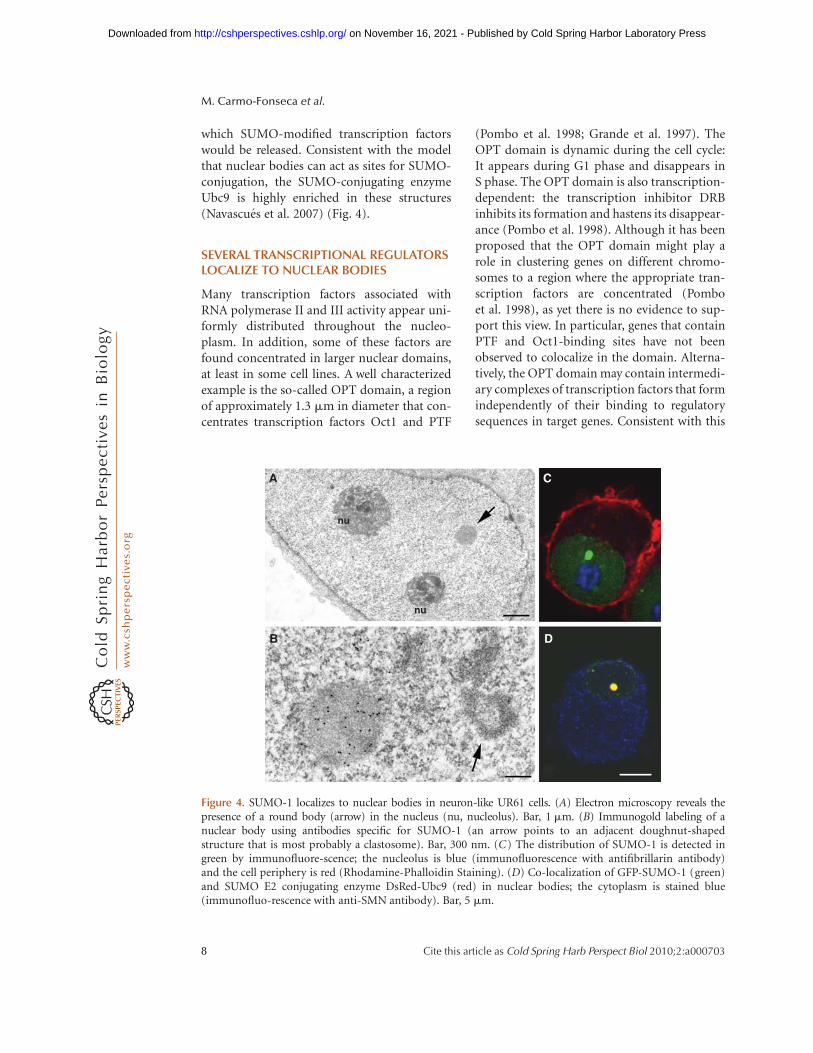
which SUMO-modified transcription factorswould be released. Consistent with the modelthat nuclear bodies can act as sites for SUMO-conjugation, the SUMO-conjugating enzymeUbc9 is highly enriched in these structures(Navascues et al. 2007) (Fig. 4).
SEVERAL TRANSCRIPTIONAL REGULATORSLOCALIZE TO NUCLEAR BODIES
Many transcription factors associated withRNA polymerase II and III activity appear uni-formly distributed throughout the nucleo-plasm. In addition, some of these factors arefound concentrated in larger nuclear domains,at least in some cell lines. A well characterizedexample is the so-called OPT domain, a regionof approximately 1.3 mm in diameter that con-centrates transcription factors Oct1 and PTF
(Pombo et al. 1998; Grande et al. 1997). TheOPT domain is dynamic during the cell cycle:It appears during G1 phase and disappears inS phase. The OPT domain is also transcription-dependent: the transcription inhibitor DRBinhibits its formation and hastens its disappear-ance (Pombo et al. 1998). Although it has beenproposed that the OPT domain might play arole in clustering genes on different chromo-somes to a region where the appropriate tran-scription factors are concentrated (Pomboet al. 1998), as yet there is no evidence to sup-port this view. In particular, genes that containPTF and Oct1-binding sites have not beenobserved to colocalize in the domain. Alterna-tively, the OPT domain may contain intermedi-ary complexes of transcription factors that formindependently of their binding to regulatorysequences in target genes. Consistent with this
A
B
C
D
nu
nu
Figure 4. SUMO-1 localizes to nuclear bodies in neuron-like UR61 cells. (A) Electron microscopy reveals thepresence of a round body (arrow) in the nucleus (nu, nucleolus). Bar, 1 mm. (B) Immunogold labeling of anuclear body using antibodies specific for SUMO-1 (an arrow points to an adjacent doughnut-shapedstructure that is most probably a clastosome). Bar, 300 nm. (C) The distribution of SUMO-1 is detected ingreen by immunofluore-scence; the nucleolus is blue (immunofluorescence with antifibrillarin antibody)and the cell periphery is red (Rhodamine-Phalloidin Staining). (D) Co-localization of GFP-SUMO-1 (green)and SUMO E2 conjugating enzyme DsRed-Ubc9 (red) in nuclear bodies; the cytoplasm is stained blue(immunofluo-rescence with anti-SMN antibody). Bar, 5 mm.
M. Carmo-Fonseca et al.
8 Cite this article as Cold Spring Harb Perspect Biol 2010;2:a000703
on November 16, 2021 - Published by Cold Spring Harbor Laboratory Press http://cshperspectives.cshlp.org/Downloaded from
view, nuclear domains enriched in transcriptionfactors could form as a result of protein modifi-cation, namely SUMOylation, as suggested forPML bodies and more recently, for a noveltype of nuclear body that concentrates KRAB-zinc finger transcriptional regulators (Brierset al. 2009).
RNA-BINDING PROTEINS INNUCLEAR BODIES
Members of three major classes of RNA-bindingproteins localize to orphan nuclear bodies. Theseinclude Sam68 and the related SLM-1 and SLM-2proteins of the signal transduction and activationof RNA (STAR) family, two subunits of the cleav-age stimulation factor (CstF) and the cleavageand polyadenylation specificity factor (CPSF)components of the mRNA 30-end processingmachinery, and DDX1, a member of the DEADbox protein family of RNA helicases.
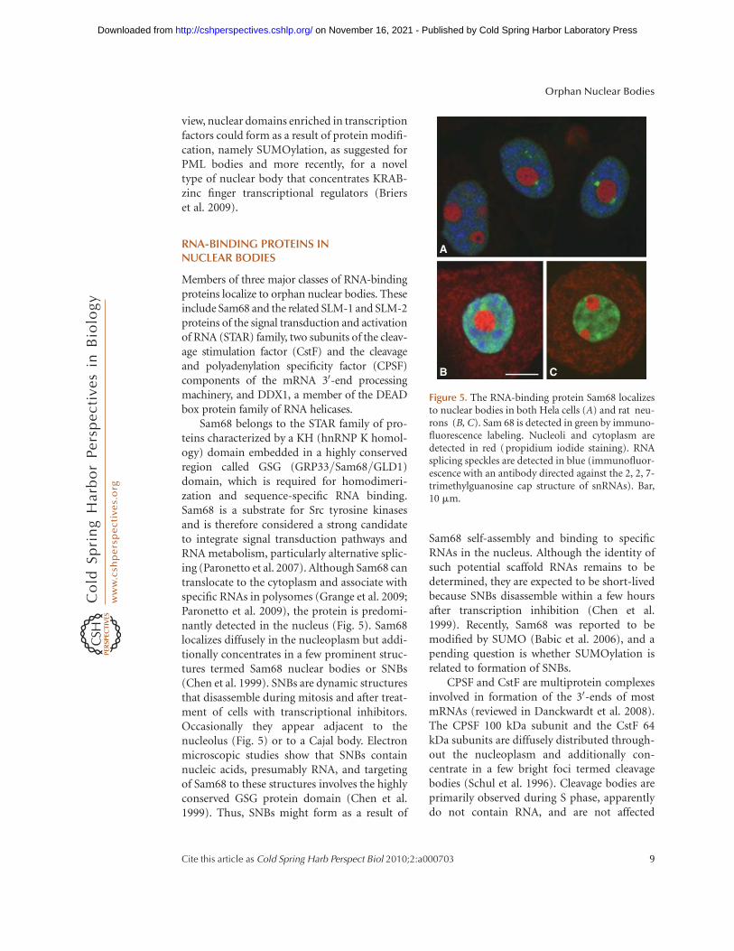
Sam68 belongs to the STAR family of pro-teins characterized by a KH (hnRNP K homol-ogy) domain embedded in a highly conservedregion called GSG (GRP33/Sam68/GLD1)domain, which is required for homodimeri-zation and sequence-specific RNA binding.Sam68 is a substrate for Src tyrosine kinasesand is therefore considered a strong candidateto integrate signal transduction pathways andRNA metabolism, particularly alternative splic-ing (Paronetto et al. 2007). Although Sam68 cantranslocate to the cytoplasm and associate withspecific RNAs in polysomes (Grange et al. 2009;Paronetto et al. 2009), the protein is predomi-nantly detected in the nucleus (Fig. 5). Sam68localizes diffusely in the nucleoplasm but addi-tionally concentrates in a few prominent struc-tures termed Sam68 nuclear bodies or SNBs(Chen et al. 1999). SNBs are dynamic structuresthat disassemble during mitosis and after treat-ment of cells with transcriptional inhibitors.Occasionally they appear adjacent to thenucleolus (Fig. 5) or to a Cajal body. Electronmicroscopic studies show that SNBs containnucleic acids, presumably RNA, and targetingof Sam68 to these structures involves the highlyconserved GSG protein domain (Chen et al.1999). Thus, SNBs might form as a result of
Sam68 self-assembly and binding to specificRNAs in the nucleus. Although the identity ofsuch potential scaffold RNAs remains to bedetermined, they are expected to be short-livedbecause SNBs disassemble within a few hoursafter transcription inhibition (Chen et al.1999). Recently, Sam68 was reported to bemodified by SUMO (Babic et al. 2006), and apending question is whether SUMOylation isrelated to formation of SNBs.
CPSF and CstF are multiprotein complexesinvolved in formation of the 30-ends of mostmRNAs (reviewed in Danckwardt et al. 2008).The CPSF 100 kDa subunit and the CstF 64kDa subunits are diffusely distributed through-out the nucleoplasm and additionally con-centrate in a few bright foci termed cleavagebodies (Schul et al. 1996). Cleavage bodies areprimarily observed during S phase, apparentlydo not contain RNA, and are not affected
A
B C
Figure 5. The RNA-binding protein Sam68 localizesto nuclear bodies in both Hela cells (A) and rat neu-rons (B, C). Sam 68 is detected in green by immuno-fluorescence labeling. Nucleoli and cytoplasm aredetected in red (propidium iodide staining). RNAsplicing speckles are detected in blue (immunofluor-escence with an antibody directed against the 2, 2, 7-trimethylguanosine cap structure of snRNAs). Bar,10 mm.
Orphan Nuclear Bodies
Cite this article as Cold Spring Harb Perspect Biol 2010;2:a000703 9
on November 16, 2021 - Published by Cold Spring Harbor Laboratory Press http://cshperspectives.cshlp.org/Downloaded from
by inhibitors of RNA transcription (Li et al.2006). Cleavage bodies often concentrate theDEAD box protein DDX1 (Bleoo et al. 2001)and are frequently found adjacent to Cajalbodies (Li 2006).
CONCLUDING REMARKS
Unlike the nucleolus, most types of orphannuclear bodies are not detected in all cell types.It is therefore unlikely that these structures playan essential role in the nucleus. In general,orphan nuclear bodies are dynamic structuresthat assemble and disassemble at specific stagesof the cell cycle or in response to changesin environmental conditions. A fundamentalquestion is whether bodies form under thesespecific conditions as a result of aggregation ofexcessive protein that is not used, or contributeto increase the capacity of cells to survive undervarying environmental stresses. Noteworthy,SUMOylation of the Caenorhabditis elegansPcG protein SOP-2 is required for both its lo-calization to nuclear bodies and its physio-logical repression of Hox genes (Zhang et al.2004). In further agreement with the view thatnuclear bodies are functionally relevant, severallines of evidence implicate SUMOylation inassembly of PML nuclear bodies and mainte-nance of cell viability (Heun 2007).
Taken together, these results suggest thatsome nuclear bodies might represent sites ofSUMO-conjugation and/or protein degra-dation by the ubiquitin-proteasome system,whereas others are more likely associated withtranscription regulation and RNA metabolism.Further studies are needed to understand pre-cisely what roles these different types of bodiesplay in the nucleus.
ACKNOWLEDGMENTS
We thank our colleagues Joao Ferreira and JoseRino for stimulating discussions and help insome figures. Our laboratories are supportedby grants from Fundacao para a Ciencia e Tec-nologia, Portugal (PTDC/BIA-BCM/101575/2008), the European Commission (LSHG-CT-2005-518238 and MRTN-CT-2006-035733),
“Direccion General de Investigacion” (BFU2008-00175, Spain) and CIBERNED (CB06/05/0037, Spain).
REFERENCES
Adori C, Low P, Moszkovkin G, Bagdy G, Laszlo L, KovacsGG. 2006. Subcellular distribution of components ofthe ubiquitin-proteasome system in non-diseasedhuman and rat brain. J Histochem Cytochem 54: 263–267.
Babic I, Cherry E, Fujita DJ. 2006. SUMO modification ofSam68 enhances its ability to repress cyclin D1 expressionand inhibits its ability to induce apoptosis. Oncogene 25:4955–4964.
Baltrons MA, Pifarre P, Berciano MT, Lafarga M, Garcıa A.2008. LPS-induced down-regulation of NO-sensitiveguanylyl cyclase in astrocytes occurs by proteasomal deg-radation in clastosomes. Mol Cell Neurosci 37: 494–506.
Bennett EJ, Bence NF, Jayakumar R, Kopito RR. 2005. Globalimpairment of the ubiquitin-proteasome system bynuclear or cytoplasmic protein aggregates precedes inclu-sion body formation. Mol Cell 17: 351–365.
Bingol B, Schuman EM. 2006. Activity-dependent dynam-ics and sequestration of proteasomes in dentritic spines.Nature 441: 1144–1148.
Bleoo S, Sun X, Hendzel M, Rowe JM, Packer M, Godbout R.2001. Association of human DEAD box protein DDX1with a cleavage stimulation factor involved in 30-endprocessing of pre-mRNA. Mol Biol Cell 12: 3046–3059.
Bouteille M, Laval M, Dupuy-Coin AM. 1974. Localizationof nuclear functions as revealed by ultrastructural autora-diography and cytochemistry. In The cell nucleus, (ed.H. Busch), Vol. I, pp. 3–71. Academic Press, New York.
Briers S, Crawford C, Bickmore WA, Sutherland HG. 2009.KRAB zinc-finger proteins localize to novel KAP1-containing foci that are adjacent to PML nuclear bodies.J Cell Sci 122: 937–946.
Buchenau P, Hodgson J, Strutt H, Arndt-Jovin DJ. 1998. Thedistribution of polycomb-group proteins during celldivision and development in Drosophila embryos: impacton models for silencing. J Cell Biol 141: 469–481.
Chen T, Boisvert F-M, Bazett-Jones DP, Richard S. 1999. Arole for the GSG domain in localizing Sam68 to novelnuclear structures in cancer cell lines. Mol Biol Cell 10:3015–3033.
Cheng Y. 2009. Toward an atomic model of the 26S protea-some. Curr Opin Struct Biol 19: 203–208.
Danckwardt S, Hentze MW, Kulozik AE. 2008. 30 end mRNAprocessing: Molecular mechanisms and implications forhealth and disease. EMBO J 27: 482–498.
Grande MA, van der Kran I, de Jong L, van Driel R. 1997.Nuclear distribution of transcription factors in relationto sites of transcription and RNA polymerase II. J CellSci 110: 1781–1791.
Grimaud C, Bantignies F, Pal-Bhadra M, Ghana P, Bhadra U,Cavalli G. 2006. RNAi components are required fornuclear clustering of Polycomb group response elements.Cell 124: 957–971.
Grange J, Belly A, Dupas S, Trembleau A, Sadoul R., Gold-berg Y. 2009. Specific interaction between San68 and
M. Carmo-Fonseca et al.
10 Cite this article as Cold Spring Harb Perspect Biol 2010;2:a000703
on November 16, 2021 - Published by Cold Spring Harbor Laboratory Press http://cshperspectives.cshlp.org/Downloaded from
neuronal mRNAs: Implication for the activity-depend-ent biosynthesis of elongation factor eEF1A. J NeurosciRes 87: 12–25.
Hanna J, Finley D. 2007. A proteassome for all occasions.FEBS Lett 581: 2854–2861.
Hanna J, Meides A, Zhang DP, Finley D. 2007. A ubiquitinstress response induces altered proteasome composition.Cell 129: 747–759.
Hay RT. 2005. SUMO: a history of modification. Mol Cell 18:1–12.
Heun P. 2007. SUMOrganization of the nucleus. Curr OpinCell Biol 19: 350–355.
Hugle B, Kleinschmidt JA, Franke WW. 1983. The 22S cylin-der particles of Xenopus laevis. II. Immunological charac-terization and localization of their proteins in tissues andcultured cells. Eur J Cell Biol 32: 157–163.
Janer A, Martin E, Muriel M-P, Latouche M, Fujigasaki H,Ruberg M, Brice A, Trottier Y, Sittler A. 2006. PML clasto-somes prevent nuclear accumulation of mutant ataxin-7and other polyglutamine proteins. J Cell Biol 174: 65–76.
Kagey MH, Melhuish TA, Wotton D. 2003. The polycombprotein Pc2 is a SUMO E3. Cell 113: 127–137.
Kagey MH, Melhuish TA, Powers SE, Wotton D. 2004. Mul-tiple activities contribute to Pc2 E3 function. EMBO J 24:108–119.
Kleinschmidt JA, Hugle B, Grund C, Franke WW. 1983. The22S cylinder particles of Xenopus laevis. I. Biochemicaland electron microscopic characterization. Eur J CellBiol 32: 143–156.
Lafarga L, Berciano MT, Pena E, Mayo I, Castano JG, Boh-mann D, Rodrigues JP, Tavanez JP, Carmo-Fonseca M.2002. Clastosome: a subtype of nuclear body enrichedin 19S and 20S proteasomes, ubiquitin, and protein sub-strates of proteasome. Mol Biol Cell 13: 2771–2782.
Lallemand-Breitenbach V, Zhu J, Puvion F, Koken M, Hon-ore N, Doubeikovsky A, Duprez E, Pandolfi PP, Puvion E,Freemenot P, et al. 2001. Role of promyelocytic leukemia(PML) sumolation in nuclear body formation, 11S pro-teasome recruitment, and As2O3-induced PML orPML/retinoic acid receptor a degradation. J Exp Med193: 1361–1371.
Lallemand-Breitenbach V, Jeanne M, Benhenda S, Nasr R,Lei M, Peres L, Zhou J, Zhu J, Raught B, de The H.2008. Arsenic degrades PML or PML-RARalpha througha SUMO-triggered RNF4/ubiquitin-mediated pathway.Nat Cell Biol 10: 547–555.
Li L, Roy K, Katyal S, Sun X, Bleoo S, Godbout R. 2006.Dynamic nature of cleavage bodies and their spatial rela-tionship to DDX1 bodies, Cajal bodies and Gems. MolBiol Cell 17: 1126–1140.
MacPherson MJ, Beatty LG, Zhou W, Du M, Sadowski PD.2009. The CTCF insulator protein is posttranslationallymodified by SUMO. Mol Cell Biol 29: 714–725.
Murata S, Yashiroda H, Tanaka K. 2009. Molecular mecha-nisms of proteasome assembly. Nat Rev Mol Cell Biol10: 104–115.
Nacerddine K, Lehembre F, Bhaumik M, Artus J, Cohen-Tannoudji M, Babinet C, Pandolfi PP, Dejean A. 2005.The SUMO pathway is essential for nuclear integrityand chromosome segregation in mice. Dev Cell 9:769–779.
Navascues J, Bengoechea R, Tapia O, Vaque JP, Lafarga M,Berciano MT. 2007. Characterization of a new SUMO-1nuclear body (SNB) enriched in pCREB, CBP, c-Jun inneuron-like UR61 cells. Chromosoma 116: 441–451.
Orr HT. 2010. Nuclear ataxias. Cold Spring Harb PerspectBiol 2: a000786.
Paronetto MP, Achsel T, Massiello A, Chalfant CE, Sette C.2007. The RNA-binding protein Sam68 modulates thealternative splicing of Bcl-x. J Cell Biol 176: 929–939.
Paronetto MP, Messina V, Bianchi E, Barchi M, Vogel G,Moretti C, Palombi F, Stefanini M, Geremia R, RichardS, et al. 2009. Sam68 regulates translation of targetmRNAs in male germ cells, necessary for mouse sperma-togenesis. J Cell Biol 185: 235–249.
Pombo A, Cuello P, Schul W, Yoon J-B, Roeder RG, Cook PR,Murphy S. 1998. Regional and temporal specialization inthe nucleus: a transcriptionally-active nuclear domainrich in PTF, Oct1 and PIKA antigens associates with spe-cific chromosomes early in the cell cycle. EMBO J 17:1768–1778.
Ravid T, Hochstrasser M. 2008. Degradation signal diversityin the ubiquitin-proteasome system. Nat Rev Mol CellBiol 9: 679–690.
Rockel TD, Stuhlmann D, von Mikecz A. 2005 Proteasomesdegrade proteins in focal subdomains of the human cellnucleus. J Cell Sci 118: 5231–5242.
Saurin AJ, Shiels C, Williamson J, Satijn DP, Otte AP, SheerD, Freemont PS. 1998. The human polycomb group com-plex associates with pericentromeric heterochromatin toform a novel nuclear domain. J Cell Biol 142: 887–898.
Schuettengruber B, Chourrout D, Vervoort M, Leblanc B,Cavalli G. 2007. Genome regulation by Polycomb andTrithorax proteins. Cell 128: 735–745.
Schul W, Groenhout B, Koberna K, Takagaki Y, Jenny A,Manders EMM, Raska I, van Driel R, de Jong L. 1996.The RNA 30 cleavage factors CstF 64 kDa and CPSF100 kDa are concentrated in nuclear domains closelyassociated with coiled bodies and newly synthesizedRNA. EMBO J 15: 2883–2892.
Spector DL. 2006. SnapShot: cellular bodies. Cell 127:1070–1070.e1.
Tai HC, Schuman EM. 2008. Ubiquitin, the proteasome andprotein degradation in neuronal function and dysfunc-tion. Nat Rev Neurosci 9: 826–838.
Tao LZ, Cheung AY, Nibau C, Wu HM. 2005. RAC GTPases intobacco and Arabidopsis mediate auxin-induced forma-tion of proteolytically active nuclear protein bodies thatcontain AUX/IAA proteins. Plant Cell 17: 2369–2383.
Tatham MH, Geoffroy MC, Shen L, Plechanovova A, Hat-tersley N, Jaffray EG, Palvimo JJ, Hay RT. 2008. RNF4 isa poly-SUMO-specific E3 ubiquitin ligase required forarsenic-induced PML degradation. Nat Cell Biol 10:538–546.
Zhang H, Smolen GA, Palmer R, Christoforu A, van denHeuvel S, Haber DA. 2004. SUMO modification isrequired for in vivo Hox gene regulation by the Caeno-rhabditis elegans Polycomb group protein SOP-2. NatGenet 36: 507–511.
Zhong S, Salomoni P, Pandolfi PP. 2000. The transcriptionalrole of PML and the nuclear body. Nat Cell Biol 2:E85–E90.
Orphan Nuclear Bodies
Cite this article as Cold Spring Harb Perspect Biol 2010;2:a000703 11
on November 16, 2021 - Published by Cold Spring Harbor Laboratory Press http://cshperspectives.cshlp.org/Downloaded from
7, 20102010; doi: 10.1101/cshperspect.a000703 originally published online JulyCold Spring Harb Perspect Biol
Maria Carmo-Fonseca, Maria T. Berciano and Miguel Lafarga Orphan Nuclear Bodies
Subject Collection The Nucleus
Transcription Factor DynamicsFeiyue Lu and Timothée Lionnet Expression
The Stochastic Genome and Its Role in Gene
Christopher H. Bohrer and Daniel R. Larson
Organization: Their Interplay and Open QuestionsMechanisms of Chromosome Folding and Nuclear
Leonid Mirny and Job DekkerMembrane ProteinsThe Diverse Cellular Functions of Inner Nuclear
Sumit Pawar and Ulrike Kutay
Organization Relative to Nuclear ArchitectureNuclear Compartments, Bodies, and Genome Nuclear Compartments: An Incomplete Primer to
Andrew S. Belmont
The Nuclear Lamina
Karen L. ReddyXianrong Wong, Ashley J. Melendez-Perez and
OrganizationEssential Roles for RNA in Shaping Nuclear
Sofia A. Quinodoz and Mitchell Guttman3D Chromatin ModelingUncovering the Principles of Genome Folding by
et al.Asli Yildirim, Lorenzo Boninsegna, Yuxiang Zhan,
DevelopmentEpigenetic Reprogramming in Early Animal
Zhenhai Du, Ke Zhang and Wei Xie
Viruses in the NucleusBojana Lucic, Ines J. de Castro and Marina Lusic
Eukaryotic GenomeNuclear Pore Complexes: The Gatekeepers of the Structure, Maintenance, and Regulation of
Marcela Raices and Maximiliano A. D'Angelo
X-Chromosome InactivationThe Molecular and Nuclear Dynamics of
François Dossin and Edith Heard
Development3D or Not 3D: Shaping the Genome during
Juliane Glaser and Stefan MundlosOutput of the GenomeThe Impact of Space and Time on the Functional
al.Marcelo Nollmann, Isma Bennabi, Markus Götz, et
Mammalian DNA Replication TimingAthanasios E. Vouzas and David M. Gilbert
Chromatin Mechanisms Driving Cancer
al.Berkley Gryder, Peter C. Scacheri, Thomas Ried, et
http://cshperspectives.cshlp.org/cgi/collection/ For additional articles in this collection, see
Copyright © 2010 Cold Spring Harbor Laboratory Press; all rights reserved
on November 16, 2021 - Published by Cold Spring Harbor Laboratory Press http://cshperspectives.cshlp.org/Downloaded from
http://cshperspectives.cshlp.org/cgi/collection/ For additional articles in this collection, see
Copyright © 2010 Cold Spring Harbor Laboratory Press; all rights reserved
on November 16, 2021 - Published by Cold Spring Harbor Laboratory Press http://cshperspectives.cshlp.org/Downloaded from