![Leesite, K(H2O)2[(UO2)4O2(OH)5]∙3H2O, a new K-bearing …asimonet/PUBLICATIONS/Olds_et_al... · 2018. 1. 10. · O (Schoep and Stradiot 1947), paulscherrerite, UO 2 (OH) 2 (previously](https://static.fdocuments.us/doc/165x107/6138f210a4cdb41a985b62d5/leesite-kh2o2uo24o2oh5a3h2o-a-new-k-bearing-asimonetpublicationsoldsetal.jpg)
Original paper Plášilite, Na(UO )(SO )(OH)·2H O, a new ...
10
www.jgeosci.org Journal of Geosciences, 60 (2015), 1–10 DOI: 10.3190/jgeosci.184 Original paper Plášilite, Na(UO 2 )(SO 4 )(OH)·2H 2 O, a new uranyl sulfate mineral from the Blue Lizard mine, San Juan County, Utah, USA Anthony R. KAMPF 1 *, Anatoly V. KASATKIN 2 , Jiří ČEJKA 3 , Joe MARTY 4 1 Mineral Sciences Department, Natural History Museum of Los Angeles County, 900 Exposition Boulevard, Los Angeles, CA 90007, USA; [email protected] 2 V/O “Almazjuvelirexport”, Ostozhenka str., 22, block 1, 119034 Moscow, Russia 3 Department of Mineralogy and Petrology, National Museum, Cirkusová 1740, 193 00 Prague 9, Czech Republic 4 5199 E. Silver Oak Rd., Salt Lake City, UT 84108, USA * Corresponding author Plášilite (IMA 2014-021), Na(UO 2 )(SO 4 )(OH)·2H 2 O, is a new uranyl sulfate mineral from the Blue Lizard mine, San Juan County, Utah, USA. The new mineral occurs in and on sandstone matrix in close association with atacamite, blödite, brochantite, calcite, chalcanthite, dickite, gerhardtite, gypsum, hexahydrite, johannite, manganoblödite, natrozippeite and tamarugite. It is a low-temperature, secondary mineral formed by the post-mining weathering of uraninite. Plášilite is monoclinic, with the space group P2 1 /c, and unit cell parameters a = 8.7122(6), b = 13.8368(4), c = 7.0465(2) Å, β = 112.126(8)°, V = 786.89(7) Å 3 and Z = 4. Crystals are long, thin blades, elongated on [001] and flattened on {100}; rarely occur as prisms, also elongated on [001]. Crystals exhibit the forms {100}, {010} and {011}, and are commonly twinned on {100}. Plášilite is greenish yellow, has a white streak and fluoresces bluish white under both long-wave and short-wave UV. It is transparent with vitreous luster. The mineral has a Mohs hardness probably between 2 and 3, brittle tenacity, even fracture and two perfect cleavages, {010} and {001}. The calculated density based on the em- pirical formula is 3.726 g/cm 3 . The mineral is optically biaxial (+), with α = 1.556(1), β = 1.581(1) and γ = 1.608(1) (measured with white light). The measured 2V is 88(1)° and the calculated 2V is 89°. Dispersion is moderate, r < v. The mineral is pleochroic with X = nearly colourless, Y = very pale yellow, Z = pale yellow; X < Y < Z. The optical orienta- tion is X = b, Y ^ c = 4° in obtuse β. The empirical formula of plášilite is Na 0.94 (UO 2 )(S 1.01 O 4 )(OH)(H 2 O) 2 (based on 9 O apfu). Prominent features in the Raman spectrum include the symmetric stretching vibrations of the uranyl (UO 2 2+ ) group and sulfate tetrahedra and the O–H stretching and bending vibrations of the H 2 O molecules. The eight strongest powder X-ray diffraction lines are [d obs Å(I )(hkl) ]: 6.90(100)(020), 5.85(99)(011,¯111), 4.024(57)(200,130), 3.492(82) (¯102,220,040), 3.136(40)(¯122), 2.690(25)(141,102,¯241,032), 2.618(34)(240,150,¯302), 1.9212(30)(mult.). The crystal structure of plášilite (R 1 = 0.019 for 1603 reflections with F obs > 4σF) contains uranyl sulfate sheets of composition [(UO 2 ) 2 (SO 4 ) 2 (OH) 2 ] 2– parallel to (010). Between the sheets and linking them to one another are chains of edge-sharing NaO 2 (H 2 O) 4 octahedra parallel to [001]. The uranyl sulfate sheet is based on the phosphuranylite anion topology. The sheets in plášilite and deliensite are geometrical isomers. Keywords: plášilite, new mineral, crystal structure, Raman spectroscopy, uranyl sulfate, Blue Lizard mine Received: 25 June 2014; accepted: 23 November 2014; handling editor: F. Laufek The online version of this article (doi: 10.3190/jgeosci.184) contains supplementary electronic material. (Kampf et al. 2014), and herein we introduce plášilite, the fourth new sodium uranyl sulfate. In addition, six other new uranyl sulfates from this mine, are currently under study and all, except one, contain essential Na. The discovery of such a wealth of new uranyl sulfates at a single mine is unprecedented. The classic Jáchymov deposit has yielded more uranyl sulfates (Ondruš et al. 1997; Tvrdý and Plášil 2010); however, they have been described from several mines and over a lengthy time period. Furthermore, it is remarkable that seven of the ten new phases from the Blue Lizard mine feature only Na as the additional cation and only one contains no essential Na. The only sodium uranyl sulfate mineral known previ- ously was natrozippeite (Frondel et al. 1976). 1. Introduction Uranyl sulfate minerals typically form by hydration–oxi- dation weathering of primary uranium minerals, mainly uraninite, by acidic solutions derived from the decompo- sition of associated sulfides (Finch and Murakami 1999; Krivovichev and Plášil 2013). They are found in most uranium deposits world-wide. Our mineralogical investigations of the Blue Lizard mine over the last few years have revealed a remarkable secondary assemblage of uranyl sulfates, the majority of which are new to science. We have already described the sodium uranyl sulfates meisserite (Plášil et al. 2013), bluelizardite (Plášil et al. 2014a) and belakovskiite
Transcript of Original paper Plášilite, Na(UO )(SO )(OH)·2H O, a new ...

www.jgeosci.org
Journal of Geosciences, 60 (2015), 1–10 DOI: 10.3190/jgeosci.184
Original paper
Plášilite, Na(UO2)(SO4)(OH)·2H2O, a new uranyl sulfate mineral from the Blue Lizard mine, San Juan County, Utah, USA
Anthony R. KAmpf1*, Anatoly V. KAsAtKIn2, Jiří ČeJKA3, Joe mARty4
1 Mineral Sciences Department, Natural History Museum of Los Angeles County, 900 Exposition Boulevard, Los Angeles, CA 90007, USA; [email protected] V/O “Almazjuvelirexport”, Ostozhenka str., 22, block 1, 119034 Moscow, Russia3 Department of Mineralogy and Petrology, National Museum, Cirkusová 1740, 193 00 Prague 9, Czech Republic 4 5199 E. Silver Oak Rd., Salt Lake City, UT 84108, USA* Corresponding author
Plášilite (IMA 2014-021), Na(UO2)(SO4)(OH)·2H2O, is a new uranyl sulfate mineral from the Blue Lizard mine, San Juan County, Utah, USA. The new mineral occurs in and on sandstone matrix in close association with atacamite, blödite, brochantite, calcite, chalcanthite, dickite, gerhardtite, gypsum, hexahydrite, johannite, manganoblödite, natrozippeite and tamarugite. It is a low-temperature, secondary mineral formed by the post-mining weathering of uraninite. Plášilite is monoclinic, with the space group P21/c, and unit cell parameters a = 8.7122(6), b = 13.8368(4), c = 7.0465(2) Å, β = 112.126(8)°, V = 786.89(7) Å3 and Z = 4. Crystals are long, thin blades, elongated on [001] and flattened on {100}; rarely occur as prisms, also elongated on [001]. Crystals exhibit the forms {100}, {010} and {011}, and are commonly twinned on {100}. Plášilite is greenish yellow, has a white streak and fluoresces bluish white under both long-wave and short-wave UV. It is transparent with vitreous luster. The mineral has a Mohs hardness probably between 2 and 3, brittle tenacity, even fracture and two perfect cleavages, {010} and {001}. The calculated density based on the em-pirical formula is 3.726 g/cm3. The mineral is optically biaxial (+), with α = 1.556(1), β = 1.581(1) and γ = 1.608(1) (measured with white light). The measured 2V is 88(1)° and the calculated 2V is 89°. Dispersion is moderate, r < v. The mineral is pleochroic with X = nearly colourless, Y = very pale yellow, Z = pale yellow; X < Y < Z. The optical orienta-tion is X = b, Y ^ c = 4° in obtuse β. The empirical formula of plášilite is Na0.94(UO2)(S1.01O4)(OH)(H2O)2 (based on 9 O apfu). Prominent features in the Raman spectrum include the symmetric stretching vibrations of the uranyl (UO2
2+) group and sulfate tetrahedra and the O–H stretching and bending vibrations of the H2O molecules. The eight strongest powder X-ray diffraction lines are [dobs Å(I )(hkl) ]: 6.90(100)(020), 5.85(99)(011,1̄11), 4.024(57)(200,130), 3.492(82)(1̄02,220,040), 3.136(40)(1̄22), 2.690(25)(141,102,2̄41,032), 2.618(34)(240,150,3̄02), 1.9212(30)(mult.). The crystal structure of plášilite (R1 = 0.019 for 1603 reflections with Fobs > 4σF) contains uranyl sulfate sheets of composition [(UO2)2(SO4)2(OH)2]
2– parallel to (010). Between the sheets and linking them to one another are chains of edge-sharing NaO2(H2O)4 octahedra parallel to [001]. The uranyl sulfate sheet is based on the phosphuranylite anion topology. The sheets in plášilite and deliensite are geometrical isomers.
Keywords: plášilite, new mineral, crystal structure, Raman spectroscopy, uranyl sulfate, Blue Lizard mineReceived: 25 June 2014; accepted: 23 November 2014; handling editor: F. LaufekThe online version of this article (doi: 10.3190/jgeosci.184) contains supplementary electronic material.
(Kampf et al. 2014), and herein we introduce plášilite, the fourth new sodium uranyl sulfate. In addition, six other new uranyl sulfates from this mine, are currently under study and all, except one, contain essential Na.
The discovery of such a wealth of new uranyl sulfates at a single mine is unprecedented. The classic Jáchymov deposit has yielded more uranyl sulfates (Ondruš et al. 1997; Tvrdý and Plášil 2010); however, they have been described from several mines and over a lengthy time period. Furthermore, it is remarkable that seven of the ten new phases from the Blue Lizard mine feature only Na as the additional cation and only one contains no essential Na. The only sodium uranyl sulfate mineral known previ-ously was natrozippeite (Frondel et al. 1976).
1. Introduction
Uranyl sulfate minerals typically form by hydration–oxi-dation weathering of primary uranium minerals, mainly uraninite, by acidic solutions derived from the decompo-sition of associated sulfides (Finch and Murakami 1999; Krivovichev and Plášil 2013). They are found in most uranium deposits world-wide.
Our mineralogical investigations of the Blue Lizard mine over the last few years have revealed a remarkable secondary assemblage of uranyl sulfates, the majority of which are new to science. We have already described the sodium uranyl sulfates meisserite (Plášil et al. 2013), bluelizardite (Plášil et al. 2014a) and belakovskiite
Anthony R. Kampf, Anatoly V. Kasatkin, Jiří Čejka, Joe Marty
2
Plášilite (‘pla: shil ait) is named for Jakub Plášil (born 1984), a researcher in the Department of Structure Analy-sis at the Institute of Physics, Academy of Sciences of the Czech Republic. His scientific research is focused on the crystal chemistry of hydrated oxysalts and hexavalent uranium compounds. He is author or co-author of more than 80 publications, including the descriptions of 23 new mineral species.
The new mineral and the name were approved by the Commission on New Minerals, Nomenclature and Clas-sification of the International Mineralogical Association (IMA2014-021). The description is based on six cotype specimens. Five of them are deposited in the collections of the Natural History Museum of Los Angeles County, catalogue numbers 64126, 64127, 64128, 64129 and 64130. A sixth cotype specimen is housed in the collec-tions of the Fersman Mineralogical Museum of the Rus-sian Academy of Sciences, Moscow, Russia, registration number 4548/1. Crystals from that specimen were used for the chemical analyses.
2. Occurrence
Plášilite was found underground in the Blue Lizard Mine, Red Canyon, White Canyon District, San Juan County, Utah, USA (37°33'26"N 110°17'44"W) by one of the authors (JM). The Blue Lizard mine is located c. 72 km west of the town of Blanding, Utah, and c. 22 km south-east of Good Hope Bay on Lake Powell. It is on the northern side of Red Canyon and close to the Markey mine. Information on the history and geology of the de-posit is taken largely from Chenoweth (1993).
The deposit was first recognized in the summer of 1898 by John Wetherill, while leading an archeological expedi-tion into Red Canyon. He noted yellow stains around a petrified tree. At that spot, he built a rock monument, in which he placed a piece of paper to claim the minerals. Although he never officially recorded his claim, 45 years later, in 1943, he described the spot to Preston V. Redd of Blanding, Utah, who went to the site, found Wetherill’s monument and claimed the area as the Blue Lizzard claim
(note alternate spelling). Underground workings to mine uranium were not developed until the 1950s.
Mineralized channels are found in the Shinarump Member of the Chinle Fm. The Shinarump Member consists of medium- to coarse-grained sandstone, con-glomeratic sandstone beds and thick siltstone lenses. Ore minerals were deposited as replacements of wood and other organic material and as disseminations in the enclos-ing sandstone. Since the mine closed in 1978, oxidation of primary ores in the humid underground environment has produced a variety of secondary minerals, mainly sulfates, as efflorescent crusts on the surfaces of mine walls.
Plášilite is a relatively widespread mineral in the sec-ondary uranyl sulfate mineral assemblages at the Blue Lizard mine. Other secondary minerals found in direct association with plášilite include atacamite, blödite, bro-chantite, calcite, chalcanthite, dickite, gerhardtite, gypsum, hexahydrite, johannite, manganoblödite, natrozippeite and tamarugite. Primary quartz (sandstone) comprises the bulk of the matrix. Additional primary minerals include baryte, bornite, chalcopyrite, covellite, feldspar, pyrite and urani-nite. Other secondary minerals in the general assemblage include belakovskiite, bluelizardite, boyleite, cobalto-blödite, copiapite, coquimbite, cyanotrichite, d’ansite-(Mn), deliensite, ferrinatrite, halite, kröhnkite, lishizhenite, meisserite, metavoltine, pickeringite, pseudojohannite, rhomboclase, römerite and sideronatrite.
Plášilite has formed late in the secondary uranyl sulfate mineral assemblages. Of the associated phases noted above, only hexahydrite has been observed growing on plášilite. Crystals of plášilite placed upon damp pH paper provide a pH between 2 and 3, indicative of strongly to moderately acidic conditions during the formation of the mineral.
3. Physical and optical properties
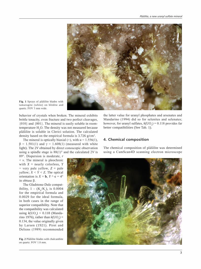
Plášilite occurs as long, thin blades (Fig. 1), elongated on [001] and flattened on {100}; rarely as prisms (Fig. 2), also elongated on [001]. Crystals exhibit the forms {100}, {010} and {011} (Fig. 3). Twinning is com-mon on {100}; prismatic crystals show polysynthetic
twinning. Plášilite is greenish yellow, has a white streak and fluo-resces bluish white under both long-wave and short-wave UV. It is transparent with vitreous lustre. The Mohs hardness could not be measured, but is probably between 2 and 3, based upon the
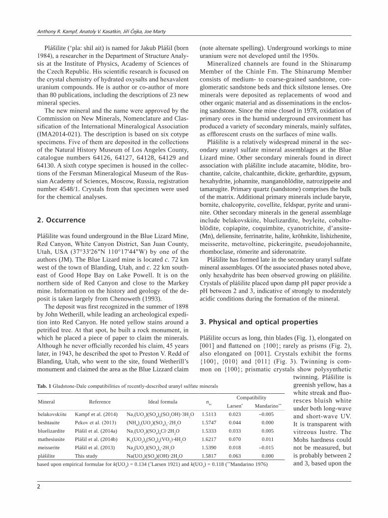
Tab. 1 Gladstone-Dale compatibilities of recently-described uranyl sulfate minerals
Mineral Reference Ideal formula nav
CompatibilityLarsen* Mandarino**
belakovskiite Kampf et al. (2014) Na7(UO2)(SO4)4(SO3OH)·3H2O 1.5113 0.023 –0.005beshtauite Pekov et al. (2013) (NH4)2(UO2)(SO4)2·2H2O 1.5747 0.044 0.000bluelizardite Plášil et al. (2014a) Na7(UO2)(SO4)4Cl·2H2O 1.5333 0.033 0.005mathesiusite Plášil et al. (2014b) K5(UO2)4(SO4)4(VO5)∙4H2O 1.6217 0.070 0.011meisserite Plášil et al. (2013) Na4(UO2)(SO4)4·2H2O 1.5390 0.018 –0.015plášilite This study Na(UO2)(SO4)(OH)·2H2O 1.5817 0.063 0.000based upon empirical formulae for k(UO3) = 0.134 (*Larsen 1921) and k(UO3) = 0.118 (**Mandarino 1976)
Plášilite, a new uranyl sulfate mineral
3
behavior of crystals when broken. The mineral exhibits brittle tenacity, even fracture and two perfect cleavages, {010} and {001}. The mineral is easily soluble in room-temperature H2O. The density was not measured because plášilite is soluble in Clerici solution. The calculated density based on the empirical formula is 3.726 g/cm3.
The mineral is optically biaxial (+), with α = 1.556(1), β = 1.581(1) and γ = 1.608(1) (measured with white light). The 2V obtained by direct conoscopic observation using a spindle stage is 88(1)° and the calculated 2V is 89°. Dispersion is moderate, r < v. The mineral is pleochroic with X = nearly colorless, Y = very pale yellow, Z = pale yellow; X < Y < Z. The optical orientation is X = b, Y ^ c = 4° in obtuse β.
The Gladstone-Dale compat-ibility, 1 – (Kp/Kc), is 0.0004 for the empirical formula and 0.0029 for the ideal formula, in both cases in the range of superior compatibility. Note that the compatibility was calculated using k(UO3) = 0.118 (Manda-rino 1976), rather than k(UO3) = 0.134, the value originally given by Larsen (1921). Piret and Deliens (1989) recommended
the latter value for uranyl phosphates and arsenates and Mandarino (1994) did so for selenites and selenates; however, for uranyl sulfates, k(UO3) = 0.118 provides far better compatibilities (See Tab. 1).
4. Chemical composition
The chemical composition of plášilite was determined using a CamScan4D scanning electron microscope
Fig. 1 Sprays of plášilite blades with tamarugite (white) on blödite and quartz. FOV 3 mm wide.
Fig. 2 Plášilite blades with chalcanthite on quartz. FOV 1.8 mm.

Anthony R. Kampf, Anatoly V. Kasatkin, Jiří Čejka, Joe Marty
4
(SEM) equipped with an Oxford Link ISIS energy-dispersive X-ray spectrometer. An operating voltage of 20 kV was used with a beam current of 1 nA and a 1 μm beam diameter. The EDS mode on the SEM was chosen for the analysis instead of the WDS mode on the elec-tron microprobe because of the instability of plášilite under the electron beam caused by high contents of both Na and H2O. Attempts to use the WDS mode were un-successful due to rapid and significant decomposition of the mineral under the electron beam. Although plášilite is a relatively widespread mineral in the secondary uranyl sulfate mineral assemblages at the Blue Lizard mine, it is present in very small quantities, making it impossible to separate an adequate amount of pure ma-terial for direct determination of H2O. Consequently, H2O was calculated by stoichiometry on the basis of 9 O apfu as indicated by the crystal structure determination. Raman spectroscopy confirmed the absence of CO2 and the presence of H2O and OH in the mineral. Other than U, Na and S, no elements with atomic numbers higher than 8 were observed. Nine spot analyses provided the data reported in Tab. 2.
The empirical formula of plášilite, calculated on the basis of 9 O apfu, is Na0.94(UO2)(S1.01O4)(OH)(H2O)2. The ideal formula is Na(UO2)(SO4)(OH)·2H2O, which requires (in wt. %) Na2O 7.01, UO3 64.70, SO3 18.11 and H2O 10.19, total 100.
5. Raman spectroscopy
A Raman spectrum (Fig. 4) of plášilite crystal was col-lected using a DXR dispersive Raman spectrometer (Thermo Scientific) mounted on a confocal Olympus mi-croscope (100× objective). The Raman signal was excited by a 532 nm diode-pumped solid-state laser and detected by a CCD detector. The experimental parameters were: 5s exposure time; 32 exposures, 400 lines/mm grating, 25 micron pinhole spectrograph aperture and 3.0 mW laser power level. The instrument was calibrated by a software-controlled calibration procedure using multiple neon emission lines (wavelength calibration), multiple polystyrene Raman bands (laser frequency calibration) and standardized white light sources (intensity calibra-tion).
The foregoing interpretation of the Raman spectrum of plášilite is based upon Nakamoto (1986), Volod’ko et al. (1981) and Čejka (1999, 2004, 2007). The crystal structure of plášilite contains one symmetrically distinct U6+, one symmetrically distinct S6+ and one symmetri-cally distinct Na+. The free uranyl ion, (UO2)
2+ with the point symmetry Dαh, exhibits three fundamental modes: the ν1 (Σg
+) symmetric stretching vibration, Raman ac-tive at 900–750 cm–1, the ν2 (δ) (Πu) doubly degenerate bending vibration, infrared active at 300–200 cm–1, and the ν3 (Σu) antisymmetric stretching vibration, infrared active at 1000–850 cm–1. The symmetry decrease from Dαh to Cαv or C2v may cause the infrared activation of the ν1 vibration, Raman activation of the ν2 and ν3 vibrations as well as splitting of the ν2 doubly degenerate bending vibration. Sulfate anion, (SO4)
2–, in Td symmetry, exhibits four fundamental modes: the ν1 (A1) symmetric stretch-ing vibration, Raman active at ~983 cm–1, the ν2 (δ) (E) doubly degenerate bending vibration, Raman active at ~450 cm–1, the ν3 (F2) triply degenerate antisymmetric stretching vibration, infrared and Raman active at ~1105
cm–1, and the ν4 (δ) (F2) triply degenerate bending vibration, infrared and Raman active at ~611 cm–1.
A band at 3600 cm–1 was assigned to the ν(OH) stretching vibration of free or weakly hydrogen-bonded hydroxyl ions. Bands at 3530 and 3385 cm–1 are attributed to the ν(OH) stretching vibrations of structurally nonequivalent hydrogen bonded water mol-ecules. According to Libowitzky (1999),
100 010
011011_
Fig. 3 Crystal drawing of plášilite; clinographic projection in standard orientation.
Tab. 2 Analytical data for plášilite (wt. %)
Constituent Mean (n = 9) Range SD Probe standardNa2O 6.61 5.42–7.63 0.59 ChkaloviteUO3 65.15 64.47–66.30 0.60 UO2
SO3 18.33 17.63–19.19 0.45 ZnSH2O 10.24*
Total 100.33* calculated from structure SD – standard deviation

Plášilite, a new uranyl sulfate mineral
5
approximate O–H···O hydrogen bond lengths are > 3.2 Å/3599 cm–1, 2.96 Å/3532 cm–1 and 2.79 Å/3385 cm–1. No bands were observed in the region of bending vibrations of water molecules (~1600 cm–1).
Bands at 1180, 1069 and 1035 cm–1 are assigned to the split triply degenerate ν3(SO4)
2– antisymmetric stretching vibrations and a strong band at 997 cm–1 to the ν1(SO4)
2– symmetric stretching vibration. A shoulder at 986.5 cm–1 may be assigned to the δU–OH bending vibration or the ν1(SO4)
2– vibration. A weak band at 905 cm–1 is connected with the
ν3(UO2)2+ antisymmetric stretching vibration. A very strong
band at 838 cm–1 was attributed to the ν1(UO2)2+ symmetric
stretching vibration, while a shoulder at 824 cm–1 may be assigned to the same vibration or to the δ U–OH bending vibration. According to Bartlett and Cooney (1989), ap-proximate U–O bond lengths in the uranyl ion are 1.78 Å/904 cm–1, 1.78 Å/838 cm–1 and 1.79 Å/824 cm–1. All these U–O bond lengths are close to those inferred by Burns (2005) for uranyl pentagonal dipyramidal coordi-nation polyhedra and agree with U–O bond lengths in the plášilite structure determined by X-ray diffraction.
Bands at 645 and 603 cm–1 are connected with triply degenerate ν4(δ)(SO4)
2– bending vibrations, and bands at 480 and 445 cm–1 to the doubly degenerate ν2(δ)(SO4)
2– bending vibrations. A band at 349 cm–1 may probably be assigned to ν(U–Oequatorial) stretching vibrations. The doubly degenerate ν2(δ)(UO2)
2+ bending vibration is characterized by a band at 243 cm–1. Remaining bands at 210, 186, 170, 137, 88 and 70 cm–1 may be assigned to lattice modes.
6. X-ray crystallography and structure determination
6.1. powder diffraction
Powder X-ray diffraction data for plášilite were obtained using a Rigaku R-Axis Rapid II curved imaging plate microdiffractometer utilizing monochromatized MoKα radiation (Gandolfi method). Observed powder dhkl val-ues and intensities were derived by profile fitting using JADE 2010 software (Materials Data Inc.). Data (in Å)
O–H
Fig. 4 The Raman spectrum of plášilite.
Anthony R. Kampf, Anatoly V. Kasatkin, Jiří Čejka, Joe Marty
6
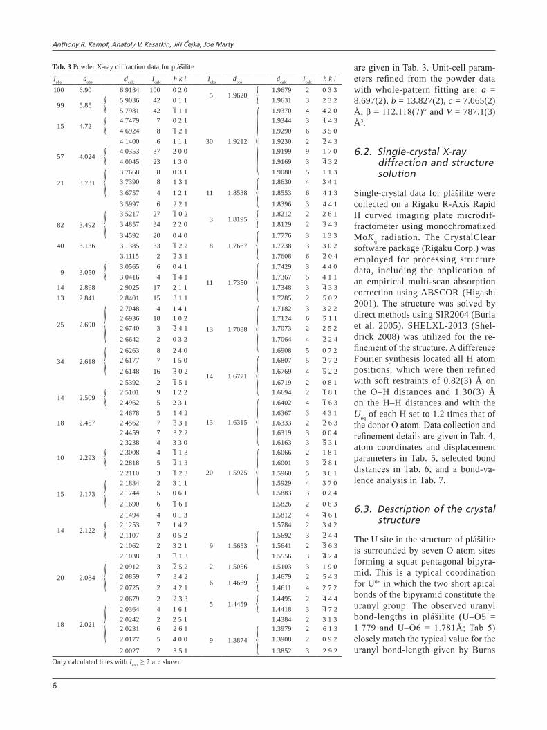
are given in Tab. 3. Unit-cell param-eters refined from the powder data with whole-pattern fitting are: a = 8.697(2), b = 13.827(2), c = 7.065(2) Å, β = 112.118(7)° and V = 787.1(3) Å3.
6.2. single-crystal X-ray diffraction and structure solution
Single-crystal data for plášilite were collected on a Rigaku R-Axis Rapid II curved imaging plate microdif-fractometer using monochromatized MoKα radiation. The CrystalClear software package (Rigaku Corp.) was employed for processing structure data, including the application of an empirical multi-scan absorption correction using ABSCOR (Higashi 2001). The structure was solved by direct methods using SIR2004 (Burla et al. 2005). SHELXL-2013 (Shel-drick 2008) was utilized for the re-finement of the structure. A difference Fourier synthesis located all H atom positions, which were then refined with soft restraints of 0.82(3) Å on the O–H distances and 1.30(3) Å on the H–H distances and with the Ueq of each H set to 1.2 times that of the donor O atom. Data collection and refinement details are given in Tab. 4, atom coordinates and displacement parameters in Tab. 5, selected bond distances in Tab. 6, and a bond-va-lence analysis in Tab. 7.
6.3. Description of the crystal structure
The U site in the structure of plášilite is surrounded by seven O atom sites forming a squat pentagonal bipyra-mid. This is a typical coordination for U6+ in which the two short apical bonds of the bipyramid constitute the uranyl group. The observed uranyl bond-lengths in plášilite (U–O5 = 1.779 and U–O6 = 1.781Å; Tab 5) closely match the typical value for the uranyl bond-length given by Burns
Tab. 3 Powder X-ray diffraction data for plášilite
Iobs dobs dcalc Icalc h k l Iobs dobs dcalc Icalc h k l100 6.90 6.9184 100 0 2 0
5 1.96201.9679 2 0 3 3
99 5.855.9036 42 0 1 1 1.9631 3 2 3 25.7981 42 1̄ 1 1
30 1.9212
1.9370 4 4 2 0
15 4.724.7479 7 0 2 1 1.9344 3 1̄ 4 34.6924 8 1̄ 2 1 1.9290 6 3 5 04.1400 6 1 1 1 1.9230 2 2̄ 4 3
57 4.0244.0353 37 2 0 0 1.9199 9 1 7 04.0045 23 1 3 0 1.9169 3 4̄ 3 2
21 3.7313.7668 8 0 3 1 1.9080 5 1 1 33.7390 8 1̄ 3 1
11 1.85381.8630 4 3 4 1
3.6757 4 1 2 1 1.8553 6 4̄ 1 33.5997 6 2̄ 2 1 1.8396 3 4̄ 4 1
82 3.4923.5217 27 1̄ 0 2
3 1.81951.8212 2 2 6 1
3.4857 34 2 2 0 1.8129 2 3̄ 4 33.4592 20 0 4 0
8 1.76671.7776 3 1 3 3
40 3.136 3.1385 33 1̄ 2 2 1.7738 3 3 0 23.1115 2 2̄ 3 1 1.7608 6 2̄ 0 4
9 3.0503.0565 6 0 4 1
11 1.7350
1.7429 3 4 4 03.0416 4 1̄ 4 1 1.7367 5 4 1 1
14 2.898 2.9025 17 2 1 1 1.7348 3 4̄ 3 3 13 2.841 2.8401 15 3̄ 1 1 1.7285 2 5̄ 0 2
25 2.690
2.7048 4 1 4 1
13 1.7088
1.7182 3 3 2 22.6936 18 1 0 2 1.7124 6 5̄ 1 12.6740 3 2̄ 4 1 1.7073 2 2 5 22.6642 2 0 3 2 1.7064 4 2̄ 2 4
34 2.6182.6263 8 2 4 0 1.6908 5 0 7 22.6177 7 1 5 0
14 1.6771
1.6807 5 2̄ 7 22.6148 16 3̄ 0 2 1.6769 4 5̄ 2 22.5392 2 1̄ 5 1 1.6719 2 0 8 1
14 2.5092.5101 9 1 2 2 1.6694 2 1̄ 8 12.4962 5 2 3 1
13 1.6315
1.6402 4 1̄ 6 3
18 2.4572.4678 5 1̄ 4 2 1.6367 3 4 3 12.4562 7 3̄ 3 1 1.6333 2 2̄ 6 32.4459 7 3̄ 2 2 1.6319 3 0 0 42.3238 4 3 3 0 1.6163 3 5̄ 3 1
10 2.2932.3008 4 1̄ 1 3
20 1.5925
1.6066 2 1 8 12.2818 5 2̄ 1 3 1.6001 3 2̄ 8 12.2110 3 1̄ 2 3 1.5960 5 3 6 1
15 2.1732.1834 2 3 1 1 1.5929 4 3 7 02.1744 5 0 6 1 1.5883 3 0 2 42.1690 6 1̄ 6 1 1.5826 2 0 6 32.1494 4 0 1 3 1.5812 4 4̄ 6 1
14 2.1222.1253 7 1 4 2 1.5784 2 3 4 22.1107 3 0 5 2
9 1.56531.5692 3 2̄ 4 4
2.1062 2 3 2 1 1.5641 2 3̄ 6 32.1038 3 3̄ 1 3 1.5556 3 4̄ 2 4
20 2.0842.0912 3 2̄ 5 2 2 1.5056 1.5103 3 1 9 02.0859 7 3̄ 4 2
6 1.46691.4679 2 5̄ 4 3
2.0725 2 4̄ 2 1 1.4611 4 2 7 22.0679 2 2̄ 3 3
5 1.44591.4495 2 4̄ 4 4
18 2.021
2.0364 4 1 6 1 1.4418 3 4̄ 7 22.0242 2 2 5 1 1.4384 2 3 1 32.0231 6 2̄ 6 1
9 1.3874
1.3979 2 6̄ 1 32.0177 5 4 0 0 1.3908 2 0 9 2
2.0027 2 3̄ 5 1 1.3852 3 2̄ 9 2Only calculated lines with Icalc ≥ 2 are shown

Plášilite, a new uranyl sulfate mineral
7
et al. (1997), based on a large-number of well-refined crystal structures. In the structure, pairs of pentagonal bipyramids share a common edge, forming dimers. The dimers are linked by sharing corners with SO4 groups, yielding a [(UO2)2(SO4)2(OH)2]
2– sheet parallel to (010) (Fig. 5). Between the sheets are NaO2(H2O)4 octahedra, which link by sharing trans H2O–H2O edges to form chains along [001]. Sodium octahedra link the sheets in the [010] direction; each Na octahedron shares one O corner with an SO4 group in the sheet and the opposite O corner with a uranyl group. The framework thereby created is further linked via relatively weak hydrogen bonds (Fig. 6).
The [(UO2)2(SO4)2(OH)2]2– sheet is based on the
phosphuranylite anion topology (Burns 2005), with a ring symbol 61524232 (Krivovichev and Burns 2007). The sheets in johannite, Cu[(UO2)2(OH)2(SO4)2](H2O)8, (Me-reiter 1982) and deliensite, Fe(UO2)2(SO4)2(OH)2·7H2O, (Plášil et al. 2012) are topologically identical to that in plášilite; however, the sheets are geometrical isomers, differing in the orientation of the SO4 groups (Fig. 5). It is worth noting that plášilite and johannite occur in direct association at the Blue Lizard mine, and deliensite has been found in the general assemblage. None of the other uranyl sulfates recorded at the Blue Lizard mine (e.g. belakovskiite, bluelizardite, meisserite and several
Tab. 4 Data collection and structure refinement details for plášilite
Temperature 298(2) KStructural formula Na(UO2)(SO4)(OH)(H2O)2
Space group P21/cUnit-cell dimensions a = 8.7122(6) Å
b = 13.8368(4) Åc = 7.04650(17) Åβ = 112.126(8)°
V 786.89(7) Å3
Z 4Density (for above formula) 3.723 g cm–3
Absorption coefficient 20.965 mm–1
F(000) 780Crystal size 80 × 20 × 5 µmθ range 3.45 to 27.48°Index ranges –11 ≤ h ≤ 11, –15 ≤ k ≤ 17, –7 ≤ l ≤ 9Reflections collected/unique 7709/1794; Rint = 0.0295Reflections with Fo > 4σ(F) 1609Completeness to θ = 27.48° 99.4%Refinement method Full-matrix least-squares on F2
Parameters refined 124GoF 1.081Final R indices [Fo > 4σ(F)] R1 = 0.0194, wR2 = 0.0446R indices (all data) R1 = 0.0231, wR2 = 0.0462Largest diff. peak/hole +3.07/–0.70 e A–3
*Rint = Σ|Fo2–Fo
2(mean)|/Σ[Fo2]. GoF = S = {Σ[w(Fo
2–Fc2)2]/(n–p)}1/2.
R 1 = Σ| |F o|– |F c| | /Σ|F o| . wR 2 = {Σ[w(F o2–F c
2)2]/Σ[w(F o2)2]} 1/2;
w = 1/[σ2(Fo2) + (aP)2 + bP] where a is 0.026, b is 0.4537 and
P is [2Fc2 + Max(Fo
2,0)]/3.
Tab. 5 Atom coordinates and displacement parameters (Å2) for plášilite
x/a y/b z/c Ueq U11 U22 U33 U23 U13 U12
U 0.23417(2) 0.46715(2) 0.12219(2) 0.01131(7) 0.00812(10) 0.01565(10) 0.00985(10) –0.00007(5) 0.00306(6) 0.00043(6)S 0.32042(13) 0.50289(8) 0.66758(15) 0.01204(19) 0.0089(5) 0.0173(5) 0.0097(5) 0.0013(4) 0.0033(4) 0.0011(4)Na 0.2793(3) 0.73085(15) 0.6584(3) 0.0305(5) 0.0401(12) 0.0251(10) 0.0272(10) –0.0035(8) 0.0136(9) 0.0062(9)O1 0.1796(4) 0.5684(2) 0.6139(5) 0.0187(6) 0.0107(15) 0.0222(15) 0.0231(16) 0.0033(13) 0.0062(12) 0.0038(13)O2 0.3124(4) 0.4419(2) 0.4914(4) 0.0205(7) 0.0261(18) 0.0238(16) 0.0104(15) –0.0004(12) 0.0055(13) 0.0040(14)O3 0.4736(4) 0.5623(2) 0.7334(5) 0.0186(6) 0.0076(15) 0.0242(15) 0.0208(16) 0.0025(13) 0.0018(12) 0.0012(12)O4 0.3242(4) 0.4368(2) 0.8366(4) 0.0173(6) 0.0236(17) 0.0207(15) 0.0099(14) 0.0047(12) 0.0090(13) 0.0065(13)O5 0.1933(4) 0.3410(2) 0.1168(4) 0.0229(7) 0.0236(18) 0.0204(16) 0.0229(17) –0.0032(13) 0.0069(14) 0.0072(14)O6 0.2856(4) 0.5921(2) 0.1325(5) 0.0225(7) 0.0179(16) 0.0186(16) 0.0309(18) –0.0006(13) 0.0092(14) 0.0015(13)OH7 –0.0008(4) 0.5113(3) 0.1785(5) 0.0240(7) 0.0120(16) 0.048(2) 0.0127(16) 0.0048(15) 0.0053(13) 0.0057(15)H7 –0.002(8) 0.483(4) 0.276(7) 0.029OW8 –0.0797(5) 0.2716(3) 0.6917(6) 0.0326(8) 0.028(2) 0.034(2) 0.035(2) –0.0005(18) 0.0121(16) 0.0040(16)H8A 0.006(5) 0.242(3) 0.702(9) 0.039H8B –0.044(7) 0.328(2) 0.719(9) 0.039OW9 0.5186(5) 0.2503(3) 0.0025(6) 0.0319(8) 0.033(2) 0.0272(18) 0.034(2) –0.0076(17) 0.0115(16) 0.0021(17)H9A 0.475(7) 0.296(3) –0.063(8) 0.038H9B 0.448(6) 0.210(3) –0.020(8) 0.038
Tab. 6 Selected bond distances (Å) and angles (°) for plášilite
U–O5 1.779(3) Na–O1 2.387(4) S–O1 1.456(3) Hydrogen bonds (D = donor, A = acceptor)U–O6 1.781(3) Na–OW9 2.389(4) S–O2 1.481(3) D–H d(D–H) d(H…A) <DHA d(D…A) A <HDHU–OH7 2.310(3) Na–OW8 2.417(4) S–O3 1.485(3) OH7–H7 0.80(3) 2.10(5) 137(6) 2.739(5) O1U–OH7 2.338(3) Na–OW8 2.425(4) S–O4 1.492(3) OW8–H8a 0.82(3) 2.26(3) 161(5) 3.050(5) O5U–O3 2.394(3) Na–OW9 2.442(4) <S–O> 1.479 OW8–H8b 0.84(3) 2.32(3) 166(5) 3.141(5) O7 101U–O2 2.454(3) Na–O6 2.459(4) OW9–H9a 0.79(3) 2.31(3) 162(6) 3.066(5) O4U–O4 2.456(3) <Na–O> 2.420 OW9–H9b 0.80(3) 2.43(3) 161(5) 3.193(5) O2 120<U–O> 2.289

Anthony R. Kampf, Anatoly V. Kasatkin, Jiří Čejka, Joe Marty
8
others under study) have structures that contain uranyl dimers.
Acknowledgements. Reviewer Stuart Mills, an anony-mous reviewer and handling editor František Laufek are thanked for their constructive comments on the manu-script. Ken Krahulec of the Utah Geological Survey is thanked for providing information about the genesis of the U deposits in the area. The recording of the plášilite Raman spectrum by Ladislav Lapčák (Institute of Chemi-cal Technology, Prague) is highly appreciated. A portion of this study was funded by the John Jago Trelawney Endowment to the Mineral Sciences Department of the Natural History Museum of Los Angeles County. Fur-ther support was provided by long-term project DKRVO
a
c
US
US
c
b
plášilite deliensite johannite
US
c
b
Fig. 5 Sheets in the structures of plášilite, deliensite and johannite. The unit is cell shown by dashed lines. For plášilite, H atoms are represented by white spheres. O–H bonds are drawn as sticks.
Tab. 7 Bond-valence analysis for plášilite (in valence units)
O1 O2 O3 O4 O5 O6 OH7 OW8 OW9 Σc
U 0.46 0.52 0.46 1.69 1.68 0.61, 0.58 5.99Na 0.18 0.15 0.17, 0.16 0.18, 0.16 1.00S 1.57 1.47 1.46 1.43 5.93H7 0.10 0.90 1.00H8a 0.08 0.92 1.00H8b 0.07 0.93 1.00H9a 0.07 0.93 1.00H9b 0.06 0.94 1.00Σa 1.86 1.99 1.97 1.96 1.77 1.83 2.15 2.18 2.21Bond strengths are as follows:*S6+–O from Brown and Altermatt (1985); Na+–O from Wood and Palenik (1999); U6+–O from Burns et al. (1997); hydrogen based on H…O bond lengths from Brown and Altermatt (1985).
ab
Na
S
U
Fig. 6 The crystal structure of plášilite. The unit is cell indicated by dashed lines. Hydrogen atoms are shown as white spheres; O–H bonds are drawn as sticks. Hydrogen bonds are shown as thin lines. Note the chains of [NaO2(H2O)4] octahedra running along [001].
Plášilite, a new uranyl sulfate mineral
9
2014/02 of the Ministry of Culture of the Czech Republic (National Museum 000232782) to J. Č.
Electronic supplementary material. Supplementary crys-tallographic data for this paper are available online at the Journal web site (http://dx.doi.org/10.3190/jgeosci. 184).
References
Bartlett Jr, Cooney rP (1989) On the determination of uranium–oxygen bond lengths in dioxouranium(VI) compounds by Raman spectroscopy. J Mol Struct 193: 295–300
Brown ID, altermatt D (1985) Bond-valence parameters obtained from a systematic analysis of the inorganic crystal structure database. Acta Cryst B41: 244–248
Burla mC, CalIanDro r, CamallI m, CarrozzInI B, CasCarano Gl, De Caro l, GIaCovazzo C, PolIDorI G, sPaGna r (2005) SIR2004: an improved tool for crystal structure determination and refinement. J Appl Cryst 38: 381–388
Burns PC (2005) U6+ minerals and inorganic compounds: insights into an expanded structural hierarchy of crystal structures. Canad Mineral 43: 1839–1894
Burns PC, ewInG rC, HawtHorne, FC (1997) The crystal chemistry of hexavalent uranium: polyhedron geo-metries, bond-valence parameters, and polymerization of polyhedra. Canad Mineral 35: 1551–1570
CHenowetH wl (1993) The Geology and Production History of the Uranium Deposits in the White Canyon Mining District, San Juan County, Utah. Utah Geological Survey Miscellaneous Publications 93–3: pp 1–26
Čejka j (1999) Infrared spectroscopy and thermal analysis of the uranyl minerals. In: Burns PC, ewInG rC (eds) Uranium: Mineralogy, Geochemistry and the Environ-ment. Mineralogical Society of America and Geochemi-cal Society Reviews in Mineralogy and Geochemistry 38: pp 521–622
Čejka j (2004) Vibrational spectroscopy of uranyl miner-als – infrared and Raman spectra of uranyl minerals. I. Uranyl, UO2
2+. Bull mineral–petrolog Odd Nár Muz (Praha) 12: 44–51 (in Czech)
Čejka j (2007) Vibrational spectroscopy of uranyl miner-als – infrared and Raman spectra of uranyl minerals. III. Uranyl sulfates. Bull mineral–petrolog Odd Nár Muz (Praha) 14–15: 40–46 (in Czech)
Finch Rj, MuRakaMi T (1999) Systematics and paragenesis of uranium minerals. In: Burns PC, ewInG rC (eds) Uranium: Mineralogy, Geochemistry and the Environment. Min-eralogical Society of America and Geochemical Society Reviews in Mineralogy and Geochemistry 38: pp 91–179
FRondel c, iTo j, honea RM, Weeks aM (1976) Mineral-ogy of the Zippeite Group. Canad Mineral 14: 429–436
kaMpF aR, plášil j, kasaTkin aV, MaRTy j (2014) Bela-kovskiite, Na7(UO2)(SO4)4(SO3OH)(H2O)3, a new uranyl sulfate mineral from the Blue Lizard mine, San Juan County, Utah, USA. Mineral Mag 78: 639–649
HIGasHI t (2001) ABSCOR. Rigaku Corporation, TokyokRiVoVicheV sV, BuRns pc (2007) Actinide compounds
containing hexavalent cations of the VI group ele-ments (S, Se, Mo, Cr, W). In: kRiVoVicheV sV, BuRns PC,tananaev IG (eds) Structural Chemistry of Inorganic Actinide Compounds. Elsevier, Amsterdam, pp 95–182
kRiVoVicheV sV, plášil j (2013) Mineralogy and Crystal-lography of Uranium. In: Burns PC, sIGmon Ge (eds) Uranium: From Cradle to Grave. Mineralogical Associa-tion of Canada Short Courses 43: pp 15–119
larsen es (1921) The microscopic determination of the nonopaque minerals. US Geol Surv Bull 679: pp 1–294
liBoWiTzky e (1999) Correlation of O–H stretching frequen-cies and O–H···O hydrogen bond lengths in minerals. Monat Chem 130: 1047–1059
manDarIno Ja (1976) The Gladstone-Dale relationship – Part I: derivation of new constants. Canad Mineral 14: 498–502
manDarIno Ja (1994) Natural and synthetic selenites and selenates and their Gladstone-Dale compatibility. Eur J Mineral 6: 337–349
MeReiTeR k (1982) Die Kristallstrukturs des Johannits, Cu(UO2)2(OH)2(SO4)2·8H2O. Tschermaks Mineral Petrol Mitt 30: 47–57
nakaMoTo k (1986) Infrared and Raman Spectra of Inor-ganic and Coordination Compounds. J. Wiley and Sons, New York, pp 1–496
ondRuš p, VeseloVský F, skála R, císařoVá i, hloušek j, FRýda j, VaVřín i, Čejka j, GaBašoVá a (1997) New naturally occurring phases of secondary origin from Jáchymov (Joachimsthal). J Czech Geol Soc 42: 77–108
pekoV iV, kRiVoVicheV sV, yapaskuRT Vo, chukanoV nV, BelakoVskiy di (2014) Beshtauite, (NH4)2(UO2)(SO4)2 ·2H2O, a new mineral from Mount Beshtau, Northern Caucasus, Russia. Amer Miner 99: 1783–1787
PIret P, DelIens m (1989) The Gladstone-Dale constant k(UO3) for uranyl phosphates and arsenates. Canad Mineral 27: 533–534
plášil j, hauseR j, peTříČek V, MeisseR n, Mills sj, škoda R, FejFaRoVá k, Čejka j, sejkoRa j, hloušek j, johanneT j.-M, MachoViČ V, lapČák l (2012) Crystal structure and formula revision of deliensite, Fe[(UO2)2(SO4)2(OH)2](H2O)7. Mineral Mag 76: 2837–2860
plášil j, kaMpF aR, kasaTkin aV, MaRTy j, škoda R, silVa s, Čejka j (2013) Meisserite, Na5(UO2)(SO4)3(SO3OH)(H2O), a new uranyl sulfate mineral from the Blue Lizard mine, San Juan County, Utah, USA. Mineral Mag 77: 2975–2988
plášil j, kaMpF aR, kasaTkin aV, MaRTy j (2014a) Blue-lizardite, Na7(UO2)(SO4)4Cl(H2O)2, a new uranyl sulfate
Anthony R. Kampf, Anatoly V. Kasatkin, Jiří Čejka, Joe Marty
10
mineral from the Blue Lizard mine, San Juan County, Utah, USA. J Geosci 59: 145–158
plášil j, VeseloVský F, hloušek j, škoda R, noVák M, sejkoRa j, Čejka j, škácha p, kasaTkin aV (2014b) Mathesiusite, K5(UO2)4(SO4)4(VO5)(H2O)4, a new uranyl vanadate–sulfate from Jáchymov, Czech Republic. Amer Miner 99: 625–632
sheldRick GM (2008) A short history of SHELX. Acta Cryst A64: 112–122
TVRdý j, plášil j (2010) Jáchymov – reiche Erzlagerstätte und Radonbad im böhmischen Westerzgebirge. Auf-schluss 61: 277–292
Volod’ko lV, koMyak ai, uMReyko ds (1981) Uranyl Compounds Vol. I, Spectra and Structure. Belorussian State University, Minsk, pp 1–620 (in Russian)
Wood RM, palenik Gj (1999) Bond valence sums in co-ordination chemistry. Sodium–oxygen complexes. Inorg Chem 38: 3926–3930