Original Article - Welcome to IAS Fellows Publicationsrepository.ias.ac.in/20472/1/323.pdf ·...
14
Original Article Trend of Japanese encephalitis in North India: evidence from thirty-eight acute encephalitis cases and appraisal of niceties Shailendra K. Saxena 1 , Niraj Mishra 1 , Rakhi Saxena 1 , Maneesh Singh 1 , Asha Mathur 2 1 Laboratory of Infectious Diseases & Molecular Virology, Centre for Cellular and Molecular Biology (CSIR), Uppal Road, Hyderabad 500 007, India 2 Department of General Pathology and Microbiology, Saraswati Medical and Dental College, Lucknow 227 105, India Abstract Background: In the year 2005, an epidemic of Japanese encephalitis (JE) occurred in the northern states of India. The present study was planned to reconfirm the circulation of JE in the area and to assess the trend of the disease to slow down the burden of JE. Methodology: Surveillance was conducted to identify patients with acute encephalitis. Blood and cerebrospinal fluid specimens from suspected cases underwent pathological, serological, and demographic investigations. Viral testing for evidence of Japanese encephalitis virus (JEV) infection was also performed, either by IgM capture ELISA/RT-PCR or both. To identify circulating JEV strains, RT-PCR, sequencing and phylogenetic analysis was performed. Based on clinical cases reported between 1992 and 2008, the trend of JE infection in the state was analyzed to examine the dynamics of infection. Results: Our investigations (n = 38) revealed that only 55.3% cases were positive for JE. Pathological examination revealed marked pleocytosis in CSF (90 76.9 cells/mm 3 ), and peripheral leucocytosis (64.7 8.86% neutrophils) with mild anemia. Males were more susceptible than females with a ratio of 1.63:1 and significant gender difference (P 0.05) was observed in patients below six years. In the patient group younger than six years, the rate of infection per million was six-fold higher (P 0.005) in males as compared to females. Our phylogenetic study suggests that the circulating strain during the 2005 JE epidemic was close to GP78, and in the future a larger epidemic may occur. Conclusions: The 2005 JE epidemic was possibly caused by JEV GP78 and it is spreading into newer areas. The trend of JE suggests that the problem in North India is escalating and larger epidemics may occur in the future; therefore, serious steps are necessary to combat JE, including the development of more efficient surveillance methods and differential diagnosis. Key Words: encephalitis; Japanese encephalitis; virus; outbreak; seasonal incidence; Uttar Pradesh; India; trend J Infect Dev Ctries 2009; 3(7):517-530. Received 6 April 2009 - Accepted 27 June 2009 Copyright © 2009 Saxena et al. This is an open-access article distributed under the Creative Commons Attribution License, which permits unrestricted use, distribution, and reproduction in any medium, provided the original work is properly cited. Introduction Almost every year, so-called “undiagnosed illnesses” invade India and unfailingly claim thousands of lives. Viral encephalitis, a major global emerging public health problem, is among them. Although viruses are the most common cause of encephalitis, bacteria, fungus, and parasites may also be responsible for infection. In India, although many encephalitis outbreaks have been reported since 1955 [1], several have remained undiagnosed. In the absence of a defined cause, these outbreaks were tentatively attributed to Reye’s syndrome, dengue, chikungunya, Japanese encephalitis (JE) and measles. Between July and December 2005, a large and severe epidemic of viral encephalitis was seen in northern India. The disease gripped Uttar Pradesh, border areas of Bihar and Nepal. During this interval a total of 6097 JE cases with 1,398 deaths were reported [2,3]. The case fatality rate (CFR) was 22.9%. Uttar Pradesh was most affected with 5,737 JE cases and 1,334 deaths (CFR 23.3%) and Bihar experienced 360 cases with 64 deaths (CFR 17.8%) [2]. However, the true incidence was much higher, as most of the cases remained asymptomatic and undiagnosed. The majority of the cases during this epidemic came from eastern Uttar Pradesh (Gorakhpur and adjoining areas), which is the paddy growing “Terai area.” Uttar Pradesh (a northern state of India) lies between latitudes 24º and 31º north and longitudes 77º and 84º east (Figure 1) and is surrounded by Uttaranchal in the northeast, Haryana and Himachal Pradesh in the north, Delhi and Rajasthan in the west, Madhya Pradesh in the southwest, Chhattisgarh in the south, Bihar in the southeast, and
Transcript of Original Article - Welcome to IAS Fellows Publicationsrepository.ias.ac.in/20472/1/323.pdf ·...

Original Article
Trend of Japanese encephalitis in North India: evidence from thirty-eight acute encephalitis cases and appraisal of niceties Shailendra K. Saxena1, Niraj Mishra1, Rakhi Saxena1, Maneesh Singh1, Asha Mathur2
1Laboratory of Infectious Diseases & Molecular Virology, Centre for Cellular and Molecular Biology (CSIR), Uppal Road,
Hyderabad 500 007, India 2Department of General Pathology and Microbiology, Saraswati Medical and Dental College, Lucknow 227 105, India
Abstract Background: In the year 2005, an epidemic of Japanese encephalitis (JE) occurred in the northern states of India. The present study was
planned to reconfirm the circulation of JE in the area and to assess the trend of the disease to slow down the burden of JE.
Methodology: Surveillance was conducted to identify patients with acute encephalitis. Blood and cerebrospinal fluid specimens from
suspected cases underwent pathological, serological, and demographic investigations. Viral testing for evidence of Japanese encephalitis
virus (JEV) infection was also performed, either by IgM capture ELISA/RT-PCR or both. To identify circulating JEV strains, RT-PCR,
sequencing and phylogenetic analysis was performed. Based on clinical cases reported between 1992 and 2008, the trend of JE infection in
the state was analyzed to examine the dynamics of infection.
Results: Our investigations (n = 38) revealed that only 55.3% cases were positive for JE. Pathological examination revealed marked
pleocytosis in CSF (90 76.9 cells/mm3), and peripheral leucocytosis (64.7 8.86% neutrophils) with mild anemia. Males were more
susceptible than females with a ratio of 1.63:1 and significant gender difference (P 0.05) was observed in patients below six years. In the
patient group younger than six years, the rate of infection per million was six-fold higher (P 0.005) in males as compared to females. Our
phylogenetic study suggests that the circulating strain during the 2005 JE epidemic was close to GP78, and in the future a larger epidemic
may occur.
Conclusions: The 2005 JE epidemic was possibly caused by JEV GP78 and it is spreading into newer areas. The trend of JE suggests that
the problem in North India is escalating and larger epidemics may occur in the future; therefore, serious steps are necessary to combat JE,
including the development of more efficient surveillance methods and differential diagnosis.
Key Words: encephalitis; Japanese encephalitis; virus; outbreak; seasonal incidence; Uttar Pradesh; India; trend
J Infect Dev Ctries 2009; 3(7):517-530.
Received 6 April 2009 - Accepted 27 June 2009
Copyright © 2009 Saxena et al. This is an open-access article distributed under the Creative Commons Attribution License, which permits unrestricted use,
distribution, and reproduction in any medium, provided the original work is properly cited.
Introduction Almost every year, so-called “undiagnosed
illnesses” invade India and unfailingly claim
thousands of lives. Viral encephalitis, a major global
emerging public health problem, is among them.
Although viruses are the most common cause of
encephalitis, bacteria, fungus, and parasites may also
be responsible for infection. In India, although many
encephalitis outbreaks have been reported since
1955 [1], several have remained undiagnosed. In the
absence of a defined cause, these outbreaks were
tentatively attributed to Reye’s syndrome, dengue,
chikungunya, Japanese encephalitis (JE) and
measles. Between July and December 2005, a large
and severe epidemic of viral encephalitis was seen in
northern India. The disease gripped Uttar Pradesh,
border areas of Bihar and Nepal. During this interval
a total of 6097 JE cases with 1,398 deaths were
reported [2,3]. The case fatality rate (CFR) was
22.9%. Uttar Pradesh was most affected with 5,737
JE cases and 1,334 deaths (CFR 23.3%) and Bihar
experienced 360 cases with 64 deaths (CFR 17.8%)
[2]. However, the true incidence was much higher,
as most of the cases remained asymptomatic and
undiagnosed.
The majority of the cases during this epidemic
came from eastern Uttar Pradesh (Gorakhpur and
adjoining areas), which is the paddy growing “Terai
area.” Uttar Pradesh (a northern state of India) lies
between latitudes 24º and 31º north and longitudes
77º and 84º east (Figure 1) and is surrounded by
Uttaranchal in the northeast, Haryana and Himachal
Pradesh in the north, Delhi and Rajasthan in the
west, Madhya Pradesh in the southwest,
Chhattisgarh in the south, Bihar in the southeast, and

Saxena et al. - Escalating JE in North India. J Infect Dev Ctries 2009; 3(7):517-530.
518
Nepal along the East. After the 2001 census, Uttar
Pradesh is the most populous state in India,
accounting for 16.4% of the total population of the
country. Population density of this state is 689
persons per square kilometer, while it is 324 persons
per square kilometer for the country. Children aged
0 to 6 years make up 18.35% of the population, of
which 9.58% are male and 8.77% are females, while
approximately 40% of the total population belongs
to the 0-12 year age group. The rural population is
79.22% [4]. There are three distinct seasons:
Summer (March to June, with temperatures ranging
from 27.5º-32.5 ºC, with a max 45 ºC); Monsoon
(July to October, with rainfall of 1,000-2,000 mm in
the east, and 600-1,000 mm in the west); and Winter
(November to February, with temperatures ranging
from 12.5º-17.5 ºC). The entire state has a tropical
monsoon climate.
JE is an acute viral zoonotic infection of the
central nervous system (CNS), which produces
meningomyelo-encephalitis. It poses a serious public
health problem with an increasing frequency of
epidemics and outbreaks in many parts of the Indian
subcontinent and South East Asian countries over
the last four decades. Now approaching newer areas
such as Papua New Guinea and Australia, it has been
classified as new emerging disease [5,6]. JE virus
(JEV) is an arthropod-borne flavivirus and is
transmitted to human beings by Culex
tritaeniorrhynchus and other related rice field-
breeding mosquitoes of genus Culex. The disease
commonly affects children and is a major cause of
acute childhood encephalopathy. JEV or
antigenically related virus has been identified
serologically in different parts of India since the
mid-fifties. Although initial cases of JE were
reported in the 1950s in India, Uttar Pradesh saw its
first epidemic in 1978 [7]. Since then, this
encephalitis has taken more than 10,000 lives in the
state [8,9]. As in 2005, most of the cases of acute
encephalitis came from Gorakhpur and adjoining
areas. In the present study, patients with acute
encephalitis were enrolled from this area and
examined for the evidence of JE infection with
clinicopathological, serological, and demographical
features. Based on current evidence and historical
epidemics of JEV in Uttar Pradesh (India), we will
discuss the current trend of the disease to prepare
ourselves for the possibility of a future epidemic.
Materials and Methods
Outbreak Surveillance and sample collection
After approval from the Institutional Review
Board of the Centre for Cellular and Molecular
Biology, Hyderabad, surveillance was conducted at
Gorakhpur and adjoining areas to identify patients
with acute encephalopathic illness during July to
November 2005. A patient was suspected to have
encephalitis if all the following criteria were met: (i)
a recent history of fever; (ii) cerebrospinal fluid
(CSF) protein concentration of at least 40 mg per
deciliter or white blood cell (WBC) count of at least
5 per cubic millimeter; (iii) no other clinical
diagnosis; (iv) neurological impairment. Mild and
acute encephalitis were categorized on the basis of
severity of symptoms. Mild encephalitis was
characterized by mild fever and headache, and the
patient testing positive for JE-ELISA. However, a
Figure 1. Japanese encephalitis affected areas of India, 2005. The map of India shows Uttar Pradesh outlined in red, the
area which was worst affected by JE in 2005 (Map modified and used with permission from Google Maps, CA, USA).

Saxena et al. - Escalating JE in North India. J Infect Dev Ctries 2009; 3(7):517-530.
519
recent history of any one or more symptoms
including altered sensorium, headache, stiffness of
the neck, tremors, vomiting, altered mental status,
seizure, myalgia, abdominal pain and depressed
level of consciousness with coma and paralysis
along with fever along with positive testing for JE-
ELISA indicated acute encephalitis. No tests were
performed for Reye's syndrome, dengue,
chikungunya or any other flavivirus infections. A
total of 38 patients with symptoms of acute
encephalopathic illness were enrolled for this study
(Table 1). After the written informed consent of the
patient/patient’s guardian was obtained, blood and/or
CSF samples were collected and stored at 20 C. No
patient had history of JE vaccination. All JE patients
were examined completely for clinico-pathological
symptoms as described above, and previous clinical
history and demographic information about the
patients were obtained.
Laboratory investigation Blood and/or CSF
specimens from patients underwent serological and
viral testing for evidence of arboviral infection by
IgM-capture ELISA. For every examination, serum
and/or CSF samples were collected by single aseptic
venipuncture or lumber puncture respectively. All
the serum and CSF samples were tested for the
presence JEV by IgM-capture ELISA (JEV CheX
IgM ELISA Kit, XCyton Diagnostics Ltd.,
Bangalore, India) according to the manufacturer’s
instructions. Briefly, CSF and serum samples were
diluted 10- and 20-fold respectively and 100 µl of
test CSF/serum samples with appropriate controls
were incubated in ELISA plates for one hour at
37ºC. To each well 100 µl of JEV antigen was added
and incubated at 37ºC for one hour and washed
again. Next, 100 µl of biotinylated-monoclonal
antibody was added and incubated for 30 minutes at
37ºC and 100 µl of streptavidin-peroxidase
conjugate was added. The plate was kept at room
temperature for 15 minutes and 100µl of substrate
was added to each well and the plate was left for 10
minutes at room temperature. Finally, 100 µl of stop
solution was added to each well to arrest the
reaction. The plate was read at 450 nm in an ELISA
reader (SpectraMax 190, Molecular Devices,
Sunnyvale, California, USA) within one hour of
reaction. A positive reaction of the CSF sample
confirmed JEV infection and a positive reaction of
the serum sample suggested a recent JEV infection.
Samples were considered as JE positive if the
ELISA value was more than 100 ELISA units;
otherwise, samples were considered as JE negative
[10] (Table 1). This kit is already evaluated for its
sensitivity, specificity, and usability against JE
patient samples by various workers [11,12]. If the
blood and CSF samples drawn from a patient within
first five days of fever were found non-reactive, a
second serum/CSF sample was collected from that
patient after seven days of the occurrence of the
fever. Clinical investigations of JE positive patients
included the following: complete peripheral blood
examination; blood glucose level; CSF examination
for appearance with cell count; measurement of
glucose and protein; bacterial culture; and gram
staining.
Molecular and Phylogenetic analysis
Notably, we could also extract viral RNA,
specifically GP05, from a CSF sample (I.D. 17).
Viral RNA was isolated using a GF-1 Viral
RNA/DNA Extraction Kit (Vivantis Inc., California,
USA) according to the manufacturer’s instructions.
The full env gene of GP05 was amplified by
standard reverse-transcriptase polymerase chain
reaction (RT-PCR). RT-PCR was performed with an
RT-PCR Kit (Promega, Madison, WI, USA) using
5’CTGTTGGTCGCTCCGGCTTACAG3’ as the
forward primer and 5’
AGCATGCACATTGGTCGCTAAGAA3’ as the
reverse primer. Primers were designed against the
env gene of JEV GP78 (AF075723) as a reference
sequence by using Gene Tool software (BioTools
Inc., Edmonton, Canada) and synthesized by
Bioserve Biotechnologies India Pvt. Ltd., India.
Briefly, for a 20 µl RT-reaction, 100 mM MgCl2,
10X RT Buffer, 10mM dNTPs, 0.5 unit RNasin, 2.5
unit of reverse transcriptase, 15 picomolar reverse
primer and 200ng viral RNA were added. RT was
performed at 47°C for one hour. Second strand
synthesis was performed by PCR for a 20 µl reaction
using 10X PCR Buffer (Applied Biosystems, USA),
37 mM MgCl2 (Applied Biosystems, USA), 5 mM
dNTP mix (Eppendorf, Germany), 2 pM each of
forward and reverse primers, 2 units of AmpliTaq
Gold DNA Polymerase (Applied Biosystems, USA),
and 1µl of cDNA. Amplification was carried out for
35 cycles in Eppendorf Mastercycler EP (Eppendorf,
Germany), each consisting of denaturation at 95 C
for 30 seconds, annealing at 60 C for 30 seconds,
Saxena et al. - Escalating JE in North India. J Infect Dev Ctries 2009; 3(7):517-530.
520
and extension at 72 C for 2 minutes. Pre-
denaturation and a further extension were performed
at 95 C for 10 minutes and 72 C for 10 minutes
respectively. Sequencing of the amplicon was
performed on an ABI 3130 sequencer (Applied
Biosystems, USA) with the BigDye Terminator
cycle sequencing ready reaction kit with the primers
mentioned above. Phylogenetic analysis of GP05
was performed with other known Indian as well as
Southeast Asian JEV strains considering other
flaviviruses as outlined. All the sequences were
retrieved from NCBI (Table 2) and a neighbour-
I.D. Sex Age Sample JEV-IgM EU
Result Serum CSF Serum CSF
1 F 11Y N Y N -2.69 JE -ve
2 M 8Y N Y N 3.07 JE –ve
3 F 10Y N Y N 3.07 JE –ve
4 M 4Y Y N 699.22 N JE +ve
5 F 11Y N Y N 3.46 JE -ve
6 M 8Y N Y N -0.76 JE –ve
7 F 8Y Y N 61.47 N JE –ve
8 F 6Y N Y N -4.46 JE –ve
9 M 6.3Y N Y N -2.74 JE –ve
10 M 11Y N Y N 168.04 JE +ve
11 M 8Y N Y N 250.51 JE +ve
12 M 5Y N Y N 293.12 JE +ve
13 F 7Y N Y N 275.25 JE +ve
14 F 6Y N Y N 494.84 JE +ve
15 F 6Y N Y N 91.75 JE –ve
16 M 10Y N Y N -0.68 JE –ve
17 F 8Y N Y N 103.09 JE +ve
18 M 4.6Y Y N 499.59 N JE +ve
19 F 12Y Y N 416.06 N JE +ve
20 M 5Y Y N 6.82 N JE –ve
21 M 7Y Y N 285.54 N JE +ve
22 M 10Y Y N 223.92 N JE +ve
23 M 8.6Y Y N 592.8 N JE +ve
24 F 8Y Y N 300.34 N JE +ve
25 M 6Y Y N 182.19 N JE +ve
26 M Y N 13.7 N JE –ve
27 F 8Y Y N 167.51 N JE +ve
28 F Y N 46.19 N JE –ve
29 M Y N 13.7 N JE –ve
30 F 9Y Y N 85.88 N JE –ve
31 M 6Y Y N 534.7 N JE +ve
32 M 7Y Y N 171.76 N JE +ve
33 F 8Y Y N 255.29 N JE +ve
34 F 8Y Y N 277.19 N JE +ve
35 M 8.6Y Y N 793.56 N JE +ve
36 M 4Y Y N 769 N JE +ve
37 M 7Y Y N 11.11 N JE –ve
38 M 1Y Y N 29.23 N JE –ve
Table 1. Information sheet of patients (n = 38) with acute encephalitis in Uttar Pradesh (India), 2005.

Saxena et al. - Escalating JE in North India. J Infect Dev Ctries 2009; 3(7):517-530.
521
joining tree was constructed using the ClustalX
(1.83) alignments program in Phylip (3.68) and
visualized by Treeview [Win32] version 1.6.6
(Figure 2).
Rigorous steps were taken to avoid risk of
contamination of samples, or cross-contamination
between samples. All the epidemiological samples
were handled separately in a biosafety cabinet hood
in a BSL3 facility to avoid any lab contamination.
All RT-PCR and product analysis procedures
described were independently repeated. Standard
precautions to avoid product contamination were
taken for all RT-PCR assays. Reaction mixtures for
the reverse transcription and PCR stages were
always prepared in a laminar flow-hood. The
remainder of aliquots of the PCR master mixes were
disposed after the first use. In addition to the use of
non-infected control samples, all steps up to and
including the analysis of the RT-PCR products were
physically separated by working in a laboratory that
did not use related products and all work was
undertaken with designated equipment.
Analysis of Trend of JE in Uttar Pradesh
In order to investigate the possible trend of
occurrence of JE, we compared the 2005 JE
epidemic with the 1978 and 1980 epidemics, and
also plotted a graph of cases/deaths and year of
occurrence for 1992-2008. Data in this study was
obtained from the World Health Organization
(WHO) [2], National Institute of Communicable
diseases (NICD) [8], National Vector Borne Disease
Control Programme (NVBDCP) [9] and Indian
Council of Medical Research (ICMR) [13]. Graphs
were plotted between numbers of cases/deaths and
time. WHO is the international surveillance agency
and NICD, NVBDCP and ICMR are the Indian
Government surveillance agencies that maintain
such records.
Statistical analysis
The serological and CSF analysis were
represented as arithmetic means (A.M.) SD. The
data was analyzed using student t-test and 2-test P
0.05. Rate of infection per million population (RM)
and relative ratio (RR) was calculated as mentioned
by Nash et. al. [14].
Results Demographic characteristics
A demographic investigation of all 38
Figure 2. Phylogeny of Japanese encephalitis virus GP05 isolated from the Gorakhpur 2005 epidemic, with reference
to other Southeast Asian isolates and flaviviruses based on partial env gene sequence. The tree was generated by neighbor-
joining method. Bootstrap values are indicated at the branch points. DEN, WNV, KUN, SLEV and MVE denotes Dengue virus, West
Nile virus, Kunjin virus, Saint Louis encephalitis virus and Murray Valley encephalitis virus respectively.

Saxena et al. - Escalating JE in North India. J Infect Dev Ctries 2009; 3(7):517-530.
522
S.N. Virus I.D. Accession No. Country Year of Isolation Host Genotype
1 691004 Z34097 Sri Lanka 1969 Human III
2 733913 Z34095 India 1973 Human III
3 782219 U70402 India 1978b Human III
4 826309 U70403 India 1982a Human III
5 7812474 U70387 India 1978c Human III
6 B2524 U70392 Nepal 1985 Human III
7 Beijing-1 L48961 China 1949 Human III
8 Dengue M29095 New Guinea 1944 Human
9 FU AF217620 Australia 1995 Human II
10 G8924 U70394 India 1956 Mosquito III
11 GP-05 FJ979830 India 2005 Human III
12 GP-14 DQ914531 India 2005 Human III
13 GP-48 DQ914532 India 2005 Human III
14 GP-55 DQ914529 India 2005 Human III
15 GP-67 DQ914530 India 2005 Human III
16 GP78 AF075723 India 1978 Human III
17 GP-82 DQ914528 India 2005 Human III
18 H49778 U70395 Sri Lanka 1987 Human III
19 JaNAr0102 AY377577 Japan 2002 Mosquito I
20 014178 EF623987 India 2001 Human III
21 04940-4 EF623989 India 2002 Mosquito III
22 057434 EF623988 India 2005 Human III
23 JKT5441 U70406 Indonesia 1981 Mosquito II
24 JKT7003 U70408 Indonesia 1981 Mosquito IV
25 JKT9092 U70409 Indonesia 1981 Mosquito IV
26 K91P55 U34928 Korea 1991 Mosquito I
27 Kunjin D00246 Australia 1960 Mosquito
28 MVE AF161266 Australia 1951 Human
29 826309 U03689 India 1982b Human III
30 Nakayama U03694 Japan 1935 Human III
31 NO L43566 Australia 1995 Human II
32 P20778 Z34096 India 1958 Human III
33 R53567 U70418 India NA NA III
34 SLEV NC_007580 USA 1975 Birds
35 Usutu NC_006551 Australia 2001 Blackbird
36 WNV AF206968 Egypt 1950 Human
Table 2. Sequence information of JEV and other flaviviruses used in phylogenetic study.

Saxena et al. - Escalating JE in North India. J Infect Dev Ctries 2009; 3(7):517-530.
523
* Population figures are from the 2001 India Census.
S.N Patient group No. of
patients (%)
Population at
risk (million)
Population at
risk (%) RM RR 95% CI
A
JE ( ve) patients below
six 7 18.4
14.015329 8.4
0.5
1.09 0.05-24.9 JE ( ve) patients below
six 5 13.2 0.36
JE ( ve) patient above 6 14 36.8 60.929021 36.7
0.23
JE ( ve) patient above 6 12 31.6 0.2
B
JE ( ve) female patient
below 6 1 2.6
6.771362 4.1
0.15
0.62 0.01-67.1
JE ( ve) female patient
below 6 2 5.3 0.3
JE ( ve) female patient
above 6 7 18.4
29.059214 17.5
0.24
JE ( ve) female Patient
above 6 6 13.2 0.21
C
JE ( ve) male patient
below 6 6 18.4
7.243967 4.4
0.83
1.24 0.78-1.97
JE ( ve) male patient
below 6 3 7.9 0.41
JE ( ve) male patient
above 6 7 18.4
31.869807 19.2
0.22
JE ( ve) male patient
above 6 6 15.8 0.19
D
Total JE ( ve) female
patient 8 21.1
35.830576 21.6
0.22
0.85 0.02-37.5
Total JE ( ve) female
patient 8 21.1 0.22
Total JE ( ve) male
patient 13 34.2
39.113774 23.5
0.33
Total JE ( ve) male
patient 9 23.7 0.23
E
JE ( ve) male patient
below six 6 15.8
6.771362 4.1
0.89
2 0.1-55.7
JE ( ve) male patient
below six 3 7.9 0.44
JE ( ve) female patient
below six 1 2.6
7.243967 4.4
0.14
JE ( ve) female patient
below six 2 5.3 0.28
Total population of U.P. * 166197921
Table 3. Demographic characteristics of patients (n = 38) with acute encephalitis in Uttar Pradesh (India), 2005.

Saxena et al. - Escalating JE in North India. J Infect Dev Ctries 2009; 3(7):517-530.
524
encephalitis patients was conducted and the
population attack rate was calculated. Our study
shows that the disease affected predominantly the
low socio-economic, rural children aged 3 months to
15 years, in far-flung paddy fields, with a male to
female ratio of 1.63:1. Estimated population at risk
was 45.1% in the endemic areas. As per the 2001
census, the patients were classified into two groups
(below and above six years of age) and RM and RR
were calculated as presented in Table 3. RM
suggests that patients below six years of age were
more prone (RM 0.5) than patients above age six
(RM 0.23). Males (RM 0.83) were found to be
almost six times more susceptible to encephalitis
than females (RM 0.15) below age six. Females
above six years of age (RM 0.24) were more
susceptible to JEV infection than females aged six or
younger (RM 0.15). Data suggest that all patients
and males below six years of age were more
susceptible to JE infection, and males were more
susceptible than females. The demographic features
of encephalitis patients were consistent with JEV
infection.
Clinical characteristics
This study suggests that acute encephalitis was
marked by high-grade fever (100%), altered
sensorium (100%), headache (71.4%), stiffness of
the neck (47.6%), tremors (81%), vomiting (57.1%),
altered mental status (47.6%), seizure (76%),
myalgia (19%), abdominal pain (19%), depressed
level of consciousness with coma, and paralysis
followed by death. 76% patients suffered slurred
speech (Table 4). Notably, the patients who survived
showed evidence of mental retardation and/or
neurological deficit. The clinical symptoms are
suggestive of JEV infection among 21 patients out
of 38 patients diagnosed as encephalitis.
Figure 3. Occurrence of Japanese encephalitis in Uttar Pradesh, India, in
Presentation of cases (epidemic curve) of acute encephalitis in the 2005 epidemic. The graph represents the total
) reported during the acute encephalitis epidemic in 2005 in eastern Uttar Pradesh. (b)
together suggest an increase in the number of patients ( , early occurrence ( and early shift in peak ( as in 2005
encephalitis started in late July and the peak (n = 2,554) timing of the epidemic was mid-September, while in 1980
encephalitis started in early September and the peak (n = 56) timing of the epidemic was early October, and in 1978
the epidemic started in late September and peaked (n = 55) in late October. (Values from ref. 2,7,13).

Saxena et al. - Escalating JE in North India. J Infect Dev Ctries 2009; 3(7):517-530.
525
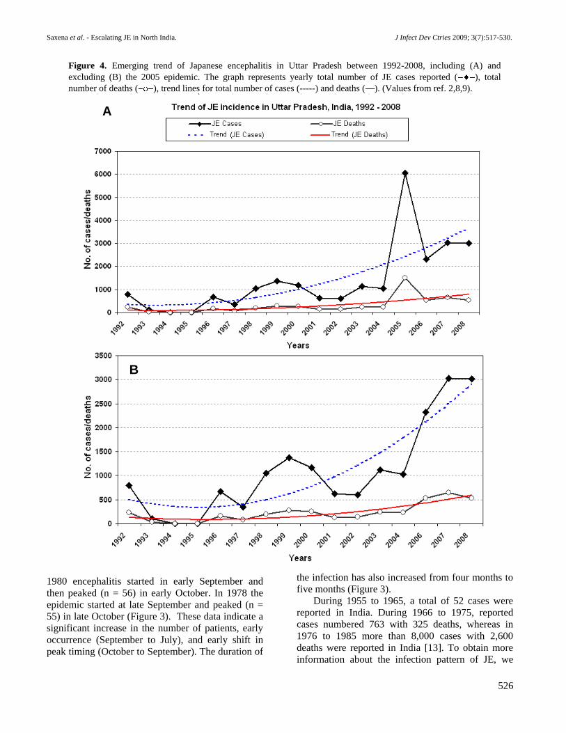
The pathological investigations revealed that out
of 38 cases, 55.3% (n=21) samples were positive for
JE (Table 1) when tested by rapid IgM capture
ELISA (JEV CheX) as described in materials and
methods. Notably, during the epidemic of 2005,
WHO reported only 37% cases of encephalitis were
JE positive, which includes clinical as well as
laboratory confirmed cases both [2]. A serological
finding of our study is presented in Table 5. The
infection could be characterized by marked
pleocytosis in cerebrospinal fluid (90 76.9
cells/cubic mm), and peripheral leucocytosis (64.7
8.86% neutrophils) with mild anaemia (hemoglobin;
11.2 ± 3.25 g/dL). The data also exhibit alteration in
lymphocyte (28.9 ± 8.4%) and polymorph (64.7 ±
8.86%). No significant difference was observed in
white blood cell count (8416.7 ± 1192.6 cells/ L),
eosinophil (1.95 ± 0.67%), reticulocyte count (1.92
± 0.56%) and blood glucose (95.4 ± 25.1mg/dL).
The CSF was collected within 2 to 7 days (4.2 ±
1.36 days) of onset of disease. Shifting of CSF
protein content (49.5 ± 30.1mg/dL) towards the
upper side of normal range (20.0-50.0 mg/dL) was
also observed. Mild anaemia and leucocytosis with
neutrophilia is one of the symptoms of JE infection.
All the 21 cases, which were found JE positive by
pathological investigations, were also confirmed to
be JE positive by the serological investigation (Table
5).
Phylogenetic analysis of GP05
The 2005 encephalitis epidemic surpassed all the
pervious epidemics in its intensity; therefore, we
sequenced GP05 and performed phylogenetic
analysis. Our results (shown in Figure 2) suggest
that the circulating strain GP05 was close to GP78
(the known endemic strain isolated in 1978) [7], and
all the Indian JEV strains clustered together and fell
in Genotype-III. The phylogenetic tree also
proposes that the JEV strain 057434 isolated in 2005
from Gorakhpur (Uttar Pradesh), and 04940-4
isolated in 2002 from Bhandara (Maharashtra), are
close to GP05. However, the JEV strain 014178
isolated in 2001 from Lakhimpur (Uttar Pradesh), is
distinct from all the above-mentioned strains and lies
between other 2005 strains and GP05.
Trend of JE occurrence
Since its first detection in India in 1955, JE has
shown an increasing trend in its occurrence;
however, it was not reported in Uttar Pradesh until
1978. There were two epidemics back to back in
Uttar Pradesh in 1978 and 1980 preceded by heavy
rainfall. We further compared the 2005 epidemic
with the previous epidemics of 1978 and 1980 to
predict the trend of JE epidemics in the area. In
2005, the encephalitis outbreak started in late July
and peaked (n = 2,554) in mid-September, while in
Clinical Characteristics No. of Patients (%)
Syndromes
Encephalitis
With weakness 7 33
Without weakness 6 28.6
Aseptic meningitis without encephalitis 6 28.6
Fever and headache 15 71.4
Signs and symptoms
Fever (temperature > 37.8) 21 100
Weakness 15 71.4
Vomiting 12 57.1
Headache 15 71.4
Altered mental status 10 47.6
Stiff Neck 10 47.6
Myalgia 4 19
Tremor 17 81
Slurred speech 16 76
Abdominal pain 4 19
Seizures 16 76
Altered sensorium 21 100
Table 4. Presentation of clinical features in patients (n = 21) with Japanese encephalitis in Uttar Pradesh (India), 2005.
Saxena et al. - Escalating JE in North India. J Infect Dev Ctries 2009; 3(7):517-530.
526
1980 encephalitis started in early September and
then peaked (n = 56) in early October. In 1978 the
epidemic started at late September and peaked (n =
55) in late October (Figure 3). These data indicate a
significant increase in the number of patients, early
occurrence (September to July), and early shift in
peak timing (October to September). The duration of
the infection has also increased from four months to
five months (Figure 3).
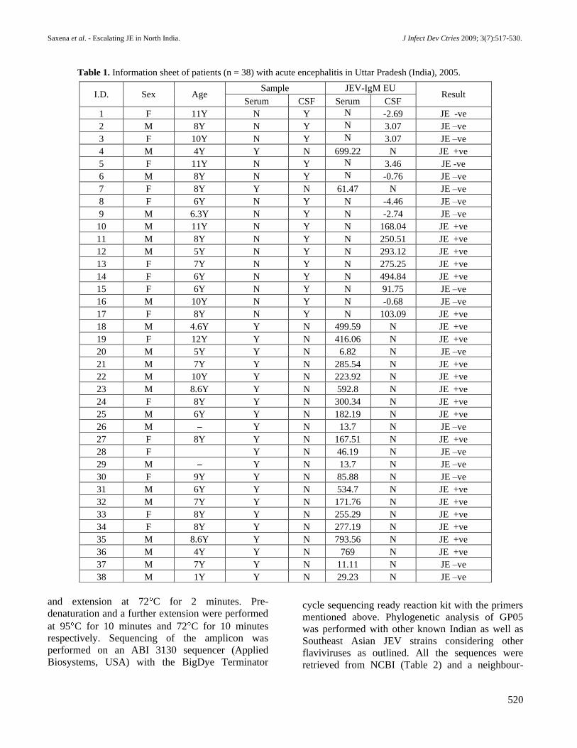
During 1955 to 1965, a total of 52 cases were
reported in India. During 1966 to 1975, reported
cases numbered 763 with 325 deaths, whereas in
1976 to 1985 more than 8,000 cases with 2,600
deaths were reported in India [13]. To obtain more
information about the infection pattern of JE, we
Figure 4. Emerging trend of Japanese encephalitis in Uttar Pradesh between 1992-2008, including (A) and
excluding (B) the 2005 epidemic. The graph represents yearly total number of JE cases reported ( ), total
number of deaths ( ), trend lines for total number of cases (-----) and deaths ( ). (Values from ref. 2,8,9).
A
B
Saxena et al. - Escalating JE in North India. J Infect Dev Ctries 2009; 3(7):517-530.
527
plotted a graph (Figure 4A) for the total number of
JE cases/deaths in Uttar Pradesh for seventeen years
(1992-2008) based on the numerical values available
from NICD and NVBDCP. Our results (presented in
Figure 4A) suggest that JE in North India is
escalating and larger epidemics may occur in the
future. We found a similar trend even when we
excluded data from the 2005 epidemic (Figure 4B).
Discussion Since the mid-1950s, JEV activity has been
reported in various parts of India, both in the form of
outbreaks of encephalitis and the level of antibodies
found in the population [7, 15]. JEV, a flavivirus, is
the major cause of encephalitis in Southeast Asia,
including India, with high mortality. Among all viral
encephalitis that is encountered in India, JE appears
to be of greater significance. Only one of every 300
persons infected with JEV develops clinical
encephalitis. The mortality in this disease has varied
from 20-40% in different parts of India. The
majority of the deaths occur during the first week of
illness. A deadly outbreak of undiagnosed brain
fever was reported in Uttar Pradesh and adjoining
areas during 2005. In intensity and magnitude, it
surpassed all the previously reported epidemics.
Our pathological investigations revealed marked
pleocytosis in cerebrospinal fluid and peripheral
leucocytosis with mild anaemia, which is in
accordance with earlier studies done by Kumar et al.
[16] in the same area. In CSF the elevated level of
cells and alteration of protein level indicates
penetration of the blood-brain barrier. The migration
of neutrophils at the site of injury may be attributed
to the production of soluble macrophage-derived
factor (MDF), which is one of the key mediators in
the host-innate immune response during JEV
infection [17]. The mild anaemia may be because a
result of MDF-induced hyperferretimia in serum
[18]. The malfunctioning of the liver and kidney
reported by Kumar et al. [16], may be caused by
replication of the virus in these organs [19].
Presence of virus in CSF from most of the fatal
cases might suggest that the virus is actively
multiplying in the central nervous system. This also
suggests that virus isolation or demonstration of CSF
is an indicator of poor prognosis because some of
the patients with low levels of IgM antibodies in
CSF/serum expired. The present findings are
comparable to the previous reports that isolation of
virus from CSF and low levels of JEV-specific IgG
and IgM in CSF and serum correlated significantly
with mortality [16].
Our demographic, clinical, pathological, and
serological investigations confirm the circulation of
JEV in Gorakhpur and adjoining areas. We have
earlier reported the prevalence of JEV-GP78 in the
affected area [7] and our present results (Figure 2)
reconfirm the presence of GP78 in North India,
especially in Uttar Pradesh during the JE outbreak of
2005. Clustering of JEV strains GP05 and 057434
(reported from another laboratory during the same
epidemic) with GP78 again concurs with our results
and also suggests that identification of GP05 by RT-
Variable Mean SD Range Normal Range
Blood
White -cell count (cells/ L) 8416.7 1192.6 6300 11000 3530 13050
Hemoglobin (g/dL) 11.2 3.25 6.0 17.5 11.5 18.0
Lymphocyte (%) 28.9 8.4 13.0 43.0 25.0 50.0
Polymorph (%) 64.7 8.86 50.0 83.0 30.0 60.0
Eosinophil (%) 1.95 0.67 1.0 3.0 0.3 5.0
Reticulocyte Count (%) 1.92 0.56 1.0 3.0 1.0 2.0
Blood glucose (mg/dL) 95.4 25.1 60.0 140.0 70.0 140.0
Cerebrospinal fluid
Interval between onset and
collection (days) 4.2 1.36 2 7
Cell count (per mm3) 90 76.9 10.0 260.0 < 4.0
Protein (mg/dL) 49.5 30.1 20.0 100.0 20.0 50.0
Glucose (mg/dL) 50.57 9.4 36.0 68.0 40.0 70.0
Table 5. Laboratory results of patients (n = 21) with Japanese encephalitis in Uttar Pradesh (India), 2005.

Saxena et al. - Escalating JE in North India. J Infect Dev Ctries 2009; 3(7):517-530.
528
PCR is real positivity and not laboratory
contamination during the PCR procedure. JEV strain
014178, isolated in 2001 from Lakhimpur (Uttar
Pradesh), caused high fatality in pediatrics claiming
443 cases with 96 deaths (CFR 21.7%) [20].
Therefore, it appears that further evolution of the
2001 strain may have caused the large JE outbreak
of 2005 with high fatality (CFR 22.9%). Similarly,
JEV strains 057434 and 04940-4 caused high fatality
and were also found to be closely associated with
GP05. Few JEV strains isolated from the same
epidemic differ from GP05 [21]. This may have two
possible explanations: (i) the co-circulation of JEV
GP05 with other already circulating strains in the
area, and (ii) the lack of proofreading activity of
viral RNA dependent RNA polymerase leading to a
mutation in viral genome during RNA replication
resulting in diverse evolution of 2001 JEV strain
(014178). If this is the case, the possibility of further
evolution/emergence of new JEV strains cannot be
excluded. The presence of JEV strain 04940-4 in
Bhandara, (Maharashtra State) in 2002 suggests that
JE is spreading to new areas, which is an alarming
concern.
Considering the large number of JE negative
results, it is important to understand that different
diseases of children affecting the brain and
sensorium and causing death should not be clubbed
together just because they occurred in the same time
period, assuming that all of them represented one
epidemic. There were a few cases with low platelet
counts with fever. This clinical picture was clearly
different from JE, and was most probably due to
Dengue [3,16]. Earlier studies have shown that in
addition to JEV few other serologically related
flaviviruses, such as Dengue and West Nile viruses,
are active in Uttar Pradesh [7]. Another reason for
the large number of non-JE patients may be the low
level of viremia and rapid development of
neutralizing antibody. It is very difficult to detect
genomic RNA or to isolate virus from serum
samples; hence IgM capture ELISA for JE antibody
or reverse transcription after virus culture are
recommended [21,22,23]. However, cross-reactivity
of antibody and mutation during culture passage and
reverse transcription are the main drawbacks. These
difficulties also emphasize the need for CSF
collection for epidemiological studies and clinical
diagnosis. Consequently, there is a need for effective
and efficient methods for diagnosis and
identification of JEV in patients.
Commencing in the last week of July, the 2005
encephalitis epidemic started about a month earlier
as compared to the previous epidemics of 1978 and
1980 (Figure 3). The early detection of JE could be
attributed to the improved surveillance/diagnoses,
but ecological issues such as seasonal drift also need
consideration. Even though the data points for the
1978 and 1980 epidemics are far less in comparison
to the 2005 epidemic, the graph suggests that further
investigation in the future will help us to understand
JE circulation kinetics for better management during
future epidemics. Interestingly, our graph (Figures
4A and 4B) suggests an escalating trend of JE year
by year in Uttar Pradesh and indicates that there
might be an even larger JE epidemic in the future.
This observation is in accordance with a recent
prediction of substantial risk of zoonotic and vector-
borne emerging infectious diseases (such as JE) in
North India, especially in Uttar Pradesh and
adjoining areas [24]. Deeper insights are required to
determine why JE is showing an intensifying trend
in North India. The affected area in North India is
known as the “Terai area.” Floods are an annual
feature in the region giving rise to water logging.
The warm, humid climate of the region provides an
excellent breeding ground for Culex
tritaeniorrhynchus and Culex vishnui mosquitoes,
which are vectors of JE. Therefore, during the rainy
season, an increase in the population density of the
mosquito in this division is observed. The area is
densely populated so mosquito-human contact is
very frequent. Villages in this rice-growing region
abound in stray and reared pigs. Studies from
peninsular and eastern parts of India indicate that
pigs are the main vertebrate host of the virus and the
major reservoir of the infection [25]. Pigs, besides
other animals, are widely prevalent in both rural and
urban areas of Uttar Pradesh. However,
epidemiological and ecological aspects of the illness
are yet to be studied in this part of the country. Due
to the evolution of new viral strains and/or
reemergence of older viral strains, children lack herd
immunity. Although health management facilities
are improved in the area, there is still a lack of
adequate resources and proper facilities for health
care and hygiene. These factors may be the
responsible for the intensifying trend of JE in North
India.
JE is still a major health problem in North India.
No proper antiviral against JEV is available
[26,27,28]. The following preventive measures may
largely mitigate the disease: proper sample

Saxena et al. - Escalating JE in North India. J Infect Dev Ctries 2009; 3(7):517-530.
529
collection; prediction of future strain(s); educating
public health workers and professionals;
vaccination; trials of new antiviral therapy;
improvement in clinical management, especially
early and specific detection; nutritional support to
the affected patients. Wider issues, including current
agricultural practices, water management systems,
and human behavioral patterns, need to be
investigated. Alterations in ecological issues such as
seasonal drift, which may cause shifts in the early
occurrence of the disease, should also be taken into
consideration. There is a need to monitor JE in birds,
mammals and its vector(s), along with human, to
obtain the proper information about climatic,
entomological, viral, and human host factors
affecting JE. The remedy lies in tackling the cause
and there is a need for a joint venture among health
officials, researchers, clinicians and ecologists who
can save lives by resolving the causative factors and
solve the escalating problem of JE in India. To this
end, a combination of strategies is required and we
need to proceed with a sense of urgency in this
matter.
Our results show that JEV infection led to
marked pleocytosis in CSF and peripheral
leucocytosis with mild anemia. All the JEV strains
reported in India fall under Genotype III. The JEV
strain circulating during 2005 was similar to GP78
and possibly originated due to the diverse evolution
of the 2001 JEV strain. JE is also spreading to newer
areas. The trend of JE suggested that the problem in
North India is escalating and larger epidemics may
occur in the future. Considering the possibility of a
larger epidemic in the future, serious steps should be
taken to combat JE, including the development of
more efficient surveillance methods and differential
diagnosis.
Gene Accession Number
The sequencing result leads to identification of 1524
bp ss-RNA (JEV GP05 envelope gene), which was
submitted in NCBI with FJ979830 accession number.
Acknowledgments The authors are grateful to the Council of Scientific
and Industrial Research (CSIR), India, and Dr. Lalji
Singh, Director, Centre for Cellular and Molecular
Biology, for his encouragement and support for this work.
We also thank Dr. Sunil K. Verma and Ms Ira Bhatnagar
for reviewing this manuscript. N.M. gratefully
acknowledges CSIR-JRF/NET research fellowship.
References 1. Webb JKG, Perriera S (1956) Clinical diagnosis of an
arthropod borne type virus encephalitis in children in north
Arcot district, Madras state, India. Indian J Med Sci 10:
573-580.
2. Immunization and Vaccine Development, Japanese
Encephalitis. World Health Organization: Regional Office
for South East Asia. Available:
http://www.searo.who.int/en/section1226/section2073.asp.
Accessed 09 February 2008.
3. Saxena SK, Singh M, Pathak AK, Mathur A (2006) Reply
to 'Encephalitis outbreak finds Indian officials unprepared'.
Nat Med 12: 269-270.
4. Government of Uttar Pradesh, Official website. Available:
http://www.upgov.nic.in/. Accessed 09 February 2008.
5. Umenai T, Krzysko R, Bektimirov TA, Assaad FA (1985)
Japanese encephalitis: current worldwide status. Bull
World Health Organ 63: 625-631.
6. Mackenzie JS, Gubler DJ, Petersen LR (2004) Emerging
flaviviruses: the spread and resurgence of Japanese
encephalitis, West Nile and dengue viruses. Nat Med 10:
S98-109.
7. Mathur A, Chaturvedi UC, Tandon HO et al (1982)
Japanese encephalitis epidemic in Uttar Pradesh, India
during 1978. Indian J Med Res 75: 161-169.
8. Investigation reports, National Institute of Communicable
diseases. Available: http://nicd.org/
InvestigationReports.asp. Accessed 09 February 2009.
9. National Vector Borne Disease Control Programme.
Directorate General of Health services, Ministry of Health
and Family Welfare. Available: http://nvbdcp.gov.in/je-
cd.html. Accessed April 4, 2009.
10. Japanese Encephalitis (JEV CheX) ACTION SHEET
GENLisa. Available: http://keragen.co.in/PDFs/Genlisa/
JEV-CheX.pdf.
11. Ravi V, Desai A, Balaji M, Apte MP, Lakshman L,
Subbakrishna DK, Sridharan G, Dhole TN, Ravikumar BV
(2006) Development and evaluation of a rapid IgM capture
ELISA (JEV-Chex) for the diagnosis of Japanese
encephalitis. J Clin Virol 35: 429-434.
12. Jacobson JA, Hills SL, Winkler JL, Mammen M,
Thaisomboonsuk B, Marfin AA, Gibbons RV (2007)
Evaluation of three immunoglobulin M antibody capture
enzyme-linked immunosorbent assays for diagnosis of
Japanese encephalitis. Am J Trop Med Hyg 7: 164-168.
13. Namachivayam V, Umayal K (1982) Proceedings of
National Conference on Japanese Encephalitis. ICMR: 30-
33.
14. Nash D, Mostashari F, Fine A, Miller J, O'Leary D, Murray
K, Huang A, Rosenberg A, Greenberg A, Sherman M,
Wong S, Layton M; 1999 West Nile Outbreak Response
Working Group (2001) The outbreak of West Nile virus
infection in the New York City area in 1999. N Engl J Med
344:1807-1814.
15. Banerjee K, Sengupta SN, Dandawate CN, Tongaonkar SS,
Gupta NP (1976) Virological and serological investigations
of an epidemic of encephalitis which occurred at Bankura
district, West Bengal. Indian J Med Res 64: 121-130.
16. Kumar R, Tripathi P, Singh S, Bannerji G (2006) Clinical
features in children hospitalized during the 2005 epidemic
of Japanese encephalitis in Uttar Pradesh, India. Clin Infect
Dis 43: 123-131.

Saxena et al. - Escalating JE in North India. J Infect Dev Ctries 2009; 3(7):517-530.
530
17. Saxena SK, Singh A, Mathur A (2000) Antiviral effect of
nitric oxide during Japanese encephalitis virus infection. Int
J Exp Pathol 81: 165-172.
18. Bharadwaj M, Khanna N, Mathur A, Chaturvedi UC (1991)
Effect of macrophage-derived factor on hypoferraemia
induced by Japanese encephalitis virus in mice. Clin Exp
Immunol. 83:215-218.
19. Mathur A, Arora KL, Chaturvedi UC (1981) Congenital
infection of mice with Japanese encephalitis virus. Infect
Immun 34: 26-29.
20. Outbreak investigations, Japanese Encephalitis Division,
National Institute of Virology.
Available:http://www.niv.co.in/departments/Japanese_Ence
phalitis/outbreak_investigation.htm. Accessed 14 June
2009.
21. Parida M, Dash PK, Tripathi NK , Ambuj, Sannarangaiah
S, Saxena P, Agarwal S, Sahni AK, Singh SP, Rathi AK,
Bhargava R, Abhyankar A, Verma SK, Rao PV, Sekhar K
(2006) Japanese Encephalitis Outbreak, India, 2005. Emerg
Infect Dis 12: 1427-1430.
22. Gajanana A, Samuel PP, Thenmozhi V, Rajendran R
(1996) An appraisal of some recent diagnostic assays for
Japanese encephalitis. Southeast Asian J Trop Med Public
Health 27: 673-679.
23. Kabilan L, Vrati S, Ramesh S, Srinivasan S, Appaiahgari
MB, Arunachalam N, Thenmozhi V, Kumaravel SM,
Samuel PP, Rajendran R (2004) Japanese encephalitis virus
(JEV) is an important cause of encephalitis among children
in Cuddalore district, Tamil Nadu, India. J Clin Virol 31:
153-159.
24. Jones KE, Patel NG, Levy MA et al (2008) Global trends in
emerging infectious diseases. Nature 451: 990-993.
25. Kumar R, Mathur A, Kumar A, Sharma S, Saksena PN,
Chaturvedi UC (1988) Japanese encephalitis an important
cause of acute childhood encephalopathy in Lucknow,
India. Postgrad Med J; 64: 18-22.
26. Saxena, SK (2008): Japanese encephalitis: perspectives and
new development. Future Neurol 3: 515-521.
27. Saxena V, Dhole TN (2008) Preventive strategies for
frequent outbreaks of Japanese encephalitis in Northern.
India. J Biosci 33: 505-514.
28. Kabilan L, Rajendran R, Arunachalam N, Ramesh S,
Srinivasan S, Samuel PP, Dash AP (2004) Japanese
encephalitis in India: an overview. Indian J Pediatr 71: 609-
615.
Corresponding Author Dr. Shailendra K. Saxena
Laboratory of Infectious Diseases & Molecular Virology
Centre for Cellular and Molecular Biology (CSIR)
Uppal Road, Hyderabad 500 007 (AP)
India
Phone: +91-40-27192630 (Direct); 27160222 41 Ext.
2630
Fax: +91-40-27160591; 27160311
E-mail: [email protected]; [email protected]
Conflict of interest: No conflict of interest is declared.