Original Article The anti-ulcerative colitis effects of ... · PDF filespread of the injurious...
10
Int J Clin Exp Med 2015;8(11):21861-21870 www.ijcem.com /ISSN:1940-5901/IJCEM0014443 Original Article The anti-ulcerative colitis effects of Annona squamosa Linn. leaf aqueous extract in experimental animal model Rasha YM Ibrahim, Amal I Hassan, Eithar K AL-Adham Department of Radioisotopes, Nuclear Research Centre, Atomic Energy Authority, Egypt Received August 13, 2015; Accepted October 7, 2015; Epub November 15, 2015; Published November 30, 2015 Abstract: This study aimed to evaluate the anti-inflammatory effects of Annona squamosa (A. squamosa) leaf aque- ous extract against acetic acid induced colitis in rats with a trial to explore its use for the treatment of colon inflam- mation. Sprague Dawley rats weighing 180-200 g were used in this study. Treatment with A. squamosa extract at dose 300 mg/kg for 4 weeks counteracted acetic acid induced ulcerative colitis by a significant decrease (P<0.05) of colonic tissue of malondialdehyde (MDA) and significant increases of catalase (CAT), glutathione (GSH) and glu- tathione peroxidase (Gpx) compared to ulcerative colitis control group. Furthermore, induction of oxidative stress was observed in the colonic tissue through the levels of 8-hydroxy-2’-deoxyguanosine (8-OHdG) which significant increase in colonic tissue DNA by acetic acid. Moreover AA induced significant increase in serum interleukin-10 (IL10), tumor necrosis factor-α (TNF-α), transforming growth factor (TGF 1β), and C reactive protein (CRP) as com- pared to the control group. On the contrary, our results showed AA induced significant decrease of vascular endo- thelial growth factor (VEGF) and thyroid hormones triiodothyronin and thyroxin (T3 & T4) in installed group with AA as compared to control which significantly improved after treatment with A. squamosa leaf extract. Histopathological observation in our study confirmed the biochemical study. Thus, therapeutic method offer a sign to analyze further the effectiveness of A. squamosa as a unique agent for alleviating colitis. Keywords: Annona squamosa, natural remedies, ulcerative colitis, free radicals Introduction Ulcerative colitis (UC), Crohn’s disease (CD) are common diseases affecting large intestine and characterized by an inflammatory process with remissions and exacerbations. These disorders represent the inflammatory bowel disease (IBDS) [1]. In pediatric age group the disease presents by both intestinal and extra intes- tinal manifestations serious complications occur like anemia and stunted growth. Ulcerative Colitis activity index (PUCAI) was established to assess the activity of the dis- ease and therapeutic response of children [2]. Inflammation happens as a natural defense mechanism of the mammalian tissues against injuries in order to eliminate or restrict the spread of the injurious agents. A large number of medicinal plants throughout the world are attributed with having various medicinal prop- erties as they contain different classes of phy- tochemicals [3]. Many studies prove that every part of A. squa- mosa possess medicinal property [4]. Folkloric record reported the use of Annona squamosa as an insecticidal, an antitumor agent, anti- diabetic, antioxidant, anti-lipidemic and anti- inflammatory agent which has been character - ized due to the presence of the cyclic peptides. In addition, the crushed leaves were inhaled to overcome the hysteria and fainting spells, and they were also applied on the ulcers and wounds [5]. All parts of this plant (leaf, bark, shoot and roots) have various compounds of medicinal importance and hence were used in different kinds of health problems. The ulcer is a major health hazard both in terms of morbidity and mortality. Some results have observed that leaves of Annona species exhibit antioxidant activity in different in vitro models due to the
Transcript of Original Article The anti-ulcerative colitis effects of ... · PDF filespread of the injurious...

Int J Clin Exp Med 2015;8(11):21861-21870www.ijcem.com /ISSN:1940-5901/IJCEM0014443
Original Article The anti-ulcerative colitis effects of Annona squamosa Linn. leaf aqueous extract in experimental animal model
Rasha YM Ibrahim, Amal I Hassan, Eithar K AL-Adham
Department of Radioisotopes, Nuclear Research Centre, Atomic Energy Authority, Egypt
Received August 13, 2015; Accepted October 7, 2015; Epub November 15, 2015; Published November 30, 2015
Abstract: This study aimed to evaluate the anti-inflammatory effects of Annona squamosa (A. squamosa) leaf aque-ous extract against acetic acid induced colitis in rats with a trial to explore its use for the treatment of colon inflam-mation. Sprague Dawley rats weighing 180-200 g were used in this study. Treatment with A. squamosa extract at dose 300 mg/kg for 4 weeks counteracted acetic acid induced ulcerative colitis by a significant decrease (P<0.05) of colonic tissue of malondialdehyde (MDA) and significant increases of catalase (CAT), glutathione (GSH) and glu-tathione peroxidase (Gpx) compared to ulcerative colitis control group. Furthermore, induction of oxidative stress was observed in the colonic tissue through the levels of 8-hydroxy-2’-deoxyguanosine (8-OHdG) which significant increase in colonic tissue DNA by acetic acid. Moreover AA induced significant increase in serum interleukin-10 (IL10), tumor necrosis factor-α (TNF-α), transforming growth factor (TGF 1β), and C reactive protein (CRP) as com-pared to the control group. On the contrary, our results showed AA induced significant decrease of vascular endo-thelial growth factor (VEGF) and thyroid hormones triiodothyronin and thyroxin (T3 & T4) in installed group with AA as compared to control which significantly improved after treatment with A. squamosa leaf extract. Histopathological observation in our study confirmed the biochemical study. Thus, therapeutic method offer a sign to analyze further the effectiveness of A. squamosa as a unique agent for alleviating colitis.
Keywords: Annona squamosa, natural remedies, ulcerative colitis, free radicals
Introduction
Ulcerative colitis (UC), Crohn’s disease (CD) are common diseases affecting large intestine and characterized by an inflammatory process with remissions and exacerbations. These disorders represent the inflammatory bowel disease (IBDS) [1]. In pediatric age group the disease presents by both intestinal and extra intes- tinal manifestations serious complications occur like anemia and stunted growth. Ulcerative Colitis activity index (PUCAI) was established to assess the activity of the dis-ease and therapeutic response of children [2].
Inflammation happens as a natural defense mechanism of the mammalian tissues against injuries in order to eliminate or restrict the spread of the injurious agents. A large number of medicinal plants throughout the world are attributed with having various medicinal prop-
erties as they contain different classes of phy-tochemicals [3].
Many studies prove that every part of A. squa-mosa possess medicinal property [4]. Folkloric record reported the use of Annona squamosa as an insecticidal, an antitumor agent, anti- diabetic, antioxidant, anti-lipidemic and anti-inflammatory agent which has been character-ized due to the presence of the cyclic peptides. In addition, the crushed leaves were inhaled to overcome the hysteria and fainting spells, and they were also applied on the ulcers and wounds [5]. All parts of this plant (leaf, bark, shoot and roots) have various compounds of medicinal importance and hence were used in different kinds of health problems. The ulcer is a major health hazard both in terms of morbidity and mortality. Some results have observed that leaves of Annona species exhibit antioxidant activity in different in vitro models due to the

Natural remedies
21862 Int J Clin Exp Med 2015;8(11):21861-21870
presence of flavanoids like rutin and hypero-side [6, 7].
The altered oxidant/antioxidant status in inflamed colon has received attention in both human and animals. Evidence suggests that reactive oxygen species (ROS) are produced in excess in the inflamed mucosa and may be pathogenic in inflammatory ulcerative disease [8]. The main sources of ROS in inflamed muco-sa are activated phagocytic leucocytes and neutrophils, capable of producing superoxide and cascade of various reactive species lead-ing to very reactive hydroxyl radicals and perox-ide. These products cause impairment in cell membrane stability and death of cells by lipid peroxidation in inflammatory ulcerative disease [9]. These intermediate products of oxygen metabolism (super oxide, hydroxyl radicals and H2O2) are controlled by various cellular defense mechanisms comprising enzymatic (superoxide dismutase and catalase) and non- enzymatic (GSH) scavenger component.
Protection versus free radicals can be achieved by preventing their formation, by blocking the chain reactions, or by repairing the oxidative damaged biomolecules [10]. The present study focuses on the inhibition of ulcerative colitis by A. squamosa leaf extract which might make it, enable to be a source of developing novel anti-ulcer medicinal product from nature. As well as, to achieve better control clinical and laboratory manifestations of the disease using a natural product to minimize the use of medications and to avoid their side effects especially with the long term use in pediatric age group.
Materials and methods
Preparation of plant extract
Leaves of A. squamosa were collected during December 2013 from the medicinal garden of Ghabor Farm. These were washed with water and 50 g of fresh leaves (kept at 25°C for 5 days in absence of sunlight) were extract-ed in 1 L of boiling water for 2 h and concen-trated the volume by boiling in a water bath. The brown extract thus obtained was cooled, filtered using Whatman No. 1 filter paper, and centrifuged at 10,000 rpm at 25°C. The super-natant was concentrated up to 100 ml on rota-ry vapor under reduced pressure. The lyophi-
lized concentrated crude extract was used for the study [11].
Determination of DPPH radical scavenging activity
A 1,1-diphenyl-2-picrylhydrazyl radical (DPPH) method was performed as described by Koleva et al. [12]. Approximately 10 μL of different con-centrations (15-500 μg/mL) of test sample solutions were added to 190 μL DPPH (150 μM) in ethanol solution. The solutions were vor- texed and incubated for 20 minutes at 37°C. The solvent alone acted as a blank. The decrease in absorbance of test mixtures (due to quenching of DPPH free radicals) was deter-mined as being 517 nm, and the percentage of inhibition was calculated. Ascorbic acid was used as a standard. The IC50 values were deter-mined as the concentration of the test mixture that gave 50% reduction in absorbance from that of the control blank.
Determination of total antioxidant activity (TTA)
The total antioxidant activity was evaluated using the method described by Prieto et al. [13]. An aliquot of sample solution/vitamin E (equivalent to 500 mg) was combined with reagent solution (0.6 M sulfuric acid, 28 mM sodium phosphate and 4 mM ammonium molybdate). The samples were then cooled at room temperature, and the absorbance was measured at 695 nm against the blank in a spectrophotometer. The total antioxidant activ-ity was expressed as gram equivalents of vita-min E.
Induction of experimental colitis
Twenty four hour fasted rats were lightly anes-thetized with diethyl ether inhalation. Colitis was induced by 4% acetic acid through a 1 ml sterile syringe coupled to a No. 6 polyethylene catheter, externally lubricated with lidocaine gel. The catheter was introduced anally to a dis-tance of 7 cm, in accordance with the standard method of this laboratory [14, 15]. The animals in the control group received an endoanal infu-sion of 1 ml normal saline solution.
Experimental design
Twenty-eight adult male (12 weeks old) albino rats, weighing 180-200 g, were used in the
Natural remedies
21863 Int J Clin Exp Med 2015;8(11):21861-21870
present study. They were housed in stainless steel cages, maintained at room temperature and provided with water and standard feed ad libitum.
After an adaptation period of one week, the rats were randomly divided into four equal groups (eight rats each). Group 1 (G1) sham group; without colitis induction and treated with extract vehicle (normal saline), GII AA group: treated with 4% AA colitis induction which con-tinued for 3 consecutive days, GIII extract groups treated with A squamosa extract orally (300 mg/kg) and GIV, rats were treated A squa-mosa extract (300 mg/kg) following by AA treatment.
Sampling
The proximal 5 cm of the dissected colon specimen was used for biochemical analysis of tissue catalase (CAT) according to Beers and Sizer [16]. Glutathione peroxidase (GPx) activi- ty was determined by using a colorimetric kit (BioVision, USA.). The serum samples were processed in a single batch using a commer-cially available sandwich VEGF ELISA detec- tion kit (R&D Systems, Minneapolis, MN) acc- ording to manufacturer instructions. The colon-ic samples were minced and homogenized using a polytron homogenizer. The supernatant was obtained by centrifuging at 3000 rpm for 20 minutes. Serum 8-hydroxy-deoxyguanosine (8-OHDG), reduced glutathione-GSH [17] and Malondialdehyde (MDA) level MDA was estimat-ed by Satoh [18]. 75 mg of Thiobarbituric acid (TBA) was dissolved in 15% TCA, to this 2.08 ml of 0.2N HCl was added, the volume was made up to 100 ml using 15% TCA. 3.0 ml of this reagent was added to 0.75 mL of serum of the rats. The test tubes were kept in a boiling wa- ter bath for 15 minutes. They were cooled and centrifuged for 10 minutes at 10000 rpm. Ab- sorbance of the supernatant was read against the blank at 535 nm. The results were ex- pressed in nmol/mL of serum. Interleukin-10 (IL-10), tumor necrosis factor-α (TNF-α), and transforming growth factor beta (TGF 1β) test kits were obtained from R&D (USA). C-reactive protein (CRP) levels were determined using ELISA commercial kit (Quantikine M murine; R&D Systems, Minneapolis, MN). Serum levels of C-reactive protein (CRP) were quantified by a highly sensitive ELISA kit (Life Diagnostics, West Chester, UK). Besides, serum activities
of total T3 & T4 by radioimmunoassay kit pur-chased from (Diagnosttic Product Corporation (DPC), 96th St., Los Angeles).
Ethic statement
The study was conducted accordance with the guidelines set by the European Economic Community (EEC) regulations (Revised Directive 86/609/EEC) and approved by the Ethical Committee at National Center for Radiation Research, Egyptian Atomic Energy Authority, Cairo, Egypt (NCRR-EAEA).
Histological examination
Autopsy samples were taken from the aorta of rats in different groups and fixed in 10% formal saline for twenty four hours. Washing was done in tap water then serial dilutions of alcohol (methyl, ethyl and absolute ethyl) were used for dehydration. Specimens were cleared in xylene and embedded in paraffin at 56 degrees in hot air oven for twenty four hours. Paraffin bees wax tissue blocks were prepared for sectioning at 4 microns thickness by slide microtome. The obtained tissue sections were collected on glass slides, deparaffinized, and stained by hematoxylin & eosin stain for examination through the light electric microscope [19].
Immunohistochemical study: proliferating cell nuclear antigen (PCNA)
Colonic tissue was immediately placed in 4% paraformaldehyde solution for histological stu- dies. Evaluation of epithelial cell proliferation was performed by immunohistochemistry us- ing a mouse monoclonal anti-PCNA antibody (Santa Cruz Corporation, USA). PCNA index were recorded by the image analyzer (Leica Q500 MC program) in the Histology Department, Faculty of Medicine, Ain Shams University.
PCNA indexTotal number of nuclei/cryptsPCNA p sitive nuclei/crypts
100= #q
Total number of nuclei/crypts.
Statistical analysis
The data analysis was carried out with SPSS Inc. software (version 22). One-way ANOVA was used to study a significant difference between means of the experimental groups with a sig-nificance level of P<0.05. Tukey-Kramer’s test was used to compare the significance among
Natural remedies
21864 Int J Clin Exp Med 2015;8(11):21861-21870
the rat groups. All data are presented as ± standard error of means (SE).
Results
DPPH free radical scavenging
The results pertaining to the free radical scav-enging activity of the two different extracts, along with the standard reference vitamin C. The model of stable DPPH free radicals can be used to evaluate the antioxidant activity in a relatively short time. The concentration of the sample necessary to reduce the initial concen-tration of DPPH by 50% (IC50) under the experi-mental conditions was determined. A lower value of IC50 indicates higher antioxidant activi-ty. The aqueous extract showed comparable levels of free radical scavenging activity with an IC50 value of (157.2 μg/mL).
Total antioxidant
The total antioxidant activity of aqueous ex- tracts was 189 μg, against the standard α-to- copherol.
In vivo results
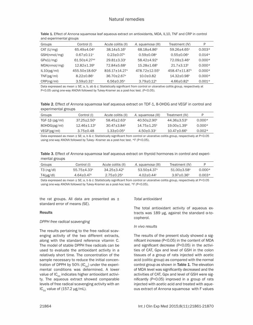
The results of the present study showed a sig-nificant increase (P<0.05) in the content of MDA and significant decrease (P<0.05) in the activi-ties of CAT, Gpx and level of GSH in the colon tissues of a group of rats injected with acetic acid (colitic group) as compared with the normal control group as shown in Table 1. The elevation of MDA level was significantly decreased and the activities of CAT, Gpx and level of GSH were sig-nificantly (P<0.05) improved in a group of rats injected with acetic acid and treated with aque-ous extract of Annona squamosa with F values
Table 1. Effect of Annona squamosa leaf aqueous extract on antioxidants, MDA, IL10, TNF and CRP in control and experimental groups
Groups Control (I) Acute colitis (II) A. squamosa (III) Treatment (IV) PCAT (U/mg) 65.49±4.04a 38.14±5.16b 68.18±4.86b 59.26±4.65a 0.003*GSH(nmol/mg) 0.67±0.11a 0.23±0.07b 0.59±0.08a 0.55±0.06a 0.014*GPx(U/mg) 61.50±4.27ab 29.81±3.33c 58.42±4.92b 72.09±3.46a 0.000*MDA(nmol/mg) 12.82±1.39b 72.84±5.68a 15.28±1.68b 21.7±3.13b 0.000*IL10(pg/ml) 455.50±18.60b 816.17±14.27a 478.72±12.55b 458.47±11.87b 0.000*TNF(pg/ml) 8.22±0.86c 36.70±2.67a 10.0±0.82 14.32±0.98b 0.000*CRP(ng/ml) 3.59±0.31b 6.56±0.35a 3.79±0.12b 4.66±0.82b 0.001*Data expressed as mean ± SE; a, b, ab & c: Statistically significant from control or ulcerative colitis group, respectively at P<0.05 using one-way ANOVA followed by Tukey-Kramer as a post-hoc test. (P<0.05).
Table 2. Effect of Annona squamosa leaf aqueous extract on TDF-1, 8-OHDG and VEGF in control and experimental groupsGroups Control (I) Acute colitis (II) A. squamosa (III) Treatment (IV) PTGF-1β (pg/ml) 37.25±2.50b 58.45±2.63a 40.50±2.90b 44.36±3.53b 0.000*8OHDG(pg/ml) 12.46±1.13b 30.47±3.84a 14.75±1.25b 19.00±1.39b 0.000*VEGF(pg/ml) 3.75±0.48 1.33±0.05d 4.50±0.33c 10.47±0.66b 0.002*Data expressed as mean ± SE; a, b & c: Statistically significant from control or ulcerative colitis group, respectively at P<0.05 using one-way ANOVA followed by Tukey–Kramer as a post-hoc test. *F (P<0.05).
Table 3. Effect of Annona squamosa leaf aqueous extract on thyroid hormones in control and experi-mental groupsGroups Control (I) Acute colitis (II) A. squamosa (III) Treatment (IV) PT3 (ng/dl) 55.75±4.33a 34.25±3.42b 53.50±4.37a 51.00±3.58a 0.000*T4(µg/dl) 4.64±0.47c 2.75±0.25a 4.02±0.44c 3.97±0.36b 0.003*Data expressed as mean ± SE; a, b & c: Statistically significant from control or ulcerative colitis group, respectively at P<0.05 using one-way ANOVA followed by Tukey-Kramer as a post-hoc test. *F (P<0.05).

Natural remedies
21865 Int J Clin Exp Med 2015;8(11):21861-21870

Natural remedies
21866 Int J Clin Exp Med 2015;8(11):21861-21870
(68.75, 8.401, 19.95 and 5.33) respectively at P<0.05.
As shown in Table 2 which revealed significant elevation (P<0.05) in the concentrations of serum interleukin-10 (IL10), tumor necrosis factor-α (TNF-α), transforming growth factor (TGF 1β), C reactive protein (CRP) and 8-hydroxy-2’-deoxyguanosine (8-OHDG) in a group of rats injected with acetic acid as compared to the normal control group. It was noticed that the elevation in the concentration of these param-eters significantly (P<0.05) decreased in the group of rats treated with extract of Annona squamosa as compared with the colitic rats group. Our results showed AA induced signifi-cant decrease of vascular endothelial growth factor (VEGF) which significantly (P<0.05) in- creased in the group of rats treated with extract of Annona squamosa. On the other hand the levels of thyroid hormones triiodothyronine and thyroxin (T3 & T4) in an installed group with AA showed a significant decrease (P<0.05) as com-pared to control which significantly improved after treatment with A. squamosa leaves extract as shown in Table 3.
Histopathological findings
There was no histopathological alteration, and the normal histological structure of the mucosa
and sub-mucosa were recorded in the normal control group and the group administrated with A. squamosa leaves extract only as shown in Figure 1A, 1B. But in a group of rats injected with AA and induced colitis the sub-mucosal layer showed massive numbers of focal inflam-matory cells infiltration (Figure 1C). On the con-trary, the group of rats injected with AA and treated with A. squamosa leaves extract showed significant Protective effects on colon where Goblet cells formation were noticed in the lining mucosal epithelium (Figure 1D).
Immunohistochemistry for PCNA
Our results demonstrated that PCNA was posi-tively stained in cellular nucleus of colonic glands; particular in the lower third of the crypts (Figure 1E, 1F). Immunohistochemical staining for PCNA showed that PCNA labeled nuclei nearly reached up to the surface in many crypts in GII (Figure 1G). Moreover, the PCNA index showed significant increase compared to group I (Table 4). Positively stained nuclei for PCNA remained at the lower third of the crypts in GIII. The PCNA index showed non-significant increase compared to control group (Figure 1H; Table 4). By PCNA, positively stained nuclei were located in the lower third of the crypts. Besides, damaged cells have strongly positive PCNA immunohistochemical stain with irregu-lar in the shape of crypt. By PCNA, positively stained nuclei were located in the lower third of the crypts in G IV (Figure 1I, 1J). However, they appeared significant decrease compared to group II (Table 4). The percentage of PCNA-positive cells of colon mucosa in normal control group were 8.96±0.71, whereas the percent-age of PCNA-positive cells in AA control group were 38.51 ± 2.84, A squamosa-treated group was 13.0±1.58 (Table 4). Our results revealed that cell proliferation in colonic glands was increased in rats with experimental colitis, as determined by the biomarker, PCNA.
Figure 1. A. Photomicrograph of a section of colon of a control rat showing closely packed simple tubular colonic crypts extending down to muscularis mucosa (mu). The lamina propria appears filling the space between the colonic crypts and contains mononuclear cells. B. Photomicrograph of a section of the colon in group II (colitis). Some crypts show dilated lumen in their basal parts (↑). The sub-mucosal layer showed massive number of focal inflammatory cells infiltration. C. Annona squamosa group showing the luminal surface is lined by simple columnar cells with basal oval nuclei and apical brush border. Goblet cells predominate in the glands and show basal nuclei. D. The group of rats injected with AA and treated with A. squamosa leaves extract. Goblet cells formation was noticed in lining mucosal epithelium. E, F. Immunohistochemical staining for PCNA in control colon. G. Immunohistochemical staining for PCNA in GII (AA group). H. Immunohistochemical staining for PCNA in GIII (Annona squamosa) only. I, J. Immunohistochemical staining for PCNA in GIV (AA+ Annona squamosa).
Table 4. PCNA index in control and experi-mental groupsGroup No Mean ± SE PI 5 8.96±0.71b 0.012II 5 38.51±2.84a 0.002III 5 10.40±1.33b 0.003IV 5 13.0±0.87b 0.002Data expressed as mean ± SE; a, b: Statistically signifi-cant from control or ulcerative colitis group, respectively at P<0.05 using one-way ANOVA followed by Tukey-Kram-er as a post-hoc test. P<0.05.

Natural remedies
21867 Int J Clin Exp Med 2015;8(11):21861-21870
Discussion
Many factors have been caused in the events of ulcerative colitis, such as neutrophil infiltra-tion and the over-production of proinflammato-ry mediators, including cytokines, and reactive oxygen species (ROS). The tissue injury pro-duced by neutrophils and macrophages has been attributed to their ability to release ROS, nitrogen metabolites, cytotoxic proteins, lytic enzymes and cytokines as well as their disrupt-ing effects on the epithelial integrity [20].
Acetic acid induced colitis model is similar to human ulcerative colitis in terms of histological features. It affects the distal colon portion and induces non-transmural inflammation, massive necrosis of mucosal and sub-mucosal layers, mucosal edema, neutrophil infiltration of the mucosa and sub-mucosal ulceration [21].
This study reported that the technique resulted in ulcerative colitis as was evident from the acetic acid administration. Annona squamosa Linn is a multi-target tree with edible fruits and is a source of the medicinal & industrial prod-ucts. Annona squamosa Linn is used as an antioxidant, anti-diabetics, hepato-protective, cytotoxic activity, gene toxicity, anti-tumor activ-ity, anti-lice agent. It is related to contain alka-loids, carbohydrates, stable oils, tannins & phe-nolic compounds. Earlier works on the phyto-chemisrty of Annona squamosa leaf, reported the presence of alkaloids, falvonoids, phenols, saponins, glycosides in water, methanol, chlo-roform, and petroleum ether extracts [22, 23].
The results of our study showed a significant increase (P<0.05) in the content of MDA and sig-nificant decrease (P<0.05) in the activities of CAT, Gpx and level of GSH in the colon tissues of a group of rats injected with acetic acid sug-gesting that AA may aggravate oxidative and inflammatory injury in rats induced colitis. In this study, A squamosa decreased oxidative damage compared with ulcerative colitis. There are multiple evidences suggesting the role of oxygen free radicals in colitis. CAT, GSH and GPx are an important antioxidant enzyme which may be used for the free radical destruction and may be prevented by the free radical spe-cies the results are consistent with William and Juneja [24] who hypothesize that aqueous extract of A. squamous may cause the allevia-tion of ROS generation through its effect on
these antioxidants in colon. This study also pro-vides an indication to further explore the anti-oxidative effects of A. squamosa species for alleviating chronic colitis. Annona squamosa L (Annonaceae family), having high content of nutritional compounds such as vitamin C, thia-mine, niacin, niacinamide, dietary fiber, potas-sium and iron in considerably significant amounts [25].
Compared with those of the control group, the levels of IL-10, TNF-a, CRP and the TGF-1B in the colonic tissues were significantly increased in the group of rats installed with AA, implying that AA may aggravate colonic damage by trig-gering the release of inflammatory factors in rats with colitis, the results in accordance with [26]. These inflammatory mediators significant-ly improved after treatment with an extract of Annona squamosa, this extract possesses cen-trally and peripherally mediated analgesic prop-erties. The central analgesic action may be mediated via inhibition of central pain recep-tors, while the peripheral analgesic effect may be mediated through inhibition of cyclooxygen-ase and/or lipoxygenase (and other inflamma-tory mediators). This hypothesis is in conso-nance with those of Wagner et al. [27] who pos-tulated that acetic acid writhing and hot plate test methods are useful techniques for the evaluation of centrally and peripherally acting analgesic drugs, respectively.
Annona squamosa leaves was found to pos-sess significant analgesic and anti-inflammato-ry activity which is quite comparable to Pen- tozocine and Aceclofenac there by leading to believe in a mixed central and peripheral mech-anism for its action [28]. Furthermore a detail- ed investigation on the extract is underway to determine the phytoconstituents that are res- ponsible for these activities as well as to define the exact mechanism of action of the herbal drug.
The results indicate the leaf extract of Annona squamosa L enhance the plasma levels of thy-roid hormones and there is evidence that leaf extract of this plant has thyroid inhibitory and antiperoxidative in nature [29] which also effec-tive in proper functioning of carbohydrate as well as lipid metabolism [30].
PCNA had been used in the past in an attempt to measure the proliferative activity of odonto-

Natural remedies
21868 Int J Clin Exp Med 2015;8(11):21861-21870
genic epithelium cysts [28]. At late S phase, PCNA is outstanding within the nucleoli. PCNA is seen to be powerfully associated within the nuclear regions wherever DNA synthesis is hap-pening [31]. In the present study, PCNA expres-sion in the lower third of the crypts of colon was significantly increased in GII. This suggests that the highest proliferative activity is in the suprabasal cell layers [31, 32]. This could also reflect a unique epithelial differentiation pro-cess, in which the basal cells assume some characteristics of preameloblasts, which indi-cate that it might have entered to some extent towards ameloblast differentiation [33]. The appearance of differentiated cell in the basal layer probably accounts for the fact that the major proliferation compartment is supra-basally. Immunohistochemical study detected decrease in the number of PCNA positive in group (IV) which indicated a decrease in the cell division in the crypt region. While, there was increase in the number of PCNA positive nuclei in group (II). The term of proliferative antigens is correlated with various degrees of epithelial dysplasia and inflammatory changes in biopsy specimens from patients with long-standing ulcerative colitis [34]. In the present work, A. squamosa at either treatment level significantly reduced the increase in the percentages of PCNA-positive cells seen in rats treated with AA.
The products of lipid peroxidation, particularly the cytotoxic aldehydes like malondialdehyde (MDA), are important because they can also induce damage DNA [35]. A highly active hydroxyl radical can break the DNA threads cre-ating (8-OHdG). Thus, this DNA damage may compromise gene expression [36]. More- over, the 8-OHdG adduct was determined as a marker of oxidative DNA harm because this lesion is a frequently found adduct in mutated oncogenes and tumor suppressor genes [37]. Thus, our observation of the increased oxida-tive DNA damage in the colon is highly signifi-cant, and could lead to serious consequences, if unrepaired.
Histological examination showed that mucosa and submucosa were the main sites of colonic injury, with abundant inflammatory cell infiltra-tion, local erosion and ulcer formation com-pared with those in the control group, while a group of rats treated with leaf extracts of Annona squamosa Goblet cells formation were
noticed in the lining mucosal epithelium these cells are mucus comprises an important barrier that prevents bacteria and other inflammatory agents from invading the mucosa [38]. Goblet cells are reduced in number and size in ulcer-ative colitis. Recent studies in mouse models of colitis focus the significance of the role of mucin in preserving the integrity of protective mucus barriers which breakdown can result in colitis [39].
Medical treatment of ulcerative colitis in pedi-atric population includes anti-inflammatory therapy, corticosteroids and immunomodulato-ry agents.
Although medications can achieve a good laboratory and clinical control yet, there are lim-iting factors to their use due to the adverse effects. Corticosteroids are effective in the acute exacerbation but on the long term side effects appear like osteoporosis, compression fractures, weight gain, cushingoid appearance restlessness as well as personality changes and emotional liability [40]. Immunomodulatory agents as purine are useful in treatment of ulcerative colitis yet they have serious draw backs as pancreatitis, hepatitis and bone mar-row suppression. Patients should be monitored for leukopenia. Treatment of fulminant cases includes cyclosporine tacrolimus and infliximab are nephrotoxic causing irreversible renal dam-age and insufficiency. Tacrolimus although it is a potent inhibitor of T-lymphocytes but, still it is nephrotoxic, causes bone marrow suppression and neurological symptoms [41].
In children elective colectomy is indicated in severe cases with growth retardation and mal-nutrition when dysplasia is detected which may causes malignancy [42]. So, the need for a nat-urally occurring anti-ulcer medicinal product as Annona squamosal leaves extract in an impor-tant issue especially in pediatric patients to avoid the adverse effect of anti-inflammatory. immunomodulatory drugs and steroids on the long term.
Conclusion
Ulcerative colitis is a chronic and inflammatory disease caused by the inflammation and sores in the lining of large intestine. Annona squamo-sa leaves extract have potential anti-ulcer activity and can be used as anti -ulcer medici-

Natural remedies
21869 Int J Clin Exp Med 2015;8(11):21861-21870
nal product from nature. However, inhibition of ulceration events by A. squamosa is suggestive of an important balance between antioxidant enzymes and anti-inflammatory cytokine products.
Acknowledgements
The authors are grateful to Dr. Adel M. Bakeer Kholoussy, Professor of Pathology, Faculty of Veterinary Medicine, Cairo University for his helping in the examination of the histopatho-logical slides and for his valuable comments.
Disclosure of conflict of interest
None.
Address correspondence to: Dr. Amal I Hassan, Department of Radioisotopes, Nuclear Research Centre, Atomic Energy Authority, 9 Ahmed El-Zayat St. Dokki, P.O. 12311, Giza, Egypt. E-mail: [email protected]
References
[1] Baumaqart DC, Sandborn WJ. Inflammatory bowel disease: clinical aspects and estab-lished and evolving therapies. Lancet 2007; 369: 1641-1657.
[2] Turner D, Otley AR, Mack D, Hyams J, de Bruijne J, Uusoue K, Walters TD, Zachos M, Mamula P, Beaton DE, Steinhart AH, Griffiths AM. Development, validation, and evaluation of a pediatric ulcerative colitis activity index: a prospective multicenter study. Gastroentero- logy 2007; 133: 423-32.
[3] Saleem M TS, Christina AJM, Chidambarana- than N, Ravi V, Gauthaman K. Hepatoprotective activity of Annona squamosa Linn. on experi-mental animal model. International Journal of Applied Research in Natural Products 2008; 1: 1-7.
[4] Soni H, Malik SS, Singhai AK. Evaluation of leaf of aqueous extract of MeOH extract of Aannon squamosa on cell viability American. J Pharm Tech Res 2012; 2: 936-944.
[5] Gajalakshmi S, Divya R, Deepika VD, Mythili S, Sathiavelu A. Pharmacological activities of an-nona squamosa. Int J Pharm Sci Rev Res 2011; 10: 24-29.
[6] Pélissler Y, Marion C, Kone DD, Lamaty G, Menut C, Besslere JM. Volatile components of Annonamuricata L. J Essent Oil Res 1994; 6: 411-414.
[7] Pandey N, Dushyant B. Phytochemical and Pharmacological review on Annona squamosa Linn. International Journal of Research in Ph-
armaceutical and Biomedical Sciences 2011; 2: 1404-1412.
[8] Grisham MB. Oxidants and free radicals in in-flammatory bowel diseases. Lancet 1994; 344: 859-861.
[9] Buffinton GD, Doe WF. Depleted mucosal anti-oxidant defenses in inflammatory bowel dis-ease. Free Radic Boil Med 1995; 19: 911-918.
[10] Halliwell B, Gutteridge JM. Free radicals in biol-ogy and medicine. 3rd edition. Oxford University Press; 1999. pp. 140-63.
[11] Gupta RK, Kesari AN, Watal G, Murthy PS, Chandra R, Maithal K. Hypoglycaemic and an-tidiabetic effect of aqueous extract of leaves of Annona squamosa (L.) in experimental animal. Curr Sci 2005; 88: 1244-54.
[12] Koleva II, Niederlander HA, van Beek TA. An on-line HPLC method for detection of radical scav-enging compounds in complex mixtures. Anal Chem 2000; 72: 2323-2328.
[13] Prieto P, Pineda M, Aguilar M. Spectropho- tometric quantitation of antioxidant capacity through the formation of a phosphomolybde-num complex: specific application to the deter-mination of vitamin E. Analytical Biochemistry 1999; 269: 337-341.
[14] Campos FG, Waitzberg DL, Logulo AF, Cukier C, Soares S, Oliveira TS. Padronização técnica e histológica de colite experimental com ácido trinitrobenzenosulfônico (TNBS). Rev Hosp Clin Fac Med S Paulo 1997; 52: 180-86.
[15] Niu X, Fan T, Huang H, Zhang Y, Xing W. Protective effect of sanguinarine against ace-tic acid-induced ulcerative colitis in mice. Toxicol Appl Pharmacol 2013; 267: 256-65.
[16] Beers RF, Sizer IW. A spectrophotometric meth-od for measuring the breakdown of hydrogen peroxide by catalase. J Biol Chem 1952; 195: 133-40.
[17] Moron MA, Mannervick B. levels of glutathi-one, glutathione s-transferase activities in rat liver. Biochemical Et Biophysica Acta 1979; 582: 67-78.
[18] Satoh K. Serum lipid peroxide in cerebrovascu-lar disorders determined by a new colorimetric method. Clin Chim Acta 1978; 90: 37-43.
[19] Banchroft JD, Stevens A, Turner DR. Theory and practice of histological techniques. 4th edition. New York, London, San Francisco, and Tokyo: Churchill Livingstone; 1996.
[20] Pravda J. Radical induction theory of ulcerative colitis. World J Gastroenterol 2005; 11: 2371-2384.
[21] Randhawa PK, Kavinder S, Nirmal S, Amteshwar SL. A Review on Chemical-Induced Inflammatory Bowel Disease Models in Ro- dents. Korean J Physiol Pharmacol 2014; 18: 279-288.

Natural remedies
21870 Int J Clin Exp Med 2015;8(11):21861-21870
[22] Saha R. Pharmacognosy and pharmacology of Annona squamosa: A review. Int J Pharm Life Sci 2011; 2: 1183-1189.
[23] Xu RH. Annona squamosa Linn: cytotoxic activ-ity found in leaf extract against human tumor cell lines. Pak J Pharm Sci 2014; 27: 1559-1563.
[24] William GK, Juneja V. Medical nutrition therapy for renal disorder. In: Mahan KL, Stump ES, editors. Krause’s Food and Nutrition Therapy. 12th edition. Toronto: Elsevier Canada; 2008. pp. 921-48.
[25] Wang S, Ghazala HR, Guo H, Ahmed M, Ahmed M, Hassan SZ, Hassan A, Chen ZS, Xu RH. Annona squamosa Linn: Cytotoxic activity found in leaf extract against human tumor cell lines. Pak J Pharm Sci 2014; 27: 1559-1563.
[26] Ding H, MeiQ, Gan HZ, Cao LY, Liu XC, Xu JM. Gastroenterology report: effect of homocyste-ine on intestinal permeability in rats with ex-perimental colitis, and its mechanism. Gas- troenterol Rep 2014; 2: 215-20.
[27] Wagner H, Reiter M, Frest W, Drrogen NL. Zur chemie und pharmacologie des herzwirksame prinzips von Annona squamosa. Planta Med 1980; 40: 77-85.
[28] Singh DP, Brijeshkunvar M, Richa M. Anti-nociceptive and Anti-inflammatory Activity of Annon squamosa L. Leaf Extract in Mice and Rats. Res J Pharm Phytochem 2013; 4: 182-185.
[29] Panda S, Kar A. Squamosa leaf extract causing possible ammetioration of hyperthyroidism. Curr Sci 2003; 84: 1402-1404.
[30] Ganong WF. The thyroid gland in Review of medical Physiology. 17th edition. Philadelphia: Appleton and Lange; 1994. pp. 290-305.
[31] de Oliveira DM, de Souza Andrade ES, da Silveira MM, Camargo IB. Correlation of the ra-diographic and morphological features of the dental follicle of third molars with incomplete root formation. Int J Med Sci 2008; 5: 36-40.
[32] Nairooz S, Ibrahim HS, Omar MM, Mohammad A. Structural changes of the colonic mucosa induced by orlistat: Experimental study. Egypt J Histol 2010; 33: 635-648.
[33] Browne RM. The odontogenic keratocyst 40 years on. Ann R Coll Surg Engl 1996; 78: 426-33.
[34] Maga G, Hübscher U. Proliferating cell nuclear antigen (PCNA): a dancer with many partners. J Cell Sci 2003; 116: 3051-3060.
[35] Barrera G. Oxidative Stress and Lipid Pe- roxidation Products in Cancer. Progression and therapy. ISRN Oncol 2012; 2012: 137289.
[36] Frederico B, Calegare A, Tufik S, D’Almeida V. DNA methylation and oxidative stress in sleep deprived rats. Sleep Sci 2012; 5: 84-88.
[37] Lee WL, Huang JY, Shyur LF. Phytoagents for Cancer Management: Regulation of Nucleic Acid Oxidation, ROS, and Related Mechanisms. Oxid Med Cell Longev 2013; 2013: 925804.
[38] Davis CW, Dickey BF. Regulated airway goblet cell mucin secretion. Annu Rev Physiol 2008; 70: 487-512.
[39] Young S, Kim YS, Ho SB. Intestinal Goblet Cells and Mucins in Health and Disease: Recent Insights and Progress. Curr Gastroenterol Rep 2010; 12: 319-30.
[40] Bressler B, Marshall JK, Bernstein CN, Bitton A, Jones J, Leontiadis GI, Panaccione R, Stein-hart AH, Tse F, Feagan B; Toronto Ulcerative Colitis Consensus Group. Clinical practice guidelines for the medical management of nonhospitalized ulcerative colitis: the Toronto consensus. Gastroenterology 2015; 148: 1035-1058.
[41] Kam L. Ulcerative colitis in young adults. Com-plexities of diagnosis and management. Post-grad Med 1998; 103: 45-49.
[42] Ziring DA, Wu SS, Mow WS, Martín MG, Mehra M, Ament ME. Oral tacrolimus for steroid-de-pendent and steroid-resistant ulcerative colitis in children. J Pediatr Gastroenterol Nutr 2007; 45: 306-311.