Original Article Synthesis, radiolabelling and in vitro in vivo
21
Introduction Positron emission tomography (PET) is a power- ful non-invasive imaging technique which allows quantification of biochemical and pharmacody- namic processes in healthy and diseased states. In drug discovery and development, PET offers opportunities to investigate drug-target interactions in vivo and to monitor the effects of a drug candidate on the progression of a dis- ease [1, 2]. The design of PET probes which selectively target a particular receptor, enzyme or transporter is a major challenge in further advancement of PET technique. Besides ana- logues of endogenous substrates such as [ 18 F]- fluorodeoxyglucose ([ 18 F]-FDG) or [ 11 C]- or [ 18 F]- labelled amino acids [3, 4], small synthetic or- ganic molecules are emerging as promising scaffolds for the development of highly selective PET probes. Metabotropic glutamate receptor subtype 5 (mGluR5) is a seven transmembrane domain, G- protein coupled receptor, belonging to group I of the metabotropic glutamate receptors and it is mainly located postsynaptically [5-7]. mGluR5 is involved in long-term potentiation processes [8] and plays a role in several disorders of central nervous system (CNS) such as schizophrenia, depression, anxiety, Alzheimer and Parkinson’s Am J Nucl Med Mol Imaging 2012;2(1):14-28 www.ajnmmi.us /ISSN:2160-8407/ajnmmi1111002 Original Article Synthesis, radiolabelling and in vitro and in vivo evaluation of a novel fluorinated ABP688 derivative for the PET imag- ing of metabotropic glutamate receptor subtype 5 Selena Milicevic Sephton 1 , Patrick Dennler 1 , Dominique S. Leutwiler 1 , Linjing Mu 2 , Cindy A. Wanger- Baumann 1 , Roger Schibli 1 , Stefanie D. Krämer 1 , Simon M. Ametamey 1 1 Center for Radiopharmaceutical Sciences of ETH, PSI and USZ, Department of Chemistry and Applied Biosciences of ETH Zurich, Wolfgang-Pauli Strasse 10, 8093 Zurich, Switzerland; 2 Center for Radiopharmaceutical Sciences ETH-PSI -USZ, Department of Nuclear Medicine, University Hospital Zurich, Switzerland. Received November 6, 2011; accepted November 20, 2011; Epub December 10, 2011; Published January 1, 2012 Abstract: (E)-3-(Pyridin-2-ylethynyl)cyclohex-2-enone O-(2-(3- 18 F-fluoropropoxy)ethyl) oxime ([ 18 F]-PSS223) was evalu- ated in vitro and in vivo to establish its potential as a PET tracer for imaging metabotropic glutamate receptor subtype 5 (mGluR5). [ 18 F]-PSS223 was obtained in 20% decay corrected radiochemical yield whereas the non-radioactive PSS223 was accomplished in 70% chemical yield in a SN2 reaction of common intermediate mesylate 8 with potas- sium fluoride. The in vitro binding affinity of [ 18 F]-PSS223 was measured directly in a Scatchard assay to give Kd = 3.34 ± 2.05 nM. [ 18 F]-PSS223 was stable in PBS and rat plasma but was significantly metabolized by rat liver micro- somal enzymes, but to a lesser extent by human liver microsomes. Within 60 min, 90% and 20% of [ 18 F]-PSS223 was metabolized by rat and human microsome enzymes, respectively. In vitro autoradiography on horizontal rat brain slices showed heterogeneous distribution of [ 18 F]-PSS223 with the highest accumulation in brain regions where mGluR5 is highly expressed (hippocampus, striatum and cortex). Autoradiography in vitro under blockade conditions with ABP688 confirmed the high specificity of [ 18 F]-PSS223 for mGluR5. Under the same blocking conditions but us- ing the mGluR1 antagonist, JNJ16259685, no blockade was observed demonstrating the selectivity of [ 18 F]-PSS223 for mGluR5 over mGluR1. Despite favourable in vitro properties of [ 18 F]-PSS223, a clear-cut visualization of mGluR5- rich brain regions in vivo in rats was not possible mainly due to a fast clearance from the brain and low metabolic stability of [ 18 F]-PSS223. Keywords: mGluR5, PET imaging, [ 18 F]-PSS223, [ 11 C]-ABP688, [ 18 F]-FDEGPECO, autoradiography, microsome en- zymes
Transcript of Original Article Synthesis, radiolabelling and in vitro in vivo

Introduction Positron emission tomography (PET) is a power-ful non-invasive imaging technique which allows quantification of biochemical and pharmacody-namic processes in healthy and diseased states. In drug discovery and development, PET offers opportunities to investigate drug-target interactions in vivo and to monitor the effects of a drug candidate on the progression of a dis-ease [1, 2]. The design of PET probes which selectively target a particular receptor, enzyme or transporter is a major challenge in further advancement of PET technique. Besides ana-logues of endogenous substrates such as [18F]-
fluorodeoxyglucose ([18F]-FDG) or [11C]- or [18F]-labelled amino acids [3, 4], small synthetic or-ganic molecules are emerging as promising scaffolds for the development of highly selective PET probes. Metabotropic glutamate receptor subtype 5 (mGluR5) is a seven transmembrane domain, G-protein coupled receptor, belonging to group I of the metabotropic glutamate receptors and it is mainly located postsynaptically [5-7]. mGluR5 is involved in long-term potentiation processes [8] and plays a role in several disorders of central nervous system (CNS) such as schizophrenia, depression, anxiety, Alzheimer and Parkinson’s
Am J Nucl Med Mol Imaging 2012;2(1):14-28 www.ajnmmi.us /ISSN:2160-8407/ajnmmi1111002
Original Article Synthesis, radiolabelling and in vitro and in vivo evaluation of a novel fluorinated ABP688 derivative for the PET imag-ing of metabotropic glutamate receptor subtype 5 Selena Milicevic Sephton1, Patrick Dennler1, Dominique S. Leutwiler1, Linjing Mu2, Cindy A. Wanger-Baumann1, Roger Schibli1, Stefanie D. Krämer1, Simon M. Ametamey1 1Center for Radiopharmaceutical Sciences of ETH, PSI and USZ, Department of Chemistry and Applied Biosciences of ETH Zurich, Wolfgang-Pauli Strasse 10, 8093 Zurich, Switzerland; 2Center for Radiopharmaceutical Sciences ETH-PSI-USZ, Department of Nuclear Medicine, University Hospital Zurich, Switzerland. Received November 6, 2011; accepted November 20, 2011; Epub December 10, 2011; Published January 1, 2012 Abstract: (E)-3-(Pyridin-2-ylethynyl)cyclohex-2-enone O-(2-(3-18F-fluoropropoxy)ethyl) oxime ([18F]-PSS223) was evalu-ated in vitro and in vivo to establish its potential as a PET tracer for imaging metabotropic glutamate receptor subtype 5 (mGluR5). [18F]-PSS223 was obtained in 20% decay corrected radiochemical yield whereas the non-radioactive PSS223 was accomplished in 70% chemical yield in a SN2 reaction of common intermediate mesylate 8 with potas-sium fluoride. The in vitro binding affinity of [18F]-PSS223 was measured directly in a Scatchard assay to give Kd = 3.34 ± 2.05 nM. [18F]-PSS223 was stable in PBS and rat plasma but was significantly metabolized by rat liver micro-somal enzymes, but to a lesser extent by human liver microsomes. Within 60 min, 90% and 20% of [18F]-PSS223 was metabolized by rat and human microsome enzymes, respectively. In vitro autoradiography on horizontal rat brain slices showed heterogeneous distribution of [18F]-PSS223 with the highest accumulation in brain regions where mGluR5 is highly expressed (hippocampus, striatum and cortex). Autoradiography in vitro under blockade conditions with ABP688 confirmed the high specificity of [18F]-PSS223 for mGluR5. Under the same blocking conditions but us-ing the mGluR1 antagonist, JNJ16259685, no blockade was observed demonstrating the selectivity of [18F]-PSS223 for mGluR5 over mGluR1. Despite favourable in vitro properties of [18F]-PSS223, a clear-cut visualization of mGluR5-rich brain regions in vivo in rats was not possible mainly due to a fast clearance from the brain and low metabolic stability of [18F]-PSS223. Keywords: mGluR5, PET imaging, [18F]-PSS223, [11C]-ABP688, [18F]-FDEGPECO, autoradiography, microsome en-zymes

Development of novel mGluR5 PET tracer
15 Am J Nucl Med Mol Imaging 2012;2(1):14-28
disease [9-14]. It is considered an important future drug target and much attention has been given to the development of an optimal PET tracer for imaging of mGluR5 [15]. It was not until the development of synthetic mGluR5 antagonist 2-methyl-6-( phenylethynyl) pyridine (MPEP) [16, 17], which unlike amino acid derived ligands acts via the non-competitive mechanism and not at the con-served glutamate binding site, that significant research efforts were made. This led to the de-velopment of (E)-3-((6-methylpyridin-2-yl)ethynyl)cyclohex-2-enone-O-11C-methyl oxime ([11C]-ABP688, Figure 1) by the Ametamey group [18, 19] which is to date the most successful clini-cally applied mGluR5 PET tracer. Despite its excellent properties as a PET imaging agent in human subjects [19], [11C]-ABP688 does have one limitation and this is due to the short physi-cal half-life (20 min) of carbon-11 which limits its application to facilities with an on-site cyclo-tron. This has provoked further research efforts towards the development of 18F-labelled PET tracers, which have a longer physical half-life (109.8 min). To date, the two most successful mGluR5 18F-labelled PET tracers are 3-fluoro-5-(2-(2-[18F](fluoromethyl)-thiazol-4-yl)-ethynyl)benzonitrile ([18F]-SP203) developed by the Pike group [20-22] and 3-[18F]fluoro-5-(2-pyridinylethynyl)benzonitrile ([18F]-FPEB) from the Hamill group [23-25] (Figure 1). However, the first undergoes significant defluorination in rodents and monkeys and to a lesser extent in humans and the latter is typically obtained in
low radiochemical yields. Our group aimed to develop a fluorine-18 ana-logue of [11C]-ABP688 with the same excellent imaging properties as [11C]-ABP688, but with added advantage of a longer physical half-life. Several derivatives were prepared [26, 27] and evaluated which finally led to the development of (E)-3-(pyridin-2-ylethynyl)cyclohex-2-enone O-(2-(2-18F-fluoroethoxy)ethyl) oxime ([18F]-FDEGPECO) [28, 29], which maintained the same [11C]-ABP688 scaffold, but the oxime functionality was additionally adorned with a six-atom long lipophilic chain (as oppose to a methyl in [11C]-ABP688) [1, 2, 30, 31]. The in vivo evaluation of [18F]-FDEGPECO allowed visu-alization of mGluR5 in the rat brain, albeit with a lower relative accumulation and shorter resi-dence time in mGluR5-rich brain regions than observed for [11C]-ABP688, which prompted us to re-evaluate the structure of [18F]-FDEGPECO. The lipophilicity of [18F]-FDEGPECO is signifi-cantly lower than that of [11C]-ABP688 [18], and we chose to pursue an [18F]-FDEGPECO ana-logue (E)-3-(Pyridin-2-ylethynyl)cyclohex-2-enone O-(2-(3-18F-fluoropropoxy)ethyl) oxime ([18F]-PSS223, Figure 1) in which the side chain func-tionality is extended by one methylene group with the aim to increase the lipophilicity towards that of [11C]-ABP688 [28, 32]. In this manuscript we report on the synthesis, radiolabelling, in vitro and in vivo evaluation of this novel compound ([18F]-PSS223). The PET images of ([18F]-PSS223 are also compared with
Figure 1. Structures of MPEP and selected PET tracers for imaging of mGluR5.

Development of novel mGluR5 PET tracer
16 Am J Nucl Med Mol Imaging 2012;2(1):14-28
those obtained with [11C]-ABP688 and [18F]-FDEGPECO. Materials and methods General All reactions requiring anhydrous conditions were conducted in flame-dried glass apparatus under an atmosphere of inert gas. All chemicals and anhydrous solvents were purchased from Aldrich or ABCR and used as received unless otherwise noted. [3H]-ABP688 (2.405 GBq/mmol, 37 MBq/mL solution in EtOH) was ob-tained from AstraZeneca. 3-Ethynylcyclohex-2-enone, (E)-3-ethynylcyclohex-2-enone oxime and (E)-3-(pyridin-2-ylethynyl)cyclohex-2-enone oxime (5) were prepared as previously reported and the characterization data is in complete agreement with those previously reported [28, 33]. To obtain (E)-3-((6-Methylpyridin-2-yl)ethynyl)cyclohex-2-enone O-[11C]-methyl oxime ([11C]-ABP688) radiolabelling was performed using module system as previously described [18, 34]. Preparative chromatographic separations were performed on Aldrich Science silica gel 60 (35-75 μm) and reactions followed by TLC analysis using Sigma-Aldrich silica gel 60 plates (2-25 μm) with fluorescent indicator (254 nm) and visualized with UV or potassium permanganate. Infrared spectra were recorded on a JASCO FT/IR 6200 (OmniLab) spectrometer using a chloro-form solution of compound. 1H and 13C NMR spectra were recorded in Fourier transform mode at the field strength specified on Bruker Avance FT-NMR spectrometers. Spectra were obtained from the specified deuterated solvents in 5 mm diameter tubes. Chemical shift in ppm is quoted relative to residual solvent signals calibrated as follows: CDCl3 δH (CHCl3) = 7.26 ppm, δC = 77.2 ppm; (CD3)2SO δH (CD3SOCHD2) = 2.50 ppm, δC = 39.5 ppm. Multiplicities in the 1H NMR spectra are described as: s = singlet, d = doublet, t = triplet, q = quartet, quint. = quin-tet, m = multiplet, b = broad; coupling constants are reported in Hz. Numbers in parentheses following carbon atom chemical shifts refer to the number of attached hydrogen atoms as re-vealed by the DEPT spectral editing technique. Electrospray (ES) mass spectra (LRMS) were obtained with a Micromass Quattro micro API LC electrospray ionization and electrospray (ES)
mass spectra (HRMS) were obtained with a Bruker FTMS 4.7 T BioAPEXII spectrometer. Electron-impact (EI) and chemical ionisation (CI) mass spectra (LRMS and HRMS) were obtained with a Waters Micromass AutoSpec Ultima MassLynx 4.0 spectrometer. Ion mass/charge (m/z) ratios are reported as values in atomic mass units. Semi-preparative purification of radiolabelled material was performed on a Merck-Hitachi L6200A system equipped with Knauer variable wavelength detector and an Eberline radiation detector using a reverse phase column (C18 Phenomenex Gemini, 5 mm, 250x10 mm) and eluting with gradient: 0-5 min 5% aq. MeCN, 5-15 min 5-50% aq. MeCN, 15-30 min 50% aq. MeCN, 30-50 min, 50-90% aq. MeCN, 50-65 min, 65% MeCN at flow rate 5 mL/min. Analyti-cal HPLC samples were analyzed by Agilent HPLC 1100 system equipped with UV multi-wavelength detector and Raytest Gabi star ra-diation detector using reverse phase column (ACE 111-0546, C18, 3 mm, 50x4.6 mm) and eluting with 45% aq. MeCN at flow rate 1 mL/min. Samples for PBS, plasma and microsome stability were analyzed by Waters ultra-performance liquid chromatography (UPLCTM) system equipped with Berthold coincidence de-tector (FlowStar LB513) using UPLC column (Waters Acquity BEH C18, 1.7 mm, 50x2.1 mm) and eluting with gradient 0-70% aq. MeCN over 5 min at flow 0.7 mL/min or for microsome as-say with 30% aq. MeCN over 5 min at flow 0.7 mL/min. Bovine serum albumin was purchased from Acros Organics. Pooled human liver micro-somes, pooled liver microsomes from male Spargue Dawley rats (20 mg protein per mL) and NADPH regenerating system (A: 31 mM NADP+, 66 mM glucose-6-phosphate, 66 mM MgCl2; B: 40 U/mL glucose-6-phosphate dehy-drogenase) were obtained from BD Biosciences. Male Wistar rats were obtained from Charles River (Sulzfeld, Germany). Animal care and all experimental procedures were approved by the Cantonal Veterinary Office in Zurich, Switzer-land. The animals were allowed free access to food and water. Radiosynthesis (E)-3-(Pyridin-2-ylethynyl)cyclohex-2-enone O-(2-

Development of novel mGluR5 PET tracer
17 Am J Nucl Med Mol Imaging 2012;2(1):14-28
(3-18F-fluoropropoxy)ethyl) oxime ([18F]-PSS223): No-carrier-added [18F]-fluoride was produced via nuclear 18O(p, n)18F reaction from enriched 18O-water using IBA cyclone 18/9 cy-clotron and it was immediately trapped on a QMA cartridge (preconditioned with 0.5 M aq. K2CO3 (1x5 mL), then H2O (1x5 mL) and dried in air). The trapped [18F]-fluoride was eluted from the cartridge with 0.25% wt Kryptofix-222® solu-tion (1 mL) in basic (0.05% wt K2CO3) aq MeCN (75% vv) into a tightly closed reaction vial. The solvents were evaporated in vacuo (130 mbar) with gentle stream of N2 gas at 110 °C over 5 min. To the resulting solid residue anhydrous MeCN (1 mL) was then added and the mixture was azeotropically dried in vacuo (130 mbar) with gentle stream of N2 at 110 °C. To the dried Kryptofix-222®/[18F–] complex a solution of (E)-3-(2-(((3-(pyridin-2-ylethynyl)cyclohex-2-en-1-ylidene)amino)oxy)ethoxy)propyl methanesul-fonate (2.44 mg, 6.22 µmol) in anhydrous N,N`-dimethylformamide (0.3 mL) was added and the dark brown mixture was heated at 90 °C for 10 min. The crude mixture was diluted with 50% vv aq. MeCN (2 mL) and purified via semi-preparative HPLC. The desired product was col-lected (retention time: 31.9 min) and immedi-ately diluted with H2O (10 mL). The aqueous solution was passed through a C18 cartridge (preconditioned with EtOH (1x5 mL), then H2O (1x5 mL) and dried in air) and the cartridge was washed with H2O (2x1 mL) and the product was eluted from the C18 cartridge with EtOH (1x0.3 mL) in a sterile vial containing 50% aq. PEG200 (5 mL) to afford the radiolabelled title com-pound in 20% decay corrected yield. Typically starting from ca. 30 GBq of activity, 5-6 GBq of product was obtained. The radiochemical purity was >97% and specific activity in the range of 100-320 GBq/mmol. Determination of logD For purposes of logD determination a further product elution from the C18 cartridge was car-ried out using EtOH (0.3 mL) and the solvent was evaporated in vacuo (100 mbar) at 90 °C for 5 min. The determination of logD value was performed using the shake-flask method as previously re-ported [28, 35]. [18F]-PSS223 (453 MBq) was partitioned between phosphate buffer (pH 7.4) saturated with 1-octanol (2 mL) and 1-octanol saturated with phosphate (pH 7.4) buffer (2
mL). The octanol (top) phase (0.5 mL) was washed with phosphate (pH 7.4) buffer satu-rated with 1-octanol (2x0.5 mL). The purity of the material (14 MBq) was confirmed by HPLC analysis. Finally washed octanol phase (0.1 mL) was diluted with phosphate (pH 7.4) buffer satu-rated with 1-octanol (0.1 mL) and the two phases were shaken and radioactivity in each phase was measured in a -counter. Pharmacology Competition Binding Assay Brain membranes were prepared from Sprague Dawley rat brains as described previously [28]. Frozen membranes were thawed on ice and pelleted at 45000xg at 4 °C for 5 min. The membranes were washed twice with HEPES buffer (30 mM HEPES, 110 mM NaCl, 5 mM KCl, 2.5 mM CaCl2, 1.2 mM MgCl2, pH 8 at 4°C) and resuspended in HEPES buffer at a protein concentration of 1.3 mg/mL. The binding assay was performed as previously described [28]. In brief, brain membranes (0.1 mg protein) were incubated in triplicates at ambient temperature with 2 nM [3H]ABP688 and PSS223 at concen-trations between 10 pM and 100 M in a total volume of 0.2 ml HEPES. PSS223 was diluted from a 1 mM ethanolic (50%) solution. The cor-responding EtOH concentrations did not affect [3H]ABP688 binding (data not shown). Unspe-cific binding of [3H]ABP688 was estimated with 100 M MMPEP. After 45 min, the samples were filtrated and the filters containing the membranes with bound [3H]ABP688 were measured in a β-counter (Beckman LS6500). Bound [3H]ABP688 (B, pmol per mg protein) was fitted with Excel solver to Equation 1 to esti-mate IC50. B = Bmin + ((Bmax – Bmin)/(1 + (C/IC50))) Eq. 1 where C is the total PSS223 concentration, Bmax is the maximal B, i.e., the plateau in the B/C plot at low log C and Bmin is the minimal B, i.e., the plateau at high log C. The inhibition con-stant Ki of PSS223 was estimated from IC50 and Kd of ABP688 (1.7±0.2 nM) [18] with the Cheng-Prusoff equation. Saturation Binding Assay Brain membranes were incubated with [18F]-PSS223 at concentrations between 0.25 and

Development of novel mGluR5 PET tracer
18 Am J Nucl Med Mol Imaging 2012;2(1):14-28
100 nM as described above. Unspecific binding was determined with 100 M MMPEP. After filtration, bound [18F]-PSS223 was quantified in a g-counter (PerkinElmer, Wizard) and Kd was estimated according to Equation 2,
Bspec = (Bmax,spec x 1/Kd x Cu)/(1 + 1/Kd x Cu) Eq. 2 where Bspec is the difference between B and the unspecific binding (at 100 M MMPEP) and Cu is the unbound concentration, i.e. the difference between the total and the bound concentra-tions. Bmax,spec is the maximal Bspec at receptor saturation. Three independent experiments were performed with [18F]-PSS223 obtained from three independent radiolabelling produc-tions. In vitro autoradiography Frozen horizontal brain slices (20 mm) from a male Wistar rat (492 g) adsorbed to SuperFrost Plus slides were thawed at ambient tempera-ture and preincubated on ice for 10 min in HEPES buffer (see above) containing 0.1% bo-vine serum albumin (BSA). Excess solution was carefully removed and slides were incubated with 1 nM [18F]-PSS223 alone or together with 100 nM ABP688 or 100 nM JNJ16259685 in HEPES buffer for 45 min at ambient tempera-ture. After incubation, the solutions were de-canted and the slides washed on ice in HEPES buffer containing 0.1% BSA, and twice in HEPES buffer (3 minutes each) and finally dipped in H2O. Dried slides were exposed to a phosphor imager plate for 30 min and the plate was scanned in a BAS5000 reader (Fuji). Stability in PBS and plasma [18F]-PSS223 (7-8 MBq) was incubated in phos-phate buffer (4 mM KH2PO4/Na2HPO4, 155 mM NaCl, pH 7.4) or rat plasma at 37 °C for up to 2 h. At different time points, samples were diluted and enzymatic reactions stopped with ice cold MeCN (140 mL). Plasma samples were centri-fuged at 12000xg for 10 min. The samples were filtered and supernatants were analyzed by UPLC. Microsome stability assay [18F]-PSS223 (15 MBq) was preincubated for 5 min at 37 °C with 50 mL NADPH regenerating system A, 10 mL NADPH regenerating system B,
200 mL phosphate buffer (0.5 M, pH 7.4) and H2O in a total volume of 975 mL. Rat or human liver microsomes (25 mL) were added at a final protein concentration of 0.5 mg/mL at time 0. The mixture was incubated at 37 °C at 600 rpm and 100 mL aliquots were removed at the indi-cated time points and immediately quenched with the same volume of ice-cold MeCN. Pro-teins were precipitated at 12000xg (3 min) and the supernatants were filtered and analyzed by UPLC. NADPH regenerating system and micro-somes were replaced by water, respectively in two control experiments. PET scans Rats were immobilized by anaesthesia with 2-3% isoflurane in oxygen/air on a GE Vista ex-plore PET/CT scanner with the head in the field of view. Body temperature was controlled with a rectal probe connected to a 37 °C air blower and respiratory frequency was monitored with a 1025T Small Animal Monitoring and Gating Sys-tem from SA Instruments (Sony Brook, NY, USA). At the start of data acquisition 15-30 MBq [18F]-PSS223 or [11C]-ABP688 was injected into a tail vein and data were collected in list mode for 90 and 60 min, respectively. After the PET scan, a CT was performed for anatomical orientation. PET data were reconstructed with 2D ordered subset expectation maximization (2D OSEM) and analyzed with PMOD 3.2 (PMOD, Zurich, Switzerland). Results Synthesis and radiolabelling The syntheses of the cold reference compound PSS223 and the desired radiolabelled analogue were accomplished via key intermediate 8 which was envisioned to originate from oxime 5 via SN2 reaction with bromoether 4. Oxime 5 was prepared by a Sonogashira reaction [36] using commercially available 2-bromopyridine and (E)-3-ethynylcyclohex-2-enone oxime as previously reported [33] (Supporting data). Monoprotection of commercially available pro-panediol with TBSCl, followed by esterification of the free hydroxyl group afforded ester 3 in 40% yield. Ester 3 was then reduced to the cor-responding ether with triethylsilane [37] in a reaction catalyzed by indium(III)-bromide (Figure 2).

Development of novel mGluR5 PET tracer
19 Am J Nucl Med Mol Imaging 2012;2(1):14-28
Bromoethers 4a and 4b were reacted with oxime 5 to afford alcohol 7 after deprotection of silyl ethers with TBAF in 48% yield. Finally, me-sylation of 7 afforded the desired precursor 8 which was used for the syntheses of PSS223 and [18F]-PSS223 (Figure 2). In the presence of Kryptofix-222®, nucleophilic substitution of me-sylate 8 with potassium fluoride gave reference compound PSS223 in 70% yield. Under analo-gous conditions [18F]-PSS223 was obtained in 20% radiochemical decay corrected yield. The identity of [18F]-PSS223 was confirmed by HPLC analysis via co-injection with cold PSS223. In vitro evaluation The experimentally determined logDpH7.4 of [18F]-PSS223 was 1.89±0.05. The binding affinity of
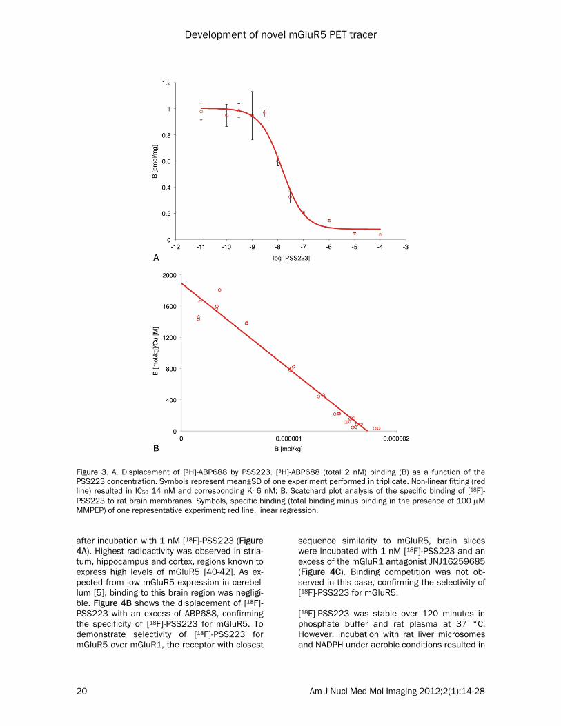
PSS223 was first estimated from a single com-petition binding experiment with 2 nM [3H]-ABP688 (Figure 3A). The estimated IC50 value was 14 nM and the respective Ki value amounted to 6 nM. The maximal displacement of [3H]-ABP688 by PSS223 was similar as ob-served with 100 M MMPEP, i.e., about 96% of total binding. Next, we determined Kd of [18F]-PSS223 in a saturation binding assay. Non-linear curve fitting of three independent experi-ments revealed a Kd of 3.34±2.05 nM and Bmax ranging from 2 to 6 pmol/mg. Linearization in the Scatchard plot confirmed a single binding site (Figure 3B). Non-specific binding was esti-mated with an excess of MMPEP [38, 39]. In vitro autoradiography with rat brain slices showed heterogeneous radioactivity distribution
Figure 2. Syntheses of PSS223 and [18F]-PSS223 from key intermediate 8 via SN2 reaction.
Development of novel mGluR5 PET tracer
20 Am J Nucl Med Mol Imaging 2012;2(1):14-28
after incubation with 1 nM [18F]-PSS223 (Figure 4A). Highest radioactivity was observed in stria-tum, hippocampus and cortex, regions known to express high levels of mGluR5 [40-42]. As ex-pected from low mGluR5 expression in cerebel-lum [5], binding to this brain region was negligi-ble. Figure 4B shows the displacement of [18F]-PSS223 with an excess of ABP688, confirming the specificity of [18F]-PSS223 for mGluR5. To demonstrate selectivity of [18F]-PSS223 for mGluR5 over mGluR1, the receptor with closest
sequence similarity to mGluR5, brain slices were incubated with 1 nM [18F]-PSS223 and an excess of the mGluR1 antagonist JNJ16259685 (Figure 4C). Binding competition was not ob-served in this case, confirming the selectivity of [18F]-PSS223 for mGluR5. [18F]-PSS223 was stable over 120 minutes in phosphate buffer and rat plasma at 37 °C. However, incubation with rat liver microsomes and NADPH under aerobic conditions resulted in
Figure 3. A. Displacement of [3H]-ABP688 by PSS223. [3H]-ABP688 (total 2 nM) binding (B) as a function of the PSS223 concentration. Symbols represent mean±SD of one experiment performed in triplicate. Non-linear fitting (red line) resulted in IC50 14 nM and corresponding Ki 6 nM; B. Scatchard plot analysis of the specific binding of [18F]-PSS223 to rat brain membranes. Symbols, specific binding (total binding minus binding in the presence of 100 M MMPEP) of one representative experiment; red line, linear regression.

Development of novel mGluR5 PET tracer
21 Am J Nucl Med Mol Imaging 2012;2(1):14-28
a significant decrease of [18F]-PSS223 and the appearance of a polar radiometabolite, co-
eluting with co-injected [18F]-fluoride. After one hour, about 90% of [18F]-PSS223 was metabo-
Figure 4. In vitro autoradiography with rat brain slices. A. Incubation with 1 nM [18F]-PSS223 showing heterogeneous distribution with highest expression in striatum, hippocampus and cortex; B. Brain slices incubated with 1 nM [18F]-PSS223 and 100 nM ABP688; C. Brain slices incubated with 1 nM [18F]-PSS223 and 100 nM mGluR1 antagonist JNJ16259685. Blue, green, orange, red, dark red indicate radioactivity from low to high. Columns represent same experiment performed in duplicate.

Development of novel mGluR5 PET tracer
22 Am J Nucl Med Mol Imaging 2012;2(1):14-28
lized under the chosen experimental conditions (Figure 5A). Incubation of [18F]-PSS223 with human liver microsomes under otherwise analo-gous conditions resulted in 20% decrease of [18F]-PSS223 after one hour (Figure 5B) and the generation of two polar radiometabolites, the more polar one co-eluting with [18F]-fluoride. None of the polar compounds were detected in the absence of microsomes or NADPH.
In vivo evaluation Dynamic PET scans were performed with two rats to determine whether [18F]-PSS223 could be used to image brain regions express-ing mGluR5. Figure 6A shows a transversal plane across the head region over time. In the early time frames, brain perfusion re-sulted in high homogenous radioactivity in the brain. This was followed by a rapid washout from all brain re-gions and radioactivity accu-mulation in the skull and jaws. The time activity curves of mGluR5-rich brain regions and cerebellum show accumulation of [18F]-PSS223 in striatum, hippo-campus and cortex (Figure 6B). However, the mean residence time was rela-tively short and radioactivity approached cerebellum lev-els already after about 20 minutes. High accumulation of 18F-radioactivity in bone is demonstrated by the time activity curve of the jaws. Figure 7 shows a compari-son of PET scans with [11C]-ABP688, [18F]-FDEGPECO and [18F]-PSS223, all aver-aged from 2 to 45 min after tracer injection. [18F]-PSS223 data are from the same scan shown in Figures 6A and B. The [18F]-FDEGPECO image was re-constructed from the data shown in our recent publica-
tion [29, 43]. Comparing the three tracers, [11C]-ABP688 showed the highest relative radioactiv-ity accumulation in mGluR5-rich brain regions such as hippocampus and striatum. [18F]-FDEGPECO also allows visualization of mGluR5-rich regions in the brain, however, at a higher background radioactivity. Our newest derivative, [18F]-PSS223 shows only weak accumulation in mGluR5-rich brain regions and significant accu-
Figure 5. A. Metabolism of [18F]-PSS223 by rat liver microsomes. Green symbols, [18F]-PSS223; blue symbols, polar 18F-radiometabolite; lines, fitted exponential functions; B. Metabolism of [18F]-PSS223 by human liver microsomes. Green symbols, [18F]-PSS223; blue and red symbols, polar 18F-radiometabolites.

Development of novel mGluR5 PET tracer
23 Am J Nucl Med Mol Imaging 2012;2(1):14-28
Figure 6. A. PET dynamic scan of the head of a male Wistar rat injected with [18F]-PSS223. Transverse planes through the head summarized for the indicated time windows. White, CT; B. Time activity curves of [18F]-PSS223 uptake in a male Wistar rat in different brain regions showing increasing uptake in jaw due to rapid defluorination.

Development of novel mGluR5 PET tracer
24 Am J Nucl Med Mol Imaging 2012;2(1):14-28
mulation of radioactivity in the skull and jaws as a result of rapid defluorination. Discussion Reference compound PSS223 was prepared in an overall satisfactory yield and all compounds en route were fully characterized (NMR, IR, MS). The synthesis of bromoether 4 proved to be more challenging than initially anticipated. After several unsuccessful attempts to obtain com-pound 4 via direct functionalization with diha-loethane, an alternative approach was em-ployed and yielded the desired bromoether 4 in 5% overall yield from three steps. During the reduction of the ester functionality in 3, the TBS silyl ether was partially substituted by the TES group (originating from the reagent triethylsi-lane) to afford a mixture of TBS:TES silylethers in a ratio of 2:1 (4a:4b, Figure 2). For our pur-
poses, it was not necessary to separate the silyl ethers 4a and 4b since they were removed to afford free hydroxyl group in the following step. The radiosynthesis of [18F]-PSS223 was accom-plished in a single step from mesylate precursor 8 reproducibly in a good radiochemical yield. Pure radiotracer was obtained after HPLC semi-preparative purification in a total radiosynthesis time of 90 min (from the end of bombardment). Experimentally determined logD value of [18F]-PSS223 was 0.2 log units higher than that of [18F]-FDEGPECO (1.7±0.1) [28] and 0.5 log units lower than that of [11C]-ABP688 [18]. This was within the optimal lipophilicity range of high affinity ABP688 analogues, (i.e., ClogP value below 2.5 and logP between 0.9 and 2.5, range of values desired for fast blood-brain barrier passage) [32]. One may assume that for those ABP688 analogues logDpH7.4 corresponds to
Figure 7. Rat brain PET images of [11C]-ABP688, [18F]-FDEGPECO and [18F]-PSS223. Planes (crosshairs) and maximal intensity projections (MIP) averaged from 2 to 45 min after tracer injection.

Development of novel mGluR5 PET tracer
25 Am J Nucl Med Mol Imaging 2012;2(1):14-28
logP as both the pyridine and the oxime nitro-gen atoms are non-protonated at pH 7.4. [18F]-PSS223 exhibited similarly high affinity for the mGluR5 as [11C]-ABP688, i.e., 3.3 nM com-pared to 1.7 nM [18], suggesting that the long chain in [18F]-PSS223 did not significantly affect the binding affinity. The value of Bmax deter-mined for [18F]-PSS223 (range: 2-6 pmol/mg) compared favourably to the Bmax values of [11C]-ABP688 (231±18 fmol/mg) [18] and [18F]-FDEGPECO (range: 0.5-4 pmol/mg) [28]. Spe-cific binding of [18F]-PSS223 to mGluR5-rich brain regions in the in vitro autoradiography experiments prompted further in vitro and in vivo characterization of [18F]-PSS223. [18F]-PSS223 was stable in buffer and rat plasma; however, it was significantly metabo-lized by the rat liver microsomal enzymes and a polar radiometabolite co-eluting with [18F]-fluoride was observed by UPLC. In the experi-ments with human liver microsomes, two polar radiometabolites were detected albeit in the amounts significantly lower than those observed with rat microsomal enzymes. Based on the control experiments, it was concluded that the process was NADPH-dependent, implying the involvement of oxidoreductases. One likely mechanism involves defluorination preceded by the oxygenation of the carbon atom in the α-position to the fluorine atom [44]. The possible mechanism is depicted in Figure 8. A lower de-gree of metabolic activity of the human com-pared to that of rat liver microsomes would be in agreement with several other studies [45-48].
Considering the rapid metabolism in vitro and the possibility of defluorination in vivo, we per-formed two dynamic PET scans. PET analysis demonstrated rapid wash-out of radioactivity from the brain and high accumulation in the skull and jaws. Accumulation of [18F]-PSS223 in mGluR5-rich brain regions was significantly lower than observed for [18F]-FDEGPECO. [18F]-PSS223 was therefore, not further investigated in vivo. The observed radioactivity accumulation in bone supports the conclusion that the radi-ometabolite (or one of the radiometabolites) observed with liver microsomes was [18F]-fluoride. Based on the higher stability of [18F]-PSS223 in human liver microsomes, less exten-sive defluorination is expected in humans. The difference in the in vivo behaviour between [18F]-PSS223 and [18F]-FDEGPECO could be at-tributed to the β-heteroatom effect [49, 50], by which primary aliphatic 18F-atoms in a β-position to heteroatom (e.g., [18F]-FCH2CH2OR) are found to be metabolized at a slower rate. This ration-ale supports absence of defluorination for [18F]-FDEGPECO. Conclusions In conclusion, the radiosynthesis of [18F]-PSS223 was accomplished in good radiochemi-cal yields and high specific radioactivity. The new radioligand binds mGluR5 in vitro with low nanomolar affinity and shows heterogeneous and specific accumulation in vitro in mGluR5-rich brain regions in autoradiographic studies. Although the PET studies with [18F]-PSS223 did
Figure 8. Likely mechanism of defluorination of [18F]-PSS223 involves cytochrome P450-catalyzed C-oxygenation. The aldehyde 9 is oxidized or reduced to the respective carboxylic acid or alcohol while [18F]-fluoride is accumulated in bone.

Development of novel mGluR5 PET tracer
26 Am J Nucl Med Mol Imaging 2012;2(1):14-28
not allow a clear-cut visualization of mGluR5 in vivo in the rat brain, due to low metabolic stabil-ity and rapid wash-out, [18F]-PSS223 could po-tentially find utility in higher animals considering that [18F]-PSS223 exhibited higher stability in human microsomes. Acknowledgements The authors would like to acknowledge Mrs. Claudia Keller and Mrs. Petra Wirth for perform-ing the PET scans. Mr. Bruno Mancosu is ac-knowledged for technical assistance with 11C-module. Prof. P. A. Schubiger, Mrs. Cindy Fischer and Dr. Thomas Betzel are acknowl-edged for support and many fruitful discussions. Experimental procedures and characterization data for all compounds, and HPLC chromato-graphs of [18F]-PSS223 are provided. Disclaimer of conflict of interest none. Address correspondence to: Dr. Simon M. Ametamey, Center for Radiopharmaceutical Sciences of ETH, PSI and USZ, Department of Chemistry and Applied Bio-sciences of ETH Zurich, Wolfgang-Pauli Strasse 10, 8093 Zurich, Switzerland; Phone: +41 44 6337463; Fax: +41 44 6331367. References [1] Ametamey SM, Honer M and Schubiger PA.
Molecular imaging with PET. Chem Rev 2008; 108: 1501-1516.
[2] Fowler JS, Volkow ND, Wang GJ, Ding YS and Dewey SL. PET and drug research and devel-opment. J Nucl Med 1999; 40: 1154-1163.
[3] Fowden L. Fluoroamino acids and protein synthesis. Ciba Foundation Symposium, Car-bon-Fluorine Compounds 1972; 2: 141-159.
[4] Weygand F and Oettmeier W. Fluorine-containing amino acids. Russian Chem Rev 1970; 39: 290-300.
[5] Masu M, Tanabe Y, Tsuchida K, Shigemoto R and Nakanishi S. Sequence and expression of a metabotropic glutamate receptor. Nature 1991; 349: 760-765.
[6] Pin JP and Duvoisin R. The metabotropic gluta-mate receptors - structure and functions. Neu-ropharmacology 1995; 34: 1-26.
[7] Tanabe Y, Masu M, Ishii T, Shigemoto R and Nakanishi S. A family of metabotropic gluta-mate receptors. Neuron 1992; 8: 169-179.
[8] Lu YM, Jia Z, Janus C, Henderson JT, Gerlai R, Wojtowicz JM and Roder JC. Mice lacking me-tabotropic glutamate receptor 5 show im-
paired learning and reduced CA1 long-term potentiation (LTP) but normal CA3 LTP. J Neu-rosci 1997; 17: 5196-5205.
[9] Chiamulera C, Epping-Jordan MP, Zocchi A, Marcon C, Cottiny CC, Tacconi S, Corsi M, Orzi F and Conquet FO. Reinforcing and locomotor stimulat effects of cocaine are absent in mGluR5 null mutant mice. Nat Neurosci 2001; 4: 873-874.
[10] Conn PJ and Pin JP. Pharmacology and func-tions of metabotropic glutamate receptors. Annu Rev Pharmacol 1997; 37: 205-237.
[11] Daggett LP, Sacaan AI, Akong M, Rao SP, Hess SD, Liaw C, Urrutia A, Jachec C, Ellis SB, Dreessen J, Knopfel T, Landwehrmeyer GB, Testa CM, Young AB, Varney M, Johnson EC and Velicelebi G. Molecular and functional characterization of recombinant human me-tabotropic glutamate receptor subtype 5. Neu-ropharmacology 1995; 34: 871-886.
[12] Dorri F, Hampson DR, Baskys A and Wojtowicz JM. Down-regulation of mGluR5 by antisense deoxynucleotides alters pharmacological re-sponses to application of ACPD in the rat hip-pocampus. Exp Neurol 1997; 147: 48-54.
[13] Ohnuma T, Augood SJ, arai H, McKenna PJ and Emson PC. Expression of the human exci-tatory amino acid transporter 2 and me-tabotropic glutamate receptors 3 and 5 in the prefrontal cortex from normal individuals and patients with schizophrenia. Mol Brain Res 1998; 56: 207-217.
[14] Rouse ST, Marino MJ, Bradley SR, Awad H, Wittmann M and Conn PJ. Distribution and roles of metabotropic glutamate receptors in the basal ganglia motor circuit: implications for treatment of Parkinson's disease and re-lated disorders. Pharmacol Ther 2000; 88: 427-435.
[15] Mu L, Schubiger PA and Ametamey SM. Radio-ligands for the PET imaging of metabotropic glutamate receptor subtype 5 (mGluR5). Curr Top Med Chem 2010; 10: 1558-1568.
[16] Gasparini F, Lingenhohl K, Stoehr N, Flor PJ, Heinrich M, Vranesic I, Biollaz M, Allgeier H, Heckendorn R, Urwyler S, Varney MA, Johnson EC, Hess SD, Rao SP, Sacaan AI, Santori EM, Velicelebi G and Kuhn R. 2-Methyl-6-(phenylethynyl)-pyridine (MPEP), a potent, selective and systemically active mGluR5 re-ceptor antagonist. Neuropharmacology 1999; 38: 1493-1503.
[17] Ritzen A, Mathiesen JM and Thomsen C. Mo-lecular pharmacology and therapeutic pros-pects of metabotropic glutamate receptor allosteric modulators. Basic Clin Pharmacol 2005; 97: 202-213.
[18] Ametamey SM, Kessler LJ, Honer M, Wyss MT, Buck A, Hintermann S, Auberson YP, Gasparini F and Schubiger PA. Radiosynthesis and pre-clinical evaluation of 11C-ABP688 as a probe for imaging the metabotropic glutamate recep-

Development of novel mGluR5 PET tracer
27 Am J Nucl Med Mol Imaging 2012;2(1):14-28
tor subtype 5. J Nucl Med 2006; 47: 698-705. [19] Ametamey SM, Treyer V, Streffer J, Wyss MT,
Schmidt M, Blagoev M, Hintermann S, Auber-son Y, Gasparini F, Fischer UC and Buck A. Human PET studies of metabotropic gluta-mate receptor subtype 5 with 11C-ABP688. J Nucl Med 2007; 48: 247-252.
[20] Brown AK, Kimura Y, Zoghbi SS, Simeon FG, Liow JS, Kreisl WC, Taku A, Fujita M, Pike VW and Innis RB. Metabotropic glutamate subtype 5 receptors are quantified in the human brain with a novel radioligand for PET. J Nucl Med 2008; 49: 2042-2048.
[21] Shetty HU, Zoghbi SS, Simeon FG, Liow JS, Brown AK, Kannan P, Innis RB and Pike VW. Radiodefluorination of 3-fluoro-5-(2-(2-[18F](fluoromethyl)-thiazol-4-yl)ethynyl)benzonitrile ([18F]SP203), a radioligand for imaging brain metabotropic glutamate subtype-5 receptors with positron emission tomography, occurs by glutathionylation in rat brain. J Pharmacol Exp Ther 2008; 327: 727-735.
[22] Simeon FG, Brown AK, Zoghbi SS, Patterson VM, Innis RB and Pike VW. Synthesis and sim-ple 18F-radiolabelling of 3-fluoro-5-(2-(2-fluoromethyl)thiazol-4-yl)ethynyl)benzonitrile as a high affinity radioligand for imaging mon-key brain metabotropic glutamate subtype-5 receptors with positron emission tomography. J Med Chem 2007; 50: 3256-3266.
[23] Barret O, Tamagnan G, Batis J, Jennings D, Zubal G, Russell D, Marek K and Seibyl J. Quantitation of glutamate mGluR5 receptor with 18F-FPEB PET in humans. J Nucl Med Meet Abstr 2010; 51: 215.
[24] Hamill TG, Krause SRC, Bonnefous C, Govek S, Seiders TG, Cosford NPD, Roppe J, Kame-necka T, Patel S, Gibson RE, Sanabria S, Riffel K, Eng W, King C, Yang X, Green MD, O'Malley SS, Hargreaves R and Burns HD. Synthesis, characterization, and first successful monkey imaging studies of metabotropic glutamate receptor subtype 5 (mGluR5) PET radio-tracers. Synapse 2005; 56: 205-216.
[25] Wang JQ, Tueckmantel W, Zhu A, Pellegrino D and Brownell AL. Synthesis and preliminary bilogical evaluation of 3-[18F]fluoro-5-(2-pyridinylethynyl)benzonitrile as a PET radio-tracer for imaging metabotropic glutamate receptor subtype 5. Synapse 2007; 61: 951-961.
[26] Baumann CA, Mu L, Wertli N, Kramer SD, Honer M, Schubiger PA and Ametamey SM. Syntheses and pharmacological charazteriza-tion of novel thiazole derivatives as potential mGluR5 PET ligands. Bioorg Med Chem 2010; 18: 6044-6054.
[27] Honer M, Stoffel A, Kessler LJ, Schubiger PA and Ametamey SM. Radiolabeling and in vitro and in vivo evaluation of [18F]-FE-DABP688 as a PET radioligand for the metabotropic gluta-mate receptor subtype 5. Nucl Med Biol 2007;
34: 973-980. [28] Baumann CA, Mu L, Johannsen S, Honer M,
Schubiger PA and Ametamey SM. Structure-activity relationships of fluorinated (E)-3-((6-methylpyridin-2-yl)ethynyl)cyclohex-2-enone-O-methyloxime (ABP688) derivatives and the discovery of a high affinity analogue as a po-tential candidate for imaging metabotropic glutamate receptor subtype 5 (mGluR5) with positron emission tomography (PET). J Med Chem 2010; 53: 4009-4017.
[29] Wanger-Baumann CA, Mu L, Honer M, Belli S, Alf MF, Schubiger PA, Kramer SD and Ametamey SM. In vitro and in vivo evaluation of [18F]-FDEGPECO as a PET tracer for imaging the metabotropic glutamate receptor subtype 5 (mGluR5). Neuroimage 2011; 56: 984-991.
[30] Jacobs AH, Li H, Winkeler A, Hilker R, Knoess C, Ruger A, Galldiks N, Schaller B, Sobesky J, Kracht L, Monfared P, Klein M, Vollmar S, Bauer B, Wagner R, Graf R, Wienhard K, Her-holz K and Heiss WD. PET-based molecular imaging in neuroscience. Eur J Nucl Med Mol Imaging 2003; 30: 1051-1065.
[31] Reichel A. Addressing central nervous system (CNS) penetration in drug discovery: basics and implications of the evolving new concept. Chem Biodivers 2009; 6: 2030-2049.
[32] Dischino DD, Welch MJ, Kilbourn MR and Raichle ME. Relationship between lipophilicity and brain extraction of C-11-labeled radio-pharmaceuticals. J Nucl Med 1983; 24: 1030-1038.
[33] Lucatelli C, Honer M, Salazar JF, Ross TL, Schubiger PA and Ametamey SM. Synthesis, radiolabeling, in vitro and in vivo evaluation of [18F]-FPECMO as a positron emission tomogra-phy radioligand for imaging the metabotropic glutamate receptor subtype 5. Nucl Med Biol 2009; 36: 613-622.
[34] Gasparini F, Auberson Y, Kessler L and Ametamey SM. A preparation of pyridylacety-lene derivatives, useful as radiotracers and imaging agents. WO 2005030723 A1 2005;
[35] Wilson AA, Jin L, Garcia A, DaSilva JN and Houle S. An admonition when measuring the lipophilicity of radiotracers using counting techniques. Applied Radiation and Isotopes 2001; 54: 203-208.
[36] Sonogashira K, Tohda Y and Hagihara N. Con-venient synthesis of acetylenes - catalytic sub-stitutions of acetylenic hydrogen with bromoal-kenes, iodoarenes and bromopyridines Tetra-hedron Lett 1975; 50: 4467-4470.
[37] Sakai N, Moriya T, Fujii K and Konakahara T. Direct reduction of esters to ethers with an indium(III)bromide/triethylsilane catalytic sys-tem. Synthesis 2008; 21: 3533-3536.
[38] Cosford NDP, Tehrani L, Roppe J, Schweiger E, Smith ND, Anderson J, Bristow L, Brodkin J, Jiang XH, McDonald I, Rao S, Washburn M and Varney MA. 3-[(2-Methyl-1,3-thiazol-4-yl)

Development of novel mGluR5 PET tracer
28 Am J Nucl Med Mol Imaging 2012;2(1):14-28
ethynyl]-pyridine: a potent and highly selective metabotropic glutamate subtype 5 receptor antagonist with anxiolytic activity. J Med Chem 2003; 46: 204-206.
[39] Pagano A, Ruegg D, Litschig S, Stoehr N, Stier-lin C, Heinrich M, Floersheim P, Prezeau L, Carroll F, Pin JP, Cambria A, Vranesic I, Flor PJ, Gasparini F and Kuhn R. The non-competitive antagonists 2-methyl-6-(phenylethynyl)pyridine and 7-hydroxyiminocyclopropan[b]chromen-1a-carboxylic acid ethyl ester interact with over-lapping binding pockets in the transmem-brane region of group I metabotropic gluta-mate receptors. J. Bio. Chem. 2001; 275: 33750.
[40] Shigemoto R, Kinoshita A, Wada E, Nomura S, Ohishi H, Takada M, Flor PJ, Neki A, Abe T, Nakanishi S and Mizuno N. Differential pre-synaptic localization of metabotropic gluta-mate receptor subtypes in the rat hippocam-pus. J Neurosci 1997; 17: 7503-7522.
[41] Shigemoto R and Mizuno N. Metabotropic glutamate receptors - immunocytochemical and in situ hybridization analysis. Handbook of Chemical Neuroanatomy 2000; 18: 63-98.
[42] Shigemoto R, Nomura S, Ohishi H, Sugihara H, Nakanishi S and Mizuno N. Immunohisto-chemical localization of a metabotropic gluta-mate receptor, mGluR5, in the rat brain. Neu-rosci Lett 1993; 163: 53-57.
[43] Wanger-Baumann CA. Development of novel fluorine-18 labelled PET tracers for imaging of metabotropic glutamate receptor subtype 5 (mGluR5). PhD Thesis 2010; DISS. ETH 18915:
[44] Testa B and Kramer SD. The biochemistry of drug metabolism - An introduction: Part 2. Redox reactions and their enzymes. Chem Biodivers 2007; 4: 257-405.
[45] Berthou F, Guillois B, Riche C, Dreano Y, Jac-qzaigrain E and Beaune PH. Interspecies variations in caffeine metabolism related to cytochrome P4501A enzymes. Xenobiotica 1992; 22: 671-680.
[46] Pearce R, Greenway D and Parkinson A. Spe-cies differences and interindividual variation in liver microsomal cytochrome P450 2A en-zymes: Effects on coumarin, dicumarol, and testosterone oxidation. Arch Biochem Biophys 1992; 298: 211-225.
[47] Ratanasavanh D, Lamiable D, Biour M, Guedes Y, Gersverg M, Leutenegger E and Riche C. Metabolism and toxicity of coumarin on cultured human, rat, mouse and rabbit hepatocytes. Fundam Clin Pharmacol 1996; 10: 504-510.
[48] Kramer SD and Testa B. The biochemistry of drug metabolism - An introduction: Part 6. Inter-individual factors affecting drug mtabo-lism. Chem Biodivers 2008; 5: 2465-2578.
[49] French AN, Napolitano E, Brocklin HFV, Bro-dack JW, Hanson RN, Welch MJ and Katzenel-lenbogen JA. The beta-heteroatom effect in metabolic defluorination: The interaction of resonance and inductive effects may be a fundamental determinant in the metabolic liability of fluorine-substituted compounds. J Labelled Compd Radiopharm 1991; 30: 431-433.
[50] Purohit A, Radeke H, Azure M, Hanson K, Ben-etti R, Su F, Yalamanshili P, Yu M, Hayes M, Guaraldi M, Kagan M, Robinson S and Case-bier D. Synthesis and biological evaluation of pyridazinone analogues as potential cardiac positron emission tomography tracers. J Med Chem 2008; 51: 2954-2970.

1
Synthesis, Radiolabelling and In Vitro and In Vivo Evaluation of a Novel Fluorinated ABP688 Derivative for the PET Imaging of Metabotropic Glutamate Receptor Subtype 5
Selena Milicevic Sephton1; Patrick Dennler1; Dominique S. Leutwiler1; Linjing Mu2; Cindy A. Wanger-Baumann1; Roger Schibli1; Stefanie D. Krämer1; Simon M. Ametamey1*
Supporting Information
Chemistry
3-((Tert-butyldimethylsilyl)oxy)propan-1-ol (2): A flame dried flask was charged with anhydrous N,N`-dimethylformamide (25 mL) and at ambient temperature under N2 atmosphere 1,3-propanediol (1.00 mL, 1.05 g, 13.8 mmol, d=1.05) was added and the resulting colourless solution was treated with diisopropylethylamine (22.5 mL, 17.0 g, 131 mmol, d=0.755) in one portion and pale yellow biphasic mixture was vigorously stirred and further treated with a solution of tert-butyldimethylchlorosilane (2.08 g, 13.8 mmol) in DMF (15 mL) dropwise over 16 min during which time the mixture turned cloudy and it was allowed to stir for 6 h. After this time the mixture was partitioned between H2O (30 mL) and Et2O (50 mL) and the two layers were well shaken and separated. The aqueous phase was extracted with Et2O (2x50 mL). The combined organic extracts were washed with 2M aq. HCl (2x50 mL; CAUTION: vigorous bubbling), saturated aq. NaHCO3 (1x50 mL), brine (1x50 mL), dried (Na2SO4) and concentrated in vacuo to give crude mixture as pale yellow oil (3.78 g). The crude product was purified by chromatography on a silica gel column (eluting with EtOAc:pentane 3:7) to afford title compound (1.29 g, 6.77 mmol, 49%) as a colourless oil: IR (neat) 3358, 2954, 2930, 1741, 1472, 1373, 1251, 1094, 962, 835, 776 cm–1; 1H NMR (400 MHz, CDCl3) δ 3.84 (t, J = 5.6 Hz, 2H), 3.80 (dd, J = 10.9, 5.4 Hz, 2H), 2.54 (t, J = 5.3 Hz, 1H), 1.78 (quint, J = 5.6 Hz, 1H), 0.90 (s, 9H), 0.08 (s, 6H) ppm; 13C NMR (100 MHz, CDCl3) δ 63.0 (2), 62.6 (2), 34.3 (2), 26.1 (3C, 3), 18.4 (0), −5.3 (2C, 3) ppm; MS (ES+) m/z 191 (M + H)+; HRMS (EI+) m/z 191.1462 (calcd. for C9H23O2Si: 191.1467).
3-((Tert-butyldimethylsilyl)oxy)propyl 2-bromoacetate (3): A stirred solution of 3-((tert-butyldimethylsilyl)oxy)propan-1-ol (818 mg, 4.30 mmol) in anhydrous dichloromethane (9 mL) was allowed to cool to 0 °C (the ice bath) and under N2 atmosphere it was then treated with triethylamine (0.66 mL, 478 mg, 4.73 mmol, d=0.726) in one portion followed by 4-dimethylaminopyridine (26.0 mg, 0.22 mmol) and finally bromoacetylbromide (0.38 mL, 867 mg, 4.30 mmol, d=2.31) was added dropwise over 10 min during which time the mixture turned orange and then yellow and cloudy and it was allowed to stir and slowly warm to ambient temperature over 16 h. After this time the mixture was partitioned between H2O (20 mL) and CH2Cl2 (10 mL) and the two layers were well shaken and separated. The aqueous phase was extracted with CH2Cl2 (2x20 mL). The combined organic extracts were washed with H2O (3x20 mL), brine (1x20 mL), dried (Na2SO4) and concentrated in vacuo to give crude mixture as brown oily residue (2.49 g). The crude mixture was purified by

2
chromatography on a silica gel column (eluting with gradient EtOAc:pentane 1:18 to EtOAc:pentane 1:9) to afford title compound (489 mg, 1.57 mmol, 36%) as a colourless oil: IR (neat) 2954, 2931, 2854, 1742, 1473, 1277, 1257, 1103, 1008, 969, 834 cm–1; 1H NMR (400 MHz, CDCl3) δ 4.28 (t, J = 6.4 Hz, 2H), 3.83 (s, 2H), 3.71 (t, J = 6.0 Hz, 2H), 1.87 (quint, J = 6.2 Hz, 1H), 0.89 (s, 9H), 0.05 (s, 6H) ppm; 13C NMR (100 MHz, CDCl3) δ 167.4 (0), 63.5 (2), 59.3 (2), 31.7 (2), 26.1 (3C, 3), 26.0 (2), 18.5 (0), −5.2 (2C, 3) ppm; HRMS (EI+) m/z 252.9890 ((M – C4H9)+ calcd. for C7H14BrO3Si; 252.9896).
(3-(2-Bromoethoxy)propoxy)(tert-butyl)dimethylsilane (4a): A stirred solution of 3-((tert-butyldimethylsilyl)oxy)propyl 2-bromoacetate (709 mg, 2.28 mmol) in chloroform (2.2 mL) under N2 atmosphere was treated with indium(III) bromide (40.4 mg, 0.11 mmol) in one portion and triethylsilane (1.46 mL, 1.06 g, 9.12 mmol, d=0.728) was then added dropwise (addition time < 1 min) and the resulting heterogeneous mixture was allowed to heat at 60 °C (oil bath temperature) over 5 h in a flask equipped with condenser. Immediately upon heating mixture turned creamy and yellow. After this time the mixture was allowed to cool to ambient temperature and it was then diluted with H2O (12 mL) and CH2Cl2 (20 mL) and the two layers were well shaken and separated. The aqueous phase was further extracted with CH2Cl2 (2x20 mL). The combined organic extracts were washed with brine (1x30 mL), dried (Na2SO4) and concentrated in vacuo to give crude mixture as brown oily residue (2.49 g). The crude product was purified by chromatography on a silica gel column (eluting with EtOAc:pentane 1:18) to afford the title compound (287 mg, 0.97 mmol, 42%) as a mixture with (3-(2-bromoethoxy)propoxy)triethylsilane (4b) in a 5:1 NMR ratio, respectively: IR (neat) 2954, 2928, 2857, 1471, 1255, 1098, 1006, 836, 776, 745, 666 cm–1; 1H NMR (400 MHz, CDCl3) δ 3.74 (t, J = 6.3 Hz, 4H, overlapped), 3.71 (t, J = 6.2 Hz, 4H, overlapped), 3.59 (t, J = 6.2 Hz, 2H, 4b), 3.58 (t, J = 6.3 Hz, 2H, 4a), 3.46 (t, J = 6.3 Hz, 4H, overlapped), 1.81 (quint, J = 6.2 Hz, 4H, 4b), 1.79 (quint, J = 6.2 Hz, 4H, 4a), 0.96 (t, J = 8.0 Hz, 9H, 4b), 0.89 (s, 9H, 4a), 0.60 (q, J = 8.0 Hz, 6H, 4b), 0.05 (s, 6H, 4a) ppm; MS (EI+) m/z 239 (M – C4H9)+ for 4a and 267 (M – C2H5)+ for 4b. This mixture was used for the next step.
(E)-3-(Pyridin-2-ylethynyl)cyclohex-2-enone O-(2-(3-((tert-butyldimethylsilyl)oxy)propoxy)ethyl) oxime (6a) and (E)-3-(pyridin-2-ylethynyl)cyclohex-2-enone O-(2-(3-(triethylsilyl)oxy)propoxy)ethyl) oxime (6b): A flame dried flask was charged with (E)-3-(pyridin-2-ylethynyl)cyclohex-2-enone oxime (185 mg, 0.87 mmol) and anhydrous N,N`-dimethylformamide (10 mL) was added and clear pale yellow mixture was treated with sodiumhydride (50 mg of 60% suspension in oil, 1.25 mmol) and the resulting bright yellow heterogeneous mixture was stirred at ambient temperature under N2 atmosphere for 36 min during which time the mixture turned orange. After this time a mixture of (3-(2-bromoethoxy)propoxy)(tert-butyl)dimethylsilane and (3-(2-bromoethoxy)propoxy)triethylsilane (5:1 ratio, respectively, 287 mg, 0.96 mmol) in DMF (7.5 mL) was added dropwise over 8 min during which time the mixture turned brown and it was allowed to stir further for 70 min. After this time the crude mixture was quenched with saturated aq. NaHCO3 (10 mL) and it was diluted with H2O (40 mL) and Et2O (80 mL) and the two layers were well shaken and separated. The aqueous phase was extracted with Et2O (2x80 mL). The combined organic extracts were washed with H2O (3x40 mL), brine (1x40 mL),

3
dried (Na2SO4) and concentrated in vacuo to give the crude mixture as a brown oily residue (293 mg). The crude product was used for the next step without purification.
(E)-3-(Pyridin-2-ylethynyl)cyclohex-2-enone O-(2-(3-hydroxypropoxy)ethyl) oxime (7): At ambient temperature under N2 atmosphere round bottom flask was charged with the crude mixture of (E)-3-(pyridin-2-ylethynyl)cyclohex-2-enone O-(2-(3-((tert-butyldimethylsilyl)oxy)propoxy)ethyl) oxime and (E)-3-(pyridin-2-ylethynyl)cyclohex-2-enone O-(2-(3-((triethylsilyl)oxy)propoxy)ethyl) oxime (293 mg, 0.68 mmol) and anhydrous tetrahydrofuran (12.3 mL) was added and the resulting clear orange mixture was further treated with tetrabutylammoniumfluoride solution in THF (1.4 mL, 1.36 mmol, c=1M) dropwise over 5 min and the resulting brown mixture was allowed to stir at ambient temperature under N2 for 70 min. After this time the mixture was partitioned between H2O (30 mL) and EtOAc (40 mL) and the two layers were well shaken and separated. The aqueous phase was extracted with EtOAc (2x40 mL, slow separation of phases). The combined organic extracts were washed with H2O (3x30 mL), brine (1x40 mL), dried (Na2SO4) and concentrated in vacuo to give crude mixture as brown oil. The crude product was purified by chromatography on a silica gel column (eluting with gradient EtOAc:pentane 9:1 to 100% EtOAc) to give the title compound (131 mg, 0.42 mmol) as a pale yellow oil: IR (neat) 3412, 2936, 2869, 2196, 1581, 1463, 1428, 1358, 1248, 1122, 1059, 978, 958, 861, 779 cm–1; 1H NMR (400 MHz, CDCl3) δ 8.60 (ddd, J = 4.9, 1.7, 0.9 Hz, 1H), 7.66 (td, J = 7.8, 1.8 Hz, 1H), 7.45 (dt, J = 7.8, 1.0 Hz, 1H), 7.23 (ddd, J = 7.6, 4.9, 1.2 Hz, 1H), 6.58 (t, J = 1.6 Hz, 1H), 4.26 (bdm, J = 4.6 Hz, 2H), 3.78 (t, J = 5.5 Hz, 2H), 3.71 (bdm, J = 4.8 Hz, 2H), 3.72 (bdm, J = 3.5 Hz, 2H), 3.69 (dd, J = 5.7 Hz, 2H), 2.57 (dd, J = 6.4 Hz, 2H), 2.40 (td, J = 6.1, 1.6 Hz, 2H), 1.84 (quint, J = 5.6 Hz, 2H), 1.80 (quint, J = 6.3 Hz, 2H) ppm; 13C NMR (100 MHz, CDCl3) δ 156.0 (0), 150.2 (1), 143.4 (0), 136.4 (1), 131.2 (1), 127.6 (0), 127.5 (1), 123.0 (1), 92.0 (0), 90.2 (0), 73.5 (2), 70.9 (2), 69.8 (2), 62.4 (2), 32.0 (2), 29.6 (2), 22.4 (2), 21.0 (2) ppm; MS (ES+) m/z 315 (M + H)+; HRMS (ESI) m/z 315.1710 (calcd. for C18H23N2O3: 315.1703).
(E)-3-(2-(((3-(Pyridin-2-ylethynyl)cyclohex-2-en-1-ylidene)amino)oxy)ethoxy)propyl methanesulfonate (8): A stirred solution of (E)-3-(pyridin-2-ylethynyl)cyclohex-2-enone O-(2-(3-hydroxypropoxy)ethyl) oxime (131 mg, 0.42 mmol) in anhydrous tetrahydrofuran (4.2 mL) was treated with triethylamine (39 µL, 57.0 mg, 0.50 mmol, d=1.477) in one portion and methanesulfonylchloride (117 µL, 85.0 mg, 0.84 mmol, d=0.726) was then added dropwise (addition time < 1 min) and the resulting pale yellow mixture was allowed to stir at ambient temperature under N2 atmosphere for 22 min during which time white precipitate formed. After this time the mixture was partitioned between H2O (16 mL) and EtOAc (20 mL) and the two layers were well shaken and separated. The aqueous phase was extracted with EtOAc (2x20 mL). The combined organic extracts were washed with H2O (3x16 mL), brine (1x16 mL), dried (Na2SO4) and concentrated in vacuo to give crude mixture as pale yellow oil. The crude product was purified by chromatography on a silica gel column (eluting with gradient EtOAc:pentane 4:1 to 100% EtOAc) to afford the title compound (138 mg, 0.35 mmol, 85%) as a pale yellow oil: IR (neat) 2933, 2870, 2197, 2097, 1580, 1462, 1428, 1352, 1173, 1124, 1061, 887, 863, 842, 779, 629 cm–1; 1H NMR (400 MHz, CDCl3) δ 8.59 (bdm, J = 4.4 Hz, 1H), 7.65 (td, J = 7.8, 1.8 Hz, 1H), 7.44 (bdm, J = 7.8 Hz, 1H), 7.22 (ddd, J = 7.6, 4.9, 1.1 Hz, 1H), 6.56 (t, J = 1.5 Hz, 1H),

4
4.34 (t, J = 6.2 Hz, 2H), 4.24 (bdm, J = 4.7 Hz, 2H), 3.70 (bdm, J = 4.8 Hz, 2H), 3.59 (t, J = 5.9 Hz, 2H), 3.00 (s, 3H), 2.57 (dm, J = 6.5 Hz, 2H), 2.40 (td, J = 6.2, 1.4 Hz, 2H), 2.01 (quint, J = 6.0 Hz, 2H), 1.80 (quint, J = 6.4 Hz, 2H) ppm; 13C NMR (100MHz, CDCl3) δ 155.9 (0), 150.3 (1), 143.4 (0), 136.3 (1), 131.1 (1), 127.6 (0), 127.4 (1), 123.0 (1), 92.1 (0), 90.0 (0), 73.7 (2), 69.6 (2), 67.5 (2), 66.6 (2), 37.3 (3), 29.6 (2), 29.5 (2), 22.5 (2), 20.9 (2) ppm; MS (ES+) m/z 393 (M + H)+; HRMS (ESI) m/z 393.1489 (calcd. for C19H25N2O5S: 393.1479).
(E)-3-(Pyridin-2-ylethynyl)cyclohex-2-enone O-(2-(3-fluoropropoxy)ethyl) oxime (PSS223): A flame dried round bottom flask was charged with Kryptofix-222® (150 mg, 0.40 mmol) and potassium fluoride (23.0 mg, 0.40 mmol) and at ambient temperature under N2 atmosphere anhydrous acetonitrile (3.2 mL) was added. Resulting colourless solution was further treated with a solution of (E)-3-(2-(((3-(pyridin-2-ylethynyl)cyclohex-2-en-1-ylidene)amino)oxy)ethoxy)propyl methanesulfonate (78.0 mg, 0.20 mmol) in anhydrous acetonitrile (3.2 mL) dropwise over 2 min during which time the mixture turned pale orange and the mixture was allowed to heat at 80 °C (oil bath temperature) for 40 min. The mixture was then allowed to cool to ambient temperature and then diluted with H2O (15 mL) and EtOAc (25 mL) and the two layers were well shaken and separated. The aqueous phase was extracted with EtOAc (2x25 mL, slow separation of phases). The combined organic extracts were washed with H2O (3x15 mL), brine (1x15 mL), dried (Na2SO4) and concentrated in vacuo to give the title compound (44.1 mg, 0.14 mmol, 70%) as a colourless oil: IR (neat): 3050, 2925, 2869, 2205, 1580, 1462, 1358, 1124, 1060, 978, 864, 778, 741 cm–1; 1H NMR (400 MHz, CDCl3) δ 8.59 (ddd, J = 4.9, 1.8, 0.9 Hz, 1H), 7.65 (td, J = 7.8, 1.8 Hz, 1H), 7.44 (dt, J = 7.8, 1.0 Hz, 1H), 7.22 (ddd, J = 7.6, 5.0, 1.2 Hz, 1H), 6.57 (t, J = 1.6 Hz, 1H), 4.55 (dt, J = 47, 5.9 Hz, 2H), 4.25 (dm, J = 4.8 Hz, 2H), 3.71 (dm, J = 4.9 Hz, 2H), 3.61 (t, J = 6.2 Hz, 2H), 2.58 (ddm, J = 6.4 Hz, 2H), 2.40 (td, J = 6.0, 1.5 Hz, 2H), 1.96 (dquint, J = 26, 6.0 Hz, 2H), 1.80 (quint, J = 6.4 Hz, 2H) ppm; 13C NMR (100 MHz, CDCl3) δ 156.0 (0), 150.3 (1), 143.6 (0), 136.3 (1), 131.2 (1), 127.45 (0), 127.42 (1), 123.0 (1), 92.4 (0), 90.1 (0), 81.5 (d, J = 141 Hz, 2), 73.8 (2), 69.6 (0), 67.0 (d, J = 5.5 Hz, 2), 31.0 (d, J = 20 Hz, 2), 29.6 (2), 22.5 (2), 21.0 (2) ppm; 19F NMR (376 MHz, CDCl3) δ –221.8 (ddt, J = 46, 25 Hz) ppm; MS (ES+) m/z 317 (M + H)+; HRMS (ESI) m/z 317.1663 (calcd. for C18H22FN2O2: 317.1660).
HPLC chromatograph of [18F]-PSS223: Quality control

5
HPLC chromatograph of [18F]-PSS223: Coinjection with cold reference

6