Original Article Microscopic Characteristics of Lower ... · Original Article Microscopic...
5
344 Korean J Ophthalmol 2011;25(5):344-348 http://dx.doi.org/10.3341/kjo.2011.25.5.344 pISSN: 1011-8942 eISSN: 2092-9382 Original Article Microscopic Characteristics of Lower Eyelid Retractors in Koreans Won-Kyung Cho 1 , Ji-Sun Paik 1 , Seung-Ho Han 2 , Suk-Woo Yang 1 1 Department of Ophthalmology and Visual Science, Seoul St. Mary’s Hospital, The Catholic University of Korea College of Medicine, Seoul, Korea 2 Department of Anatomy, Catholic Institute for Applied Anatomy, Seoul St. Mary’s Hospital, The Catholic University of Korea College of Medicine, Seoul, Korea Purpose: To identify the microscopic characteristics of lower eyelid retractors in Korean individuals and to elucidate age-related changes in lower eyelid retractors. Methods: Eighteen Korean lower eyelids from formalin-fixed cadavers were stained with Masson’s trichrome. Specimens were divided into two groups based on age at death (group A, ≤65 years; group B, >65 years), and the microscopic findings were analyzed and compared by light microscopy. Results: The capsulopalpebral fascia (CPF) had distinct junctions and no fusion with orbital septum in 14 eyelids (77.8%). The CPF was fused with the orbital septum in only two eyelids (11.1%). Although not significant, the in- ferior tarsal muscle was closer to the tarsus in group A (1.24 ± 0.71 mm) than group B (2.14 ± 1.18 mm, p = 0.07), and the tarsal height tended to be longer in group B (4.71 ± 0.55 mm) than group A (4.16 ± 1.01 mm, p = 0.20). Tarsal fatty infiltration was more evident in group B. Conclusions: The CPF was rarely fused with the orbital septum in our sample of Korean lower eyelids. Although we did not identify any remarkable age-related changes in lower eyelid structures, there was a tendency for the lower retractor to loosen from the tarsus and for increased fatty infiltration in the lower eyelids from elderly individuals. Key Words: Koreans, Lower eyelid retractors, Microscopic structure ⓒ2011 The Korean Ophthalmological Society This is an Open Access article distributed under the terms of the Creative Commons Attribution Non-Commercial License (http://creativecommons.org/licenses /by-nc/3.0/) which permits unrestricted non-commercial use, distribution, and reproduction in any medium, provided the original work is properly cited. Received: July 26, 2010 Accepted: October 8, 2010 Reprint requests to Suk-Woo Yang, MD. Department of Ophthalmology and Visual Science, Seoul St. Mary’s Hospital, The Catholic University of Korea College of Medicine, #505 Banpo-dong, Seocho-gu, Seoul 137-040, Korea. Tel: 82-2-2258-6200, Fax: 82-2-599-7405, E-mail: [email protected] Several previous studies have examined the gross anatom- ical structure of Asian lower eyelids [1-5]. Doxanas and Anderson [1] identified differences between Asian and non-Asian lower eyelids based on gross dissection, and re- ported that lower eyelid retractor fusional location with the orbital septum was higher in Asians. These anatomical dif- ferences may be associated with differences in the incidence of common senile conditions, such as involutional entropion and ectropion. Lower lid entropion is much more common than ectropion in Asian individuals (11.4% vs. 1.5%, re- spectively), but ectropion is more common than entropion in non-Asians individuals (6.2% vs. 3.7%, respectively) [6,7]. Previously, Lim et al. [8] and Kakizaki et al. [9] used micro- scopy to study the fundamental differences between Asian and non-Asian lower eyelids. Lim et al. [8] demonstrated a lack of or limited fusion of capsulopalpebral fascia (CPF) with the orbital septum of Chinese subjects and Kakizaki et al. [9] reported a distinct junction between the orbital septum and the CPF of Japanese subjects. In the present paper, we report our study of lower eyelids from Korean cadavers and compared our results with pre- viously reported results for Chinese and Japanese subjects. In addition, we also determined the effects of aging on the mi- croscopic changes in Korean lower eyelids. Materials and Methods Specimens from normal-appearing lower eyelids of 18 preserved Korean cadavers were prepared (7 right, 11 left). All cadavers were donated to the Medical College of Catholic University of Korea, and written consent and approvals were obtained before use. We followed the guidelines of the Declaration of Helsinki during the preparation of specimens. All cadavers were fixed in 10% buffered formalin. We re- sected full-thickness lower eyelids at the mid-portion (10 mm horizontally, 30 mm vertically) and embedded these in liquid paraffin for several hours. The embedded specimens were sectioned into 6-㎛ sections with a microtome, and then
Transcript of Original Article Microscopic Characteristics of Lower ... · Original Article Microscopic...
344
Korean J Ophthalmol 2011;25(5):344-348http://dx.doi.org/10.3341/kjo.2011.25.5.344pISSN: 1011-8942 eISSN: 2092-9382
Original Article
Microscopic Characteristics of Lower Eyelid Retractors in Koreans
Won-Kyung Cho1, Ji-Sun Paik1, Seung-Ho Han2, Suk-Woo Yang1
1Department of Ophthalmology and Visual Science, Seoul St. Mary’s Hospital, The Catholic University of Korea College of Medicine, Seoul, Korea2Department of Anatomy, Catholic Institute for Applied Anatomy, Seoul St. Mary’s Hospital,
The Catholic University of Korea College of Medicine, Seoul, Korea
Purpose: To identify the microscopic characteristics of lower eyelid retractors in Korean individuals and to elucidate age-related changes in lower eyelid retractors.
Methods: Eighteen Korean lower eyelids from formalin-fixed cadavers were stained with Masson’s trichrome. Specimens were divided into two groups based on age at death (group A, ≤65 years; group B, >65 years), and the microscopic findings were analyzed and compared by light microscopy.
Results: The capsulopalpebral fascia (CPF) had distinct junctions and no fusion with orbital septum in 14 eyelids (77.8%). The CPF was fused with the orbital septum in only two eyelids (11.1%). Although not significant, the in-ferior tarsal muscle was closer to the tarsus in group A (1.24 ± 0.71 mm) than group B (2.14 ± 1.18 mm, p = 0.07), and the tarsal height tended to be longer in group B (4.71 ± 0.55 mm) than group A (4.16 ± 1.01 mm, p = 0.20). Tarsal fatty infiltration was more evident in group B.
Conclusions: The CPF was rarely fused with the orbital septum in our sample of Korean lower eyelids. Although we did not identify any remarkable age-related changes in lower eyelid structures, there was a tendency for the lower retractor to loosen from the tarsus and for increased fatty infiltration in the lower eyelids from elderly individuals.
Key Words: Koreans, Lower eyelid retractors, Microscopic structure
ⓒ2011 The Korean Ophthalmological SocietyThis is an Open Access article distributed under the terms of the Creative Commons Attribution Non-Commercial License (http://creativecommons.org/licenses/by-nc/3.0/) which permits unrestricted non-commercial use, distribution, and reproduction in any medium, provided the original work is properly cited.
Received: July 26, 2010 Accepted: October 8, 2010
Reprint requests to Suk-Woo Yang, MD. Department of Ophthalmology and Visual Science, Seoul St. Mary’s Hospital, The Catholic University of Korea College of Medicine, #505 Banpo-dong, Seocho-gu, Seoul 137-040, Korea. Tel: 82-2-2258-6200, Fax: 82-2-599-7405, E-mail: [email protected]
Several previous studies have examined the gross anatom-ical structure of Asian lower eyelids [1-5]. Doxanas and Anderson [1] identified differences between Asian and non-Asian lower eyelids based on gross dissection, and re-ported that lower eyelid retractor fusional location with the orbital septum was higher in Asians. These anatomical dif-ferences may be associated with differences in the incidence of common senile conditions, such as involutional entropion and ectropion. Lower lid entropion is much more common than ectropion in Asian individuals (11.4% vs. 1.5%, re-spectively), but ectropion is more common than entropion in non-Asians individuals (6.2% vs. 3.7%, respectively) [6,7]. Previously, Lim et al. [8] and Kakizaki et al. [9] used micro-scopy to study the fundamental differences between Asian and non-Asian lower eyelids. Lim et al. [8] demonstrated a
lack of or limited fusion of capsulopalpebral fascia (CPF) with the orbital septum of Chinese subjects and Kakizaki et al. [9] reported a distinct junction between the orbital septum and the CPF of Japanese subjects.
In the present paper, we report our study of lower eyelids from Korean cadavers and compared our results with pre-viously reported results for Chinese and Japanese subjects. In addition, we also determined the effects of aging on the mi-croscopic changes in Korean lower eyelids.
Materials and Methods
Specimens from normal-appearing lower eyelids of 18 preserved Korean cadavers were prepared (7 right, 11 left). All cadavers were donated to the Medical College of Catholic University of Korea, and written consent and approvals were obtained before use. We followed the guidelines of the Declaration of Helsinki during the preparation of specimens.
All cadavers were fixed in 10% buffered formalin. We re-sected full-thickness lower eyelids at the mid-portion (10 mm horizontally, 30 mm vertically) and embedded these in liquid paraffin for several hours. The embedded specimens were sectioned into 6-㎛ sections with a microtome, and then
WK Cho, et al. Lower Eyelid Retractors in Koreans
345
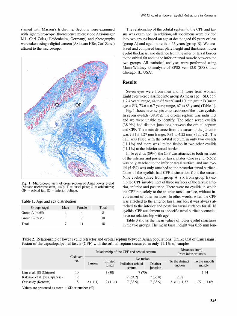
Fig. 1. Microscopic view of cross section of Asian lower eyelid (Masson trichrome stain, ×40). T = tarsal plate; O = orbicularis; OF = orbital fat; IO = inferior oblique.
Table 1. Age and sex distribution
Groups (age) Male Female TotalGroup A (≤65) 4 4 8
Group B (65<) 3 7 10
Total 7 11 18
Table 2. Relationship of lower eyelid retractor and orbital septum between Asian populations. Unlike that of Caucasians, fusion of the capsulopalpebral fascia (CPF) with the orbital septum occurred in only 11.1% of samples
Cadavers no.
Relationship of the CPF and orbital septumDistances (mm)
From inferior tarsus
Fusion Limited fusion
No fusionTo the distinct
junction To the smooth
muscleIndistinct orbital septum
Distinct junction
Lim et al. [8] (Chinese) 10 3 (30) 7 (70) 1.44 Kakizaki et al. [9] (Japanese) 19 12 (63.2) 7 (36.8) 2.38 Our study (Koreans) 18 2 (11.1) 2 (11.1) 7 (38.9) 7 (38.9) 2.31 ± 1.27 1.77 ± 1.09
Values are presented as mean ± SD or number (%).
stained with Masson’s trichrome. Sections were examined with light microscopy (fluorescence microscope Axioimager M1; Carl Zeiss, Heidenheim, Germany) and photographs were taken using a digital camera (Axiocam HRc, Carl Zeiss) affixed to the microscope.
The relationship of the orbital septum to the CPF and tar-sus was examined. In addition, all specimens were divided into two groups based on age at death: aged 65 years or less (group A) and aged more than 65 years (group B). We ana-lyzed and compared tarsal plate height and thickness, lower eyelid thickness, and distance from the inferior tarsal border to the orbital fat and to the inferior tarsal muscle between the two groups. All statistical analyses were performed using Mann-Whitney U analysis of SPSS ver. 12.0 (SPSS Inc., Chicago, IL, USA).
Results
Seven eyes were from men and 11 were from women. Eight eyes were classified into group A (mean age ± SD, 55.9 ± 7.4 years; range, 44 to 65 years) and 10 into group B (mean age ± SD, 73.6 ± 6.7 years; range, 67 to 83 years) (Table 1).
Fig. 1 shows microscopic cross-sections of the lower eyelids. In seven eyelids (38.9%), the orbital septum was indistinct and we were unable to identify. The other seven eyelids (38.9%) had distinct junctions between the orbital septum and CPF. The mean distance from the tarsus to the junction was 2.31 ± 1.27 mm (range, 0.81 to 4.22 mm) (Table 2). The CPF was fused with the orbital septum in only two eyelids (11.1%) and there was limited fusion in two other eyelids (11.1%) at the inferior tarsal border.
In 16 eyelids (89%), the CPF was attached to both surfaces of the inferior and posterior tarsal plates. One eyelid (5.5%) was only attached to the inferior tarsal surface, and one eye-lid (5.5%) was only attached to the posterior tarsal surface. None of the eyelids had CPF disinsertion from the tarsus. Nine eyelids (three from group A, six from group B) ex-hibited CPF involvement of three surfaces of the tarsus: ante-rior, inferior and posterior. There were no eyelids in which the CPF ran solely to the anterior tarsal surface, without in-volvement of other surfaces. In other words, when the CPF was attached to the anterior tarsal surface, it was always at-tached to the inferior and posterior tarsal surfaces for all 18 eyelids. CPF attachment to a specific tarsal surface seemed to have no relationship with age.
Table 3 shows the mean values of lower eyelid structures in the two groups. The mean tarsal height was 0.55 mm lon-
Korean J Ophthalmol Vol.25, No.5, 2011
346
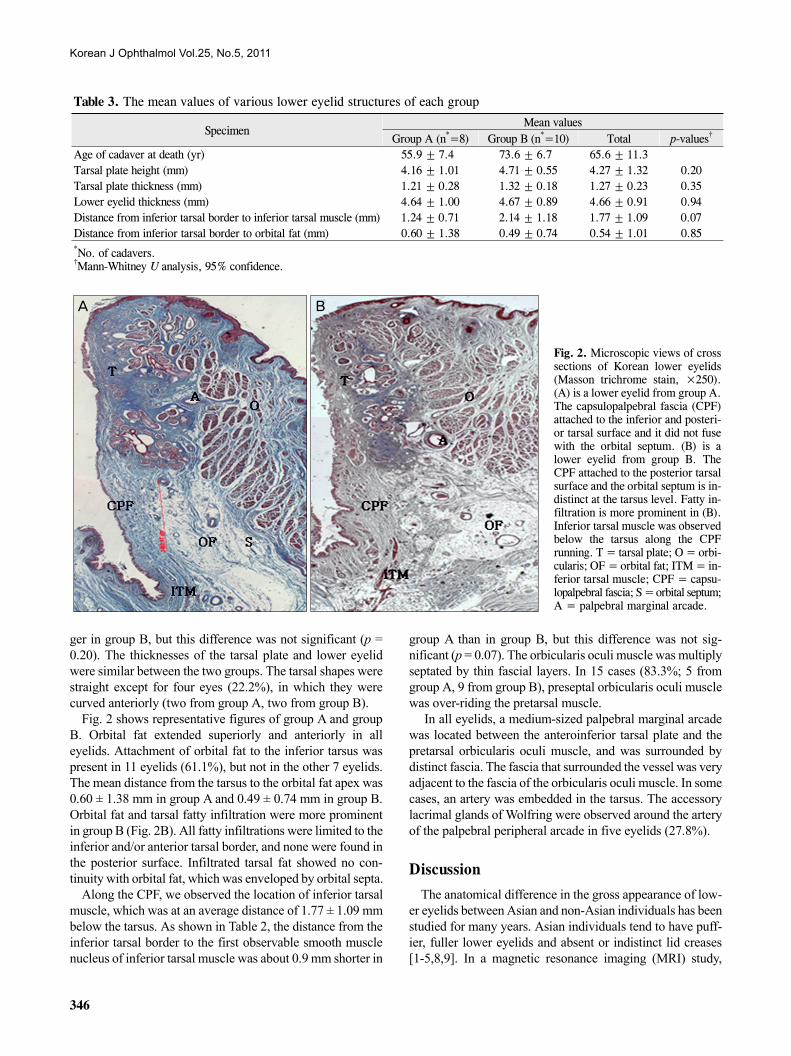
Table 3. The mean values of various lower eyelid structures of each group
SpecimenMean values
Group A (n*=8) Group B (n*=10) Total p-values†
Age of cadaver at death (yr) 55.9 ± 7.4 73.6 ± 6.7 65.6 ± 11.3Tarsal plate height (mm) 4.16 ± 1.01 4.71 ± 0.55 4.27 ± 1.32 0.20Tarsal plate thickness (mm) 1.21 ± 0.28 1.32 ± 0.18 1.27 ± 0.23 0.35Lower eyelid thickness (mm) 4.64 ± 1.00 4.67 ± 0.89 4.66 ± 0.91 0.94Distance from inferior tarsal border to inferior tarsal muscle (mm) 1.24 ± 0.71 2.14 ± 1.18 1.77 ± 1.09 0.07Distance from inferior tarsal border to orbital fat (mm) 0.60 ± 1.38 0.49 ± 0.74 0.54 ± 1.01 0.85*No. of cadavers. †Mann-Whitney U analysis, 95% confidence.
A B
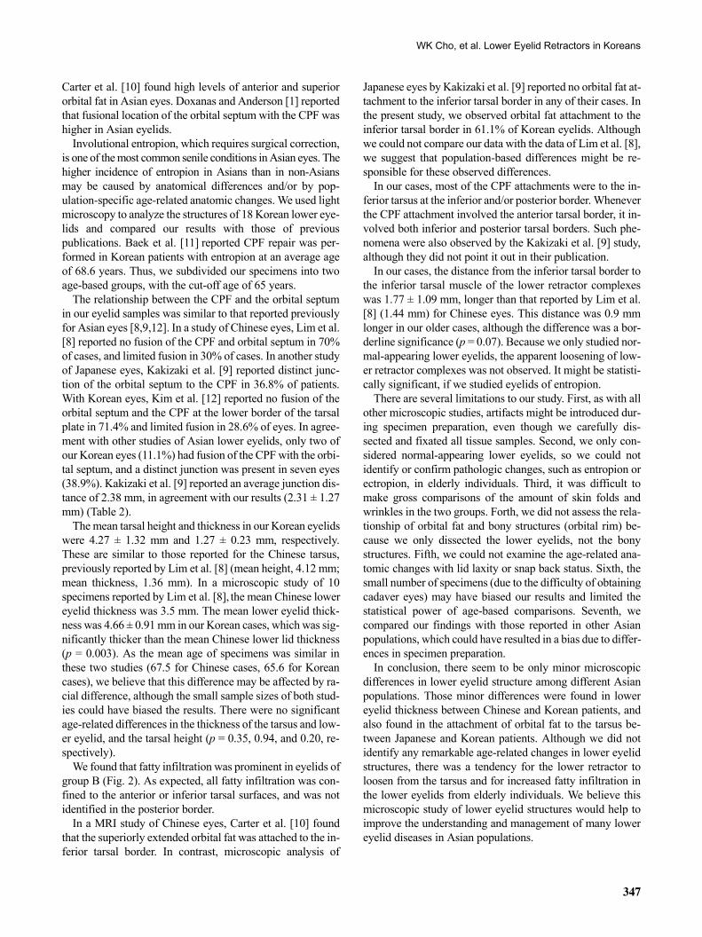
Fig. 2. Microscopic views of cross sections of Korean lower eyelids (Masson trichrome stain, ×250).(A) is a lower eyelid from group A. The capsulopalpebral fascia (CPF) attached to the inferior and posteri-or tarsal surface and it did not fuse with the orbital septum. (B) is a lower eyelid from group B. The CPF attached to the posterior tarsal surface and the orbital septum is in-distinct at the tarsus level. Fatty in-filtration is more prominent in (B). Inferior tarsal muscle was observed below the tarsus along the CPF running. T = tarsal plate; O = orbi-cularis; OF = orbital fat; ITM = in-ferior tarsal muscle; CPF = capsu-lopalpebral fascia; S = orbital septum;A = palpebral marginal arcade.
ger in group B, but this difference was not significant (p = 0.20). The thicknesses of the tarsal plate and lower eyelid were similar between the two groups. The tarsal shapes were straight except for four eyes (22.2%), in which they were curved anteriorly (two from group A, two from group B).
Fig. 2 shows representative figures of group A and group B. Orbital fat extended superiorly and anteriorly in all eyelids. Attachment of orbital fat to the inferior tarsus was present in 11 eyelids (61.1%), but not in the other 7 eyelids. The mean distance from the tarsus to the orbital fat apex was 0.60 ± 1.38 mm in group A and 0.49 ± 0.74 mm in group B. Orbital fat and tarsal fatty infiltration were more prominent in group B (Fig. 2B). All fatty infiltrations were limited to the inferior and/or anterior tarsal border, and none were found in the posterior surface. Infiltrated tarsal fat showed no con-tinuity with orbital fat, which was enveloped by orbital septa.
Along the CPF, we observed the location of inferior tarsal muscle, which was at an average distance of 1.77 ± 1.09 mm below the tarsus. As shown in Table 2, the distance from the inferior tarsal border to the first observable smooth muscle nucleus of inferior tarsal muscle was about 0.9 mm shorter in
group A than in group B, but this difference was not sig-nificant (p = 0.07). The orbicularis oculi muscle was multiply septated by thin fascial layers. In 15 cases (83.3%; 5 from group A, 9 from group B), preseptal orbicularis oculi muscle was over-riding the pretarsal muscle.
In all eyelids, a medium-sized palpebral marginal arcade was located between the anteroinferior tarsal plate and the pretarsal orbicularis oculi muscle, and was surrounded by distinct fascia. The fascia that surrounded the vessel was very adjacent to the fascia of the orbicularis oculi muscle. In some cases, an artery was embedded in the tarsus. The accessory lacrimal glands of Wolfring were observed around the artery of the palpebral peripheral arcade in five eyelids (27.8%).
Discussion
The anatomical difference in the gross appearance of low-er eyelids between Asian and non-Asian individuals has been studied for many years. Asian individuals tend to have puff-ier, fuller lower eyelids and absent or indistinct lid creases [1-5,8,9]. In a magnetic resonance imaging (MRI) study,
WK Cho, et al. Lower Eyelid Retractors in Koreans
347
Carter et al. [10] found high levels of anterior and superior orbital fat in Asian eyes. Doxanas and Anderson [1] reported that fusional location of the orbital septum with the CPF was higher in Asian eyelids.
Involutional entropion, which requires surgical correction, is one of the most common senile conditions in Asian eyes. The higher incidence of entropion in Asians than in non-Asians may be caused by anatomical differences and/or by pop-ulation-specific age-related anatomic changes. We used light microscopy to analyze the structures of 18 Korean lower eye-lids and compared our results with those of previous publications. Baek et al. [11] reported CPF repair was per-formed in Korean patients with entropion at an average age of 68.6 years. Thus, we subdivided our specimens into two age-based groups, with the cut-off age of 65 years.
The relationship between the CPF and the orbital septum in our eyelid samples was similar to that reported previously for Asian eyes [8,9,12]. In a study of Chinese eyes, Lim et al. [8] reported no fusion of the CPF and orbital septum in 70% of cases, and limited fusion in 30% of cases. In another study of Japanese eyes, Kakizaki et al. [9] reported distinct junc-tion of the orbital septum to the CPF in 36.8% of patients. With Korean eyes, Kim et al. [12] reported no fusion of the orbital septum and the CPF at the lower border of the tarsal plate in 71.4% and limited fusion in 28.6% of eyes. In agree-ment with other studies of Asian lower eyelids, only two of our Korean eyes (11.1%) had fusion of the CPF with the orbi-tal septum, and a distinct junction was present in seven eyes (38.9%). Kakizaki et al. [9] reported an average junction dis-tance of 2.38 mm, in agreement with our results (2.31 ± 1.27 mm) (Table 2).
The mean tarsal height and thickness in our Korean eyelids were 4.27 ± 1.32 mm and 1.27 ± 0.23 mm, respectively. These are similar to those reported for the Chinese tarsus, previously reported by Lim et al. [8] (mean height, 4.12 mm; mean thickness, 1.36 mm). In a microscopic study of 10 specimens reported by Lim et al. [8], the mean Chinese lower eyelid thickness was 3.5 mm. The mean lower eyelid thick-ness was 4.66 ± 0.91 mm in our Korean cases, which was sig-nificantly thicker than the mean Chinese lower lid thickness (p = 0.003). As the mean age of specimens was similar in these two studies (67.5 for Chinese cases, 65.6 for Korean cases), we believe that this difference may be affected by ra-cial difference, although the small sample sizes of both stud-ies could have biased the results. There were no significant age-related differences in the thickness of the tarsus and low-er eyelid, and the tarsal height (p = 0.35, 0.94, and 0.20, re-spectively).
We found that fatty infiltration was prominent in eyelids of group B (Fig. 2). As expected, all fatty infiltration was con-fined to the anterior or inferior tarsal surfaces, and was not identified in the posterior border.
In a MRI study of Chinese eyes, Carter et al. [10] found that the superiorly extended orbital fat was attached to the in-ferior tarsal border. In contrast, microscopic analysis of
Japanese eyes by Kakizaki et al. [9] reported no orbital fat at-tachment to the inferior tarsal border in any of their cases. In the present study, we observed orbital fat attachment to the inferior tarsal border in 61.1% of Korean eyelids. Although we could not compare our data with the data of Lim et al. [8], we suggest that population-based differences might be re-sponsible for these observed differences.
In our cases, most of the CPF attachments were to the in-ferior tarsus at the inferior and/or posterior border. Whenever the CPF attachment involved the anterior tarsal border, it in-volved both inferior and posterior tarsal borders. Such phe-nomena were also observed by the Kakizaki et al. [9] study, although they did not point it out in their publication.
In our cases, the distance from the inferior tarsal border to the inferior tarsal muscle of the lower retractor complexes was 1.77 ± 1.09 mm, longer than that reported by Lim et al. [8] (1.44 mm) for Chinese eyes. This distance was 0.9 mm longer in our older cases, although the difference was a bor-derline significance (p = 0.07). Because we only studied nor-mal-appearing lower eyelids, the apparent loosening of low-er retractor complexes was not observed. It might be statisti-cally significant, if we studied eyelids of entropion.
There are several limitations to our study. First, as with all other microscopic studies, artifacts might be introduced dur-ing specimen preparation, even though we carefully dis-sected and fixated all tissue samples. Second, we only con-sidered normal-appearing lower eyelids, so we could not identify or confirm pathologic changes, such as entropion or ectropion, in elderly individuals. Third, it was difficult to make gross comparisons of the amount of skin folds and wrinkles in the two groups. Forth, we did not assess the rela-tionship of orbital fat and bony structures (orbital rim) be-cause we only dissected the lower eyelids, not the bony structures. Fifth, we could not examine the age-related ana-tomic changes with lid laxity or snap back status. Sixth, the small number of specimens (due to the difficulty of obtaining cadaver eyes) may have biased our results and limited the statistical power of age-based comparisons. Seventh, we compared our findings with those reported in other Asian populations, which could have resulted in a bias due to differ-ences in specimen preparation.
In conclusion, there seem to be only minor microscopic differences in lower eyelid structure among different Asian populations. Those minor differences were found in lower eyelid thickness between Chinese and Korean patients, and also found in the attachment of orbital fat to the tarsus be-tween Japanese and Korean patients. Although we did not identify any remarkable age-related changes in lower eyelid structures, there was a tendency for the lower retractor to loosen from the tarsus and for increased fatty infiltration in the lower eyelids from elderly individuals. We believe this microscopic study of lower eyelid structures would help to improve the understanding and management of many lower eyelid diseases in Asian populations.
Korean J Ophthalmol Vol.25, No.5, 2011
348
Conflict of Interest
No potential conflict of interest relevant to this article was reported.
References 1. Doxanas MT, Anderson RL. Oriental eyelids. An anatomic
study. Arch Ophthalmol 1984;102:1232-5. 2. Amrith S. Oriental eyelids: anatomical and surgical considerations.
Singapore Med J 1991;32:316-8. 3. Zubiri JS. Correction of the Oriental eyelid. Clin Plast Surg
1981;8:725-37. 4. Sayoc BT. Surgery of the Oriental eyelid. Clin Plast Surg 1974;
1:157-71. 5. Weingarten CZ. Blepharoplasty in the Oriental eye. Trans Sect
Otolaryngol Am Acad Ophthalmol Otolaryngol 1976;82:ORL 442-6.
6. Vallabhanath P, Carter SR. Ectropion and entropion. Curr Opin
Ophthalmol 2000;11:345-51. 7. Carter SR, Chang J, Aguilar GL, et al. Involutional entropion
and ectropion of the Asian lower eyelid. Ophthal Plast Reconstr Surg 2000;16:45-9.
8. Lim WK, Rajendran K, Choo CT. Microscopic anatomy of the lower eyelid in asians. Ophthal Plast Reconstr Surg 2004;20: 207-11.
9. Kakizaki H, Jinsong Z, Zako M, et al. Microscopic anatomy of Asian lower eyelids. Ophthal Plast Reconstr Surg 2006;22: 430-3.
10. Carter SR, Seiff SR, Grant PE, Vigneron DB. The Asian lower eyelid: a comparative anatomic study using high-resolution magnetic resonance imaging. Ophthal Plast Reconstr Surg 1998;14:227-34.
11. Baek SH, Yang SW, Choi WC. The capsulopalpebral fascia re-pair for senile entropion. J Korean Ophthalmol Soc 2002; 43:1355-61.
12. Kim SY, Shin SJ, Yang SW, Han SH. Microscopic anatomy of the lower eyelid in Koreans. J Korean Ophthalmol Soc 2006; 47:292-6.