Original Article - Indian Prosthodontic Society · The retromylohyoid fossa is a region below and...
6
20 © 2016 The Journal of Indian Prosthodontic Society | Published by Wolters Kluwer - Medknow Lateral throat form re‑classified using a customized gauge: A clinical study N. Kalavathy, P. Roshan Kumar, Shefali Gupta, J. Sridevi, Mitha Shetty, Archana K. Sanketh Department of Prosthodoncs, D.A.P.M.R.V. Dental College, Bengaluru, Karnataka, India INTRODUCTION Successful denture therapy is a complex process demanding technical and interpersonal expertise. The prosthodontics needs to know as much as possible about each patient’s intraoral anatomy and function; expectations and experience; and likely range of physical and psychological responses to treatment; Background: A common problem faced by prosthodontists is achieving adequate retention and stability in the mandibular dentures. Recording the lateral throat form (LTF) correctly can aid in the retention and stability. Till date, Neil’s classification has been considered as the gold standard in measuring the depth of the LTF. This is a subjective classification and varies among different operators. In this study, a customized tool was used to measure the depth of the LTF, and a classification was proposed according to the measured depths. Objectives: The objective of this study is to measure the exact depth of LTF using customized gauge and to propose a classification based on the measured depth. Materials and Methods: A customized gauge was made to measure the depth of the LTF. Two different observers classified the LTFs according to Neil’s classification and according to the proposed classification in a total group of 50 patients. The customized gauge was inserted into the alveolo-lingual sulcus to measure the depth. The Pearson’s correlation statistics was carried out to observe the inter-observer relationships of sulcus depth using this customized gauge. ANOVA test was used to compare the mean depth of the sulcus as measured by observers 1 and 2. Results: There was more inter-observer variability when Neil’s classification was used as compared to the one with the proposed classification using the gauge. The inter-observer agreement for the proposed new classification was assessed by Cohen’s kappa value, with P < 0.001. The mean depth of the sulcus as calculated by observers 1 and 2 was compared with ANOVA test and found to be significant with P < 0.001. Conclusion: The proposed new classification for LTF gave consistent results and was easier to use with less variability when compared to the Neil’s classification. Key Words: Alveolo-lingual sulcus, lateral throat form, Neil’s classification, retention, stability Abstract Address for correspondence: Dr. N. Kalavathy, Department of Prosthodoncs, D.A.P.M.R.V. Dental College, No. CA‑37, 24 th Main, J.P. Nagar, 1 st Phase, Bengaluru ‑ 560 078, Karnataka, India. E‑mail: [email protected] Received: 23 rd June, 2015, Accepted: 18 th September, 2015 Access this article online Quick Response Code: Website: www.j‑ips.org DOI: 10.4103/0972‑4052.167934 Original Article How to cite this article: Kalavathy N, Kumar PR, Gupta S, Sridevi J, Shetty M, Sanketh AK. Lateral throat form re-classified using a customized gauge: A clinical study. J Indian Prosthodont Soc 2016;16:20-5. This is an open access article distributed under the terms of the Creative Commons Attribution‑NonCommercial‑ShareAlike 3.0 License, which allows others to remix, tweak, and build upon the work non‑commercially, as long as the author is credited and the new creations are licensed under the identical terms. For reprints contact: [email protected] [Downloaded free from http://www.j-ips.org on Saturday, April 02, 2016, IP: 49.206.1.43]
Transcript of Original Article - Indian Prosthodontic Society · The retromylohyoid fossa is a region below and...

20 © 2016 The Journal of Indian Prosthodontic Society | Published by Wolters Kluwer - Medknow
Lateral throat form re‑classified using a customized gauge: A clinical study
N. Kalavathy, P. Roshan Kumar, Shefali Gupta, J. Sridevi, Mitha Shetty, Archana K. SankethDepartment of Prosthodontics, D.A.P.M.R.V. Dental College, Bengaluru, Karnataka, India
INTRODUCTION
Successful denture therapy is a complex process demanding technical and interpersonal expertise. The prosthodontics needs
to know as much as possible about each patient’s intraoral anatomy and function; expectations and experience; and likely range of physical and psychological responses to treatment;
Background: A common problem faced by prosthodontists is achieving adequate retention and stability in the mandibular dentures. Recording the lateral throat form (LTF) correctly can aid in the retention and stability. Till date, Neil’s classification has been considered as the gold standard in measuring the depth of the LTF. This is a subjective classification and varies among different operators. In this study, a customized tool was used to measure the depth of the LTF, and a classification was proposed according to the measured depths.Objectives: The objective of this study is to measure the exact depth of LTF using customized gauge and to propose a classification based on the measured depth.Materials and Methods: A customized gauge was made to measure the depth of the LTF. Two different observers classified the LTFs according to Neil’s classification and according to the proposed classification in a total group of 50 patients. The customized gauge was inserted into the alveolo-lingual sulcus to measure the depth. The Pearson’s correlation statistics was carried out to observe the inter-observer relationships of sulcus depth using this customized gauge. ANOVA test was used to compare the mean depth of the sulcus as measured by observers 1 and 2.Results: There was more inter-observer variability when Neil’s classification was used as compared to the one with the proposed classification using the gauge. The inter-observer agreement for the proposed new classification was assessed by Cohen’s kappa value, with P < 0.001. The mean depth of the sulcus as calculated by observers 1 and 2 was compared with ANOVA test and found to be significant with P < 0.001.Conclusion: The proposed new classification for LTF gave consistent results and was easier to use with less variability when compared to the Neil’s classification.
Key Words: Alveolo-lingual sulcus, lateral throat form, Neil’s classification, retention, stability
Abstract
Address for correspondence: Dr. N. Kalavathy, Department of Prosthodontics, D.A.P.M.R.V. Dental College, No. CA‑37, 24th Main, J.P. Nagar, 1st Phase, Bengaluru ‑ 560 078, Karnataka, India. E‑mail: [email protected]: 23rd June, 2015, Accepted: 18th September, 2015
Access this article onlineQuick Response Code:
Website:
www.j‑ips.org
DOI:
10.4103/0972‑4052.167934
Original Article
How to cite this article: Kalavathy N, Kumar PR, Gupta S, Sridevi J, Shetty M, Sanketh AK. Lateral throat form re-classified using a customized gauge: A clinical study. J Indian Prosthodont Soc 2016;16:20-5.
This is an open access article distributed under the terms of the Creative Commons Attribution‑NonCommercial‑ShareAlike 3.0 License, which allows others to remix, tweak, and build upon the work non‑commercially, as long as the author is credited and the new creations are licensed under the identical terms.
For reprints contact: [email protected]
[Downloaded free from http://www.j-ips.org on Saturday, April 02, 2016, IP: 49.206.1.43]

Kalavathy, et al.: Lateral throat form re-classified
The Journal of Indian Prosthodontic Society | Jan-Mar 2016 | Vol 16 | Issue 1 21
and a new prosthesis. For this reason, thorough collection of relevant information regarding intraoral anatomy needs to precede the initiation of fabrication of complete dentures.[1] These parameters require that patients perceive their dentures as stationary or well retained during function. In this regard in the field of prosthodontics, retention and stability are the two major concerns for complete denture therapy, especially in lower denture because of less surface area available.[2]
Geriatric patients who present with resorbed ridges, challenge the dentist in terms of achieving proper retention and stability. Retention is defined as that quality inherent in the dental prosthesis, acting to resist the forces of dislodgement along the path of placement.[3] Thomas described three distinct spaces available on the lingual side of edentulous ridge for the extension of the denture base to get adequate retention in resorbed lower ridges. These three spaces were: (1) Sublingual crescent space (2) sublingual fossa (3) retromylohyoid fossa.[4]
The retromylohyoid fossa is a region below and behind the retromolar pad and it provides an excellent area for extending the denture for positive retention, especially when extensions into the sublingual crescent and the sublingual fossa cannot be made as in the case of resorption. Neil also mentioned that the distal end of the alveolingual sulcus (i.e. lateral throat form [LTF]) [Figure 1] can be used to achieve more vertical height of dentures in this region. Lower dentures are shallow in the mylohyoid region and turn toward the tongue and then curves back again toward ridges as we go more posteriorly. Neil classified LTF as Class I, Class II, and Class III depending on the displaceability of the instrument placed in the alveolo‑lingual sulcus on protrusion of the tongue. The perception of the displaceability of the instrument varies among different observers hence making this classification as subjective and prone to error.[5,6]
To overcome this problem, a customized gauge instrument was designed to measure the depth of the LTF and a study was conducted in the Department of Prosthodontics, Crown, and Bridge to evaluate the depth of LTF in completely edentulous patients. This instrument gives the exact depth of LTF, based on which we can classify lateral form. This measurement was then used to modify the primary impression tray in the area of interest to record the LTF more accurately during subsequent impression procedures. Keeping the above in mind, this study was conducted to measure the exact depth of the LTF using a customized gauge and propose a new classification for LTF based on the measurements obtained.
MATERIALS AND METHODS
Study designA total of 50 edentulous subjects were randomly selected from the local population who fell under the inclusion criteria:• Patientswithcompletelyedentulousmandibulararches• Patientswithgoodneuromuscularcoordination• Patientsinwhomretromolarpadcanbeeasilydistinguished.
The exclusion criteria were:• Patientwhohasundergoneanysurgicalprocedureof the
jaws, e.g., hemimandibulectomy and glossectomy• Patientwhoisnotwillingtosigntheconsentform• Anycongenitaldefectinthejaw• Anyabnormalityof oralstructures.
This instrument was checked in patients, to measure the depth of LTF on the left side, since left side has a better access for a right‑hand operator.
Two different observers classified LTF in edentulous patients using Neil’s classification [Table 1].
Instrument design (customized gauge design)The Instrument was designed with a hollow “L” shaped copper pipe with a flexible wire within it [Figure 2]. This wire was freely movable inside the pipe and was extended on both sides of the L‑shaped tube. Extension on one side would help in the measurement, and on the other side, it would move on a metal scale which is attached to the copper pipe that would accurately give us the LTF depth. A stopper
Figure 1: Lateral throat form (left side)
Table 1: Neil’s classificationClassification Description
Class I No movement to the clinician’s finger or hand mirror when patient is protruding
Class II About half as long and narrow as a Class I flange and about twice the length of a Class III
Class III The entire finger/mirror is displaced. Minimum length and thickness, usually ending the flange 2-3 mm below of just at the mylohyoid ridge
[Downloaded free from http://www.j-ips.org on Saturday, April 02, 2016, IP: 49.206.1.43]

Kalavathy, et al.: Lateral throat form re-classified
22 The Journal of Indian Prosthodontic Society | Jan-Mar 2016 | Vol 16 | Issue 1
was attached to the vertical arm which was positioned on the retromolar pad [Figure 3]. The stopper was made movable horizontally so that the same instrument could be used on either side. A scale was attached on the horizontal arm so that measurement can be made directly on the patients [Figure 4]. Mouth mirror is used to retract the tongue from the area of interest.
Method to measure the lateral throat formPatients were instructed to open their mouth and protrude their tongue so that it was ¼ inch ahead of the lower lip. Then the instrument was placed inside the patient’s mouth so that the stopper of the instrument rested on the middle third of the retromolar pad. Then the flexible wire was pushed from outside till it touches the floor of the mouth [Figure 5].
The length of wire pushed in the vertical arm was indicated on a scale attached to it and was equal to the length of wire coming out from the vertical arm which in turn reflected the LTF depth.
RESULTS
A total of 50 patients were observed by two different observers. For each observation, depth was measured using customized gauge [Table 2]. From these measurements, a classification of the LTF was proposed according to the depth measurement [Table 3]. Hence, the values obtained were denoted with the proposed classification [Table 4].
Statistical analysis was carried out to verify the significance of the proposed classification. Pearson correlation statistics to observe the relationship among the inter‑observer estimations of sulcus depth using customized gauge is shown in Table 5.
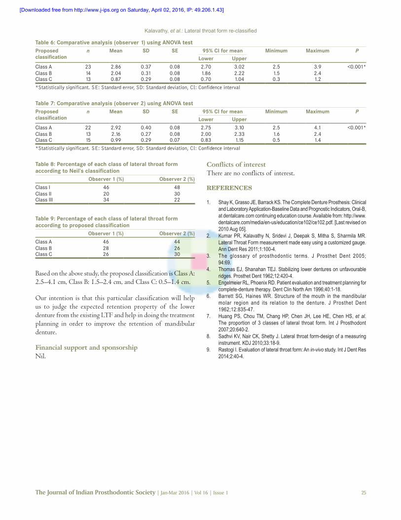
Table 6 shows the comparison of mean depth of the sulcus as measured by observer 1 using customized gauze for proposing a newer classification using ANOVA test. Comparison of mean depth of the sulcus as measured by observer 2 using customized gauze for proposing a newer classification using ANOVA test is depicted in Table 7.
The results show that there is a significant inter‑observer agreement in the proposed classification using a customized gauge.
DISCUSSION
Based on the Neil’s classification, percentage of Class I, Class II, and Class III LTF according to the observers 1
Figure 2: Customized instrument used to measure the depth of lateral throat form
Figure 3: Stopper attached to the vertical arm of the instrument
Figure 4: Metal scale attached to the horizontal arm of the instrument below the flexible wire
Figure 5: Instrument placed inside the oral cavity with the stopper resting on retromolar pad and the metal ball attached to the flexible wire touching the floor of the mouth
[Downloaded free from http://www.j-ips.org on Saturday, April 02, 2016, IP: 49.206.1.43]

Kalavathy, et al.: Lateral throat form re-classified
The Journal of Indian Prosthodontic Society | Jan-Mar 2016 | Vol 16 | Issue 1 23
and 2 was tabulated in Table 8. Based on the proposed classification, percentage of Class A, Class B, and Class C
LTF according to the observers 1 and 2 was tabulated in Table 9.
LTF, area situated at the distal end of the alveolo‑lingual sulcus, has profound influence on the fabrication of complete dentures. Yet its importance is not appreciated by most clinicians. The length and thickness of the flange in the space are different depending on the tonicity, activity, and anatomic attachments of the adjacent structures. Neil described the difference of this important area and divided it into three classifications.[5]
In the present cross‑sectional study, according to Neil’s classification, observer 1 has classified 23 patients as Class I, 10 patients as Class II, and 17 patients as Class III. Observer 2 has classified 24 patients as Class I, 15 patients Class II, and 11 patients as Class III. This proves the variability among two observers when using Neil’s classification to classify LTF. Although Neil’s has been the gold standard for classifying the LTF for many years, it is a subjective classification and varies from operator to operator. It also varies between experienced clinicians and beginners.
A study conducted by Huang et al. investigated the proportion of three classes of LTF and reported that Class I was more common than Class II or III.[7] Sadhvi et al. used a customized instrument to measure LTF intraorally and compare its efficacy with the conventional method.[8] Another study observed the significant differences between the vertical dimension of LTF measured in patients’ mouth and that of their diagnostic casts using a customized instrument.[9] However, no attempt has been made to classify the LTF based on such measurements. This study aims to propose a classification based on the measured depth.
In the present study, the customized tool described in this report gives us the exact value of LTF depth which will be helpful in classifying it and making good preliminary impressions by selecting a proper stock tray. A good preliminary cast will ensure that the custom tray is fabricated with proper extensions, which will be reflected in the final denture. This will help us achieve better retention and stability in mandibular dentures.
The statistical analysis with the Pearson’s correlation test demonstrated that there was a positive agreement between the two observers with respect to the measurement using the customized gauge. The ANOVA test gave the mean values for each class for both the observers and it was found to be roughly the same.
All these tests prove that the proposed classification is consistent with the measurements and can be used as a reliable measure for checking the LTF.
Table 2: Classification of lateral throat form using Neil’s classification and measurement using customized gaugePatients Observer 1 Observer 2
Neil’s classification
Customized gauge
Neil’s classification
Customized gauge
1 Class I 3.0 Class I 3.02 Class I 2.5 Class I 2.63 Class I 3.0 Class I 3.14 Class I 3.9 Class I 4.15 Class II 2.4 Class I 2.46 Class I 3.5 Class I 3.47 Class I 3.0 Class I 3.28 Class I 2.8 Class I 2.99 Class II 2.3 Class II 2.410 Class I 3.0 Class I 3.211 Class III 1.8 Class II 2.012 Class II 2.0 Class II 2.113 Class II 1.8 Class II 1.614 Class I 2.6 Class I 2.615 Class I 2.5 Class I 2.516 Class III 2.2 Class II 2.417 Class III 2.0 Class II 2.018 Class III 2.1 Class II 2.219 Class III 2.4 Class II 2.420 Class III 2.2 Class II 2.221 Class III 0.5 Class III 0.522 Class III 1.0 Class II 1.223 Class III 1.2 Class III 1.124 Class III 0.6 Class III 0.625 Class II 1.1 Class III 1.026 Class I 2.9 Class I 2.727 Class I 3.0 Class I 2.928 Class I 2.5 Class I 2.629 Class III 1.1 Class II 1.230 Class III 0.3 Class III 0.531 Class III 1.0 Class III 1.132 Class I 2.8 Class I 2.733 Class II 2.3 Class II 2.434 Class I 3.1 Class I 3.235 Class I 2.6 Class I 2.836 Class III 0.8 Class III 0.937 Class I 2.6 Class I 2.838 Class I 3.5 Class I 3.539 Class II 0.9 Class III 0.840 Class I 2.5 Class I 2.641 Class I 2.6 Class I 2.542 Class I 2.5 Class I 2.343 Class I 2.8 Class I 2.944 Class III 0.6 Class II 0.845 Class II 1.5 Class III 1.446 Class II 1.9 Class III 1.747 Class III 1.0 Class II 1.248 Class II 1.5 Class III 1.349 Class III 1.2 Class II 1.250 Class I 2.6 Class I 2.5
Table 3: Proposed classification for lateral throat form using customized gaugeProposed classification
Measurement range
Class A 2.5-4.1 cmClass B 1.5-2.4 cmClass C 0.5-1.4 cm
[Downloaded free from http://www.j-ips.org on Saturday, April 02, 2016, IP: 49.206.1.43]

Kalavathy, et al.: Lateral throat form re-classified
24 The Journal of Indian Prosthodontic Society | Jan-Mar 2016 | Vol 16 | Issue 1
There are a few limitations with using the instrument. Less experienced clinicians might not be able to correctly position the instrument. The metal ball might not be visible in case of an excessively large tongue. There are chances of over extending the metal ball into the alveolo‑lingual sulcus.
CONCLUSION
Instrument which was customized to measure LTF depth gave consistent results when compared against the conventional method.
Table 4: Values obtained denoted with the proposed classificationObserver 1 Observer 2
Patients Neil’s classification
Customized gauge
Proposed classification
Neil’s classification
Customized gauge
Proposed classification
1 Class I 3.0 Class A Class I 3.0 Class A2 Class I 2.5 Class A Class I 2.6 Class A3 Class I 3.0 Class A Class I 3.1 Class A4 Class I 3.9 Class A Class I 4.1 Class A5 Class II 2.4 Class B Class I 2.4 Class B6 Class I 3.5 Class A Class I 3.4 Class A7 Class I 3.0 Class A Class I 3.2 Class A8 Class I 2.8 Class A Class I 2.9 Class A9 Class II 2.3 Class B Class II 2.4 Class B10 Class I 3.0 Class A Class I 3.2 Class A11 Class III 1.8 Class B Class II 2.0 Class B12 Class II 2.0 Class B Class II 2.1 Class B13 Class II 1.8 Class B Class II 1.6 Class B14 Class I 2.6 Class A Class I 2.6 Class A15 Class I 2.5 Class A Class I 2.5 Class A16 Class III 2.2 Class B Class II 2.4 Class B17 Class III 2.0 Class B Class II 2.0 Class B18 Class III 2.1 Class B Class II 2.2 Class B19 Class III 2.4 Class B Class II 2.4 Class B20 Class III 2.2 Class B Class II 2.2 Class B21 Class III 0.5 Class C Class III 0.5 Class C22 Class III 1.0 Class C Class II 1.2 Class C23 Class III 1.2 Class C Class III 1.1 Class C24 Class III 0.6 Class C Class III 0.6 Class C25 Class II 1.1 Class C Class III 1.0 Class C26 Class I 2.9 Class A Class I 2.7 Class A27 Class I 3.0 Class A Class I 2.9 Class A28 Class I 2.5 Class A Class I 2.6 Class A29 Class III 1.1 Class C Class II 1.2 Class C30 Class III 0.3 Class C Class III 0.5 Class C31 Class III 1.0 Class C Class III 1.1 Class C32 Class I 2.8 Class A Class I 2.7 Class A33 Class II 2.3 Class B Class II 2.4 Class B34 Class I 3.1 Class A Class I 3.2 Class A35 Class I 2.6 Class A Class I 2.8 Class A36 Class III 0.8 Class C Class III 0.9 Class C37 Class I 2.6 Class A Class I 2.8 Class A38 Class I 3.5 Class A Class I 3.5 Class A39 Class II 0.9 Class C Class III 0.8 Class C40 Class I 2.5 Class A Class I 2.6 Class A41 Class I 2.6 Class A Class I 2.5 Class A42 Class I 2.5 Class A Class I 2.3 Class B43 Class I 2.8 Class A Class I 2.9 Class A44 Class III 0.6 Class C Class II 0.8 Class C45 Class II 1.5 Class B Class III 1.4 Class C46 Class II 1.9 Class B Class III 1.7 Class B47 Class III 1.0 Class C Class II 1.2 Class C48 Class II 1.5 Class B Class III 1.3 Class C49 Class III 1.2 Class C Class II 1.2 Class C50 Class I 2.6 Class A Class I 2.5 Class A
Table 5: Pearson correlation statistics to observe relationship between the inter‑observer estimation of sulcus depth using customized gauge
Values Observer 1 Observer 2
Observer 1 r 1 0.989**P <0.001n 50 50
Observer 2 r 0.989** 1P <0.001n 50 50
**Correlation is significant at the 0.01 level
[Downloaded free from http://www.j-ips.org on Saturday, April 02, 2016, IP: 49.206.1.43]
Kalavathy, et al.: Lateral throat form re-classified
The Journal of Indian Prosthodontic Society | Jan-Mar 2016 | Vol 16 | Issue 1 25
Based on the above study, the proposed classification is Class A: 2.5–4.1 cm, Class B: 1.5–2.4 cm, and Class C: 0.5–1.4 cm.
Our intention is that this particular classification will help us to judge the expected retention property of the lower denture from the existing LTF and help in doing the treatment planning in order to improve the retention of mandibular denture.
Financial support and sponsorshipNil.
Conflicts of interestThere are no conflicts of interest.
REFERENCES
1. Shay K, Grasso JE, Barrack KS. The Complete Denture Prosthesis: Clinical and Laboratory Application‑Baseline Data and Prognostic Indicators, Oral‑B, at dentalcare.com continuing education course. Available from: http://www.dentalcare.com/media/en‑us/education/ce102/ce102.pdf. [Last revised on 2010 Aug 05].
2. Kumar PR, Kalavathy N, Sridevi J, Deepak S, Mitha S, S harmila MR. Lateral Throat Form measurement made easy using a customized gauge. Ann Dent Res 2011;1:100‑4.
3. The glossary of prosthodontic terms. J Prosthet Dent 2005; 94:69.
4. Thomas EJ, Shanahan TEJ. Stabilizing lower dentures on unfavourable ridges. Prosthet Dent 1962;12:420‑4.
5. Engelmeier RL, Phoenix RD. Patient evaluation and treatment planning for complete‑denture therapy. Dent Clin North Am 1996;40:1‑18.
6. Barrett SG, Haines WR. Structure of the mouth in the mandibular molar region and its relation to the denture. J Prosthet Dent 1962;12:835‑47.
7. Huang PS, Chou TM, Chang HP, Chen JH, Lee HE, Chen HS, et al. The proportion of 3 classes of lateral throat form. Int J Prosthodont 2007;20:640‑2.
8. Sadhvi KV, Nair CK, Shetty J. Lateral throat form‑design of a measuring instrument. KDJ 2010;33:18‑9.
9. Rastogi I. Evaluation of lateral throat form: An in‑vivo study. Int J Dent Res 2014;2:40‑4.
Table 6: Comparative analysis (observer 1) using ANOVA testProposed classification
n Mean SD SE 95% CI for mean Minimum Maximum PLower Upper
Class A 23 2.86 0.37 0.08 2.70 3.02 2.5 3.9 <0.001*Class B 14 2.04 0.31 0.08 1.86 2.22 1.5 2.4Class C 13 0.87 0.29 0.08 0.70 1.04 0.3 1.2
*Statistically significant. SE: Standard error, SD: Standard deviation, CI: Confidence interval
Table 7: Comparative analysis (observer 2) using ANOVA testProposed classification
n Mean SD SE 95% CI for mean Minimum Maximum PLower Upper
Class A 22 2.92 0.40 0.08 2.75 3.10 2.5 4.1 <0.001*Class B 13 2.16 0.27 0.08 2.00 2.33 1.6 2.4Class C 15 0.99 0.29 0.07 0.83 1.15 0.5 1.4
*Statistically significant. SE: Standard error, SD: Standard deviation, CI: Confidence interval
Table 8: Percentage of each class of lateral throat form according to Neil’s classification
Observer 1 (%) Observer 2 (%)
Class I 46 48Class II 20 30Class III 34 22
Table 9: Percentage of each class of lateral throat form according to proposed classification
Observer 1 (%) Observer 2 (%)
Class A 46 44Class B 28 26Class C 26 30
[Downloaded free from http://www.j-ips.org on Saturday, April 02, 2016, IP: 49.206.1.43]