Original Article Expression and prognostic value of MAGE ... · Original Article Expression and...
9
Int J Clin Exp Pathol 2014;7(10):6734-6742 www.ijcep.com /ISSN:1936-2625/IJCEP0002124 Original Article Expression and prognostic value of MAGE-A9 in laryngeal squamous cell carcinoma Liang Han 1,2 , Bin Jiang 2 , Hao Wu 3 , Shu Zhang 4 , Xueguan Lu 1 1 Department of Radiotherapy & Oncology, Second Affiliated Hospital of Soochow University, Suzhou, Jiangsu, China; 2 Department of Head and Neck Surgery, Affiliated Tumor Hospital of Nantong University, Nantong Tumor Hospital, Nantong, Jiangsu, China; Departments of 3 Otorhinolaryngology, 4 Clinical Pathology, Affiliated Hospital of Nantong University, Nantong, Jiangsu, China Received August 27, 2014; Accepted September 18, 2014; Epub September 15, 2014; Published October 1, 2014 Abstract: Background: Melanoma-associated antigen (MAGE) family genes are reported to play important roles in the development of human cancers. However, the relationship between the expression of MAGE-A9 and clinicopath- ological characteristics in human laryngeal carcinoma remains unclear. This study aimed to examine the expression of MAGE-A9, and to evaluate the clinical significance of its expression in human laryngeal squamous cell carci- noma (LSCC). Methods: Quantitative real-time reverse transcription-PCR (qPCR) and immunohistochemistry (IHC) were performed to characterize the expression of MAGE-A9 in LSCC tissues and tumor-adjacent normal tissues. Kaplan-Meier survival and Cox regression analyses were performed to evaluate the prognosis of patients with LSCC. Results: The expression of MAGE-A9 was significantly higher in LSCC than in tumor-adjacent normal tissues. Cyto- plasmic expression of MAGE-A9 was detected in 70 of 123 (56.9%) LSCC specimens. Levels of MAGE-A9 in LSCC were related to histopathological grade (P = 0.024). Kaplan-Meier survival and Cox regression analysis revealed that MAGE-A9 expression level and lymph node metastasis were independent prognostic factors of LSCC (P = 0.005; P = 0.001, respectively). Conclusions: Our study suggests that MAGE-A9 expression is a prognostic biomarker for LSCC patients. High expression of MAGE-A9 indicates unfavorable survival outcome in LSCC patients. Keywords: Laryngeal squamous cell carcinoma, MAGE-A9, prognosis Introduction Laryngeal squamous cell carcinoma (LSCC) is a common head and neck tumor. There are more than 500 000 new cases of LSCC each year, constituting approximately 1.2% of all cancers, 25% of head and neck cancers and 99% of laryngeal malignant tumors [1-3]. The incidence of LSCC has increased in recent years. Although therapeutic strategies targeting LSCC have improved, including surgery, radiotherapy and chemotherapy, the mortality rate of LSCC has not changed [4-6]. The recurrence rate is still as high as 50%, with a local recurrence rate of 5-25% in patients with tumor stage I and 15-50% in patients with tumor stage II [7, 8]. Therefore, the identification of novel biomark- ers for LSCC tumor staging and new treatment strategies is necessary. Members of the MAGE gene family are tumor- associated antigens, which are commonly exp- ressed in various tumors of epithelial origin, including breast cancer, lung cancer and colo- rectal cancer [9-12]. MAGE-A, a subset of highly homologous MAGE genes, belongs to the chro- mosome X-clustered cancer/testis antigens [13, 14]. The MAGE-A subfamily, which contains 12 genes, is also detected in the human germ line and in various cancers [15-17]. However, their biological functions remain largely un- known. MAGE-A9, which is expressed in high- risk bladder cancer [18], is a cancer-testis gene, which exhibits restricted expression in normal tissue, but is frequently expressed in cancer and testicular germ cells. Previous studies indi- cated the oncogenic characteristics of MAGE-A9 during the development and progression of ma- lignant tumors. However, the relationship bet- ween MAGE-A9 expression and clinicopatho- logical outcome in LSCC remains unclear. In this study, we examined the expression of MAGE-A9 mRNA in LSCC and tumor-adjacent
Transcript of Original Article Expression and prognostic value of MAGE ... · Original Article Expression and...
Int J Clin Exp Pathol 2014;7(10):6734-6742www.ijcep.com /ISSN:1936-2625/IJCEP0002124
Original Article Expression and prognostic value of MAGE-A9 in laryngeal squamous cell carcinoma
Liang Han1,2, Bin Jiang2, Hao Wu3, Shu Zhang4, Xueguan Lu1
1Department of Radiotherapy & Oncology, Second Affiliated Hospital of Soochow University, Suzhou, Jiangsu, China; 2Department of Head and Neck Surgery, Affiliated Tumor Hospital of Nantong University, Nantong Tumor Hospital, Nantong, Jiangsu, China; Departments of 3Otorhinolaryngology, 4Clinical Pathology, Affiliated Hospital of Nantong University, Nantong, Jiangsu, China
Received August 27, 2014; Accepted September 18, 2014; Epub September 15, 2014; Published October 1, 2014
Abstract: Background: Melanoma-associated antigen (MAGE) family genes are reported to play important roles in the development of human cancers. However, the relationship between the expression of MAGE-A9 and clinicopath-ological characteristics in human laryngeal carcinoma remains unclear. This study aimed to examine the expression of MAGE-A9, and to evaluate the clinical significance of its expression in human laryngeal squamous cell carci-noma (LSCC). Methods: Quantitative real-time reverse transcription-PCR (qPCR) and immunohistochemistry (IHC) were performed to characterize the expression of MAGE-A9 in LSCC tissues and tumor-adjacent normal tissues. Kaplan-Meier survival and Cox regression analyses were performed to evaluate the prognosis of patients with LSCC. Results: The expression of MAGE-A9 was significantly higher in LSCC than in tumor-adjacent normal tissues. Cyto-plasmic expression of MAGE-A9 was detected in 70 of 123 (56.9%) LSCC specimens. Levels of MAGE-A9 in LSCC were related to histopathological grade (P = 0.024). Kaplan-Meier survival and Cox regression analysis revealed that MAGE-A9 expression level and lymph node metastasis were independent prognostic factors of LSCC (P = 0.005; P = 0.001, respectively). Conclusions: Our study suggests that MAGE-A9 expression is a prognostic biomarker for LSCC patients. High expression of MAGE-A9 indicates unfavorable survival outcome in LSCC patients.
Keywords: Laryngeal squamous cell carcinoma, MAGE-A9, prognosis
Introduction
Laryngeal squamous cell carcinoma (LSCC) is a common head and neck tumor. There are more than 500 000 new cases of LSCC each year, constituting approximately 1.2% of all cancers, 25% of head and neck cancers and 99% of laryngeal malignant tumors [1-3]. The incidence of LSCC has increased in recent years. Although therapeutic strategies targeting LSCC have improved, including surgery, radiotherapy and chemotherapy, the mortality rate of LSCC has not changed [4-6]. The recurrence rate is still as high as 50%, with a local recurrence rate of 5-25% in patients with tumor stage I and 15-50% in patients with tumor stage II [7, 8]. Therefore, the identification of novel biomark-ers for LSCC tumor staging and new treatment strategies is necessary.
Members of the MAGE gene family are tumor-associated antigens, which are commonly exp-
ressed in various tumors of epithelial origin, including breast cancer, lung cancer and colo- rectal cancer [9-12]. MAGE-A, a subset of highly homologous MAGE genes, belongs to the chro-mosome X-clustered cancer/testis antigens [13, 14]. The MAGE-A subfamily, which contains 12 genes, is also detected in the human germ line and in various cancers [15-17]. However, their biological functions remain largely un-known. MAGE-A9, which is expressed in high-risk bladder cancer [18], is a cancer-testis gene, which exhibits restricted expression in normal tissue, but is frequently expressed in cancer and testicular germ cells. Previous studies indi-cated the oncogenic characteristics of MAGE-A9 during the development and progression of ma- lignant tumors. However, the relationship bet- ween MAGE-A9 expression and clinicopatho-logical outcome in LSCC remains unclear.
In this study, we examined the expression of MAGE-A9 mRNA in LSCC and tumor-adjacent

MAGE-A9 and laryngeal squamous cell carcinoma
6735 Int J Clin Exp Pathol 2014;7(10):6734-6742
normal tissues via one-step quantitative rever- se transcription-polymerase chain reaction (qPCR). Furthermore, we evaluated the expres-sion of MAGE-A9 protein in LSCC by tissue microarray (TMA). Finally, we evaluated the clin-ical significance of MAGE-A9 expression in LSCC.
Materials and methods
Specimen collection
A total of 123 paraffin-embedded LSCC tissues and 22 tumor-adjacent normal tissue samples were collected from the archives of the De- partment of Pathology at the Affiliated Hospital of Nantong University, between January 2000 and May 2010. Histological diagnosis of LSCC
was performed according to the latest World Health Organization (WHO) criteria [19]. All patients were typed in accordance with the TNM stage classification system (UICC 2009) [20]. Clinical data including gender, age, alco-hol consumption, tobacco use, pTNM stage, lymph node metastasis and histopathological grade were retrospectively collected from hos-pital medical records. Clinical characteristics of 123 patients with LSCC are shown in Table 1. All patients received radical surgery. None of the patients received radiotherapy chemother-apy, and/or immunotherapy. Ethical approval to perform this study was obtained from the Human Research Ethics Committee of the local hospital, and written, informed consent was obtained from all patients participating in this study.
Table 1. Association of MAGE-A9 expression with clinicopathological factors of LSCC
Groups No.MAGE-A9 expression in cancer cells
No.MAGE-A9 in stromal cells
Low expres-sion (%)
High Expres-sion (%)
Pear-son 2
pvalue
Low expres-sion (%)
High Expres-sion (%)
Pear-son 2
pvalue
Total 123 53 (41.3) 70 (56.9) 89 70 (78.7) 19 (21.3)Gender Female 2 1 (50.0) 1 (50.0) 0.04 0.842 1 1 (100.0) 0 (0.0) 0.275 0.6 Male 121 52 (43.0) 69 (57.0) 88 69 (78.4) 19 (21.6)Age ≤ 60 years 45 20 (44.4) 25 (55.6) 0.053 0.818 28 23 (82.1) 5 (17.9) 0.297 0.586 > 60 years 78 33 (42.3) 45 (57.7) 61 47 (77.0) 14 (23.0)Smoking No 32 14 (43.7) 18 (56.3) 0.66 0.416 24 22 (91.7) 2 (8.3) 1.816 0.178 Yes 68 24 (35.3) 44 (64.7) 53 42 (79.2) 11 (20.8) Unknown 23 15 (65.2) 8 (34.8) 12 6 (50.0) 6 (50.0)Alcohol No 50 20 (40.0) 30 (60.0) 0.17 0.68 40 31 (77.5) 9 (22.5) 1.872 0.171 Yes 50 18 (36.0) 32 (64.0) 37 33 (89.2) 4 (10.8) Unknown 23 15 (65.2) 8 (34.8) 12 6 (50.0) 6 (50.0)pTNM T1 13 6 (46.1) 7 (53.9) 1.621 0.655 11 10 (90.9) 1 (9.1) 0.863 0.834 T2 54 20 (37.0) 34 (63.0) 40 33 (82.5) 7 (17.5) T3 31 12 (38.7) 19 (61.3) 25 20 (80.0) 5 (20.0) T4 2 0 (0.0) 2 (100.0) 1 1 (100.0) 0 (0.0) Unknown 23 15 (65.2) 8 (34.8) 12 6 (50.0) 6 (50.0)Lymph node metastasis No 103 44 (42.7) 59 (57.3) 0.036 0.85 77 61 (79.2) 16 (20.8) 0.11 0.74 Yes 20 9 (45.0) 11 (55.0) 12 9 (75.0) 3 (25.0)Histopathological grade High 55 31 (56.4) 24 (43.6) 7.487 0.024* 38 27 (71.0) 11 (29.0) 3.088 0.214 Middle 55 18 (31.5) 37 (68.5) 45 39 (86.7) 6 (13.3) Low 11 3 (27.3) 8 (72.7) 5 4 (80.0) 1 (20.0) Unknown 2 1 (50.0) 1 (50.0) 1 0 (0.0) 1 (100.0)*P < 0.05.
MAGE-A9 and laryngeal squamous cell carcinoma
6736 Int J Clin Exp Pathol 2014;7(10):6734-6742
One-step qPCR analysis
Eighteen samples of fresh LSCC tissues and matched tumor-adjacent normal tissues were collected. Total RNA was extracted from tissues using Trizol reagent (Invitrogen, Carlsbad, CA, USA). Total RNA (2 mg) was reverse transcribed into cDNA using Moloney murine leukemia virus retrotranscriptase (Promega, USA). RT-PCR primers were designed with the assistance of Beacon Designer 7.7 software and are as fol-lows: MAGEA9 forward: 5’-CACTGTATGTCATCT- CTG-3’; MAGEA9 reverse: 5’-ACTACTGTCATTC- ATTAACT-3’; β-actin forward: 5’-TTAATCTTCGCC- TTAATACTT-3’; β-actin reverse: 5’-AGCCTTCA- TACATCTCAA-3’. qPCR was performed using SYBR green dye and a Bio-Rad iQ50 Real-time PCR system in accordance with the manufac-turer’s instructions. Real-time PCR cycling para- meters were as follows: denaturation at 95°C for 20 s, annealing at 56°C for 30 s and exten-sion at 72°C for 30 s. Expression data were normalized to the geometric mean of the β-actin housekeeping gene and analyzed using the 2-Delta Delta Ct method as previously described.
Tissue microarray (TMA) construction and IHC analysis
Formalin-fixed, paraffin-embedded tumor sam-ples (n = 123) and normal tumor-adjacent tis-
sue specimens (n = 22) were prepared and TMAs were produced by Xinchao Biotech Co., Ltd (Shanghai, China). The TMA was cut into 4-μm sections and placed on super frost char- ged glass microscope slides.
IHC streptavidin peroxidase (SP) staining was performed as previously described [21]. Tissue microarray sections were incubated with rabbit polyclonal anti-MAGE-A9 antibody (AP6170a, 2.5 μg/ml dilution; Abgent, San Diego, CA, USA) overnight at 4°C, followed by incubation with biotinylated anti-rabbit secondary antibody at 37°C for 30 min. The same isotype of rabbit IgG was used as a negative control. Sections were then incubated with a streptavidin-horseradish peroxidase complex, colorized with 3,3-diami-nobenzidine (DAB) chromogen solution and counterstained with hematoxylin. Results were analyzed as previously described [22]. Briefly, the percentage of MAGE-A9 positive cells was scored as follow: 0 for 0%, 1 for 1-33%, 2 for 34-66% and 3 for 67-100%. The intensity of MAGE-A9 staining was also scored as follows: 0 for negative staining, 1 for yellow color staining, 2 for light brown color staining and 3 for brown color staining. Samples with a sum score of 0-2 were considered to exhibit low MAGE-A9 expres-sion, and those with a sum score of 3-6 were considered to exhibit high MAGE-A9 expre- ssion.
Statistical analysis
Statistical analysis was performed using STATA 12.0 software (Stata Corporation, College Sta- tion, TX, USA). Comparison of MAGE-A9 mRNA expression in fresh-frozen LSCC tissues with tumor-adjacent normal tissues was analyzed with the Wilcoxon signed rank nonparametric test. The association between MAGE-A9 expres-sion and clinicopathologic variables was exam-ined by chi-square test. Survival rate was esti-mated by Kaplan-Meier method and log-rank test. Multivariate analysis was performed using Cox’s proportional hazard regression model. For all tests, a two-tailed P value of less than 0.05 was considered statistically significant.
Results
Analysis of MAGE-A9 mRNA expression in LSCC by qPCR
To investigate the expression of MAGE-A9 mRNA in LSCC, we performed qPCR on RNA
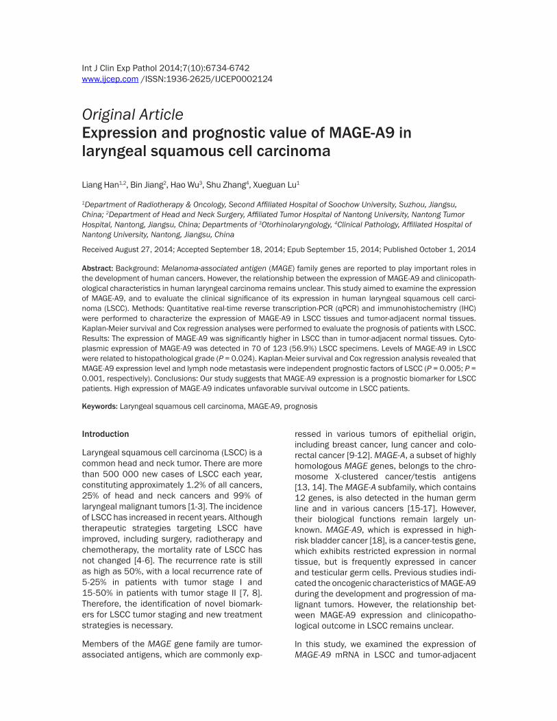
Figure 1. One-step quantitative reverse transcription-polymerase chain reaction (qPCR) was employed to evaluate MAGE-A9 mRNA expression levels in LSCC (Ca) compared with tumor adjacent tissue (N). Lev-els of MAGE-A9 mRNA in LSCC and tumor-adjacent normal tissues were 0.064 ± 0.0086 and 0.0123 ± 0.0045, respectively (t = 2.032, P = 0.028) after nor-malizing to β-actin.
MAGE-A9 and laryngeal squamous cell carcinoma
6737 Int J Clin Exp Pathol 2014;7(10):6734-6742
MAGE-A9 and laryngeal squamous cell carcinoma
6738 Int J Clin Exp Pathol 2014;7(10):6734-6742
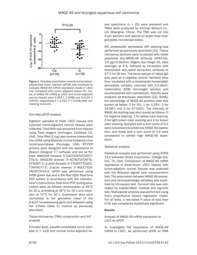
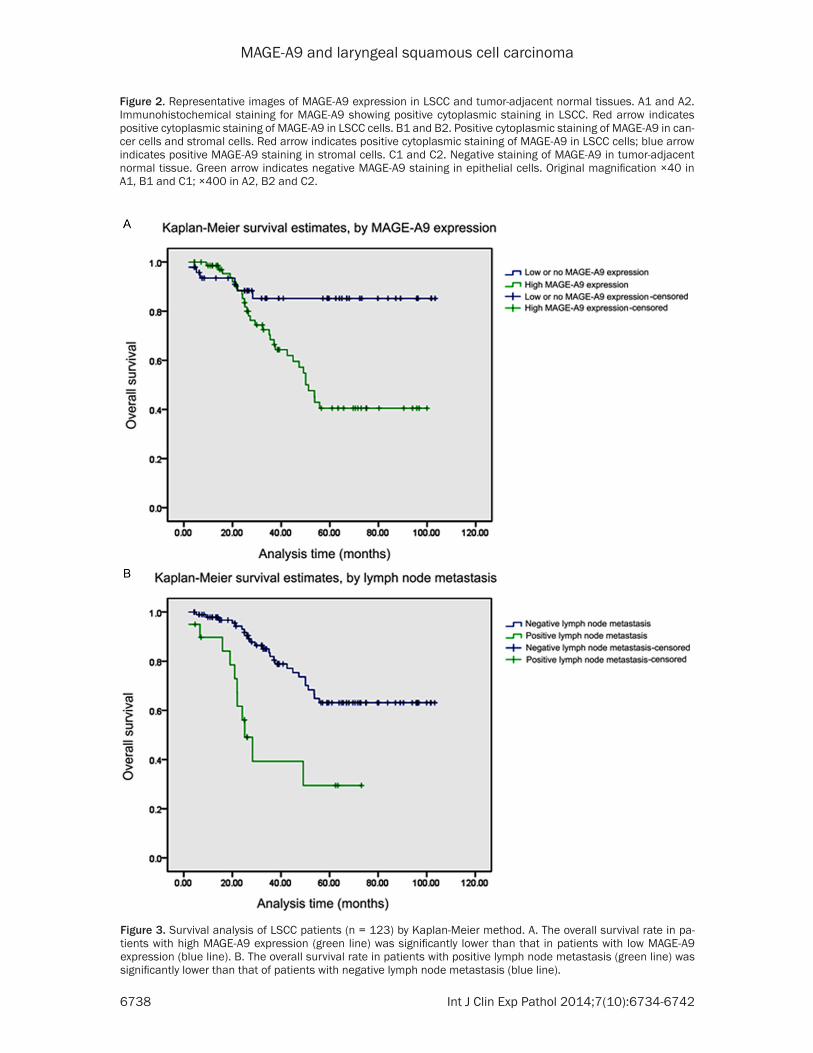
Figure 2. Representative images of MAGE-A9 expression in LSCC and tumor-adjacent normal tissues. A1 and A2. Immunohistochemical staining for MAGE-A9 showing positive cytoplasmic staining in LSCC. Red arrow indicates positive cytoplasmic staining of MAGE-A9 in LSCC cells. B1 and B2. Positive cytoplasmic staining of MAGE-A9 in can-cer cells and stromal cells. Red arrow indicates positive cytoplasmic staining of MAGE-A9 in LSCC cells; blue arrow indicates positive MAGE-A9 staining in stromal cells. C1 and C2. Negative staining of MAGE-A9 in tumor-adjacent normal tissue. Green arrow indicates negative MAGE-A9 staining in epithelial cells. Original magnification ×40 in A1, B1 and C1; ×400 in A2, B2 and C2.
Figure 3. Survival analysis of LSCC patients (n = 123) by Kaplan-Meier method. A. The overall survival rate in pa-tients with high MAGE-A9 expression (green line) was significantly lower than that in patients with low MAGE-A9 expression (blue line). B. The overall survival rate in patients with positive lymph node metastasis (green line) was significantly lower than that of patients with negative lymph node metastasis (blue line).
MAGE-A9 and laryngeal squamous cell carcinoma
6739 Int J Clin Exp Pathol 2014;7(10):6734-6742
extracted from fresh LSCC tissues (n = 18) and matched tumor-adjacent normal tissues. Follow- ing normalization to β-actin, we observed a sig-nificant increase in MAGE-A9 mRNA in LSCC compared with tumor-adjacent normal tissues (0.064 ± 0.0086 vs 0.0123 ± 0.0045, respec-tively, t = 2.032, P = 0.028). The average level of MAGE-A9 mRNA was 5.18-fold higher in LSCC compared with tumor-adjacent normal tissues (Figure 1).
Detection of MAGE-A9 expression in LSCC by IHC
We next investigated the expression of MAGEA9 protein in LSCC by IHC. MAGE-A9-positive stain-ing was predominantly localized in the cyto-plasm of cancer cells and stromal cells. Exp-ression of MAGE-A9 was significantly higher in LSCC tissues compared with tumor-adjacent normal tissues (P < 0.001). High expression of MAGEA9 was detected in 70 of 123 (56.9%) LSCC tissues, while only 6 of 22 (27.3%) tumor-adjacent normal tissues exhibited high expres-sion. High expression of MAGE-A9 in stromal cells was detected in 19 of 89 (21.3%) LSCC tissues, compared with 2 of 22 (9.1%) tumor-adjacent normal tissues. Representative IHC staining patterns of MAGE-A9 in LSCC are shown in Figure 2.
Relationship between MAGE-A9 expression and clinical parameters
The relationship between high expression of MAGE-A9 protein and LSCC patient clinical parameters is displayed in Table 1. High MAGE- A9 expression in cancer cells was significantly associated with histopathological grade (P = 0.024), while no significant correlation with other clinical parameters, including gender, age, tobacco and alcohol consumption, TNM stage and lymph node metastasis, was obser- ved. In contrast, MAGE-A9 expression in stro-mal cells was not correlated with any clinico-pathological factors.
Survival analysis
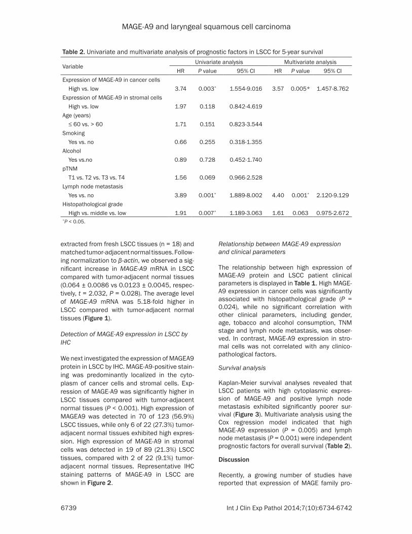
Kaplan-Meier survival analyses revealed that LSCC patients with high cytoplasmic expres-sion of MAGE-A9 and positive lymph node metastasis exhibited significantly poorer sur-vival (Figure 3). Multivariate analysis using the Cox regression model indicated that high MAGE-A9 expression (P = 0.005) and lymph node metastasis (P = 0.001) were independent prognostic factors for overall survival (Table 2).
Discussion
Recently, a growing number of studies have reported that expression of MAGE family pro-
Table 2. Univariate and multivariate analysis of prognostic factors in LSCC for 5-year survival
VariableUnivariate analysis Multivariate analysis
HR P value 95% CI HR P value 95% CIExpression of MAGE-A9 in cancer cells High vs. low 3.74 0.003* 1.554-9.016 3.57 0.005* 1.457-8.762Expression of MAGE-A9 in stromal cells High vs. low 1.97 0.118 0.842-4.619Age (years) ≤ 60 vs. > 60 1.71 0.151 0.823-3.544Smoking Yes vs. no 0.66 0.255 0.318-1.355Alcohol Yes vs.no 0.89 0.728 0.452-1.740pTNM T1 vs. T2 vs. T3 vs. T4 1.56 0.069 0.966-2.528Lymph node metastasis Yes vs. no 3.89 0.001* 1.889-8.002 4.40 0.001* 2.120-9.129Histopathological grade High vs. middle vs. low 1.91 0.007* 1.189-3.063 1.61 0.063 0.975-2.672*P < 0.05.
MAGE-A9 and laryngeal squamous cell carcinoma
6740 Int J Clin Exp Pathol 2014;7(10):6734-6742
teins is associated with tumor progression and overall survival in various cancers [23-27]. MAGE-A9 is a member of the MAGE-A gene family, which is located on chromosome X, and encodes a protein of approximately 35 kDa [28]. Although several MAGE-A family members have been reported to be potential candidates for tumor therapy [29, 30], the relationship between MAGE-A9 and LSCC remains unclear, and whether MAGE-A9 may be useful for diag-nosis and as a therapeutic target in LSCC, requires further investigation. In the present study, the clinicopathological significance of MAGE-A9 expression in patients with LSCC was evaluated, particularly the prognostic attri-butes of MAGE-A9.
The results of qPCR indicated that MAGE-A9 mRNA expression was higher in LSCC tissues than in normal cells of tumor-adjacent tissues. This result is consistent with previous studies, in which MAGE-A9 mRNA expression was sig-nificantly increased in bladder cancer tissue compared with adjacent normal tissue [17]. In this study, we also conducted IHC analysis to evaluate MAGE-A9 protein expression in LSCC TMA specimens. This analysis revealed higher MAGE-A9 expression in the cytoplasm and mesenchyme of LSCC compared with normal tumor-adjacent tissues. Previous IHC analyses demonstrated that MAGE-A is expressed in mesenchymal stem cells (hMSC-TERT20) [31], suggesting that MAGEA-9 may be a mesenchy-mal stem cell marker. In addition to LSCC, high MAGE-A9 protein expression has also been identified in malignant tumors [17, 18]. In our study, we also demonstrate that high cytoplas-mic expression of MAGE-A9 in LSCC is correlat-ed with histopathological grade.
To date, studies investigating the prognostic value of MAGE-A9 are rare; therefore, we inves-tigated the correlation between MAGE-A9 expression and overall survival in LSCC pa- tients. Univariate analysis indicated that in addition to cytoplasmic expression of MAGE-A9, lymph node metastasis and histopathological grade were also correlated with LSCC patient survival. Multivariate analysis further demon-strated that cytoplasmic MAGE-A9 expression and lymph node metastasis were also indepen-dent factors of poor prognosis in patients with LSCC. These data are in keeping with recent studies showing that high MAGE-A9 expression is independently associated with poor survival in patients with renal cell carcinoma (RCC) [32].
Interestingly, previous studies have reported nuclear MAGE-A9 staining by IHC analysis [17, 18, 28, 29]. In contrast, we did not observe MAGE-A9 expression in the nucleus of LSCC cells, although positive expression in the mes-enchyme was observed. In all 123 case of LSCC, 89 cases were witness mesenchyme tis-sue and 19 of 89 cases showed positive mes-enchyme expression of MAGE-A9. These con-flicting results may be owing to the differences in the pathological samples or the antibodies used. Although mesenchymal expression of MAGE-A9 was detected in our study, this expression was not significantly associated with pathological attributes in LSCC patients, including patient survival.
In conclusion, this study is the first to evaluate MAGE-A9 mRNA expression by qPCR and pro-tein expression with TMAs in LSCC. The present findings demonstrate high expression of MAGE-A9 in LSCC tissues, which is associated with a poor prognosis in LSCC patients. MAGE-A9 may represent a valuable prognostic biomarker of LSCC. Further research is neces-sary to elucidate the mechanisms of action of MAGE-A9 in LSCC.
Acknowledgements
This study was supported by a grant from the Jiangsu Natural Science Fund in 2014 and Jiangsu Province’s Key Medical Department in 2011.
Disclosure of conflict of interest
None.
Address correspondence to: Dr. Xueguan Lu, Department of Radiotherapy & Oncology Second Affiliated Hospital of Soochow University, 1055 Sanxiang Road, Suzhou 215004, Jiangsu, China. Tel: +86-512-67784823; Fax: +86-512-68284303; E-mail: [email protected]; [email protected]
References
[1] Jemal A, Siegel R, Ward E, Hao Y, Xu J, Thun MJ. Cancer statistics, 2009. CA Cancer J Clin 2009; 59: 225-49.
[2] Wang DS, Pan CC, Lai HC, Huang JM. Expressi-on of HMGA1 and Ezrin in laryngeal squamous cell carcinoma. Acta Otolaryngol 2013; 133: 626-32.
MAGE-A9 and laryngeal squamous cell carcinoma
6741 Int J Clin Exp Pathol 2014;7(10):6734-6742
[3] Liu XK, Li Q, Xu LH, Hu LJ, Liao WG, Zhang XR, Liu ZM, Wu D, Zeng MS. Expression and clini-cal significance of SIAH in laryngeal squamous cell carcinoma. Med Oncol 2013; 30: 485.
[4] Boyle P, Ferlay J. Cancer incidence and mortal-ity in Europe, 2004. Ann Oncol 2005; 16: 481-8.
[5] Ferlay J, Parkin DM, Steliarova-Foucher E. Esti-mates of cancer incidence and mortality in Eu-rope in 2008. Eur J Cancer 2010; 46: 765-81
[6] Li JJ, Yang XM, Wang SH, Tang QL. Prognostic role of epidermal growth factor-like domain 7 protein expression in laryngeal squamous cell carcinoma. J Laryngol Otol 2011; 125: 1152-7.
[7] Devlin JG, Langer CJ. Combined modality treat-ment of laryngeal squamous cell carcinoma. Expert Rev Anticancer Ther 2007; 7: 331-50.
[8] Lefebvre JL, Coche-Dequeant B, Degardin M, Kara A, Mallet Y, Ton Van J. Treatment of laryn-geal cancer: the permanent challenge. Expert Rev Anticancer Ther 2004; 4: 913-20.
[9] Li G, Song P, Zhang B. Expression and Signifi-cance of MAGE Genes in Human Lung Cancer. Zhongguo Fei Ai Za Zhi 2013; 16: 308-13.
[10] Lee HS, Kim SW, Hong JC, Jung SB, Jeon CH, Park JW, Park SY, Lee KD. Expression of MAGE A1-6 and the clinical characteristics of papil-lary thyroid carcinoma. Anticancer Res 2013; 33: 1731-5.
[11] Du Q, Zhang Y, Tian XX, Li Y, Fang WG. MAGE-D1 inhibits proliferation, migration and inva-sion of human breast cancer cells. Oncol Rep 2009; 22: 659-65.
[12] Otte M, Zafrakas M, Riethdorf L, Pichlmeier U, Loning T, Jänicke F, Pantel K. MAGE-A gene ex-pression pattern in primary breast cancer. Cancer Res 2001; 61: 6682-7.
[13] Simpson AJ, Caballero OL, Jungbluth A, Chen YT, Old LJ. Cancer/testis antigens, gametogen-esis and cancer. Nat Rev Cancer 2005; 5: 615-25.
[14] Chen YT, Chiu R, Lee P, Beneck D, Jin B, Old LJ. Chromosome X-encoded cancer/testis anti-gens show distinctive expression patterns in developing gonads and in testicular semino-ma. Hum Reprod 2011; 26: 3232-3243.
[15] Lee TB, Lim SC, Moon YS, Choi CH. Melanoma antigen gene family A as a molecular marker of gastric and colorectal cancers. Oncol Rep 2013; 30: 234-238.
[16] Bhan S, Negi SS, Shao C, Glazer CA, Chuang A, Gaykalova DA, Sun W, Sidransky D, Ha PK, Califano JA. BORIS binding to the promoters of cancer testis antigens, MAGEA2, MAGEA3, and MAGEA4, is associated with their transcription-al activation in lung cancer. Clin Cancer Res 2011; 17: 4267-76
[17] Picard V, Bergeron A, Larue H, Fradet Y. MAGE-A9 mRNA and protein expression in bladder cancer. Int J Cancer 2007; 120: 2170-7.
[18] Bergeron A, Picard V, LaRue H, Harel F, Hoving-ton H, Lacombe L, Fradet Y. High frequency of MAGE-A4 and MAGE-A9 expression in high-risk bladder cancer. Int J Cancer 2009; 125: 1365-1371.
[19] Thompson L. World Health Organization clas-sification of tumours: pathology and genetics of head and neck tumours. Ear Nose Throat J 2006; 85: 74.
[20] Sobin LH, Gospodarowicz MK, Wittekind Ch, editors. TNM classification of malignant tu-mours. 7 edition. John Wiley & Sons; 2009.
[21] Han L, Jiang B, Wu H, Wang X, Tang X, Huang J, Zhu J. High expression of CXCR2 is associated with tumorigenesis, progression, and progno-sis of laryngeal squamous cell carcinoma. Med Oncol 2012; 29: 2466-2472.
[22] Huang J, Zhang X, Tang Q, Zhang F, Li Y, Feng Z, Zhu J. Prognostic significance and potential therapeutic target of VEGFR2 in hepatocellular carcinoma. J Clin Pathol 2011; 64: 343-8.
[23] Svobodova S, Browning J, MacGregor D, Pol-lara G, Scolyer RA, Murali R, Thompson JF, Deb S, Azad A, Davis ID, Cebon JS. Cancer-testis antigen expression in primary cutaneous mel-anoma has independent prognostic value comparable to that of Breslow thickness, ul-ceration and mitotic rate. Eur J Cancer 2011; 47: 460-9.
[24] Shigematsu Y, Hanagiri T, Shiota H, Kuroda K, Baba T, Mizukami M, So T, Ichiki Y, Yasuda M, So T, Takenoyama M, Yasumoto K. Clinical sig-nificance of cancer/testis antigens expression in patients with non-small cell lung cancer. Lung cancer 2010; 68: 105-110.
[25] Ogata K, Aihara R, Mochiki E, Ogawa A, Yanai M, Toyomasu Y, Ando H, Ohno T, Asao T, Ku-wano H. Clinical significance of melanoma an-tigen-encoding gene-1 (MAGE-1) expression and its correlation with poor prognosis in dif-ferentiated advanced gastric cancer. Ann Surg Oncol 2011; 18: 1195-1203.
[26] Jeon CH, Shin IH, Park JB, Chae HD. Prognostic significance of MAGE in peritoneal washes in gastric carcinoma patients without peritoneal metastasis: results of a 5-year follow-up study. J Clin Gastroenterol 2010; 44: 682-686.
[27] Zhang S, Zhou X, Yu H, Yu Y. Expression of tu-mor-specific antigen MAGE, GAGE and BAGE in ovarian cancer tissues and cell lines. BMC can-cer 2010; 10: 163.
[28] Lucas S, De Smet C, Arden KC, Viars CS, Lethé B, Lurquin C, Boon T. Identification of a new MAGE gene with tumor-specific expression by representational difference analysis. Cancer Res 1998; 58: 743-752.
[29] Sang M, Lian Y, Zhou X, Shan B. MAGE-A fami-ly: attractive targets for cancer immunothera-py. Vaccine 2011; 29: 8496-8500.
MAGE-A9 and laryngeal squamous cell carcinoma
6742 Int J Clin Exp Pathol 2014;7(10):6734-6742
[32] Hatiboglu G, Pritsch M, Macher-Goeppinger S, Zoller M, Huber J, Haferkamp A, Pahernik S, Wagener N, Hohenfellner M. Prognostic value of melanoma-associated antigen A9 in renal cell carcinoma. Scand J Urol 2013; 47: 311-322.
[30] Meek DW, Marcar L. MAGE-A antigens as tar-gets in tumor therapy. Cancer Lett 2012; 324: 126-32.
[31] Gjerstorff M, Burns JS, Nielsen O, Kassem M, Ditzel H. Epigenetic modulation of cancer-germline antigen gene expression in tumori-genic human mesenchymal stem cells: impli-cations for cancer therapy. Am J Pathol 2009; 175: 314-23.