
Original Article Comparative evaluation of canal cleaning ...

ORIGINAL ARTICLE
Comparative Craniometrics Study of the Lateral Skull Base in the Adult and Pediatric PopulationArchana Srivastava1, Arulalan Mathialagan2, Ravi Sankar Manogaran3, Prabhakar Mishra4, Archana Rani5
Ab s t r Ac t Background: Lateral skull base has a complex anatomy and has numerous foramina that transmit neurovascular structure between intracranial and extracranial compartments. The pediatric skull has significant variation in anatomical dimensions with the adult counterpart and poses challenges to the otolaryngorhinologists and neurosurgeons. It is therefore important to know these anatomical surgical landmarks to perform safe surgery and give a safer outcome and also to prevent complications. Although many studies on the lateral skull base anatomy have been published, a comparative study of the anthropometric difference between adult and pediatric lateral skull base is still lacking.Aims and objective: To compare the anthropometry of the carotid canal, jugular foramen, styloid foramen, foramen ovale, foramen spinosum, foramen lacerum between adult and pediatric dry skulls.Materials and methods: A systematic measure of the anthropometric parameters of the lateral cranial base in the adult and pediatric dry skulls were performed using standardized digital callipers. The data were recorded and analyzed using SPSS.Results: The result showed that mean distances were higher in the adult patients as compared to pediatrics and the differences were statistically significant for the variables—mastoid base to stylomastoid foramen, stylomastoid foramen to foramen spinosum, foramen spinosum diameter, foramen spinosum to foramen ovale, foramen ovale width, mastoid base to base of lateral pterygoid, lateral end of bony EAC to stylomastoid foramen, stylomastoid foramen to jugular foramen, jugular foramen longitudinal diameter, jugular foramen anterior, posterior diameter, carotid canal diameter, infratemporal crest to base of lateral pterygoid, root of zygoma to foramen ovale and root of zygoma to foramen spinosum except, foramen ovale to the base of lateral pterygoid and, jugular foramen to the hypoglossal canal.Conclusion: Measures of the anthropometric parameters of the skull base have provided a profound and precise understanding of the topography of the various landmarks in the lateral skull base. Because of the presence of the landmarks in close vicinity to one another, millimeter level error in identifying them can be detrimental in the outcome of surgical procedure.Keywords: Anthropometry lateral skull base, Carotid canal, Foramen lacerum, Foramen ovale, Foramen spinosum, Styloid foramen.Journal of Postgraduate Medicine, Education and Research (2021): 10.5005/jp-journals-10028-1372
bAc kg r o u n d Skull base surgeries are considered to be highly challenging, mainly because of the complex anatomy and the high precision that is required to perform them. The foremost important step in learning these complex surgeries is to understand the relevant anatomical landmarks of the skull base and their relationship with each other. The infratemporal approach in the lateral skull base is used for the management of lesions situated in the infralabyrinthine and apical compartment of the petrous pyramid of the skull base; this approach is also used for pathologies in the infratemporal fossa (including parapharyngeal space) and the jugular foramen (JF).1,2
The foramina of the lateral cranial base act as anatomical surgical landmarks during these surgeries. The main foramina anterior to posterior are the foramen ovale (FO), foramen spinosum (FS), the carotid canal (CO), JF, and the hypoglossal canal (HC).3,4 Apart from these important foramina, other various foramina also present including foramen of vesalis (FV), canaliculus innominatus (CI), mastoid process (MP), and tympanic canaliculi (TC).3 Variations in the size, shape, and number of foraminae and their distance from each other are vital because of the delicate neurovascular structure that traverses them to perform safe surgery and give better outcome and prevent complications.
Currently, there are very few papers on the spatial relations and distances between these surgical landmarks in the adult population from our country. Also, studies in pediatric population is scarce in the world.5
This study aimed to obtain standard reference values for distances between these important surgical anatomical landmarks for both adult and pediatric population in our country and to compare the values between both the subsets of populations.
1Department of Anatomy, Prasad Institute of Medical Sciences, Lucknow, Uttar Pradesh, India2,3Department of Neurosurgery, Sanjay Gandhi Postgraduate Institute of Medical Sciences, Lucknow, Uttar Pradesh, India4Department of Biostatistics and Hospital Information, Sanjay Gandhi Postgraduate Institute of Medical Sciences, Lucknow, Uttar Pradesh, India5Department of Anatomy, King George's Medical University, Lucknow, Uttar Pradesh, IndiaCorresponding Author: Ravi Sankar Manogaran, Department of Neuro surgery, Sanjay Gandhi Postgraduate Institute of Medical Sciences, Lucknow, Uttar Pradesh, India, Phone: +8004221416, e-mail: [email protected] to cite this article: Srivastava A, Mathialagan A, Manogaran RS, et al. Comparative Craniometrics Study of the Lateral Skull Base in the Adult and Pediatric Population. J Postgrad Med Edu Res 2021;55(3):119–125.Source of support: NilConflict of interest: None
© Jaypee Brothers Medical Publishers. 2021 Open Access This article is distributed under the terms of the Creative Commons Attribution 4.0 International License (https://creativecommons.org/licenses/by-nc/4.0/), which permits unrestricted use, distribution, and non-commercial reproduction in any medium, provided you give appropriate credit to the original author(s) and the source, provide a link to the Creative Commons license, and indicate if changes were made. The Creative Commons Public Domain Dedication waiver (http://creativecommons.org/publicdomain/zero/1.0/) applies to the data made available in this article, unless otherwise stated.

Craniometry and Lateral Skull Base
Journal of Postgraduate Medicine, Education and Research, Volume 55 Issue 3 (July–September 2021)120
MAt e r i A l s A n d Me t h o d s The study was a nonrandomized analytical comparative study. The dry skull bones were analyzed concerning the lateral skull base anatomy. Both pediatric and adult skull base were analyzed (Fig. 1). Various landmarks in the lateral cranial skull base were identified, and their relation to one another was evaluated (Fig. 2). A total of 48 skulls were studied, of which the adult skulls constituted 62.5% (n = 30), and the rest of 37.5% (n = 18) were of pediatric skulls. In each skull, parameters from the right and left
sides were recorded separately. Thus, a total of 60 adult lateral cranial base and 36 pediatric lateral skull bases were studied (n = 96). The dimension of foramina and distance of key landmark from one another were measured using a standardized digital calliper. The distances measured and recorded were (i) mastoid base to stylomastoid foramen (MB-SMF), (ii) stylomastoid foramen to foramen spinosum (SMF–FS), (iii) foramen spinosum diameter (FSD), (iv) foramen spinosum to foramen ovale (FS–FO), (v) foramen ovale width (FO–W), (vi) foramen ovale to the base of lateral pterygoid (FO–BOP), (vii) mastoid base to base of lateral pterygoid (MB–BOP), (viii) lateral end of the external auditory canal to stylomastoid foramen (EAC–SF), (ix) stylomastoid foramen to jugular foramen (SF–JF), (x) jugular foramen to the hypoglossal canal (JF–HC), (xi) jugular foramen longitudinal diameter (JFLD), (xii) jugular foramen anterior-posterior diameter (JF–APD), (xiii) carotid canal diameter (CCD), (xiv) infratemporal crest to base of lateral pterygoid (ITFC–BOP), (xv) root of zygoma to foramen ovale (ROZ–FO), and (xvi) root of zygoma to foramen spinosun (ROZ–FS) (Table 1). The data were tabulated and analyzed using SPSS.
Statistical AnalysisNormality of the continuous variables was assessed and considered normally distributed when the Z score of the skewness was within ±3.29. The continuous variable is presented using mean ± standard deviation while categorical variables in frequency (%). Mean value of the distance was compared between pediatric and adult patients using independent samples t test whereas for comparisons among four groups of the patients (pediatric left, pediatric right, adult left, adult right), one-way ANOVA test Fig. 1: Comparison of a pediatric and adult skull base
Fig. 2: Middle cranial base landmarks and the parameters analyzed: (a) MB to SMF-(mastoid base to stylomastoid foramen); (b) SMF to FS-stylomastoid foramen to foramen spinosum; (c) FSD-foramen spinosum diameter; (d) FS to FO-foramen spinosum to foramen ovale; (e) FOW-foramen ovale width; (f ) FO to BOP-foramen ovale to the base of lateral pterygoid; (g) MB to BOP-fastoid base-to-base of lateral pterygoid; (h) SMF to JF-stylomastoid foramen to jugular foramen; (i) JFLD-jugular foramen longitudinal diameter; (j) JF to APD-jugular foramen anterior-posterior width; (k) CCD-carotid canal diameter; (l) ITF-c to BOP-infratemporal crest-to-base of lateral pterygoid; (m) ROZ to FO-root of zygoma to foramen ovale; (n) ROZ to FS-root of zygoma to foramen spinosum)

Craniometry and Lateral Skull Base
Journal of Postgraduate Medicine, Education and Research, Volume 55 Issue 3 (July–September 2021) 121
was used. The variables for which one-way ANOVA test was significant, multiple comparisons (using the Bonferroni method) were performed. Statistical package for social sciences, version-23 (SPSS-23, IBM, Chicago, USA), was used for data analysis. p value <0.05 was considered as statistically significant.
re s u lts The parameters measured were tabulated and analyzed. The mean value of the distances was compared between pediatric and adult patients for each of the variable using independent samples t test (Table 2). The result showed that mean distances were higher in the adult patients as compared to pediatrics for all the variables including MB–SMF, SMF–FS, FS–D, FS–FO, FO–W, MB–BOP, EAC–SF, SF–JF, JFLD, CCD, ITFC–BOP, ROZ–FO, ROZ–FS, FO–BOP, and JF–HC (Table 1). The differences were statistically significant for all the parameters except FO–BOP and JF–HC.
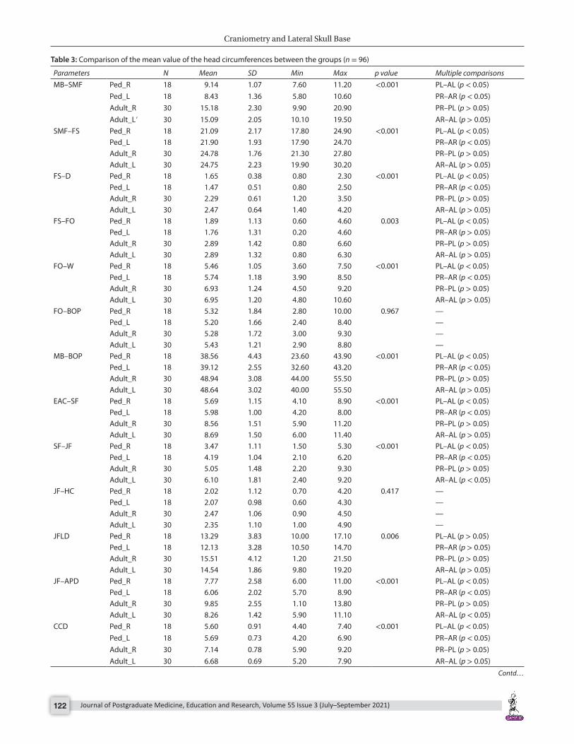
The mean values of the distances were further compared between four subgroups (pediatric left, pediatric right, adult left, adult right) for each of the variables using one-way ANOVA test (Table 3). The result showed that mean distances were significantly different in at least one pair among the groups for the variable, that is, MB–SMF, SMF–FS, FS–D, FS_FO, FO–W, MB_BOP, EAC–SF, SF–JF, JFLD, CCD, ITFC–BOP, ROZ–FO, ROZ–FS, FO–BOP, and JF–HC, while for FO–BOP and JF–HC, mean difference was not statistically significant. The variables for which the one-way ANOVA test was significant, multiple comparisons (using the Bonferroni method) were performed. The result showed that in most of the variables, mean difference was significant between the groups (adult and pediatric) that are pediatrics left to adult left and pediatric right to adult right (p < 0.05) while within the groups (within pediatrics or adults), there were no significant differences observed in the left and right measurements (p > 0.05).
di s c u s s i o n When we compared the adult skull base surgery with pediatric skull base surgery, the smaller size of the pediatric skull base has to be considered. Understanding the presence and location of specific critical landmarks of the skull base is essential in a pediatric case while making keyholes and craniotomy. Dentition and facial bone growth should be taken into consideration.6,7
Thorough knowledge of the surgical anatomy of infratemporal fossa is essential to treat the various pathological conditions of this anatomical region safely. The distal portion of the cervical internal carotid artery, internal jugular vein, and the extracranial part of cranial nerves are closely related to the infratemporal fossa.4 There is variation between right and left of the skull base foramina concerning shape and size in the same individual, besides the variation between sex and race.8 There is a distinct difference in the size skull base in children, and also there is significant inconsistency in the usual anatomical landmarks when compared with the adult skull base.9 The rate of growth of the calvarial when compared to the skull base is quite contrasting. The cranial vault grows rapidly, but
Table 1: Definition of the chosen distances
S. no. Abbreviation Distance1. MB–SMF Mastoid base to stylomastoid foramen2. SMF–FS Stylomastoid foramen to foramen
spinosum3. FS–D Foramen spinosum diameter4. FS–FO Foramen spinosum to foramen ovale5. FO–W Foramen ovale width6. FO–BOP Foramen ovale to the base of lateral ptery-
goid7. MB–BOP Mastoid base to base of lateral pterygoid8. EAC–SF Lateral end of bony EAC to stylomastoid
foramen9. SF–JF Stylomastoid foramen to jugular foramen
10. JF–HC Jugular foramen to the hypoglossal canal11. JFLD Jugular foramen longitudinal diameter 12. JF–APD Jugular foramen anterior–posterior
diameter13. CCD Carotid canal diameter 14. ITF-c–BOP Infratemporal crest to base of lateral
pterygoid15. ROZ–FO The root of zygoma to foramen ovale16. ROZ–FS The root of zygoma to foramen spinosum
Table 2: Comparisons between pediatrics and adults (n = 96)
Parameters Mean Std. deviation p valueMB–SMF Pediatric 8.78 1.25 <0.001
Adult 15.14 2.16SMF–FS Pediatric 21.49 2.07 <0.001
Adult 24.77 1.99FS–D Pediatric 1.56 0.45 <0.001
Adult 2.38 0.62FS–FO Pediatric 1.83 1.21 <0.001
Adult 2.89 1.36FO–W Pediatric 5.60 1.11 <0.001
Adult 6.94 1.21FO–BOP Pediatric 5.26 1.73 0.779
Adult 5.35 1.48MB–BOP Pediatric 38.84 3.58 <0.001
Adult 48.79 3.03EAC–SF Pediatric 5.84 1.07 <0.001
Adult 8.62 1.49SF–JF Pediatric 3.83 1.13 <0.001
Adult 5.57 1.72JF–HC Pediatric 2.04 1.04 0.103
Adult 2.41 1.07JFLD Pediatric 12.71 3.56 0.001
Adult 15.02 3.21JF–APD Pediatric 6.91 2.44 <0.001
Adult 9.06 2.20CCD Pediatric 5.64 0.81 <0.001
Adult 6.91 0.76ITF-c–BOP Pediatric 14.59 1.99 <0.001
Adult 19.05 2.12ROZ–FO Pediatric 20.91 3.71 <0.001
Adult 31.75 3.02ROZ–FS Pediatric 20.53 2.17 <0.001
Adult 28.79 2.70Independent samples t test used, p < 0.05 significant
Craniometry and Lateral Skull Base
Journal of Postgraduate Medicine, Education and Research, Volume 55 Issue 3 (July–September 2021)122
Table 3: Comparison of the mean value of the head circumferences between the groups (n = 96)
Parameters N Mean SD Min Max p value Multiple comparisonsMB–SMF Ped_R 18 9.14 1.07 7.60 11.20 <0.001 PL–AL (p < 0.05)
Ped_L 18 8.43 1.36 5.80 10.60 PR–AR (p < 0.05)Adult_R 30 15.18 2.30 9.90 20.90 PR–PL (p > 0.05)Adult_L′ 30 15.09 2.05 10.10 19.50 AR–AL (p > 0.05)
SMF–FS Ped_R 18 21.09 2.17 17.80 24.90 <0.001 PL–AL (p < 0.05)Ped_L 18 21.90 1.93 17.90 24.70 PR–AR (p < 0.05)Adult_R 30 24.78 1.76 21.30 27.80 PR–PL (p > 0.05)Adult_L 30 24.75 2.23 19.90 30.20 AR–AL (p > 0.05)
FS–D Ped_R 18 1.65 0.38 0.80 2.30 <0.001 PL–AL (p < 0.05)Ped_L 18 1.47 0.51 0.80 2.50 PR–AR (p < 0.05)Adult_R 30 2.29 0.61 1.20 3.50 PR–PL (p > 0.05)Adult_L 30 2.47 0.64 1.40 4.20 AR–AL (p > 0.05)
FS–FO Ped_R 18 1.89 1.13 0.60 4.60 0.003 PL–AL (p < 0.05)Ped_L 18 1.76 1.31 0.20 4.60 PR–AR (p > 0.05)Adult_R 30 2.89 1.42 0.80 6.60 PR–PL (p > 0.05)Adult_L 30 2.89 1.32 0.80 6.30 AR–AL (p > 0.05)
FO–W Ped_R 18 5.46 1.05 3.60 7.50 <0.001 PL–AL (p < 0.05)Ped_L 18 5.74 1.18 3.90 8.50 PR–AR (p < 0.05)Adult_R 30 6.93 1.24 4.50 9.20 PR–PL (p > 0.05)Adult_L 30 6.95 1.20 4.80 10.60 AR–AL (p > 0.05)
FO–BOP Ped_R 18 5.32 1.84 2.80 10.00 0.967 —Ped_L 18 5.20 1.66 2.40 8.40 —Adult_R 30 5.28 1.72 3.00 9.30 —Adult_L 30 5.43 1.21 2.90 8.80 —
MB–BOP Ped_R 18 38.56 4.43 23.60 43.90 <0.001 PL–AL (p < 0.05)Ped_L 18 39.12 2.55 32.60 43.20 PR–AR (p < 0.05)Adult_R 30 48.94 3.08 44.00 55.50 PR–PL (p > 0.05)Adult_L 30 48.64 3.02 40.00 55.50 AR–AL (p > 0.05)
EAC–SF Ped_R 18 5.69 1.15 4.10 8.90 <0.001 PL–AL (p < 0.05)Ped_L 18 5.98 1.00 4.20 8.00 PR–AR (p < 0.05)Adult_R 30 8.56 1.51 5.90 11.20 PR–PL (p > 0.05)Adult_L 30 8.69 1.50 6.00 11.40 AR–AL (p > 0.05)
SF–JF Ped_R 18 3.47 1.11 1.50 5.30 <0.001 PL–AL (p < 0.05)Ped_L 18 4.19 1.04 2.10 6.20 PR–AR (p < 0.05)Adult_R 30 5.05 1.48 2.20 9.30 PR–PL (p > 0.05)Adult_L 30 6.10 1.81 2.40 9.20 AR–AL (p < 0.05)
JF–HC Ped_R 18 2.02 1.12 0.70 4.20 0.417 —Ped_L 18 2.07 0.98 0.60 4.30 —Adult_R 30 2.47 1.06 0.90 4.50 —Adult_L 30 2.35 1.10 1.00 4.90 —
JFLD Ped_R 18 13.29 3.83 10.00 17.10 0.006 PL–AL (p > 0.05)Ped_L 18 12.13 3.28 10.50 14.70 PR–AR (p > 0.05)Adult_R 30 15.51 4.12 1.20 21.50 PR–PL (p > 0.05)Adult_L 30 14.54 1.86 9.80 19.20 AR–AL (p > 0.05)
JF–APD Ped_R 18 7.77 2.58 6.00 11.00 <0.001 PL–AL (p < 0.05)Ped_L 18 6.06 2.02 5.70 8.90 PR–AR (p < 0.05)Adult_R 30 9.85 2.55 1.10 13.80 PR–PL (p > 0.05)Adult_L 30 8.26 1.42 5.90 11.10 AR–AL (p < 0.05)
CCD Ped_R 18 5.60 0.91 4.40 7.40 <0.001 PL–AL (p < 0.05)Ped_L 18 5.69 0.73 4.20 6.90 PR–AR (p < 0.05)Adult_R 30 7.14 0.78 5.90 9.20 PR–PL (p > 0.05)Adult_L 30 6.68 0.69 5.20 7.90 AR–AL (p > 0.05)
Contd…

Craniometry and Lateral Skull Base
Journal of Postgraduate Medicine, Education and Research, Volume 55 Issue 3 (July–September 2021) 123
the base of the skull grows relatively slowly.10,11 The rapid growth of skull base occurs in the first half of the first decade of life, after which the growth is gradual and continues until the adolescent age. Genetics plays a vital role in the growth of the skull base and is independent of the neural elements.10
While performing a systematic measurement of various landmarks of the skull base, we compared the values with those in the literature. A study by Bejjani et al.4 describes the dimensions of the foramina in the roof of the infratemporal fossa. This study included only adult dry skulls. We could not find a study describing the skull base landmarks in a pediatric skull.
The distance between the mastoid base and stylomastoid foramen was measured; the mean value in the adult skull bone was 15.14mm with a standard deviation of 2.16 mm, Bejjani et al.4 have reported this parameter to be 10 mm in their study. The pediatric skull had a mean value of 8.78 mm with a standard deviation of 1.25 mm. Comparison of the distance between mastoid base to stylomastoid foramen between the adult and pediatric skull bones showed a significant p value (p < 0.05). In contrast, a comparison of the distance between the two sides in adult and pediatric skull bones did not show statistical significance. The distance between the lateral end of bony EAC to stylomastoid foramen was measured in the adult skull bone and the pediatric skull bone. The mean value in the adult skull bone was 8.62 mm with a standard deviation of 1.49 mm. Bejjani et al.4 have reported this parameter to be 8.7 mm. The pediatric skull had a mean value of 5.84 mm with a standard deviation of 1.07 mm. Comparison of the distance from the lateral end of bony EAC to the stylomastoid foramen between the adult and pediatric skull bones showed a significant p value (p < 0.05). Comparison of the distances between the right and left sides in adult as well as pediatric skull bones did not show statistical significance. These parameters are of primary importance because of its relevance with the location of the facial nerve. It is well-known that the mastoid process is absent at birth and its growth occurs in the first decade of life; this makes the facial nerve more superficial and interferes during surgery.12 In pediatric skulls, the mastoid tip is not fully developed, and with our study clearly stating the reduced distance between the mastoid tip and the stylomastoid foramen, the facial nerve exit point is more lateral when compared to that of the adults. Thus, we should anticipate the facial nerve to be more lateral in a pediatric patient to avoid iatrogenic injury during the surgical procedure.
The next landmark, when proceeding from lateral to the medial direction along the skull base, is the FS. The distance between the stylomastoid foramen to the FS was measured; the mean value in the adult skull bone was 24.77 mm with a standard deviation of 1.99 mm while the pediatric skull had a mean value of 21.49 mm with a standard deviation of 2.07 mm. Comparison of the distance between the stylomastoid foramen to the FS was compared between the adult, and the pediatric skull bones showed a significant p value (p < 0.05). This parameter guides us in locating and thus avoiding injury to the middle meningeal artery that is encountered during infra temporal fossa (ITF) dissection. This dimension in the adult skull in our study is comparable with the study by Bejjani et al.4 The distance between the stylomastoid foramen to the FS in a pediatric skull is significantly less, thus denoting more lateral location of the middle meningeal artery in children compared to adults. The FS diameter was also measured; the mean value in the adult skull bone was 2.38 mm with a standard deviation of 0.62 mm. This value was 2.1 mm in the study by Bejjani et al.4 The pediatric skull had a mean value of 1.56 with a standard deviation of 0.45 mm. Comparison between the adult and pediatric skull bones showed a significant p value (p < 0.05), thus signifying the relatively slender caliber of the middle meningeal artery and other contents of the FS in children and hence signifies extra caution while performing pediatric surgeries.
The major landmark medial to the FS is the FO. The distance between the FS to the FO was measured in adult skull bone and pediatric skull bone. The mean value in the adult skull bones was 2.89 mm with a standard deviation of 1.36 mm, while pediatric skulls had a mean value of 1.83 with a standard deviation of 1.21 mm. Comparison of the distance between the FS to the FO and between the adult and pediatric skull bones showed a significant p value (p < 0.05) on the left side alone; there was no significant value on comparing the right side. Bejjani et al.4 had also measured this distance; their observation was 3.5 mm. A significant difference of the distance between the FS to the FO between the adult and pediatric skull on the left side maybe because of a difference in the growth patterns of the base of the skull. This inference cannot be made with this study and warrants an analysis on a larger scale and significantly large pediatric skull data.
The FO width measures showed a mean value of 6.94 mm with a standard deviation of 1.21 mm in the adult skull bone, whereas the pediatric skull had a mean value of 5.60 mm with a standard
Contd…
Parameters N Mean SD Min Max p value Multiple comparisonsITFC–BOP Ped_R 18 14.13 2.17 12.00 21.90 <0.001 PL–AL (p < 0.05)
Ped_L 18 15.05 1.75 11.70 18.20 PR–AR (p < 0.05)Adult_R 30 18.51 1.87 15.40 22.00 PR–PL (p > 0.05)Adult_L 30 19.60 2.24 14.70 24.00 AR–AL (p > 0.05)
ROZ–FO Ped_R 18 21.54 2.00 18.00 25.10 <0.001 PL–AL (p < 0.05)Ped_L 18 20.29 4.85 2.40 24.70 PR–AR (p < 0.05)Adult_R 30 31.83 3.10 25.90 37.10 PR–PL (p > 0.05)Adult_L 30 31.67 3.00 26.50 36.30 AR–AL (p > 0.05)
ROZ–FS Ped_R 18 20.59 2.15 17.40 26.00 <0.001 PL–AL (p < 0.05)Ped_L 18 20.46 2.25 16.90 26.00 PR–AR (p < 0.05)Adult_R 30 28.79 2.72 23.10 34.00 PR–PL (p > 0.05)Adult_L 30 28.80 2.71 21.80 34.00 AR–AL (p > 0.05)
One-way ANOVA test used. Multiple comparisons (Bonferroni) were used when ANOVA was significant. p < 0.05 significant (Ped, Pediatric; R, right; L, left)

Craniometry and Lateral Skull Base
Journal of Postgraduate Medicine, Education and Research, Volume 55 Issue 3 (July–September 2021)124
deviation of 1.11 mm. Comparison of the FO width between the adult and the pediatric skull bones showed a significant p value (p < 0.05), thus signifying a smaller and laterally placed mandibular nerve in the pediatric skull. The average size in various populations is 7.4 mm and 6.67 mm.3,4 Foramen ovale size is clinically crucial in procedures such as trigeminal rhizotomy done in trigeminal neuralgia and percutaneous biopsy of the cavernous sinus.3 The distance between the FO to the base of the lateral pterygoid was measured in the adult skull bone and the pediatric skull bone. The mean value in the adult skull bone was 5.35 mm with a standard deviation of 1.48 mm. The pediatric skull had a mean value of 5.26 mm with a standard deviation of 1.73 mm. This parameter was 3.08 mm in Bejjani et al.4 study. Knowledge about this parameter will give us the information about the central limit of the ITF, that is, the lateral pterygoid plates from the FO. This helps in avoiding injury to the vital vascular compartment medial to it.
The distance between the mastoid base to the base of lateral pterygoid was measured in the adult skull bone and the pediatric skull bone. The mean value in the adult skull bone was 48.79 mm with a standard deviation of 3.03 mm. This dimension was 48.5 mm in the Bejjani et al.4 study. The pediatric skull had a mean value of 38.84 with a standard deviation of 3.58 mm. Comparison of the distance from mastoid base to the base of lateral pterygoid between the adult and pediatric skull bones showed a significant p value (p < 0.05). Comparison of the distances between the right and left sides in adult as well as pediatric skull bones did not show statistical significance. This distance is the entire length of the ITF along the skull base in a posterior to anterior direction; thus guides about the extent of dissection of the ITF performed. This also marks the medial boundary of the ITF.
The JF longitudinal diameter mean value in the adult skull bone was 15.02 mm with a standard deviation of 3.21 mm. The pediatric skull had a mean value of 12.71 mm with a standard deviation of 3.56 mm. Comparison of the distance between the adult and pediatric skull bones showed no significant p value (p > 0.05). This shows that the JF attains its adult longitudinal width at a younger age. The anterior and posterior diamter of the JF was measured in the adult skull bone and the pediatric skull bone. The mean value in the adult skull bone was 9.06 mm with a standard deviation of 2.20 mm. In Bejjani et al.,4 the anteroposterior width was 15 mm. The pediatric skull had a mean value of 6.91 mm with a standard deviation of 2.44 mm. Comparison of the width between the adult and pediatric skull bones showed a significant p value (p < 0.05). Comparison of the width between the right and left sides in adult showed statistically significant value (p < 0.05), while pediatric skull bones did not show statistical significance. This shows that the right JF is wider than the left in our study population, which is in concordance with the literature13 and the dominance is not seen in the pediatric skulls. Jugular foramen is intimately related to the distal extracranial ICA and the lower cranial nerve. In JF, the luminal packing is guarded as it can transmit pressure to the lower cranial nerve and lead to its palsy. Thus, knowledge about diameter is essential.
The mean distance between the root of the zygoma and the FO in adult was 31.75 mm and the mean value in the pediatric skull was 20.91 mm. The mean distance between the root of the zygoma and FS in the adult skull was 28.79 mm and in the pediatric skull was 20.53 mm. Both the parameters showed a statistically significant difference between the adult and pediatric skull. The root of the
zygoma is a constant bony landmark; it is seldom distorted by pathologies. The pediatric parameters are almost 10 mm less than the adult parameters. Thus, the knowledge about these parameters is essential to avoid injury to the vascular and neural structures in the FS and ovale.
The distance from the infratemporal crest to the base of lateral pterygoid was measured in the adult skull bone and the pediatric skull bone. The mean value in the adult skull bone was 19.05 mm, with a standard deviation of 2.12 mm. The pediatric skull had a mean value of 14.59 mm with a standard deviation of 1.99 mm. Comparison of the distances from the infratemporal crest-to-base of the lateral pterygoid between the adult and the pediatric skull bones showed a significant p value (p < 0.05). Bejjani et al.4 study showed the distance to be 22 mm, which is comparable to our values of the adult skull. This parameter is a measure of the width of the entire ITF from the lateral margin to its medial most limit. This gives an idea about the depth of dissection performed during surgery, and the possible entry medial to the critical medial limit of ITF, that is, the plane between the styloid process and the pterygoid hamulus can be avoided, thus injury to the vascular structures can be avoided.14
The distance between the FO and the base of pterygoid measurements were similar in both the adult and pediatric skull, in the adult skull; the parameter measured a mean of 5.35 mm, while in the pediatric skull, it measured a mean of 5.26 mm, which had no statistically significant difference. Similarly, the distance between the JF and the HC also did not show any statistically significant differences between the adult and pediatric skulls. The clinical significance of this knowledge is not clear yet.
co n c lu s i o n Morphometric data of various foramen in the base of middle cranial fossa and its relation to critical anatomical landmarks analyzed between adult and pediatric skulls are useful for surgeons who perform surgeries in these regions, especially in the pediatric group of patients, to anticipate the vital structures and to prevent complications.
cl i n i c A l si g n i f i c A n c e Skull base procedures are considered to be highly complex mainly due to its complex anatomy. Understanding the anatomy and applying it in the surgical procedure is challenging even to the experienced surgeons. Thus, the comparative study gives the objective evidence of this complexity and aids in further understanding of the complex anatomy. Our study provides a standardized distance of all the vital neurovascular structures concerning the standard bony landmarks of the middle fossa base. With this knowledge, the surgeons can reduce complications by clearly anticipating the neurovascular structures and thus preserve them.
Ac k n ow l e d g M e n tsAuthors would like to thank the following institutions:
• The Department of Anatomy, King George's Medical University, Lucknow, 226003, Uttar Pradesh, India.
• The Department of Anatomy, Prasad Institute of Medical Sciences, Lucknow, 226401, Uttar Pradesh, India.

Craniometry and Lateral Skull Base
Journal of Postgraduate Medicine, Education and Research, Volume 55 Issue 3 (July–September 2021) 125
re f e r e n c e s 1. Aslan A, Balyan FR, Taibah A, et al. Anatomic relationships between
surgical landmarks in type b and type c infratemporal fossa approaches. Eur Arch Otorhinolaryngol 1998;255(5):259–264. DOI: 10.1007/s004050050054.
2. Dallan I, Lenzi R, Bignami M, et al. Endoscopic transnasal anatomy of the infratemporal fossa and upper parapharyngeal regions: correlations with traditional perspectives and surgical implications. Minim Invasive Neurosurg 2010;53(5-6):261–269. DOI: 10.1055/s-0030-1263105.
3. Karthikeyan G, Sankaran PK, Ragunath G, et al. Morphometric study of various foramina in the middle cranial fossa of the human skull. Indian J Clin Anat Physiol 2017;4(4):574–578.
4. Bejjani GK, Sullivan B, Salas-Lopez E, et al. Surgical anatomy of the infratemporal fossa: the styloid diaphragm revisited. Neurosurgery 1998;43(4):842–852. DOI: 10.1097/00006123-199810000-00072.
5. Sharma NA, Garud RS. Morphometric evaluation and a report on the aberrations of the foramina in the intermediate region of the human cranial base: A study of an indian population. Eur J Anat 2011;15(3):140–149.
6. Gil Z, Constantini S, Spektor S, et al. Skull base approaches in the pediatric population. Head Neck 2005;27(8):682–689. DOI: 10.1002/hed.20226.
7. Brockmeyer D, Gruber DP, Haller J, et al. Pediatric skull base surgery. Pediatr Neurosurg 2003;38(1):9–15. DOI: 10.1159/000067563.
8. Pereira GA, Lopes PT, Santos AM, et al. Morphometric aspects of the jugular foramen in dry skulls of adult individuals in southern brazil. J Morphol Sci 2017;27(1):3–5.
9. Kazahaya K. Lateral skull base approaches in pediatric skull base surgery. J Neurol Surg B Skull Base 2018;79(1):047–057. DOI: 10.1055/s-0038-1624572.
10. Gruber DP, Brockmeyer D. Pediatric skull base surgery. Pediatr Neurosurg 2003;38(1):2–8. DOI: 10.1159/000067558.
11. Hoyte DA. A critical analysis of the growth in length of the cranial base. Birth Defects Orig Artic Ser 1975;11(7):255–282.
12. Grinblat G, Prasad SC, Fulcheri A, et al. Lateral skull base surgery in a pediatric population: a 25-year experience in a referral skull base center. Int J Pediatr Otorhinolaryngol 2017;94:70–75. DOI: 10.1016/ j.ijporl.2017.01.017.
13. Lichtenstein D, Saifi R, Augarde R, et al. The internal jugular veins are asymmetric. Usefulness of ultrasound before catheterization. Intensive Care Med 2001;27(1):301–305. DOI: 10.1007/s001340000792.
14. Evans BT, Wiesenefld D, Clauser L, et al. Surgical approaches to the infratemporal fossa. In: Langdon JD, Berkovitz BK, Moxham BJ, ed. Surgical anatomy of the infratemporal fossa. London: Martin Dunitz; 2003. pp. 109–147.


