Original Article Colloid cyst of third ventricle: report ... · agnosed as adult hidrocephalus and...
10
Int J Clin Exp Med 2017;10(6):8819-8828 www.ijcem.com /ISSN:1940-5901/IJCEM0049832 Original Article Colloid cyst of third ventricle: report of 11 cases with transcallosal transforaminal and transcolumna fornicis approach and clinical, radiological features Ersin Hacıyakupoğlu 1 , Derviş Mansuri Yılmaz 2 , Burak Kınalı 3 , Taner Arpaç 4 , Tuana Akbaş 4 , Sebahattin Hacıyakupoğlu 5 1 Klinik für Wirbelsaulen Chirurgie und Neurotraumatologie, 8060, Zwickau, Germany; 2 Department of Neurosur- gery, Balcali Hospital, School of Medicine, Cukurova University, 01330, Adana, Turkey; 3 Department of Neuro- surgery, Tepecik Education and Research Hospital, İzmir, Turkey; Departments of 4 Radiology, 5 Neurosurgery, Acibadem Adana Hospital, School of Medicine, Acibadem University, Adana, Turkey Received January 29, 2017; Accepted April 23, 2017; Epub June 15, 2017; Published June 30, 2017 Abstract: We evaluated, the clinical findings, radiological evidences, operation technique, complications and out- come in third ventricle colloid cysts and assessed the most safe and easy surgical approach for the treatment. 11 cases of third ventricle colloid cyst who underwent transcallosal operation between 2009-2017 were analysed retrospectively. In 10 of these cases cyst was visualised through foramina monro and in nine of them cysts were easily removed. We additionally applied posterior inter fornicial approach to the 10 th case. In the 11 th case the cyst could not be visualised through foramina monro and was found to be burried in paranchyma between foramina monro and commissuria anterior. The cyst was removed through the incision performed to columna fornicis lying on the cyst. Up to our knowledge third ventricle colloid cyst at this location is not reported in literature. Transcallosal, transforaminal and interfornicial approach which enables total resection with low complication and reccurence rate can be estimated as the most reliable procedure. Keywords: Transcallosal transforaminal, transcolumna fornicis, colloid cyst, third ventricle, craniotomy, surgery, treatment Introduction Colloid cyst of 3rd ventricle comprises 0.3-2% of brain tumors. Although it is among uncom- mon and benign brain tumors neurologic dete- rioration and death is unavoidable if treatment is neglected. It is known to be an embryologic tumor but there is not a consensus about its origin. Therefore, ependymal cysts, paraphysial cyst, neuro epithelial cysts, choroid plexus cyst are the terms used for this tumor. Some authors consider that it has neuroectodermal origin and arises at the 7 th week of embrion, forms the ceiling of rostral diencephalon and diencepha- lon at the 10 th week and develops from the embrional remnant of completely regressed paraphysis. Some authors suggest that it is originated ectodermally from Rathke’s cleft cysts and suprasellar neuroenteric cysts. Ob- servation of ciliates cell, nonciliated microvilli, goblet cell, basal cell similar to respiratory and intestinal ephitelium by electron micoscopic examination supports this opinion. It is frequently seen at 2-5 th decades but cases aging between 2 months to 82 years are report- ed in literature. It is most frequently seen at the fourth decade and in males. Hereditary predis- position is not available but familarity have been reported [1-6]. It settles at the anterior half of the 3rd ventricle ceiling, between the fornixs and binds to cho- roid plexus with a loose fibrous band in 99% of the cases. Multiple cysts in different sizes can be observed in this region incidentally at autop- sy, Computed Tomography (CT) or Magnetic Resonance Imaging (MRI). Existence in posteri- or part of 3 rd ventricle, in septum pellucidum and vellum interpositum, also, rarely in 4 th ven- tricle, frontoparietal cortex, inside sella has also been reported.
Transcript of Original Article Colloid cyst of third ventricle: report ... · agnosed as adult hidrocephalus and...

Int J Clin Exp Med 2017;10(6):8819-8828www.ijcem.com /ISSN:1940-5901/IJCEM0049832
Original ArticleColloid cyst of third ventricle: report of 11 cases with transcallosal transforaminal and transcolumna fornicis approach and clinical, radiological features
Ersin Hacıyakupoğlu1, Derviş Mansuri Yılmaz2, Burak Kınalı3, Taner Arpaç4, Tuana Akbaş4, Sebahattin Hacıyakupoğlu5
1Klinik für Wirbelsaulen Chirurgie und Neurotraumatologie, 8060, Zwickau, Germany; 2Department of Neurosur-gery, Balcali Hospital, School of Medicine, Cukurova University, 01330, Adana, Turkey; 3Department of Neuro-surgery, Tepecik Education and Research Hospital, İzmir, Turkey; Departments of 4Radiology, 5Neurosurgery, Acibadem Adana Hospital, School of Medicine, Acibadem University, Adana, Turkey
Received January 29, 2017; Accepted April 23, 2017; Epub June 15, 2017; Published June 30, 2017
Abstract: We evaluated, the clinical findings, radiological evidences, operation technique, complications and out-come in third ventricle colloid cysts and assessed the most safe and easy surgical approach for the treatment. 11 cases of third ventricle colloid cyst who underwent transcallosal operation between 2009-2017 were analysed retrospectively. In 10 of these cases cyst was visualised through foramina monro and in nine of them cysts were easily removed. We additionally applied posterior inter fornicial approach to the 10th case. In the 11th case the cyst could not be visualised through foramina monro and was found to be burried in paranchyma between foramina monro and commissuria anterior. The cyst was removed through the incision performed to columna fornicis lying on the cyst. Up to our knowledge third ventricle colloid cyst at this location is not reported in literature. Transcallosal, transforaminal and interfornicial approach which enables total resection with low complication and reccurence rate can be estimated as the most reliable procedure.
Keywords: Transcallosal transforaminal, transcolumna fornicis, colloid cyst, third ventricle, craniotomy, surgery, treatment
Introduction
Colloid cyst of 3rd ventricle comprises 0.3-2% of brain tumors. Although it is among uncom-mon and benign brain tumors neurologic dete-rioration and death is unavoidable if treatment is neglected. It is known to be an embryologic tumor but there is not a consensus about its origin. Therefore, ependymal cysts, paraphysial cyst, neuro epithelial cysts, choroid plexus cyst are the terms used for this tumor. Some authors consider that it has neuroectodermal origin and arises at the 7th week of embrion, forms the ceiling of rostral diencephalon and diencepha-lon at the 10th week and develops from the embrional remnant of completely regressed paraphysis. Some authors suggest that it is originated ectodermally from Rathke’s cleft cysts and suprasellar neuroenteric cysts. Ob- servation of ciliates cell, nonciliated microvilli, goblet cell, basal cell similar to respiratory and
intestinal ephitelium by electron micoscopic examination supports this opinion.
It is frequently seen at 2-5th decades but cases aging between 2 months to 82 years are report-ed in literature. It is most frequently seen at the fourth decade and in males. Hereditary predis-position is not available but familarity have been reported [1-6].
It settles at the anterior half of the 3rd ventricle ceiling, between the fornixs and binds to cho-roid plexus with a loose fibrous band in 99% of the cases. Multiple cysts in different sizes can be observed in this region incidentally at autop-sy, Computed Tomography (CT) or Magnetic Resonance Imaging (MRI). Existence in posteri-or part of 3rd ventricle, in septum pellucidum and vellum interpositum, also, rarely in 4th ven-tricle, frontoparietal cortex, inside sella has also been reported.
Colloid cysts: management of third ventricle colloid cysts
8820 Int J Clin Exp Med 2017;10(6):8819-8828
Most of the cases do not have neurologic defi-cit and are diagnosed incidentally. The most frequent symptom is headache and vomiting, transient diplopia, blurred vision, weakness in lower extermities, drop attack, mental detoria-tion may be the accompanying symptoms. Coma and sudden death is reported [2, 5, 7-11].
We present 11 cases with 3rd ventricle colloid cyst who except one underwent transcallosal transforaminal operation between 2009-2017 at Acıbadem Adana Hospital and University of
Cukurova, Balcali Hospital. We evaluated the complaints of patients, radiological evidences, operation technique and complications of col-loid cysts.
Case report
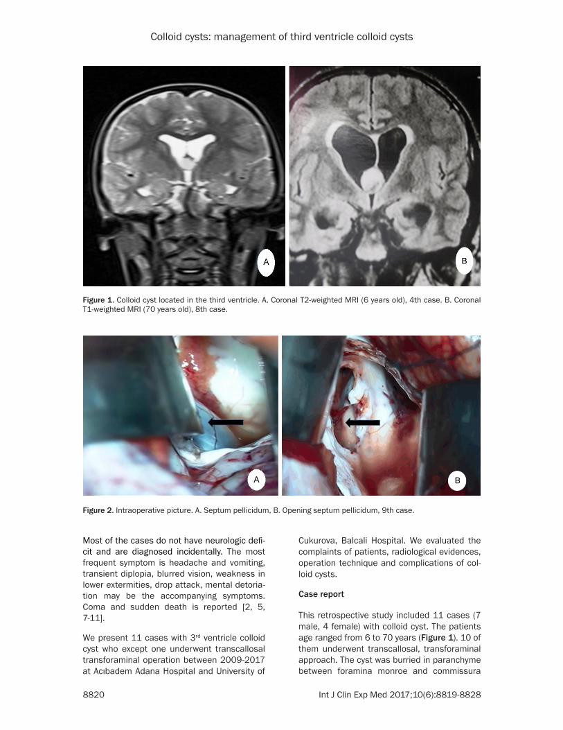
This retrospective study included 11 cases (7 male, 4 female) with colloid cyst. The patients age ranged from 6 to 70 years (Figure 1). 10 of them underwent transcallosal, transforaminal approach. The cyst was burried in paranchyme between foramina monroe and commissura
Figure 1. Colloid cyst located in the third ventricle. A. Coronal T2-weighted MRI (6 years old), 4th case. B. Coronal T1-weighted MRI (70 years old), 8th case.
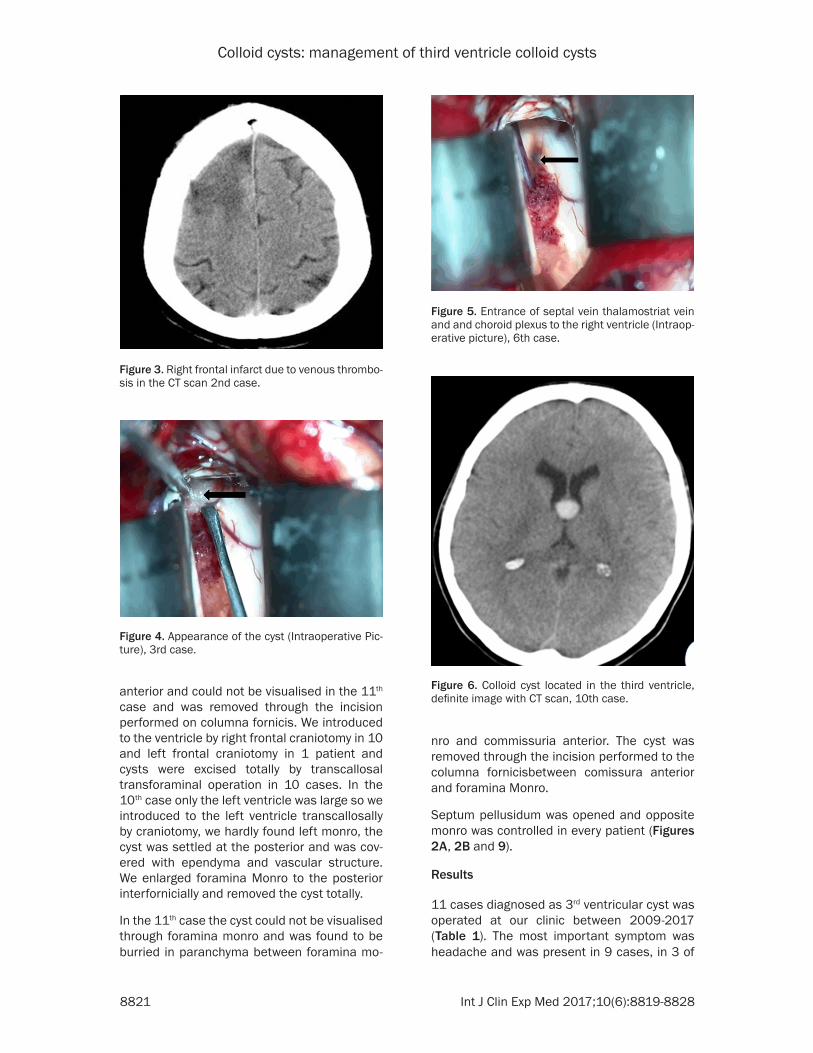
Figure 2. Intraoperative picture. A. Septum pellicidum, B. Opening septum pellicidum, 9th case.
Colloid cysts: management of third ventricle colloid cysts
8821 Int J Clin Exp Med 2017;10(6):8819-8828
anterior and could not be visualised in the 11th case and was removed through the incision performed on columna fornicis. We introduced to the ventricle by right frontal craniotomy in 10 and left frontal craniotomy in 1 patient and cysts were excised totally by transcallosal transforaminal operation in 10 cases. In the 10th case only the left ventricle was large so we introduced to the left ventricle transcallosally by craniotomy, we hardly found left monro, the cyst was settled at the posterior and was cov-ered with ependyma and vascular structure. We enlarged foramina Monro to the posterior interfornicially and removed the cyst totally.
In the 11th case the cyst could not be visualised through foramina monro and was found to be burried in paranchyma between foramina mo-
nro and commissuria anterior. The cyst was removed through the incision performed to the columna fornicisbetween comissura anterior and foramina Monro.
Septum pellusidum was opened and opposite monro was controlled in every patient (Figures 2A, 2B and 9).
Results
11 cases diagnosed as 3rd ventricular cyst was operated at our clinic between 2009-2017 (Table 1). The most important symptom was headache and was present in 9 cases, in 3 of
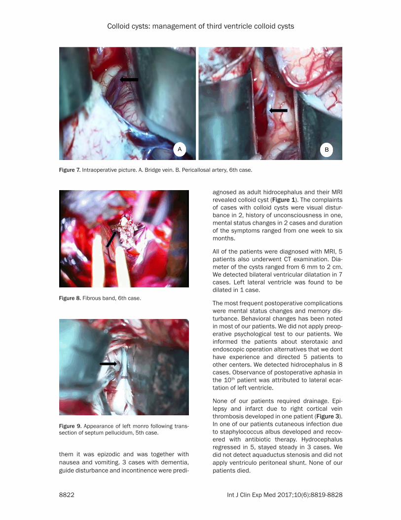
Figure 3. Right frontal infarct due to venous thrombo-sis in the CT scan 2nd case.
Figure 4. Appearance of the cyst (Intraoperative Pic-ture), 3rd case.
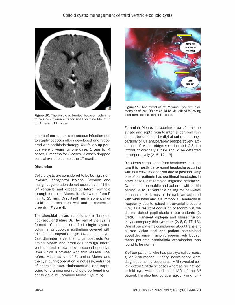
Figure 5. Entrance of septal vein thalamostriat vein and and choroid plexus to the right ventricle (Intraop-erative picture), 6th case.
Figure 6. Colloid cyst located in the third ventricle, definite image with CT scan, 10th case.
Colloid cysts: management of third ventricle colloid cysts
8822 Int J Clin Exp Med 2017;10(6):8819-8828
them it was epizodic and was together with nausea and vomiting. 3 cases with dementia, guide disturbance and incontinence were predi-
agnosed as adult hidrocephalus and their MRI revealed colloid cyst (Figure 1). The complaints of cases with colloid cysts were visual distur-bance in 2, history of unconsciousness in one, mental status changes in 2 cases and duration of the symptoms ranged from one week to six months.
All of the patients were diagnosed with MRI, 5 patients also underwent CT examination. Dia- meter of the cysts ranged from 6 mm to 2 cm. We detected bilateral ventricular dilatation in 7 cases. Left lateral ventricle was found to be dilated in 1 case.
The most frequent postoperative complications were mental status changes and memory dis-turbance. Behavioral changes has been noted in most of our patients. We did not apply preop-erative psychological test to our patients. We informed the patients about sterotaxic and endoscopic operation alternatives that we dont have experience and directed 5 patients to other centers. We detected hidrocephalus in 8 cases. Observance of postoperative aphasia in the 10th patient was attributed to lateral ecar-tation of left ventricle.
None of our patients required drainage. Epi- lepsy and infarct due to right cortical vein thrombosis developed in one patient (Figure 3). In one of our patients cutaneous infection due to staphylococcus albus developed and recov-ered with antibiotic therapy. Hydrocephalus regressed in 5, stayed steady in 3 cases. We did not detect aquaductus stenosis and did not apply ventriculo peritoneal shunt. None of our patients died.
Figure 7. Intraoperative picture. A. bridge vein. B. Pericallosal artery, 6th case.
Figure 8. Fibrous band, 6th case.
Figure 9. Appearance of left monro following trans-section of septum pellucidum, 5th case.

Colloid cysts: management of third ventricle colloid cysts
8823 Int J Clin Exp Med 2017;10(6):8819-8828
Table 1. Clinical, radiological features and treatment results of patients
Patient Age Gender Clinical Symptoms and signs
CT Scan
MRI T1 weight
MRI T2 weight Surgical Procedure Postoperative
ComplicationFollow-up
Period (Month)Clinical and
Radiological Result1 38 M HA, N, V - Hypo Hyper Transcallosal Transforaminal Mental Disturbance 36 Stable2 35 M Unconsciousness Hypo Hyper Hyper Transcallosal Transforaminal MSC, MD 12 Stable3 56 M HA, DDI Iso Hypo Hypo Transcallosal Transforaminal Behavioral Changes 6 Stable4 6 F HA, N, V Iso Hypo Hyper Transcallosal Transforaminal bC 6 Stable5 28 F HA, VD - İso Hyper Transcallosal Transforaminal BC, cutaneous infection 1 Stable6 31 F HA, MSC - Hypo Hypo Transcallosal Transforaminal MSC, bC 6 Stable7 45 M HA, VD - Hyper Hypo Transcallosal Transforaminal MSC, bC 12 Stable8 70 F DDI Iso İso Hyper Transcallosal Transforaminal bC 1 Stable9 42 M HA, N, V - Hyper Hypo Transcallosal Transforaminal MD, bC 1 Stable10 63 M HA, DDI Hyper Hypo Hypo Transcallosal İnterfornicial MSC, Aphasia 12 Stable11 39 M HA, MSC Hyper İso Hyper Transcallosal Transcolumna Fonicis Epilepsy, MD, Hemiparesis 12 StableHA: Headache, N: Nausea, V: Vomiting, BC: Behavioral changes, DDI: Demans Guide disturbance Urinary incontinance, VD: Visual disturbance, MSC: Mental status change, MD: Mental disturbance.
Colloid cysts: management of third ventricle colloid cysts
8824 Int J Clin Exp Med 2017;10(6):8819-8828
In one of our patients cutaneous infection due to staphylococcus albus developed and recov-ered with antibiotic therapy. Our follow up peri-ods were 3 years for one case, 1 year for 4 cases, 6 months for 3 cases. 3 cases dropped control examinations at the 1st month.
Discussion
Colloid cysts are considered to be benign, non-invasive, congenital lesions. Seeding and malign degeneration do not occur. It can fill the 3rd ventricle and exceed to lateral ventricle through foramina Monro. Its size varies from 5 mm to 25 mm. Cyst itself has a spherical or ovoid semi-translucent wall and its content is greenish (Figure 4).
The choroidal plexus adhesions are fibrinous, not vascular (Figure 8). The wall of the cyst is formed of pseudo stratified single layered columnar or cuboidal epithelium covered with thin fibrous capsula single layered ependym. Cyst diamater larger than 1 cm obstructs For- amina Monro and protrudes through lateral ventricle and is coated with second ependym layer which is covered with thin vessels. The- refore, visualisation of Foramina Monro and the cyst during operation is not easy, entrance of choroid plexus, thalamostriate and septal veins to foramina monro should be found inor-der to visualize Foramino Monro (Figure 5).
Foramina Monro, outpouring area of thalamo striate and septal vein to internal cerebral vein should be detected by digital subraction angi-ography or CT angiography preoperatively. Exi- stence of wide bridge vein located 2-3 cm infront of coronary suture should be detected intraoperatively [2, 8, 12, 13].
9 patients complained from headache. In litera-ture it is mostly paroxysmal headache occuring with ball-valve mechanism due to position. Only one of our patients had positional headache, in other cases it resembled migraine headache. Cyst should be mobile and adhered with a thin pedincule to 3rd ventricle ceiling for ball-valve mechanism. But, most of the cysts are adhered with wide base and are immobile. Headache is frequently due to raised intracranial pressure (ICP) as a result of occlusion of Monro but, we did not detect papil stasis in our patients [2, 14-16]. Transient diplopia and blurred vision may accompany this symptom [2, 6, 9, 17, 18]. One of our patients complained about transient blurred vision and one patient complained about decrease in vision preoperatively. Both of these patients ophthalmic examination was found to be normal.
3 of our patients who had paroxysmal demans, guide disturbance, urinary incontinance were diagnosed as hidrocephalus. MRI revealed col-loid cyst in 2 of these cases whereas iso intense colloid cyst was unnoticed in MRI of the 3rd patient. He also had cortical atrophy and lum-
Figure 10. The cyst was burried between columna fornics commisura anterior and Foramino Monro in the CT scan, 11th case.
Figure 11. Cyst infront of left Monroe. Cyst with a di-mension of 2×1.98 cm could be visualised following inter fornicial incision, 11th case.

Colloid cysts: management of third ventricle colloid cysts
8825 Int J Clin Exp Med 2017;10(6):8819-8828
bar puncture (LP) was applied considering the recovery of symptoms but following the detoria-tion of the status CT examination was per-formed and colloid cyst was diagnosed. MRI Scanning technique failed to predict the viscos-ity of the cyst contents and in this respect CT scan can be considered to be superior (Figure 6).
Mental status changes, memory disturbance, emotional, personality changes, deterioration in consciousness, ataxia, visual disturbance, symptoms resembling korsakoff syndrome may develop due to impression of the cyst to the surrounding area in cases with increased ICP. Only one of our patients complained from head-ache together with visual disturbance.
Colloid cyst have been diagnosed with pneumo-encephalography, ventriculography, angiogra-phy and brain scanning. Nowadays, brain MRI and CT is competence for diagnosis. Rounded or ovoid lesion lying in the region of the anterior third ventricle adjacent to foramina Monroe with 5-25 mm diameter is pathognomonic for colloid cyst.
MRI mostly reveals high signal and visualisa-tion of cysts containing blood and hemosiderin is better. Observation of high or low signal is not correlated with signs.
If the patient is presenting with headache or demantia, has neurologic symptoms, ventricu-lomegaly and high signal in T2 weighted MRI, cerebro spinal fluid circulation must be rees-tablished even if the patient is alert [5, 16, 18, 19].
Operation should be performed in patients with acute hidrocephalus, deep venous obstruction and acute neurologic deterioration due to impression to the hypothalamic regulatory sys-tem. Treatment of asymptomatic colloid cyst is controversial. Asymptomatic cyst in old patients with a diameter smaller than 8 mm can be fol-lowed up with MRI examination per year. Operation is decided if ventricular enlargement and neurologic symptoms develop.
8 of our cases had hydrocephalus, ventricular diameter was normal in 3 cases and no signs were present other than headache, nausea, vomiting, demantia, incontinance, guide distur-bance. The most catastrophic event associated with colloid cyst of the third ventricle is sudden death. Pallock [3] postulated that sudden death is not only due to the size of the tumor and acute ventricular dilatation or duration of symp-toms. Reflexes effecting cardiovascular centers near the third ventricle might also have played a role in these patients. Incidence of sudden death is not known and young patients should be operated because of this reason.
Figure 12. A. Colloid cyst located in the third ventricle (Preoperative Axial T1-weighted MRI); B. Appearance of 3rd ventricle through monro following the removal of colloid cyst (Postoperative CT scan), 7th case.

Colloid cysts: management of third ventricle colloid cysts
8826 Int J Clin Exp Med 2017;10(6):8819-8828
The aim of treatment is preventing the develop-ment of hydrocephalus, sudden neurologic deterioration and death. Extirpation and aspira-tion therapy is still controversial [2, 18, 20-23].
Besides transcallosal transforaminal surgery interhemispheric, frontal transcortical, subfron-tal or bifrontal anterior inter hemispheric app- roach through both lamina terminalis can be performed. Following transcallosal lateral ven-tricle insertion entrance to the 3rd ventricle can be performed through transforaminal, supra-choroidal, subchoroidal, transvellum inter posi-tum or interforniceal approach.
Endoscopic, stereotactic aspiration and exci-sion of the cyst wall are the other options for treatment. Cortical venous anatomy, pericallo-sal artery septal vein thalamo striate and inter-nal cerebral vein should be visualised and localisation of foramina Monro should be deter-mined by angiography prior to transcallosal operation. Frontal bridge vein lesion should be prevented in interhemispheric approach (Figure 7).
If the right side is not convenient entrance can be done from the left side. Following the ecarta-tion of the hemisphere infront of the coronary suture entrance to the right lateral ventricle through gyrus cinguli and corpus callosum can be performed.
Right Monro can be visualised by following cho-roid plexus. Colloid cyst looks grey if it is pro-truded to the lateral ventricle (Figure 4). However, it is not easily seen because it is cov-ered with double layered ependym, septal vein, thalamostriat vein and choroid plexus. If cho-roid plexus is not in frontal horn foramina Monro is settled posteriorly. Monro is enlarged frequently, cyst material can be punctured and aspirated inorder to perform internal decom- pression.
Fibrous bands of the capsule adhering to the ceiling of 3rd ventricule and choroid plexus can be cut following coagulation (Figure 8).
The wall of the cysts can be delivered though the foramen monro using microsurgical dissec-tion techniques, additonal techniques are rare-ly required. Following the removal of the cyst inorder to control the opposite Foramino Monro septum pellucid should be excised [4, 12, 13, 21-26] (Figure 9).
We removed 10 of the cysts with this technique foramina monro was narrow inthe 10th case only the left ventricle was large so we intro-duced to the left ventricle transcallosally by craniotomy, we hardly found left monro, the cyst was settled at the posterior and was cov-ered with ependyma and vascular structure.We enlarged foramina Monro to the posterior inter-fornicially and removed the cyst totally. Es- pecially in this patient recent memory distur-bance, motor aphasia and confusion lasting in 2 months developed. Major advantage of trans-callosal approach is lack of important deficit. Memory loss and hemiparesis are rare compli-cations. In cases without hydrocephalus reach-ing the mass is more easy, visualisation of both Foramino Monros is possible. If the lesion set-tles more posteriorly as seen in our case inter-forniceal approach can be prefferred. In this approach septum pellucidum is excised and midline forniceal raphe is identified. Excision is performed between two forniceal body begin-ing from monro to the posterior, if the excision is longer than 1-2 cm memory loss and hemipa-resis may develop due to hypocampal commis-ura trauma [8, 10, 17, 23, 27]. Although our insicion was small probably, ecartation of the ventricule in order to visualise Foramino Monro resulted with aphasia (10th case ). Recovery of aphasia in 2 months supports this wiev. Sy- mond [24] also recommends not to use ecarta-tion in ventricle because the genu of the inter-nal capsule lies in the inmediate subepandymal plane in the grove between the head of the cau-date nucleus and the thalamus.
The cyst was burried between columna fornics commisura anterior and Foramina Monro in the 11th case (Figure 10).
In order to enlarge the front of monro, posterior aspect of anterior commisura can be opened limitedly.
Therefore the cyst of the 11th case was removed totally through a plain incision at midline be- tween monro and anterior commisura (Figure 11).
More than 99% of cases with colloid cysts have been reported to occur within the third ventri-cle, almost all in the anterior half. Rare cases of colloid cysts were reported to occur in the pos-terior third ventricle and there are individual case reports of colloid cyst in the septum pel-lucidum, in the vellum inter positum, in the fourth ventricle, intra cerebral (A single case)

Colloid cysts: management of third ventricle colloid cysts
8827 Int J Clin Exp Med 2017;10(6):8819-8828
and intra sellar (A single case). We did not met a case of colloid cyst resembling the location of our 11th case in literature. Our case had indis-tinct left hemiparesis and memorial distur-bance for 1 month postoperatively.
In transcortical approach excission of conical block of cortex through middle frontal gyrus or entrance to lateral ventricule by linear cerebrot-omy, cyst can be delivered from monro. Inter- forniceal and transchoroidal entrance can not be applied with this approach, if ventricules are not dilated the risk of hemiparesis and epilepsy is high [4, 8, 19, 22, 23, 25].
Endoscopic surgery can be applied but total resection range is lower than trans callosal approach. Stereotactic aspiration can be app- lied as an urgent approach but reccurrence is known to be often [1, 12, 22, 27-32].
Cyst can be aspirated according to its viscosity [6, 9, 31].
Transcallosal approach is the most successful procedure in cerebrospinal fluid (CSF) obstruc-tion. However, despite the total excision of the cyst, obstruction can persist due to cyst con-tent and spilled blood (Figure 12).
Acqueduct stenosis may accompany colloid cyst. Therefore etiology of hydrocephalus must be clarified preoperatively. Mental status chan- ges and memory disturbance are the most com- mon complications of transcallosal operation [1, 2, 31]. These changes were observed in 9 of our patients, we don’t know if these changes were present before the operation because none of our patients underwent advanced neu-ropshchological evaluation. It develops due to the cyst itself or trauma to the fornix or sur-rounding tissue during the operation. Changes present before the operation becomes obvious following the operation. Cholinergic input flow from basal nucleus to hippocampus is effected in memory loss. Probably, fornical and hippo-campal nuclei functions deterioration due to operation. Another cause of memory loss is inferior thalamic pedincle injury lying from thal-amus dorso medial nucleus to amygdala. These pathways are related with fornix. Mc Mackin- stated that serious memory disturbance will not develop in cases with healty left fornix [19].
Following colloid cyst operation mutism, signifi-cant verbal deficit, stupor, obtundation, behav-ior changes, sectioning information, hyperter-
mia, hemiparesis, drowsiness, cranial nerve paralysis did not develop in our patients.
In one case we injured vein which was adherent widely to duramater and draining to superior sagittal sinus. Bleeding ceased with coagula-tion. Focal motor epilepsy dominant at the left arm developed at the postoperative 4th day. CT revealed venous infarct (Figure 3), antiepileptic drug was recommended for 1 month, the symp-toms were found to be recovered at the 1st month.
Conclusion
Transcallosal, transforaminal, interfornicial approach allows beter visualisation of both Foramina Monros and third ventricle. We can estimate that Transcollosal, transforaminal, interfornicial approach is an easy, safe proce-dure with low complication and reccurence rate for total resection of 3rd ventricule colloid cysts.
Disclosure of conflict of interest
None.
Address correspondence to: Dr. Derviş Mansuri Yıl- maz, Department of Neurosurgery, Balcalı Hospital, School of Medicine, Cukurova University, Adana 01330, Turkey. Tel: +905454558500; Fax: +90322- 3386988; E-mail: [email protected]
References
[1] Aggarwal A, Corbett A and Graham J. Familial colloid cyst of the third ventricle. J Clin Neuro-suci 1999; 6: 520-522.
[2] Laidlaw J and Kaye HA. Colloid cysts. Brain tu-mors. In: Kaye HA, editor. Laws RE Elsevier Ed-inburg; 2012. pp. 849-863.
[3] Pollock BE, Schreiner SA and Huston J 3rd. Atheory on the natural history of colloid cysts of the thrid ventricle. Neurosurgery 2000; 46: 1077-1083.
[4] Siwanuwatn R, Deshmukh P, Feiz-Erfan I, Rekate HL, Zabramski JM, Spetzler RF and Rosenfeld JV. Microsurgical anatomy of the transcallosal anterior interforniceal approach to the third ventricle. Neurosurgery 2005; 56: 390-396.
[5] Socin HV, Born J, Wallemacq C, Betea D, Legros JJ and Beckers A. Familial colloid cyst of the third ventricle: neuroendocrinological follow-up and rewiew of the literature. Clin Neurol Neurosurg 2002; 104: 367-370.
[6] Süzer T. Üçüncü ventrikül kolloid kistler. Türk Nöroşirurji Dergisi 2014; 24: 50-53.

Colloid cysts: management of third ventricle colloid cysts
8828 Int J Clin Exp Med 2017;10(6):8819-8828
[7] Ahmed SK and Stanworth PA. Colloid cyst of the third ventricle in indentical twins. Br J Neu-rosurg 2002; 16: 303-307.
[8] Apuzzo ML and Litofsky NS. Surgery in an around the anterior third ventricle. In: Apuzzo ML, editor. Brain surgery. New York: Churchill Livingstone; 1993. pp. 541-579.
[9] Kondziolka D and Lunsford LD. Factors predict-ing successful sterotactic aspiration of colloid cysts. Stereotact Funct Neurosurg 1992; 59: 135-138.
[10] Hingwala DR, Sangihvi DA, Shenoy AS, Dange NN and Geol AH. Colloid cyst of the velum in-terpositum: a common lesion at an uncommon site. Surg Neurol 2008; 72: 182-184.
[11] Symss NP, Ramamurthi R, Kapu R, Rao SM, Vasudevan MC, Pande A, Cugati G. Complica-tion avoidance in transcallosal transforaminal approach to colloid cysts of the anterior third ventriclen: an analysis of 80 cases. Asian J Neurosurg 2014; 9: 51-57.
[12] Abdou MS and Cohen AR. Endoscopic treat-ment of colloid cysts of the third ventricle: technical note and review of the literature. J Neurosurg 1998; 89: 106-1068.
[13] Desai KI, Nadkarni TD, Muzumdar DP and Goel AH. Surgical management of colloid cyst of the third ventricle-a study of 105 cases. Surg Neu-rol 2002; 57: 295-302.
[14] Amar PA, Albuguergue CF and Apsuzzo JLM. Anterior third ventricle lesion (In clindig colloid cysts). In: kaye HA, Black MP, editors. Opera-tive neurosurgery. London: Harcort Publishers; 2000. pp. 753-768.
[15] Coce N, Pavliša G, Nanković S, Jakovčević A, Seronia-Kuhar M and Pavliša G. Large hemor-rhagic colloid cyst in a 35-year-old male. Turk-ish Neurosurg 2012; 22: 783-784.
[16] Ganti SR, Antunes JL, Louis KM and Hilal SK. Computed tomography in the diagnosis of col-loid cysts of the third ventricle. Radiolog 1981; 138: 385-391.
[17] Camacho A and Kell JP. Colloid cyst of the third ventricle. In: Rengachary SJ, Wilkins HR, edi-tors. Principles of Neurosurgery Honkong Mos-by-Worte 1994; 36: 10.
[18] Pollock BE and Huston J 3rd. Natural history of asymptomatic colloid cysts of the third ventri-cle. J Neurosurg 1999; 91: 364-346.
[19] Mc Mackin D, Cockburn J, Anskow P and Gaf-fan D. Correlation of fornix damage with mem-ory impairment in six cases of colloid cyst re-moval. Acta Neurochir 1995; 135: 12-18.
[20] Demirci S, Doğan KH, Erkol Z and Gulmen MK. Sudden death due to colloid cyst of the third ventricle: report of there cases with a special sign at autopsy. Forensic Sci Int 2009; 189: e33-e36.
[21] De Witt Hamer, PC, Verstegen MJ, De Haan RJ, Vandertop WP, Thomeer RT, Mooij JJ and van
Furth WR. High risk of acute deterioration in patients harboring symptomatic colloid cysts of the third ventricle. J Neurosurg 2002; 96: 1041-1045.
[22] Gruen P and Appuzzo JLM. Third ventricle ex-posure by the ınterhemispheric corridor. In: Rengachary SS, Wilkins HR, editors. Neurogi-cal Operative Atlas. Illinois, The American As-sociation of Neurological Surgeon 1995; 4: 37-42.
[23] Yasargil MG and Abdlrauf SI. Surgery of intra-ventricular tumours. Neurosurgery 2008; 62: 1029-1041.
[24] Symon L, Calliauw L, Cohadon F, Guidetti B, Loew F, Nomes H. Pàsztor E, Pertuiset B, Pick-ard JD and Yaşargil MG. Surgical techniques in the management of colloid cysts of the third ventricle. Adv Tech Stand Neurosurg 1990; 17: 121-157.
[25] Türe U, Yasargil GM and Al-Mefty O. The trans-callosal-transforaminal approach to the third ventricle with regard to the venous variations in the region. J Neurosurg 1997; 87: 706-715.
[26] Easwer HV, Bhattacharya RN, Nair S, Rao BR, Menon G, Abraham M, Kumar KK. Precoronal, Paramedian minicraniotomy: a minimal access approach for microsurgical, transcallosal, transforaminal removal of colloid cysts of the third ventricle. Minim Invas Neurosurg 2008; 51: 253-257.
[27] Awasthi D and Kruse JJ. Excision of colloid cyst via the transcallosal approach. In: Rengachary SS, Wilkins HR, editors. IIIionis The American Association of Neurosurgeons 1997; 8: 227-234.
[28] Greenlee JD, Teo C, Ghahreman A and Kwok B. Purely endoscopic resection of colloid cysts. Neurosurgery 2008; 62 Suppl 1: 51-56.
[29] Hellwig D, Bauer BL, Schulte M, Gatscher S, Riegel T and Bertalanffy H. Neuroendoscopic treatment for colloid cysts of the third ventri-cle: the experience of a decade. Neurosurgery 2003; 52: 525-533.
[30] Longati P, Godano U, Gangemi M, Delitala A, Morace E, Genitori L, Alafaci C, Benvenuti L, Brunori A, Cereda C, Cipri S, Fiorindi A, Giorda-no F, Mascari C, Oppido PA, Perin A and Tripodi M. Cooperative study by the Italian neuroen-doscopy group on the treatment of 61 colloid cysts. Childs Nerv Syst 2006; 22: 1263-1267.
[31] Mathiesen T, Grane P, Lindquist C and von Holst H. High recurence rate following aspira-tion of colloid cysts in the third ventricle. J Neu-rosurg 1993; 78: 748-752.
[32] Mishra S, Chandra PS, Suri A, Rajender K, Sharma BS and Mahapatra AK. Endoscopic management of third ventricular colloid cysts: eight year’s institutional experience and de-scription of a new technique. Neurol India 2010; 58: 412-417.