ORIGINAL ARTICLE Cephalometric evaluation of the …moroortodontia.com.br/leitura/meaw 1999.pdf ·...
10
There are varieties of severe malocclusions that can be treated orthodontically but with a great deal of effort. Anterior open bite, in particular, is one mal- occlusion thought to be more difficult to treat, and therefore most have to be corrected by means of surgi- cal intervention. To solve these problems, numerous studies 1-11 pertinent to treatment modalities have been introduced with controversies on the effectiveness of treatment. Suggested treatment modalities for anterior open bite are based directly or indirectly on the neuro- muscular and morphologic features 12-18 and on the eti- ologic and the environmental factors. 19-22 Even though there have been controversies regard- ing the dentoalveolar characteristics of open bite mal- occlusions, many authors 17,19,21,23-26 report that upper and lower anterior teeth have already overerupted in skeletal open bite cases. Therefore extruding the overe- rupted anterior teeth by using anterior vertical elastics to achieve an overbite has been criticized as an invalid approach for stable results. 19,27,28 Among multiple etiologic factors, the most fre- quently discussed factor is the overeruption of the upper molars. Clinicians have emphasized the necessi- ty of reducing the vertical dimension of the upper pos- terior segments, or at least trying to prevent the extru- sion during orthodontic treatment. 2,8,9,13,26,29 According to Arat and Iseri, 30 posterior rotation of 29 ORIGINAL ARTICLE Cephalometric evaluation of the anterior open bite treatment Young Il Chang, DDS, MSD, PhD, a and Seong Cheol Moon, DDS, MSD b Seoul, Korea The present study was aimed at evaluating the treatment changes of anterior open bite malocclusion cases treated by means of the Multiloop Edgewise Arch Wire technique, which is considered one of the more effective treatment modalities for anterior open bite malocclusions. The open bite sample was composed of 16 young adults, 4 males and 12 females. The normal occlusion sample, as a controlled sample was composed of 58 young adults who had pleasing facial profiles and normal occlusions with no experience of orthodontic or prosthodontic treatment. The normal sample was subdivided by the cephalometric vertical facial relationships. Forty adults with cephalometric vertical facial relationships within the normal range of Korean standards were classified as Normal Occlusion Group 1. Eighteen adults with an increased vertical facial relationship but with normal occlusion, were classified as Normal Occlusion Group 2. Thirty-nine reference points were digitized on each film, and the computerized cephalometric analysis was obtained with 8 skeletal, 10 dentoalveolar, 17 teeth angulations, and 4 occlusal plane measurements.Treatment changes were determined by the paired t test, and the structural differences between the four groups were tabulated by the Student’s t test. The treatment changes were observed mainly in the dentoalveolar region in the upper and the lower occlusal planes, accompanied by the uprighting of the posterior teeth to the occlusal plane through the distal tipping movement of the entire dentition. After the treatment, there was a tendency for the structural feature of the open bite group to approximate those of the normal occlusion group 2. This ascertains that the treatment changes of open bite malocclusion produced by means of the multiloop edgewise arch wire technique are similar to those found in the natural dentoalveolar compensatory mechanism. (Am J Orthod Dentofacial Orthop 1999;115:29-38) a Professor of Orthodontics, College of Dentistry, Seoul National University. b Lecturer of Orthodontics, Saint Vincent Hospital, College of Medicine, The Catholic University of Korea. Reprint request to: Dr. Young Il Chang, Department of Orthodontics, College of Dentistry, Seoul National University, 28,Yeongkun-Dong, Chongro-Gu, Seoul, 110-749, Korea. Copyright © 1999 by the American Association of Orthodontists. 0889-5406/99/$8.00 + 0 8/1/89508 Fig 1. Diagram of the multiloop edgewise arch wire.
-
Upload
duongkhanh -
Category
Documents
-
view
215 -
download
0
Transcript of ORIGINAL ARTICLE Cephalometric evaluation of the …moroortodontia.com.br/leitura/meaw 1999.pdf ·...

There are varieties of severe malocclusionsthat can be treated orthodontically but with a great dealof effort. Anterior open bite, in particular, is one mal-occlusion thought to be more difficult to treat, andtherefore most have to be corrected by means of surgi-cal intervention. To solve these problems, numerousstudies1-11 pertinent to treatment modalities have beenintroduced with controversies on the effectiveness oftreatment. Suggested treatment modalities for anterioropen bite are based directly or indirectly on the neuro-muscular and morphologic features12-18and on the eti-ologic and the environmental factors.19-22
Even though there have been controversies regard-ing the dentoalveolar characteristics of open bite mal-occlusions, many authors17,19,21,23-26report that upperand lower anterior teeth have already overerupted inskeletal open bite cases. Therefore extruding the overe-rupted anterior teeth by using anterior vertical elastics to achieve an overbite has been criticized as an invalid
approach for stable results.19,27,28
Among multiple etiologic factors, the most fre-quently discussed factor is the overeruption of theupper molars. Clinicians have emphasized the necessi-ty of reducing the vertical dimension of the upper pos-terior segments, or at least trying to prevent the extru-sion during orthodontic treatment.2,8,9,13,26,29
According to Arat and Iseri,30 posterior rotation of
29
ORIGINAL ARTICLE
Cephalometric evaluation of the anterior open bite treatment
Young Il Chang, DDS, MSD, PhD, a and Seong Cheol Moon, DDS, MSD b
Seoul, Korea
The present study was aimed at evaluating the treatment changes of anterior open bite malocclusion casestreated by means of the Multiloop Edgewise Arch Wire technique, which is considered one of the moreeffective treatment modalities for anterior open bite malocclusions. The open bite sample was composed of16 young adults, 4 males and 12 females. The normal occlusion sample, as a controlled sample wascomposed of 58 young adults who had pleasing facial profiles and normal occlusions with no experience oforthodontic or prosthodontic treatment. The normal sample was subdivided by the cephalometric verticalfacial relationships. Forty adults with cephalometric vertical facial relationships within the normal range ofKorean standards were classified as Normal Occlusion Group 1. Eighteen adults with an increased verticalfacial relationship but with normal occlusion, were classified as Normal Occlusion Group 2. Thirty-ninereference points were digitized on each film, and the computerized cephalometric analysis was obtainedwith 8 skeletal, 10 dentoalveolar, 17 teeth angulations, and 4 occlusal plane measurements. Treatmentchanges were determined by the paired t test, and the structural differences between the four groups weretabulated by the Student’s t test. The treatment changes were observed mainly in the dentoalveolar regionin the upper and the lower occlusal planes, accompanied by the uprighting of the posterior teeth to theocclusal plane through the distal tipping movement of the entire dentition. After the treatment, there was atendency for the structural feature of the open bite group to approximate those of the normal occlusiongroup 2. This ascertains that the treatment changes of open bite malocclusion produced by means of themultiloop edgewise arch wire technique are similar to those found in the natural dentoalveolarcompensatory mechanism. (Am J Orthod Dentofacial Orthop 1999;115:29-38)
aProfessor of Orthodontics, College of Dentistry, Seoul National University.bLecturer of Orthodontics, Saint Vincent Hospital, College of Medicine, TheCatholic University of Korea.Reprint request to: Dr. Young Il Chang, Department of Orthodontics, College ofDentistry, Seoul National University, 28, Yeongkun-Dong, Chongro-Gu, Seoul,110-749, Korea.Copyright © 1999 by the American Association of Orthodontists.0889-5406/99/$8.00 + 08/1/89508
Fig 1. Diagram of the multiloop edgewise arch wire.
30 Chang and Moon American Journal of Orthodontics and Dentofacial OrthopedicsJanuary 1999
the mandible and increase of the lower facial heightoccur as a result of the extrusion of the posterior teeth,despite the fact that a high pull headgear was used inpremolar extraction cases. Similarly, Sato31 mentionedthat the increase of the posterior vertical dimension isdue to the mesial tipping of the molars during the pre-molar extraction treatment. Proffit32 reported that ahigh pull headgear can restrict the extrusion of uppermolars, but does allow the extrusion of the lowermolars. The favorable upper and forward rotation of themandible can hardly be achieved.
Kim33 has reported that anterior open bite is char-acterized by divergent upper and lower occlusal planesand marked mesial inclinations of the dentition in theopen bite skeletal pattern. The open bite skeletal pat-tern is determined by the overbite depth indicator(ODI)34 and the anteroposterior dysplasia indicator(APDI).35 The mean value of ODI is 74.5°, and theAPDI is 81.4°. Wardlaw,36 after scrutinizing the cepha-lometric measurements in the receiver operating char-acteristic analysis, ascertained that the ODI analysiswas found to be the most valuable analytic measure-ment among all other variables tested.
When the means of ODI and APDI, representingthe vertical and the horizontal components, are com-bined, the sum equals 155.9°, and it is designated as the
combination factor (CF).37The more the CF falls below155°, the greater the chance of an open bite. To correctthe characteristic features of open bite, Kim33 createdand developed the Multiloop Edgewise Arch Wire(MEAW) (Fig 1) technique and treated the malocclu-sion with success; a number of reports on treatment ofopen bite with MEAW therapy have appeared.38-40
The purpose of this study is to examine the treat-ment results of anterior open bite malocclusions treat-ed by means of MEAW technique and to compare thestructural changes between the normal group and theopen bite group. Appliance manipulation is discussedelsewhere.33
MATERIALTreatment Group
The treatment group is composed of 16 young adultopen bite patients treated by means of the MEAW tech-nique in the Department of Orthodontics, SeoulNational University (SNU) Hospital. To eliminate thegrowth factor, these patients were selected on the basisof wrist x-rays and the height-weight growth curve. Inaddition, extraction cases were excluded from thisstudy to eliminate any effects that may be caused bythe extraction treatment. The 16 patients were 4 malesand 12 females with the mean age of 18.1 years at the
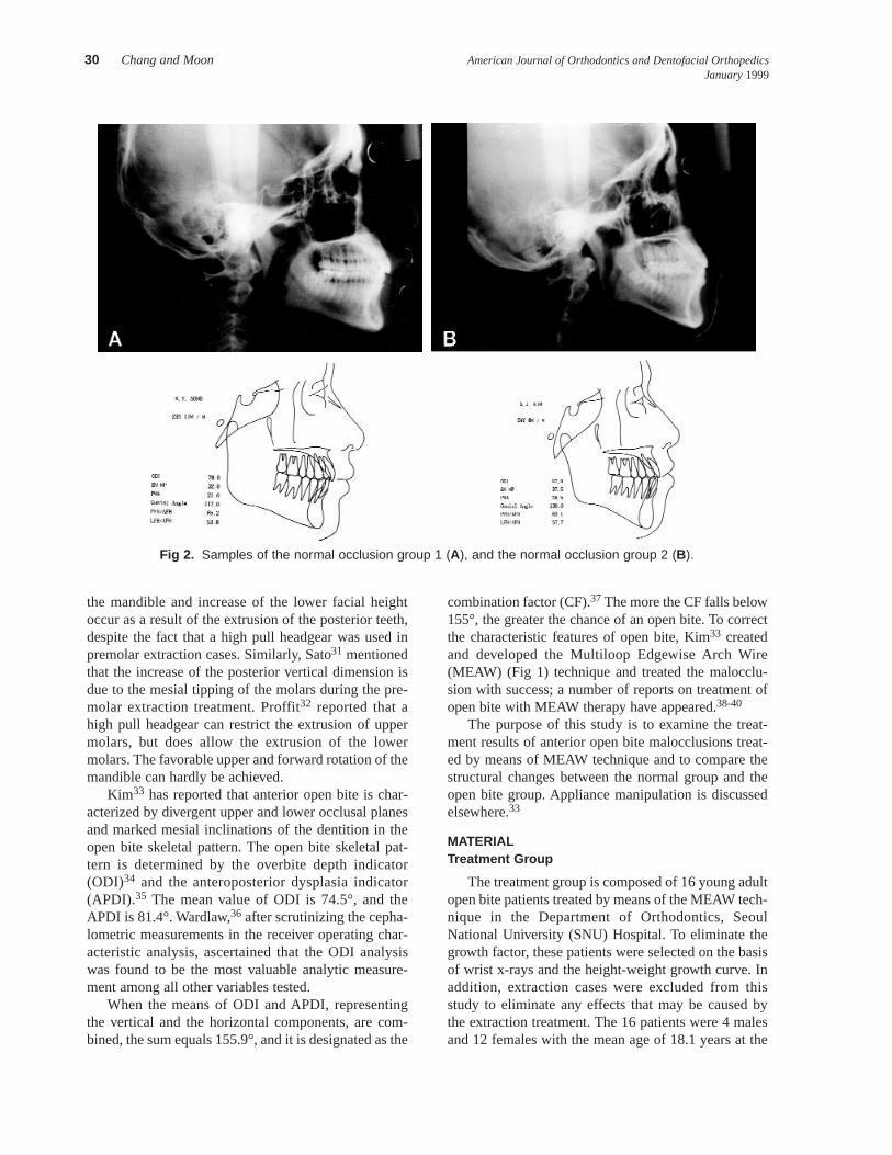
Fig 2. Samples of the normal occlusion group 1 (A), and the normal occlusion group 2 (B).

American Journal of Orthodontics and Dentofacial Orthopedics Chang and Moon 31Volume 115,Number 1
pretreatment stage and 19.8 years at the posttreatmentstage. Eight cases were Class I and the other eight wereClass II malocclusions. The mean treatment durationwas 1.6 years, and the mean duration of MEAW thera-py was 6 months.
Normal Occlusion Groups
Fifty-eight students of SNU dental school wereselected as the normal group who met the followingcriteria: (1) clinically pleasing facial profile, (2) esthet-ically and functionally favorable normal occlusion, and(3) no orthodontic or prosthodontic treatment experi-ence except fewer than three teeth restored for caries.
Because of the considerable variation that existed inthe vertical dimensions of the cephalograms, the nor-mal group was subdivided into Normal OcclusionGroup 1 (NOG 1) and Normal Occlusion Group 2(NOG 2). NOG 2 is composed of persons who showedless than 1 standard deviation (SD) from the meanvalue of the ODI and indicated an increased verticalrelationship that was verified by the following fivemeasurements; sella-nasion (SN)-mandibular planeangle, palatomandibular plane angle, gonial angle, andthe facial height ratios (posterior facial height/anteriorfacial height, anterior lower facial height/anterior totalfacial height). If more than three of the five measure-ments were more than 1 SD toward a long-face tenden-cy, these individuals were categorized as NOG 2, thegroup that has a skeletal pattern of an anterior open bitetendency (Fig 2). Eight female and 10 male subjectsfell into this group with a mean age of 21.72 years.
The remaining 40 subjects, 20 female and 20 malewith a mean age of 20.90 years, were classified asNOG 1 with the normal range of the vertical relation-ship (Fig 2).
METHODS
The cephalograms were obtained from the Depart-ment of Radiology at SNU Hospital. The enlargementfactor was found to be 5.6% in all films that ensuredthe consistency of the image. Thirty-nine referencepoints were marked on the tracing (Figs 3 and 4). Thepretreatment and posttreatment cephalograms weretraced and marked concurrently to achieve consistencyin the determination of the reference points.
The reference points were digitized into the com-puter. Eight skeletal, 10 dentoalveolar, 4 occlusalplane, and 17 tooth angular measurements were calcu-lated by a computer program created and used by ourdepartment. To reduce the error, all the measurementswere tabulated twice at 1 week intervals and the aver-age of the two measurements was recorded. In addi-tion, the intraobserver error by measuring the index ofreliability was evaluated.41 Bisected occlusal plane(BOP)-to-upper second premolar, BOP-to-lower firstmolar, BOP-to-lower second molar, and the mandibu-lar plane-to-lower second molar showed a reliability ofapproximately 0.85. All other measurements showed areliability of better than 0.9. The statistical analysiswas tabulated with SAS statistical package.
FINDINGSTreatment Changes
The means and standard deviations and the resultsof paired t test for the treatment group are listed inTable I. There were no significant changes in the skele-tal measurements, except an increase of ODI, whichwas due to the increase of the AB-to-mandibular planeangle.
Fig 3. Reference points.
Fig 4. Reference points.

32 Chang and Moon American Journal of Orthodontics and Dentofacial OrthopedicsJanuary 1999
The upper and lower anterior dentoalveolar heightswere increased, but the upper anterior dentoalveolarheight, measured along the long axis of the tooth, didnot change significantly. No significant change wasfound in the upper posterior dentoalveolar height. Thelower posterior dentoalveolar height was significantlydecreased.
There were significant changes in the distance of
the pterygomaxillary fissure (Ptm)-to-upper secondmolar and Ptm-to-lower second molar, which indicatedthe distal movement of the entire dentition. The upperocclusal plane showed downward rotation anteriorly,whereas the lower occlusal plane showed an upwardrotation.
The interincisal angle increased significantly. Allthe posterior teeth were found to be upright to the
Table I. Treatment changes of anterior open bite group
Pretreatment PosttreatmentVariables Mean SD Mean SD t Test
Skeletal measurement1. SN-MP angle 43.75 4.68 43.44 5.40 NS2. FH-PP angle 0.66 3.13 0.61 2.96 NS3. AB-MP angle 60.16 3.78 61.36 4.17 **4. ODI 60.86 5.28 61.99 5.49 *5. PP-MP angle 33.31 4.66 33.28 4.78 NS6. Gonial angle 129.01 6.34 128.45 6.01 NS7. PTFH/ATFH (%) 59.52 3.87 59.74 4.53 NS8. ALFH/ATFH (%) 56.87 2.09 57.09 1.81 NS
Dentoalveolar measurements9. U1-PP (UADH) 28.56 2.27 30.57 2.27 ***10. U7-PP (UPDH) 21.19 2.12 22.26 2.54 NS11. L1-MP (LADH) 43.14 2.31 45.92 3.50 ***12. L7-MP (LPDH) 30.31 2.07 29.27 2.45 **13. Ptm to U7 distal (mm) 4.61 1.79 3.71 2.03 **14. Ptm to L7 distal (mm) 5.91 2.66 3.98 2.58 ***15. Overbite -4.63 2.13 0.91 0.66 ***16. Overjet 2.62 1.81 3.24 1.00 NS17. UADH (long axis) 33.54 2.72 33.71 2.60 NS18. LADH (long axis) 43.59 2.44 46.66 3.75 ***
Occlusal plane measurements19. PP-UOP 6.98 3.71 9.89 4.62 **20. MP-LOP 21.22 3.26 26.20 4.39 ***21. PP-UOP/PP-MP 0.20 0.10 0.29 0.11 **22. MP-LOP/PP-MP 0.64 0.10 0.79 0.16 ***
Tooth axis measurements23. Interincisal angle 118.74 9.86 129.44 9.75 **24. BOP-U4 76.30 4.90 85.02 3.62 ***25. BOP-U5 82.86 3.74 89.78 3.04 ***26. BOP-U6 89.95 6.26 92.64 4.24 NS27. BOP-U7 98.04 7.81 99.04 6.91 NS28. BOP-L4 76.82 4.23 85.25 3.36 ***29. BOP-L5 77.99 4.72 87.05 3.39 ***30. BOP-L6 80.59 6.05 88.94 3.74 **31. BOP-L7 81.01 4.54 92.41 5.89 ***32. PP-U4 93.91 6.46 86.11 5.53 ***33. PP-U5 88.14 7.08 81.58 5.63 ***34. PP-U6 80.21 6.61 78.50 5.88 NS35. PP-U7 72.16 7.68 72.14 7.09 NS36. MP-L4 79.31 5.26 69.97 4.00 ***37. MP-L5 78.11 5.71 68.19 4.02 ***38. MP-L6 75.53 7.11 66.30 4.25 ***39. MP-L7 75.09 6.54 62.86 5.14 ***
NS, Not significant.*P < .05.** P < .01.*** P < .001.

American Journal of Orthodontics and Dentofacial Orthopedics Chang and Moon 33Volume 115,Number 1
bisected occlusal plane. The changes of the lower firstand second molars, however, were not statistically sig-nificant (P > .05). The upper premolars were upright inrelation to the palatal plane, but the lower first and sec-ond molars showed no statistical significance (P > .05).All of the lower posterior teeth were distally upright inrelation to the mandibular plane.
These findings indicate that the MEAW therapyminimally affected the skeletal pattern, correcting theopen bite by distal uprighting of the posterior teeth andby changing of the occlusal planes. In addition, thefindings indicate that the MEAW therapy not only pre-vented the extrusion of posterior teeth, but also intrud-ed them, especially lower posterior teeth.
Comparison of the Four Groups
The means and standard deviations of NOG 1,NOG 2, and the treatment group, and the results of thet test are listed in Tables II, III, IV, and V.
In the skeletal measurements, all variables showeda significant difference between the NOG 1 and NOG2, except the Frankfort horizontal (FH)-to-palatal plane
angle. There was no significant difference between theNOG 2 and the treatment group. It showed that NOG 2and the treatment group had the same verticallyincreased open bite skeletal pattern, but the verticalfacial relationship of NOG 1 was within the normalrange (Table II).
In the dentoalveolar measurements, the upper ante-rior dentoalveolar height of NOG 2 was significantlygreater than that of NOG 1. The lower posterior den-toalveolar height of NOG 2 was significantly smallerthan that of NOG 1. All of the measurements, exceptthe lower posterior dentoalveolar height of the pre-treatment group, were significantly smaller than thoseof the NOG 2. There was no significant differencebetween the NOG 2 and the posttreatment group(Table III).
The palatal plane-to-upper occlusal plane angle andthe mandibular plane-to-lower occlusal plane angle ofNOG 1 were significantly smaller than those of NOG 2.The ratio of both angles to the palatal plane-to-mandibu-lar plane angle showed no significant difference. In com-paring NOG 2 and the treatment groups, palatal plane-
Table II. Skeletal measurements
Variables NOG 1 NOG 2 Pretreatment Posttreatment
SN-MP angle 34.18 ± 5.31 41.68 ± 4.20* 43.75 ± 4.68 43.44 ± 5.40FH-PP angle 1.00 ± 2.21 0.69 ± 2.83 0.66 ± 3.13 0.61 ± 2.96AB-MP angle 69.20 ± 3.83 60.56 ± 2.95* 60.16 ± 3.78 61.36 ± 4.17ODI 70.23 ± 4.81 61.30 ± 4.01* 60.86 ± 5.28 61.99 ± 5.49PP-MP angle 23.74 ± 3.88 31.74 ± 3.53* 33.31 ± 4.66 33.28 ± 4.78Gonial angle 121.45 ± 4.16 131.56 ± 4.14* 129.01 ± 6.34 128.45 ± 6.01PFH/AFH (%) 66.99 ± 4.08 61.02 ± 3.43* 59.52 ± 3.87 59.74 ± 4.53LFH/TFH (%) 55.27 ± 1.37 56.21 ± 2.21* 56.87 ± 2.09 57.09 ± 1.81
*P < .05 (between NOG 1 and NOG 2).** P < .05 (between NOG 2 and pretreatment).*** P < .05 (between NOG 2 and posttreatment).
Table III. Dentoalveolar measurements
Variables NOG 1 NOG 2 Pretreatment Posttreatment
U1-PP (UADH) 29.99 ± 2.76 31.63 ± 2.40* 28.56 ± 2.27** 30.57 ± 2.27U7-PP (UPDH) 22.45 ± 2.48 22.94 ± 2.36 21.19 ± 2.12** 22.26 ± 2.54L1-MP (LADH) 45.36 ± 3.67 46.52 ± 3.21 43.14 ± 2.31** 45.92 ± 3.50L7-MP (LPDH) 33.16 ± 3.54 30.18 ± 2.33* 30.31 ± 2.07 29.27 ± 2.45Ptm to U7 distal (mm) 5.85 ± 2.52 5.81 ± 2.15 4.61 ± 1.79 3.71 ± 2.03***Ptm to L7 distal (mm) 5.93 ± 2.72 6.11 ± 2.68 5.91 ± 2.66 3.98 ± 2.58***Overbite 1.45 ± 0.79 1.39 ± 0.77 –4.63 ± 2.13** 0.91 ± 0.66***Overjet 3.19 ± 0.79 3.01 ± 0.80 2.62 ± 1.81 3.24 ± 1.00UADH’ (long axis) 33.70 ± 2.65 35.38 ± 2.51* 33.54 ± 2.72** 33.71 ± 2.60LADH’ (long axis) 46.22 ± 3.92 47.03 ± 3.29 43.59 ± 2.44** 46.66 ± 3.75
*P < .05 (between NOG 1 and NOG 2).** P < .05 (between NOG 2 and pretreatment).*** P < .05 (between NOG 2 and posttreatment).
34 Chang and Moon American Journal of Orthodontics and Dentofacial OrthopedicsJanuary 1999
to-upper occlusal plane angle and mandibular plane-to-lower occlusal plane angle of the pretreatment groupwere significantly smaller initially than those of NOG 2.After treatment, both angles were increased and showedno significant difference to those of NOG 2. The ratio ofthe two angles to the palatal plane-to-mandibular planeangle was increased during treatment and became simi-lar to those of NOG 2 (Table IV).
The mean interincisal angle was increased to 5.48°greater than that of NOG 2. Despite some variation inthe skeletal pattern, the NOG 1 and the NOG 2 weresimilar in the angle between all posterior teeth and theocclusal plane. After the treatment, all of the posteriorteeth were found to be upright in relation to theocclusal plane and they became more upright thanthose of NOG 2. In general, the angles between theupper posterior teeth and the palatal plane did not showany significant difference between the groups. Theseangles, however, showed a tendency to become smallerin NOG 2 and in the posttreatment group. The angles
between the lower posterior teeth and the mandibularplane of NOG 2 were found to be significantly smallerthan those of NOG 1. These angles in the pretreatmentgroup were more obtuse than those of NOG 2, butbecame acute after treatment.
In other words, the upper and the lower posteriorteeth were distally uprighted to the palatal plane andthe mandibular plane with treatment (Table V).
DISCUSSIONTreatment Changes
The ODI was increased due to the increase of theAB-to-mandibular plane angle. This change was prob-ably caused by the distal movement of the lower denti-tion and the B point during the treatment of eight ClassIII open bite patients. There were no significantchanges in other skeletal measurements. In some cases,10 of 16 cases, the upward and forward rotation of themandible and the decreasing of the lower face heightdue to the intrusion of the lower posterior teeth were
Table IV Occlusal plane measurements
Variables NOG 1 NOG 2 Pretreatment Posttreatment
PP-UOP 8.06 ± 2.96 9.84 ± 3.15* 6.98 ± 3.71** 9.89 ± 4.62MP-LOP 19.37 ± 3.21 26.22 ± 2.91* 21.22 ± 3.26** 26.20 ± 4.39PP-UOP/PP-MP 0.33 ± 0.10 0.30 ± 0.09 0.20 ± 0.10** 0.29 ± 0.11MP-LOP/PP-MP 0.82 ± 0.12 0.83 ± 0.13 0.64 ± 0.10** 0.79 ± 0.16
*P < .05 (between NOG 1 and NOG 2).** P < .05 (between NOG 2 and pretreatment).*** P < .05 (between NOG 2 and posttreatment).
Table V Tooth axis measurementsVariables NOG 1 NOG 2 Pretreatment Posttreatment
Interincisal angle 121.91 ± 7.51 123.96 ± 6.45 118.74 ± 9.86 129.44 ± 9.75BOP-U4 80.10 ± 4.17 82.83 ± 3.98* 76.30 ± 4.90** 85.02 ± 3.62BOP-U5 85.04 ± 3.85 87.56 ± 3.28* 82.86 ± 3.74** 89.78 ± 3.04BOP-U6 89.59 ± 4.00 90.37 ± 3.79 89.95 ± 6.26 92.64 ± 4.24BOP-U7 96.67 ± 5.33 95.76 ± 4.62 98.04 ± 7.81 99.04 ± 6.91BOP-L4 81.13 ± 4.24 83.02 ± 4.38 76.82 ± 4.23** 85.25 ± 3.36BOP-L5 82.38 ± 3.48 84.16 ± 4.43 77.99 ± 4.72** 87.05 ± 3.39***BOP-L6 83.58 ± 4.03 84.10 ± 3.98 80.59 ± 6.05** 88.94 ± 3.74***BOP-L7 81.27 ± 4.82 82.68 ± 4.10 81.01 ± 4.54 92.41 ± 5.89***PP-U4 93.19 ± 5.03 89.03 ± 4.62* 93.91 ± 6.46** 86.11 ± 5.53PP-U5 90.23 ± 7.29 84.36 ± 4.27* 88.14 ± 7.08 81.58 ± 5.63PP-U6 83.67 ± 4.65 81.47 ± 4.36 80.21 ± 6.61 78.50 ± 5.88PP-U7 76.65 ± 5.34 76.15 ± 4.98 72.16 ± 7.68 72.14 ± 7.09MP-L4 81.49 ± 4.57 73.01 ± 4.30* 79.31 ± 5.26** 69.97 ± 4.00***MP-L5 80.24 ± 4.16 71.87 ± 4.24* 78.11 ± 5.71** 68.19 ± 4.02***MP-L6 79.05 ± 4.70 71.88 ± 3.94* 75.53 ± 7.11 66.30 ± 4.25***MP-L7 81.35 ± 5.06 73.34 ± 3.47* 75.09 ± 6.54 62.86 ± 5.14***
*P < .05 (between NOG 1 and NOG 2).** P < .05 (between NOG 2 and pretreatment).*** P < .05 (between NOG 2 and posttreatment).

American Journal of Orthodontics and Dentofacial Orthopedics Chang and Moon 35Volume 115,Number 1
observed, but the treatment effect of the MEAWseemed confined to the dentoalveolar region.
The lower anterior teeth were extruded lingually, andthe alveolar bone showed definitive remodeling followedby the movement of the teeth. This finding is in agree-ment with the experimental study by Lee et al42 on mon-keys. It showed that marked tooth movement and con-siderable cellular activities took place in the monkeywith the use of MEAW therapy; the control monkeywithout MEAW therapy showed insignificant toothmovement and cellular activity. Proffit43 pointed out thatwhen an anterior open bite can be treated by elongatingthe incisors, it is better for both esthetics and stability toelongate the lower incisors, not the upper incisors.
As mentioned earlier, some clinicians emphasize acareful control of the posterior teeth in the treatment ofopen bites and recommend a high-pull headgear or aposterior bite-block.8,9,44According to the study on theforce system of MEAW by the finite element analy-sis,45 MEAW with second order tip-back activations onthe posterior segments exerts an uprighting force on theposterior teeth and an extrusive force on the anteriorteeth with anterior vertical elastics. In another studyusing photoelastic analysis,46 it was found that MEAWwith the activations has an intrusive effect on theincisors and the second molars without elastics, but theintrusive forces on the incisors are counteracted by theanterior vertical elastics. The consequence is the extru-sion of incisors. The molars, on the other hand, receivegreater intrusive force and a stronger uprighting effectfrom the anterior vertical elastic force.
From this study, it was evident that there was nosignificant change in the upper posterior dentoalveolarheight, but the lower posterior dentoalveolar height wassignificantly decreased. A sample case treated withMEAW therapy is shown in Figs 5, 6, 7, and 8.
Comparison of the four groups
NOG 2 and the treatment groups showed an increasedvertical dimension of the face, but NOG 1 showed thenormal range of the vertical facial relationship. Despitethe fact that NOG 2 had a similar open bite skeletal pat-tern with the treatment group, NOG 2 had normal occlu-sion. Björk and Skieller47 pointed out that the eruptionpath of the teeth should be changed to compensate for thepositional changes of the jaws during growth and devel-opment. If this process is absent or incomplete, a dyspla-sia of the occlusion and a space problem may appear.Because there is a large variation in the amount and thedirection of growth and because a perfect harmony of thegrowth cannot always be achieved in the jaws, Solow48
referred to the need for the dentoalveolar compensatorymechanism. The eruption of teeth and the change in theposition to compensate the interarch variation, therefore,can produce the normal occlusal relationship between thedental arches. This discussion suggests that the NOG 2,which has the open bite tendency skeletal pattern, canpossess a normal occlusal relationship by an adequatedentoalveolar compensation. The treatment group, whichhad a similar skeletal pattern to NOG 2, however, couldnot possess a normal occlusal relationship because of aninadequate dentoalveolar compensation.
Fig 5. Facial photographs of pretreatment (A), during (B), and after retention (C).
36 Chang and Moon American Journal of Orthodontics and Dentofacial OrthopedicsJanuary 1999
In evaluating the dentoalveolar region, NOG 2 wassignificantly greater than NOG 1 in the upper andlower anterior dentoalveolar heights. This difference isprobably due to the dentoalveolar compensation in
NOG 2. The pretreatment group showed smaller upperand lower anterior dentoalveolar heights than those ofNOG 2 but showed no difference after treatment. Thatprobably means that there was an inadequate den-
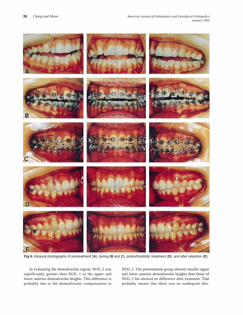
Fig 6. Intraoral photographs of pretreatment (A), during (B and C), postorthodontic treatment (D), and after retention (E).

American Journal of Orthodontics and Dentofacial Orthopedics Chang and Moon 37Volume 115,Number 1
toalveolar compensation in the pretreatment group rel-ative to the vertically increased jaw relationship andthat a proper dentoalveolar height was achieved aftertreatment.
The palatal plane-to-upper occlusal plane angle andthe mandibular plane-to-lower occlusal plane anglewere greater in NOG 2 than in NOG 1. Both angleswere smaller in the pretreatment group but showed nodifference after treatment. Nielsen49 defined the maxil-
lary zone as the palatal plane-to-upper occlusal planeangle, normally 10° ± 3°. The mandibular zone isdefined as the mandibular plane-to-lower occlusalplane angle, normally 20° ± 4°. He also reported that inthe case of an increase in the vertical jaw relationship,an increase in the maxillary zone and the mandibularzone means a favorable dentoalveolar compensation.
In the pretreatment group, the angulations of theposterior teeth to the occlusal plane were smaller thanthose of NOG 2. Kim33 noted the typical mesial incli-nation of the posterior teeth in open bite was in agree-ment with aforementioned findings. And the fact thatthose of NOG 2 tended to be larger than those of NOG1 concurs with the findings of Björk,47 who reportedthat there is a tendency of the distal inclination of theposterior teeth in the backward rotation pattern with anadequate dentoalveolar compensation. Those of thepretreatment group were increased during treatmentand became similar to those of NOG 2, or more uprightto the occlusal plane than those of NOG 2.
In summary, even though the vertical relationshipof the face is increased because of growth, the normalocclusal relationship can be achieved by the adequatedentoalveolar compensatory mechanism, but in thecase of inadequate or negative dentoalveolar compen-sation, open bite is likely to be present. If the skeletaldysplasia is too severe to be solved by orthodontictreatment alone, combined treatment with surgeryshould be done to restore the function and the estheticsof the orofacial complex. In many cases, however,orthodontic alteration of the dentition pertinent to thegiven skeletal pattern with the proper diagnosis andtreatment planning can bring satisfactory results.
Fig 7. Cephalograms of pretreatment (A), during (B),and after retention (C).
Fig 8. Superimposition of preorthodontic and post-orthodontic treatment.

38 Chang and Moon American Journal of Orthodontics and Dentofacial OrthopedicsJanuary 1999
The treatment changes with the MEAW therapyoccurred mainly in the dentoalveolar region andshowed a considerable similarity to the natural den-toalveolar compensatory mechanism. In other words,the MEAW technique allows orthodontists to producethe natural dentoalveolar compensation orthodontical-ly. The morphologic features of the posttreatmentgroup appeared to be like those of NOG 2, which hadnormal occlusion because of the natural dentoalveolarcompensation despite its open bite skeletal pattern.
Even if an open bite is corrected by the orthodonticdentoalveolar compensation suitable for the skeletalpattern, relapse may still occur from the persisting etio-logic factors that originally prohibited the natural den-toalveolar compensation. The etiologic factors shouldbe determined at the time of initial diagnosis and shouldbe controlled during treatment and retention.
CONCLUSION
The conclusions of this study are as follows:1. The treatment changes mainly occurred in the den-
toalveolar region.2. The treatment changes are alteration of occlusal
planes accompanied by uprighting of the posteriorteeth.
3. The treatment changes are similar to natural den-toalveolar compensation.
REFERENCES
1. Arvystas MG. Treatment of anterior skeletal open-bite deformity. Am J Orthod1977;72:147-64.
2. Dellinger EL. A clinical assessment of the active vertical corrector: a nonsurgicalalternative for skeletal open bite treatment. Am J Orthod Dentofacial Orthop1986;89:428-36.
3. Denison TF, Kokich VG, Shapiro PA. Stability of maxillary surgery in open bite ver-sus nonopen bite malocclusions. Angle Orthod 1989;59:5-10.
4. Fränkel R, Fränkel C. A functional approach to treatment of skeletal open bite. Am JOrthod 1983;84:54-68.
5. Fränkel R, Fränkel C. Functional aspects of molar extraction in skeletal open bite. In:Graber LW, ed. Orthodontics. state of the art: essence of the science. St Louis: CVMosby; 1986. p. 184-99.
6. Fränkel R. My way from the activator to the functional regulator. In: Hösl E, BaldaufA, editors. Mechanical and biological basics in orthodontic therapy. Heidelberg:Hüthig Buch; 1991. p. 63-78.
7. Huang GJ, Justus R, Kennedy DB, Kokich VG. Stability of anterior open bite treatedwith crib therapy. Angle Orthod 1990;60:17-25.
8. Iscan HN, Akkaya S, Koralp E. The effect of the spring-loaded posterior bite-block onthe maxillo-facial morphology. Eur J Orthod 1992;14:54-60.
9. Kuhn RJ. Control of anterior vertical dimension and proper selection of extraoralanchorage. Angle Orthod 1968;38:340-9.
10. Speidel TM, Isaacson RJ, Worms FW. Tongue-thrust therapy and anterior dental open-bite: a review of new facial growth data. Am J Orthod 1972;62:287-95.
11. Tuenge RH, Elder JR. Posttreatment changes following extraoral high-pull traction tothe maxilla of Macaca mulatta. Am J Orthod 1974;66:618-44.
12. Fields HW, Proffit WR. Facial pattern differences in long faced children and adults.Am J Orthod 1984;85:217-23.
13. Frost DE. Cephalometric diagnosis and surgical: orthodontic correction of apertog-nathia. Am J Orthod 1980;78:657-69.
14. Isaacson RJ. Extreme variation in vertical facial growth and associated variations inskeletal and dental relations. Angle Orthod 1971;41:219-29.
15. Nahoum HI. Vertical proportion and the palatal plane in anterior open-bite. Am JOrthod 1971;59:273-82.
16. Nahoum HI. Varieties of anterior open bite. Am J Orthod 1972;61:486-92.17. Nahoum HI. Vertical proportions: a guide for prognosis and treatment in anterior open-
bite. Am J Orthod 1977;72:128-46.18. Nahoum HI. Anterior open-bite: a cephalometric analysis and suggested treatment
procedures. Am J Orthod 1975;67:513-21.19. Subtelny JD. Open bite malocclusion: early and late diagnosis and correction. In:
Orthodontics: evaluation and future, proceedings of the international conference on theoccasion of the 25th anniversary of the orthodontic department of the University ofNijmegen, Netherland. Samson Stafleu, Alphen aan den Rijn, 1988. p. 239-60.
20. Linder-Aronson S. Respiratory function in relation to facial morphology and the den-tition. Br J Orthod 1979;6:59-71.
21. Straub W. Malfunction of the tongue, part 1. the abnormal swallowing habit: its caus-es, effects and results in relation to orthodontic treatment and speech therapy. Am JOrthod 1979;76:565-76.
22. Sweinhart E. A clinical study of open bite. Am J Orthod 1942;28:18-34.23. Wylie WL. The relationship between ramus height, dental height, and overbite. Am J
Orthod 1946;32:57-67.24. Ellis E, McNamara JA. Component of adult class III open-bite malocclusion. Am J
Orthod 1984;86:277-90.25. Nemeth RB, Isaacson RJ. Vertical anterior relapse. Am J Orthod 1974;65:565-85.26. Schudy FF. The rotation of the mandible resulting from growth; Its implications in
orthodontic treatment. Angle Orthod 1965;35:36-50.27. Subtelny JD, Sakuda M. Open bite; diagnosis and treatment. Am J Orthod
1964;50:337-58.28. Epker BN, Fish LC. Surgical-orthodontic correction of open-bite deformity. Am J
Orthod 1977;71:278-99.29. Schudy FF. The control of vertical overbite in clinical orthodontics. Angle Orthod
1968;38:19-39.30. Arat M, Iseri H. Orthodontic and orthopedic approach in the treatment of skeletal open
bite. Eur J Orthod 1992;14:207-15.31. Sato S, Suzuki Y. A new approach for treatment of malocclusion. Seoul: Ji-Sung Pub-
lishing Company; 1992 p. 101-10. 32. Proffit WR, Fields HW. Treatment of orthodontic problems in preadolescent children:
contemporary orthodontics, St. Louis: CV Mosby; 1986. p. 386-9.33. Kim YH. Anterior open bite malocclusion: nature, diagnosis and treatment by means
of multiloop edgewise archwire technique. Angle Orthod 1987;57:290-321.34. Kim YH. Overbite depth indicator(ODI) with particular reference to anterior open-
bite. Am J Orthod 1974;65:586-611.35. Kim YH, Vietas JJ. Anteroposterior dysplasia indicator (APDI): an adjunct to cephalo-
metric differential diagnosis. Am J Orthod 1978;73:619-33.36. Wardlaw DW. Cephalometrics of anterior open bite: a receiver operating characteris-
tic (ROC) analysis. Am J Orthod Dentofacial Orthop 1992;101:234-43.37. Kim YH, Chang YI, Chung WN. Overbite depth indicator, anteroposterior dysplasia
indicator, combination factor and extraction index. Int J MEAW 1994;1:11-32.38. Sato S. Dynamics of gnatho-facio complex and treatment approach to malocclusions.
Tokyo: Tokyo Clinical Publishing Co; 1991. p. 125-270.39. Kim YH. Treatment of malocclusions by means of multiloop edgewise archwire tech-
nic. J Assoc Orthodont Repub China 1995;7:62-72.40. Mestre JC. Reporte de un caso tratado con la retrotecnica (Mestre) y con la tecnica
(Kim). Revista Iberoamericana De Ortodoncia 1996;15:39-48.41. Houston WJB. The analysis of errors in orthodontic measurements. Am J Orthod
1983;83:382-90.42. Lee SY, Kim TW, Chang YI. Histologic changes in mandibular periodontium of the mon-
key following experimental extrusion of anterior teeth. Kor J Orthod 1995;25:415-24 .43. Proffit WR. Interarch elastics: their place in modern orthodontics. In: Mechanical and
biological basics in orthodontic therapy. Heidelberg: Hüthig Buch; 1991. p. 173-8.44. Pearson LE. Vertical control through use of mandibular posterior intrusive forces.
Angle Orthod 1973;43:194-200.45. Chun GM, Nahm DS. Mechanical analysis of the Multiloop edgewise archwire. Kor J
Orthod 1991;21:31-45.46. Yeom JB, Rhee BT. A photoelastic study of the stress distribution by multiloop edge-
wise archwire. Kor J Orthod 1990;20:267-81.47. Björk A, Skieller V. Facial development and tooth eruption: an implant study at the age
of puberty. Am J Orthod 1972;62:339-83.48. Solow B. The dentoalveolar compensatory mechanism: background and clinical
implication. Br J Orthod 1980;7:145-61.49. Nielsen IL. Vertical malocclusion: etiology, development, diagnosis and some aspects
of treatment. Angle Orthod 1991;61:247-60.