ORGANS OF IMMUNE SYSTEM PRIMARY AND SECONDARY LYMPHOID ORGANS
27
-
Upload
sruthy-chandran -
Category
Science
-
view
601 -
download
3
Transcript of ORGANS OF IMMUNE SYSTEM PRIMARY AND SECONDARY LYMPHOID ORGANS

ORGANS OF IMMUNE SYSTEM
PRIMARY AND SECONDARY
LYMPHOID ORGANS
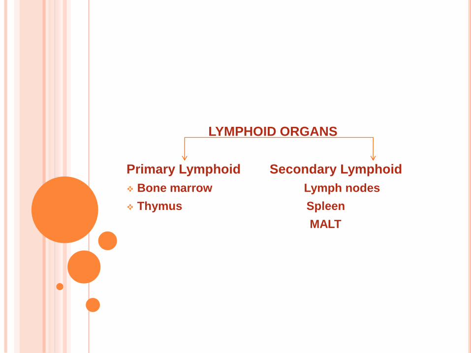
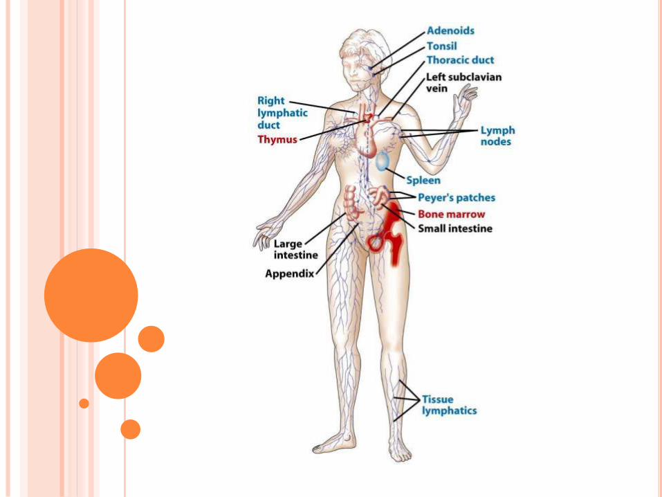
LYMPHOID ORGANS
Primary Lymphoid Secondary Lymphoid
Bone marrow Lymph nodes
Thymus Spleen
MALT
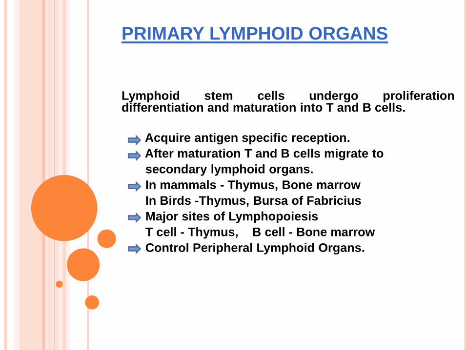
PRIMARY LYMPHOID ORGANS
Lymphoid stem cells undergo proliferationdifferentiation and maturation into T and B cells.
Acquire antigen specific reception.
After maturation T and B cells migrate to
secondary lymphoid organs.
In mammals - Thymus, Bone marrow
In Birds -Thymus, Bursa of Fabricius
Major sites of Lymphopoiesis
T cell - Thymus, B cell - Bone marrow
Control Peripheral Lymphoid Organs.
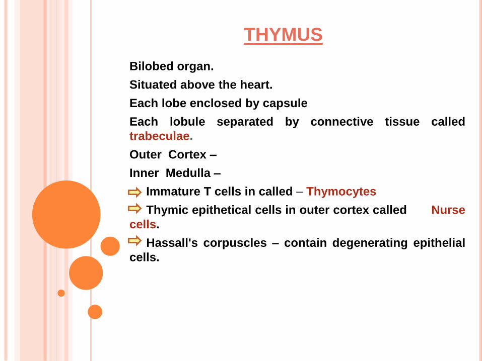
THYMUS
Bilobed organ.
Situated above the heart.
Each lobe enclosed by capsule
Each lobule separated by connective tissue called
trabeculae.
Outer Cortex –
Inner Medulla –
Immature T cells in called – Thymocytes
Thymic epithetical cells in outer cortex called Nurse
cells.
Hassall's corpuscles – contain degenerating epithelial
cells.
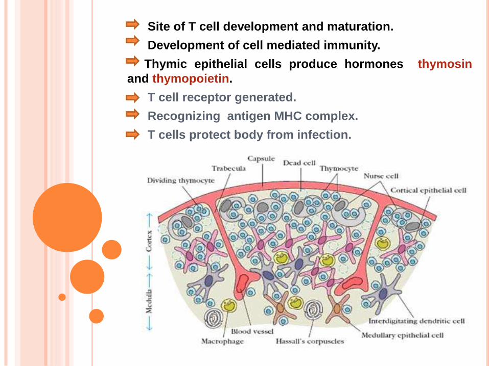
Site of T cell development and maturation.
Development of cell mediated immunity.
Thymic epithelial cells produce hormones thymosin
and thymopoietin.
T cell receptor generated.
Recognizing antigen MHC complex.
T cells protect body from infection.
Removal of thymus from newborn mice.
Decrease in circulating lymphocytes.
Absence of cell mediated immunity.
Increase in infectious disease.
Congential birth defect in humans
[ Diveorge’s Syndrome ]
Mice – mude mice
Aging – decline in thymic function
Maximal size at puberty.
BONE MARROW
Site of blood cell formation.
B cell origin and mature
E.g. Humans and Mice
Fat cells, bony tissue, dendritic cells
Stomatal cells interact with B cells
Secrete cytokines.
Selection process occur.
It is not the site of B cell development in all species.
BURSA OF FABRICIUS
Gut associated lymphoid organs.
[ Birds ]
Lymph epithelial tissue.
Hindgut of chicken.
Multiply and differentiate into B lymphocytes.
Immuno globulins synthesis.
Described by Fabricus in 1621.
Humoral immunity in birds.
Absent in mammals ( primates, rodents ).
SECONDARY LYMPHOID ORGANS
Organs in which antibodies are formed.
Antigen trapping and lymph filtration mechanism.
Receive immuno competenal cells (primary lymphoid
organ for making them and active).
Spleen
Lymph nodes
Mucosa associated lymphoid tissue.
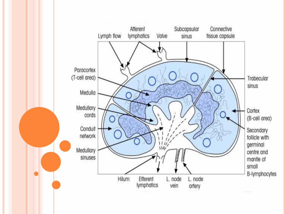
LYMPH NODES
Solid encapsulated bean shaped structure.
Seen in Armpits, Mesenteries.
Network packed with lymphocytes, macrophages, dendritic
cells.
Three concentric regions :-
Cortex , Para cortex, Medulla
CORTEX :-
Outer most layer
Contains lymphocytes, macrophage, follicular dendritic
cells arranged in primary follicle
Lymphoid tissues organized into structures - lymphoid
follicle.
Lymphoid follicle activated by antigen – primary follicle
[ Follicular Dendritic Cell, Resting B Cell ]
Primary follicle develop into secondary follicle.
In children with B cell deficiency cortex lack primary
follicles and germinal centers.
PARACORTEX :-
[ T lymphocytes, interdigiting dendritic cells ].
Thymus dependent area – Para cortex
Thymus independent area – Cortex
Class II MHC present.
MEDULLA :-
Inner most layer
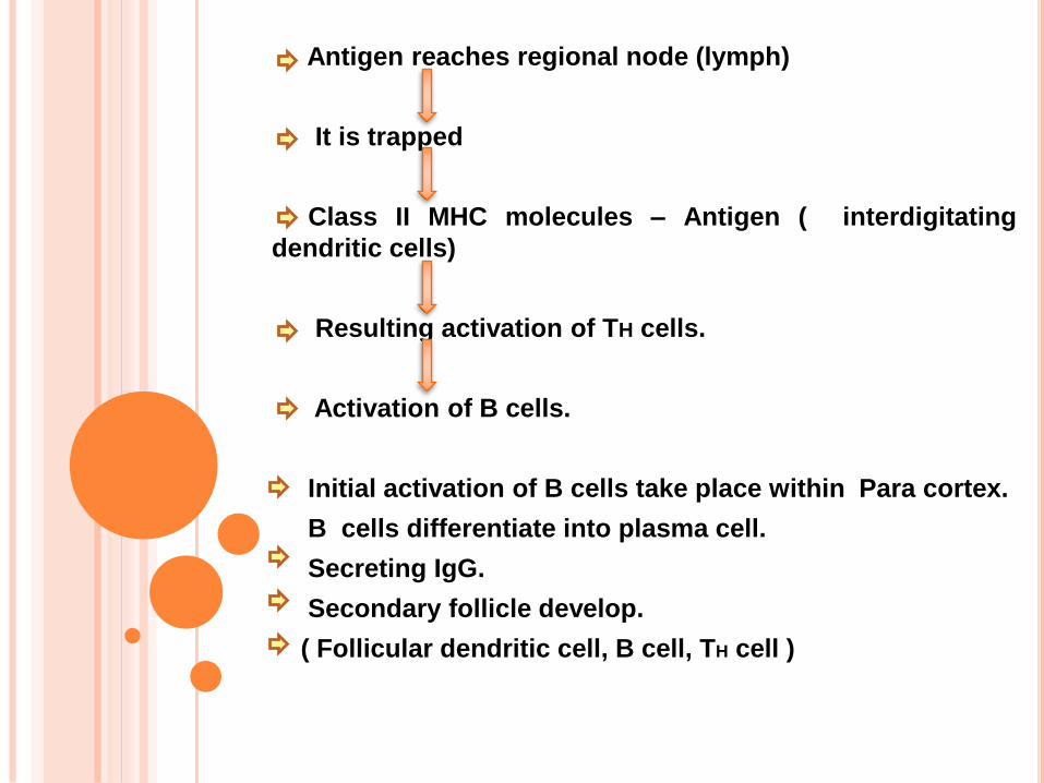
Antigen reaches regional node (lymph)
It is trapped
Class II MHC molecules – Antigen ( interdigitating
dendritic cells)
Resulting activation of TH cells.
Activation of B cells.
Initial activation of B cells take place within Para cortex.
B cells differentiate into plasma cell.
Secreting IgG.
Secondary follicle develop.
( Follicular dendritic cell, B cell, TH cell )
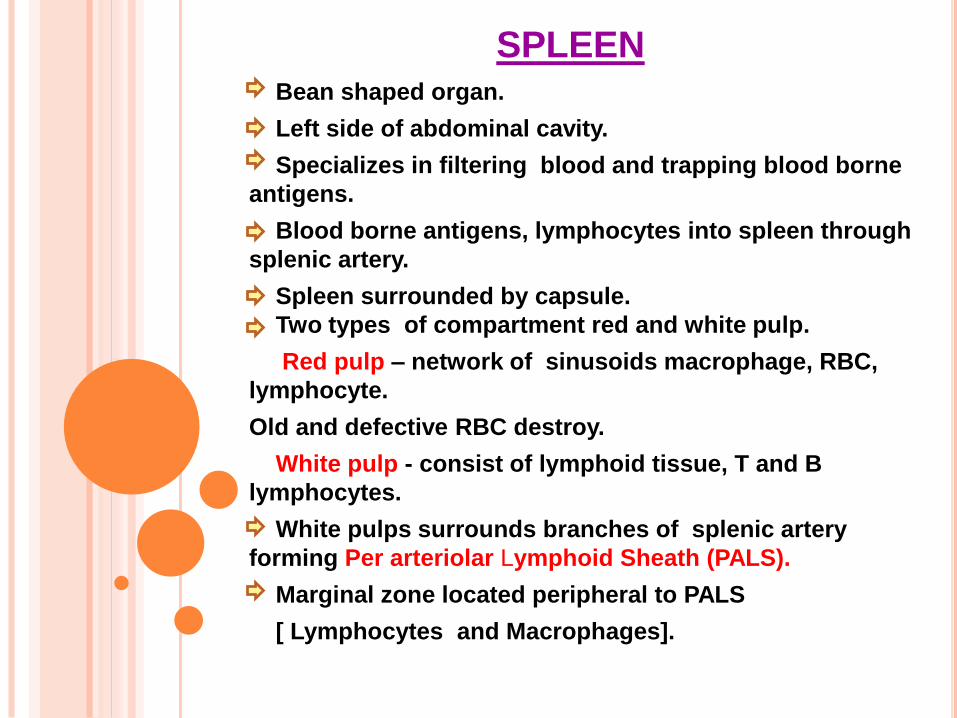
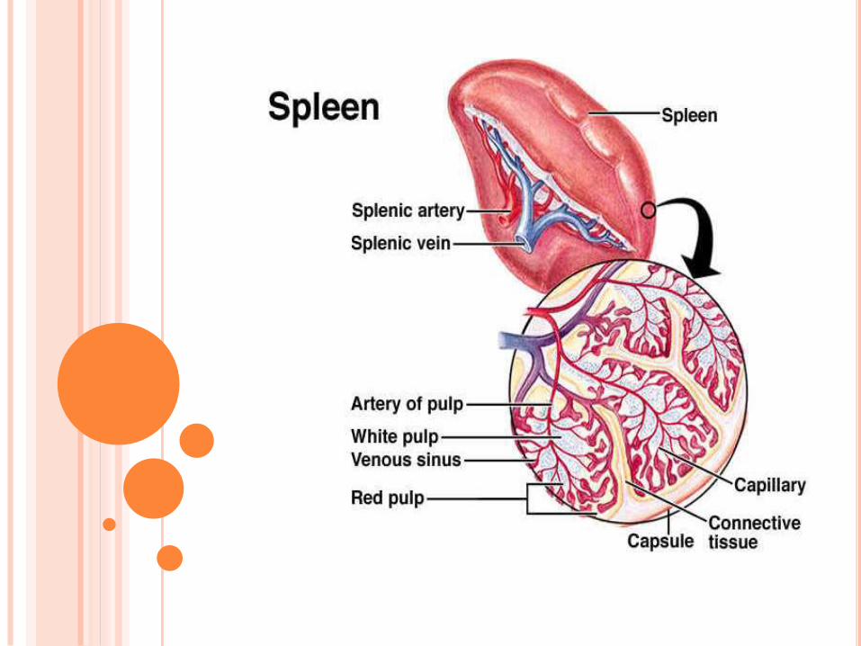
SPLEENBean shaped organ.
Left side of abdominal cavity.
Specializes in filtering blood and trapping blood borne
antigens.
Blood borne antigens, lymphocytes into spleen through
splenic artery.
Spleen surrounded by capsule.
Two types of compartment red and white pulp.
Red pulp – network of sinusoids macrophage, RBC,
lymphocyte.
Old and defective RBC destroy.
White pulp - consist of lymphoid tissue, T and B
lymphocytes.
White pulps surrounds branches of splenic artery
forming Per arteriolar Lymphoid Sheath (PALS).
Marginal zone located peripheral to PALS
[ Lymphocytes and Macrophages].
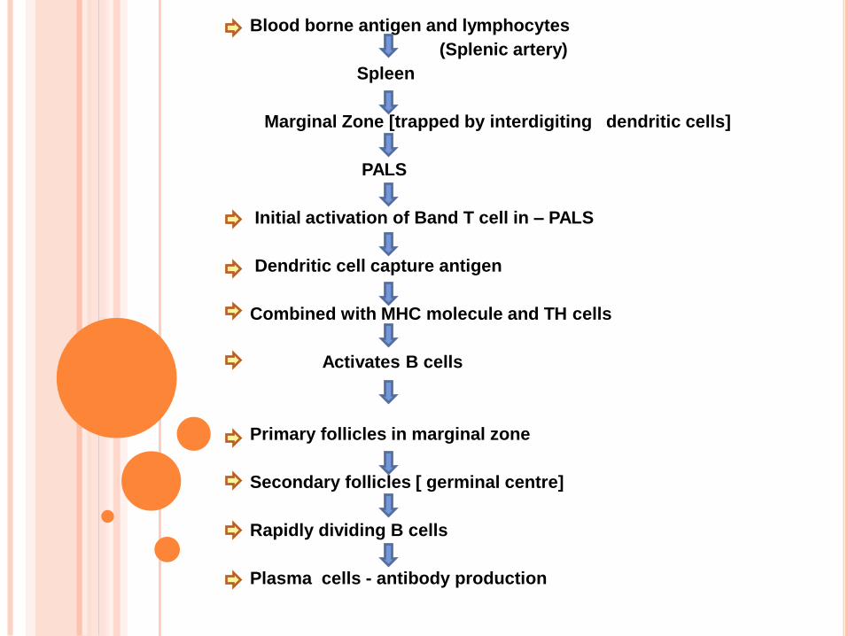
Blood borne antigen and lymphocytes
(Splenic artery)
Spleen
Marginal Zone [trapped by interdigiting dendritic cells]
PALS
Initial activation of Band T cell in – PALS
Dendritic cell capture antigen
Combined with MHC molecule and TH cells
Activates B cells
Primary follicles in marginal zone
Secondary follicles [ germinal centre]
Rapidly dividing B cells
Plasma cells - antibody production
In children, splenectomy – increases bacterial sepsis,
pneumonia, influence.
Splenectomy in adult – increase in blood borne bacterial
infection.
MUCOSA ASSOSCIATED LYMPHOID
TISSUE
Lymphoid tissue in mucosal epithelial surface – MALT
Antibody producing plasma cells.
Nasal associated lymphoid tissue – back of nose,
palate, base of tongue, tonsils
Handling airborne microbes
Tonsils defend against antigen entire through nasal
and oral epithelial route
Respiratory, Uriogential, Gastrointestinal tract
The endocytose antigen from lumen
Mucous membrane – effective barrier
Non specific immunity
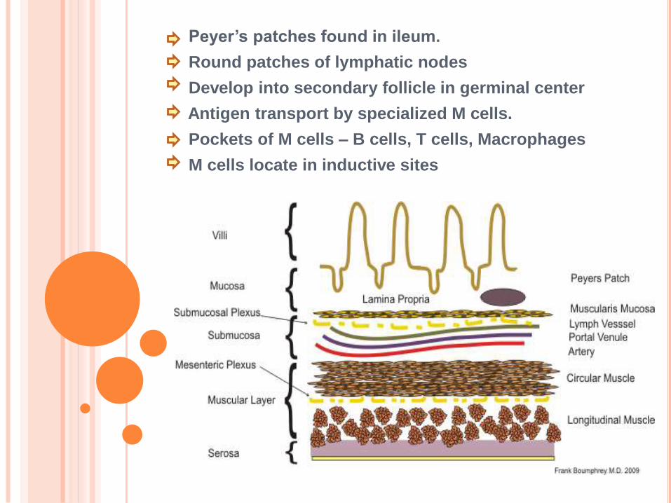
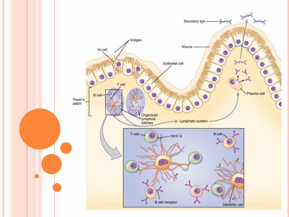
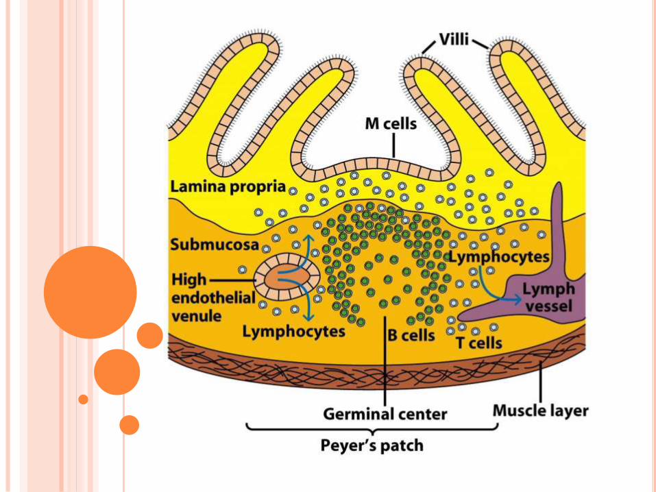
Peyer’s patches found in ileum.
Round patches of lymphatic nodes
Develop into secondary follicle in germinal center
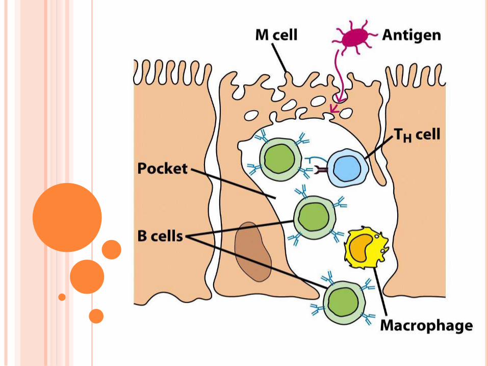
Antigen transport by specialized M cells.
Pockets of M cells – B cells, T cells, Macrophages
M cells locate in inductive sites
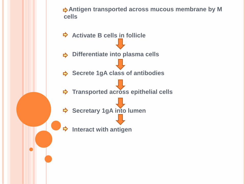
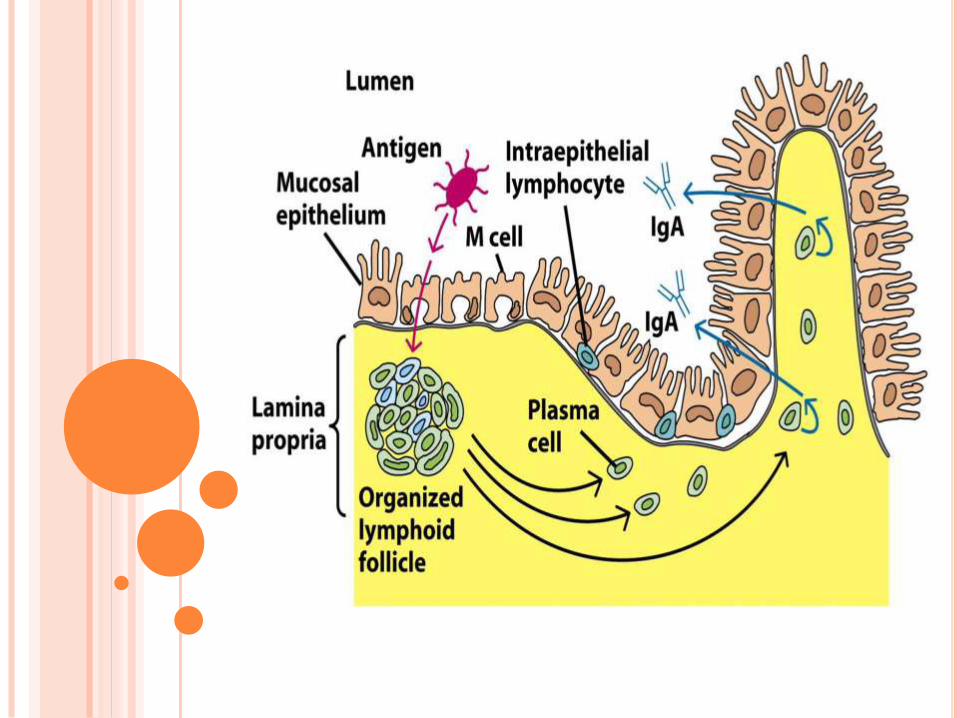
Antigen transported across mucous membrane by M
cells
Activate B cells in follicle
Differentiate into plasma cells
Secrete 1gA class of antibodies
Transported across epithelial cells
Secretary 1gA into lumen
Interact with antigen