Organogenesis and Cell Culture in Rice Plants under ... · Organogenesis and Cell Culture in Rice...
5
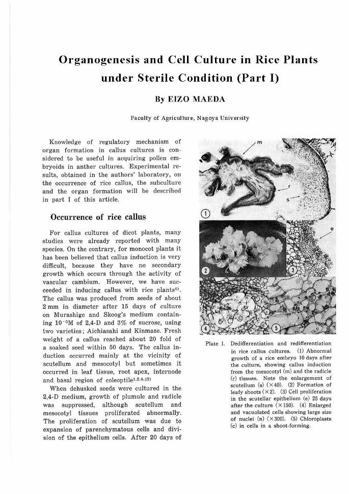
Organogenesis and Cell Culture in Rice Plants under Sterile Condition (Part I) By EIZO MAEDA Faculty of Agriculture, Nagoya University Knowledge of regulatory mechanism of organ formation in caJlus cultures is con- sidered to be useful in acquiring poJl en em- bryoids in anther cultures. Experimental re- sults, obtained in the authors' laboratory, on the occurrence of rice caJius, the subculture and the organ formation wiJI be described in part I of this article. Occurrence of rice callus For caJlus cultures of dicot plants, many studies were already reported with many species. On the contrary, for monocot plants it has been believed that callus induction is very difficult, because they have no secondary growth which occurs through the activity of vascular cambium. However, we have suc- ceeded in inducing callus with rice plants-O . The callus was produced from seeds of about 2 mm in diameter after 15 days of cultu re on Murashige and Skoog's medium contain- ing 10- r;M of 2,4-D and 3% of sucrose, using two varieties; Aichiasahi and Kinmaze. Fresh weight of a callus reached about 20 fold of a soaked seed within 50 days. The caJlu s in- duction occurred mainly at the vicinity of scuteJlum and mesocotyl but sometimes it occurred in leaf tissue, root apex, internode and basal region of coleoptiJel,M,12> When dehusked seeds were cultured in the 2,4-D medium, growth of plumule and radicle was suppressed, although scuteJium and mesocotyl tissues proliferated abnormally. The proliferation of scutellum was due to expansion of parenchymatous cells and divi- sion of the epithelium cells. After 20 days of Plate 1. Dedifferentiation and redifferentiation in rice callus cultures. (I) Abnormal growth of a rice embryo IO days after the culture, showing callus induction from t he mesocotyl (m) and the radicle (r) tissues. Note the enlargement of scutellum (s) ( X 40). (2) Formation of leafy shoots ( X 2). (3) Cell proliferation in the scutellar epithelium (e) 25 days after the culture ( X 150). (4) Enlarged and vacuolated cells showing large size of nuclei (n) ( X 300). (5) Chloroplasts (c) in cells in a shoot-forming.
Transcript of Organogenesis and Cell Culture in Rice Plants under ... · Organogenesis and Cell Culture in Rice...
Organogenesis and Cell Culture in Rice Plants
under Sterile Condition (Part I)
By EIZO MAEDA
Faculty of Agriculture, Nagoya University
Knowledge of regulatory mechanism of organ formation in caJlus cultures is considered to be useful in acquiring poJlen embryoids in anther cultures. Experimental results, obtained in the authors' laboratory, on the occurrence of rice caJius, the subculture and the organ formation wiJI be described in part I of this article.
Occurrence of rice callus
For caJlus cultures of dicot plants, many studies were already reported with many species. On the contrary, for monocot plants it has been believed that callus induction is very difficult, because they have no secondary growth which occurs through the activity of vascular cambium. However, we have succeeded in inducing callus with rice plants-O . The callus was produced from seeds of about 2 mm in diameter after 15 days of culture on Murashige and Skoog's medium containing 10- r;M of 2,4-D and 3% of sucrose, using two varieties; Aichiasahi and Kinmaze. Fresh weight of a callus reached about 20 fold of a soaked seed within 50 days. The caJlus induction occurred mainly at the vicinity of scuteJlum and mesocotyl but sometimes it occurred in leaf tissue, root apex, internode and basal region of coleoptiJel,M,12>
When dehusked seeds were cultu red in the 2,4-D medium, growth of plumule and radicle was suppressed, although scuteJium and mesocotyl tissues proliferated abnormally. The proliferation of scutellum was due to expansion of parenchymatous cells and division of the epithelium cells. After 20 days of
Plate 1. Dedifferentiation and redifferentiation in rice callus cultures. (I) Abnormal growth of a rice embryo IO days after the culture, showing callus induction from the mesocotyl (m) and the radicle (r) tissues. Note the enlargement of scutellum (s) ( X 40). (2) Formation of leafy shoots ( X 2). (3) Cell proliferation in the scutellar epithelium (e) 25 days after the culture ( X 150). (4) Enlarged and vacuolated cells showing large size of nuclei (n) ( X 300). (5) Chloroplasts (c) in cells in a shoot-forming.

culture, rapid cell division began in the epithelium to produce callus tissue (Plate 1- 3).
There are two central cylinders in rice mesocotyl, radicular side and scutellar side. In the mesocotyl of the cultured seeds, cell division began at first at the outermost layer of the central cylinder of radicular side and then extended to the radicle tips and the vascular tissue of the scutellum. After that the callus masses grew from the cell dividing areas (Plate 1-1). When the 2,4-D level was decreased to 3 X 10-GM, the outermost )ayer of the central cylinder in mesocotyl formed scattered meristematic centers and stunted primordium of lateral roots. These results show that callus tissue derived from mesocotyl is formed by abnormal proliferation of the cell layer from which lateral roots will be formed, indicating that the callus initiation resembles the initiation of adventitious roots except for the callus produced from scutellar epithelium12>. This suggestion was confirmed by the evidence that at the vicinity of vascular bundle in the leaf sheath basal regions, both callus induction and root initiation were caused by regulating 2,4-D levels used3l.
Morphology and growth of callus were examined with 15 varieties including japonica and indica types of riceoi. Variation in growth rates and in callus textures, compact or friable, was great among them, and not related to the types. Nuclear size in callus cells varied. Typical size, from 5.5 to 7.4 µm
in diameter, was estimated to account for 60.5% of the cells in callus tissues derived from seeds of 10 varieties, while giant nuclei over 11.0 p.m occurred at 0.5%. In addition, very large nuclei over 15 µm were often observed with the treatment of colchicine5l.
Subculture of rice callus
When callus tissues, derived from seeds of varieties, Te-Tep, Konansen and Tan-ginbozu, were transferred to the medium containing 0.5% of yeast extracts, fresh weight increased
5
4 to 8 fold in 14 day's culture and 27 fold in 42 day's culture. Growth rate of callus changed differently during long term subculture over 600 days with different varieties used. As the subculture was repeated, the rate decreased in Te-Tep and increased in Tan-ginbozu but no change was found in KonansenO>. Frequency distribution of nuclear size in prolonged subculture was studied with these three varieties. The mean nuclear size of Tan-ginzobu callus tissues increased from 6 µ,m to 7 µ,m after about one year of subculture. Cells having the giant nucleus above 11 µm increased to 2% (Plate 1- 4) 8>.
Glucose, maltose, fructose and soluble starch were utilized as carbon sources as well as sucrose. Sometimes they were more suitable than sucrose17>. Thiamine was required for callus subculture but not essential for callus induction. Kinetin was not necessary to the subculture. It was not effective but sometimes repressive in callus induction1>. With Tan-ginbozu, a dwarf mutant of Ginbozu, in which the dwarfism is controlled' by single recessive gene and the content of gibberellin-like substances is very low, the callus growth in the 10- 7M 2,4-D medium was
4
OF. wt.
~ D.wt.
400
e .:,[
"' "' r:: J Standard error 300:;
.c:
2 ~ 200
0 'f t:fa [:J?j tpj tij [lj tpj )'o Varieties T-T T-G T·G T-G T-G T-G 2,4-D(M) 10-s 10-s 10-1 10-1 10-1 10-1 GA3(M) 0 0 105 5,.10-s 10-4 5,clO""'
Fig. 1, Effect of GA3 concentration on the callus growth of a dwarf variety (T-G), compared with a tall variety (T-T).
J >,
Q
6
accelerated with addition of 10-0M to 10-·•M of gibberellin (Fig. I)H>.
The acceleration by gibberellin was also found in other dwarf varieties such as Sankei No. 10, Waiseishinriki and Kotaketamanishiki except Murasakidaikoku. But the acceleration was not found in normal varieties, Ginbozu, Te-Tep and Norin No. 8. These results indicate that the callus of Tan-ginbozu has an abnormal gibberellin metabolism which is found in its seedlingsH>. Such effect of gibberellin on growth of callus was observed only in the medium containing yeast extracts or casein hydrolyzates but not in the chemically defined medium. The promoting effect was recovered when the mixture of 14 amino acids was added to the defined medium, but never when each one of the amino acids was added10J. The growth-promoting effect of gibberellin was not observed in Tan-ginbozu callus after 14 generations of subcultureH>. This result is interesting in view of the change of nuclear size cansed in this variety by prolonged subcultures>.
01·gan formation in rice callus
It is noteworthy that in the year 1968 the organ formation in rice callus, i.e., redifferentiation of shoots in rice callus, was first reported from five different laboratories in Japan (Kawata's, Niizeki's, Tamura's, Yamada's and the authors laboratory independently). The author observed clearly shoot formation in the callus of Te-Tep, cultured on the 10- 7 M 2,4-D med ium (Plate 1-2)7>.
Furthermore, it was shown that kinetin promoted shoot formation in the medium containing yeast extracts and casein hydrolyzate, with four varieties, Te-Tep, Norin No. 8, Norin No. 29 and Kyotoasahi, but adenine, tyrosine and potassium biphosphate were not effective•=i> . The promoting effect of kinetin disappeared if the chemically defined medium was used 11;1 . Albino shoots were obtained at 30 to 50% of the differentiated shoots. Gibberellin inhibited shoot formation at low concentrations, even at 5x10⁻¹ M. Gibberellin
JARQ Vol. 14, No. 1, 1980
also inhibited the kinetin effect on shoot production. The regenerative 1Jotential declined with age as noted in other species of monocotyledons. The shoots differentiated vigorously in the 4th subculture generation of Aichiasahi but not in the 9th ot· further generations to> .
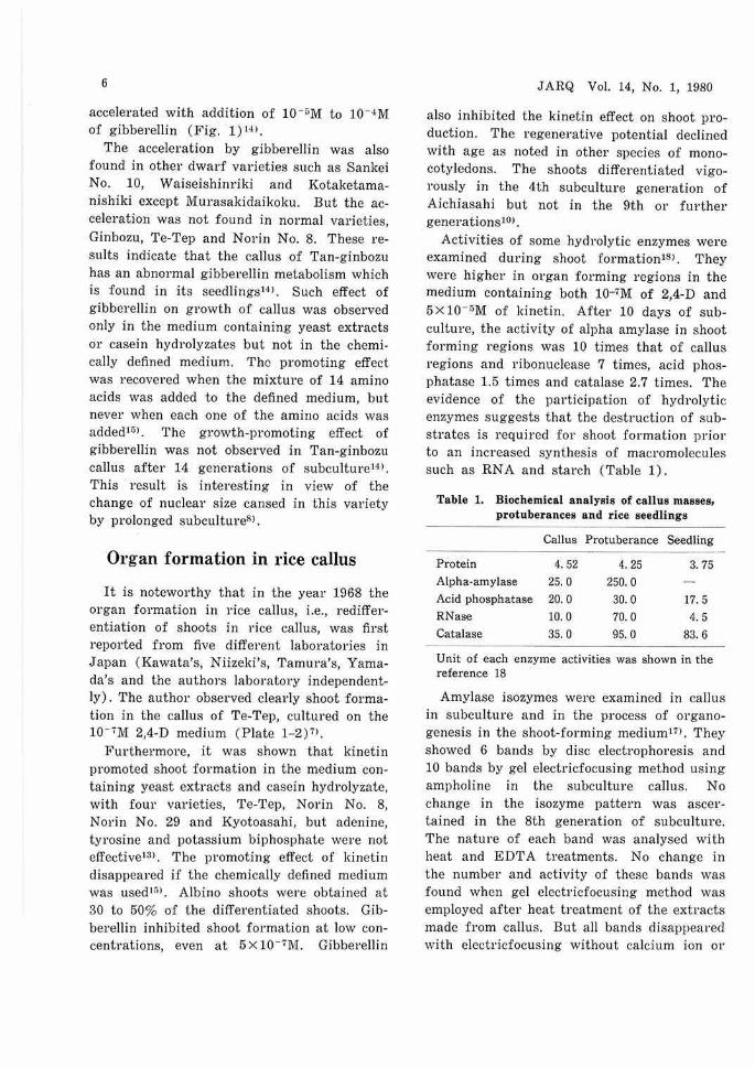
Activities of some hydrolytic enzymes were examined during shoot formation1si . They were higher in organ forming regions in the medium containing both 10-7M of 2,4-D and 5 x 10- 0M of kinetin. After 10 days of subculture, the activity of alpha amylase in shoot forming regions was 10 times that of callus regions and ribonuclease 7 times, acid phosphatase 1.5 times and catalase 2.7 times. The evidence of the participation of hydrolytic enzymes suggests that the destrnction of substrates is required for shoot formation prior to an increased synthesis of macromolecules such as RN A and starch (Table 1).
Table 1. Biochemical analysis of callus masses, protuberances and rice seedlings
Callus Protuberance Seedling
Protein 4.52 4.25 3. 75 Alpha-amylase 25. 0 250. 0 Acid phosphatase 20.0 30.0 17.5 RNase 10. 0 70.0 4.5 Catalase 35. 0 95.0 83.6
Unit of each enzyme activities was shown in the reference 18
Amylase isozymes were examined in callus in subculture and in the process of organogenesis in the shoot-forming mediumm. They showed 6 bands by disc electrophoresis and 10 bands by gel electricfocusing method using ampholine in the subculture callus. No change in the isozyme pattern was ascertained in the 8th generation of subculture. The nature of each band was analysed with heat and EDTA treatments. No change in the number and activity of these bands was found when gel electricfocusing method was employed after heat treatment of the extracts made from callus. But all bands disappeared with electricfocusing without calcium ion or

with EDTA treatment after the electricfocusing with calcium ion, except only one band which has an isoelectric point nearest the acidic side. This remaining band is considered to be an alpha amylase similar to other bands but without a specific requirement for calcium ion and with resistance to EDT A. From these results it is probable that all 10 amylase isozymes in rice callus are alpha type.
Increase in alpha amylase activity in organ forming callus was due to intensification of four bands in the isozymes and addition of one new band. In rice seedlings, on the other hand, alpha amylase activity was very low 01· undetectable. Therefore, it can be said that biochemical changes during shoot regeneration reflect an increase in hydrolytic enzyme activities, especially of alpha amylase, at the time of shoot initiation, and the decrease in the activities after the shoots regenerated18> . In the callus tissues derived from a dwarf variety, growth acceleration by gibberellin was also accompanied with changes in enzyme activitiesio>.
To clarify the changes in accumulation of starch grains during shoot differentiation, paraffin sections of callus were stained with Lugol's solution10>, which generally gives purple or reddish purple coloration of starch grains. Many starch grains were found in the greening region in the shoot-forming tissue grown on a medium containing 10-1 M 2,4-D and 5Xl0-5M kinetin after 9 days of culture. In this medium, large grains, stained to dark purple, were also observed in the enlarged parenchymatous cells in callus periphery contacting the medium. However, starch grains were not observed in regenerated shoot apices. Very few starch grains were observed in the callus after 9 days of culture in the medium containing both kinetin and gibberellin. Therefore, it is clear that gibberellin inhibited both the strach accumulation and the shoot formation in rice callus. Carbohydrate substrate must be available for shoot formation to take place. The starch deposition is considered to be an energy
7
source. Chloroplasts, starch grains, nuclei, nucleoli,
cytoplasm, cell wall and osmiophillic granules were examined using various staining schedules with semi-thin sections, prepared from materials embedded in epoxy resin (Plate 1-5). In Tan-ginbozu callus, bi-nuclear cells were often in contact with each other10>.
It has recently been indicated that callus structure is considerably more complicated than the homogenous parenchyma mass as was considered originally. This is especially true in the process of organogenesis. Thus, to clarify what changes in external morphology of callus surface occur in the shootforming process, gross-morphological observation was madell>. Various appearances observed were classified into 8 categories as follows:
(1) dry (i.e. without a watery reflection), (2) (3) (4)
(5) (6)
green, protuberant, green and protuberant (i.e. greencolored with outgrowths), white (i .e. milky white colored), white and green (i.e. with mixed white and green areas),
(7) green leaf ( i.e. areas containing green leaves),
(8) white leaf (i.e. areas containing albino leaves).
With time in culture the external morphology of shoot-forming tissue changed from one category to another. Therefore, the sequential order of morphological changes in callus surface during the shoot formation was traced. It was found that the tissue at the earliest visible stage was dry 01· white. In some cases, the tissue changed from dry to green or protuberant, to green and protuberant, and further to leafy region, where shoot apical meristems producing leaf primordia were organized. Alternatively, tissue changed from white to white and green, and finally to leafy region. In both cases, these changes were often observed in the callus region near the medium. Not all of the dry areas, white areas and others proceeded to

8
green or white leaves, but some of them disappeared showing dark-brown in color. Thus, it seems that the onset of shoot formation is induced by some physiological situation in which a sequence of morphological changes can proceed. The decreased 2,4-D level in the medium was a factor tending to promote greening of callus and occurrence of white or white and green regions. On the other hand, the increased kinetin level favoured the occurrence of dry and protuberant regions.
References 1) Inoue, M. & Maeda, E.: Effect of auxin
concent1·ation on the callus induction from various organs of rice seedlings. Proc. Crop Sci. Soc. Jap., 45, 545-557 (1967).
2) Inoue, M. & Maeda, E.: Relation between 2,4-D levels and callus initiation in the epidermal cells of scutellum and coleorhiza of rice. Proc. Crov Sci. Soc. Jap., 45, 637-638 (1976) .
3) Inoue, M. & Maeda, E.: Occurrence of adventitious roots from rice leaf sheaths treated with 2,4-D. Jciv. J. C1·op Sci., 46, 319-320 (1977).
4) Maeda, E.: Callus formation and isolation of single cells from rice seedings. P1·oc. C1·ov Sci. Soc. Jap., 34, 139-147 (1965).
5) Maeda, E.: A morphological study of nuclei and action of colchicine on rice callus cultivated in vit1·0. Jap. J. Breed., 17, 7-12 (1967).
6) Maeda, E. : Varietal difference in callus formation of rice seeds under sterile culture. P1·oc. C1·op Sci. Soc. Jap., 36, 233-239 (1967).
7) Maeda, E .: Subculture and organ formation in the callus derived from .rice embryos in vitro. Proc. C1·07> Sci. Soc. Jav., 37, 51-58
JARQ Vol. 14, No. 1, 1980
(1968). 8) Maeda, E.: Variation in nuclear size of
rice callus tissues under the aseptic subculture. Jap. J. Genetics, 44, 285-289 (1969).
9) Maeda, E . : Growth of rice callus derived from embryo under the subculture condition. Proc. Crop Sci. Soc. Jap., 40, 141- 149 (1971).
10) Maeda, E. & Saka, H.: Light microscopy of cell organelles in the shooting rice callus tissues. Proc. C1·07> ScL Soc. Jav., 42, 442-453 (1973).
11) Nakano, H. & Maeda, E.: Morphology of the process of shoot formation in the rice callus culture. P'roc. Crop Sci. Soc. Ja71., 43, 151-160 (1974) .
12) Nishimura, S. & Maeda, E. : Histological studies of callus induction in rice seed. Jap. J. C1"01J Sci., 46, 275-285 (1977).
13) Saka, H. & Maeda, E. : Effect of kinetin on organ formation in callus tissue derived from rice embryos. Proc. Crop Sci .. Soc. Jap., 38, 668-674 (1969).
14) Saka, H. & Maeda, E.: Requirement of gibberellic acid for callus growth of dwarf and normal varieties of 01·yzci sativ<i L. P1·oc. C1·07> Sci. Soc. Ja7>., 40, 27- 33 (1971) .
15) Saka, H. & Maeda, E. : Amino acids as complementary factors for the growth and redifferentiation of rice callus tissues. P1·oc. C1·ov Sci. Soc. Jap., 41, 15-21 (1972).
16) Saka, H. & Maeda, E. : Changes in enzyme activity during gibberellic acid-mediated callus growth in dwarf rice. Proc. Crop Sci. Soc. Jap., 42, 315-321 (1973).
17) Saka, H. & Maeda, E.: Characteristics and varietal differences of alpha-amylase isozymes in rice callus tissues. P1·oc. Crop Sci. Soc. Ja7>., 42, 307-314 (1973).
18) Saka, H. & Maeda, E.: Changes in some hydrolytic enzymes associated with the redifferentiation of shoots in rice callus tissues. Proc. Crov Sci. Soc. Jav., 43, 207-218 (1974) .