Orbital Trauma - Lieberman's eRadiology Learning Sites
35
Lucy Shen, 2004 Gillian Lieberman, MD Orbital Trauma Lucy Shen Harvard Medical School Year IV Gillian Lieberman, MD March, 2004
Transcript of Orbital Trauma - Lieberman's eRadiology Learning Sites
Lucy Shen, 2004Gillian Lieberman, MD
Orbital Trauma
Lucy ShenHarvard Medical School Year IV
Gillian Lieberman, MD
March, 2004
Lucy Shen, 2004Gillian Lieberman, MD
2
Orbital Trauma
• Causes: high acceleration, MVA, violent crimes, industrial accidents
• Orbital fractures• Soft tissue injuries
Lucy Shen, 2004Gillian Lieberman, MD
3
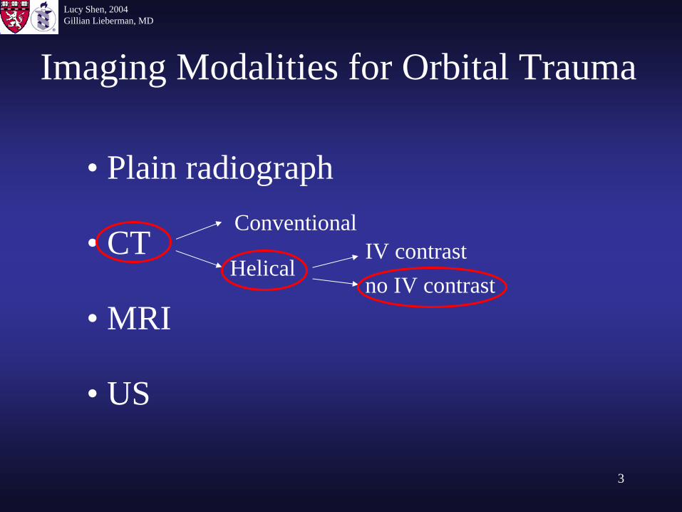
Imaging Modalities for Orbital Trauma
• Plain radiograph
• CT
• MRI
• US
Conventional
HelicalIV contrastno IV contrast
Lucy Shen, 2004Gillian Lieberman, MD
4
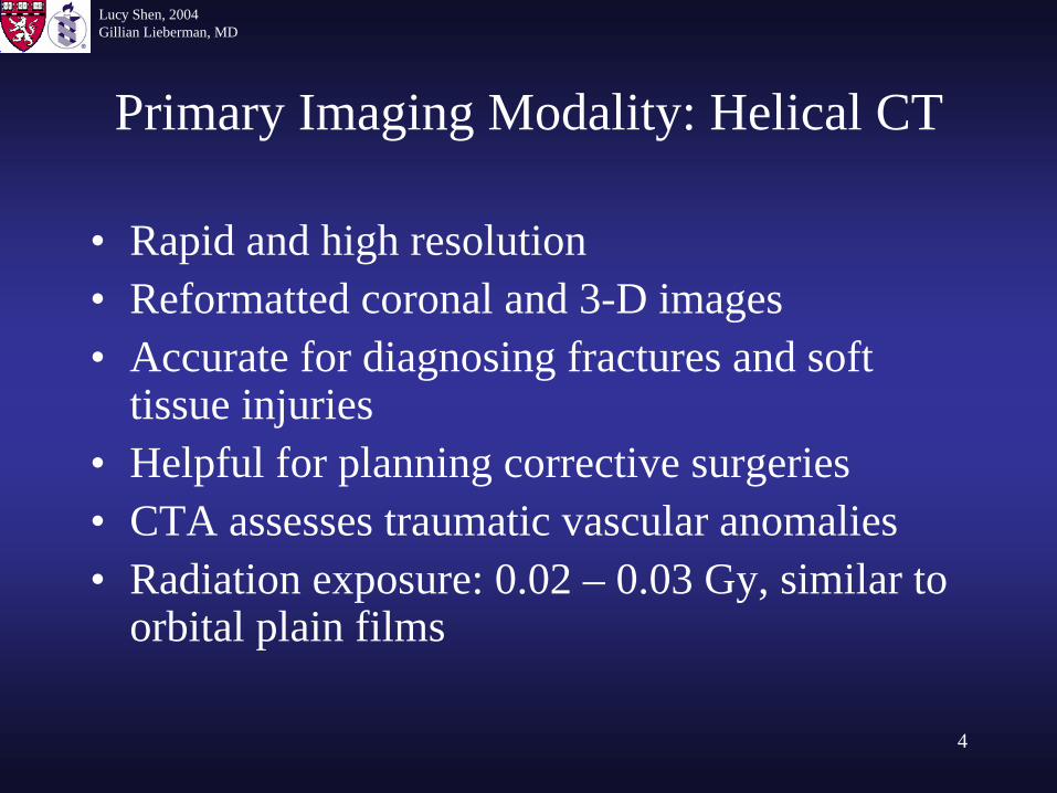
Primary Imaging Modality: Helical CT
• Rapid and high resolution• Reformatted coronal and 3-D images• Accurate for diagnosing fractures and soft
tissue injuries• Helpful for planning corrective surgeries• CTA assesses traumatic vascular anomalies• Radiation exposure: 0.02 – 0.03 Gy, similar to
orbital plain films
Lucy Shen, 2004Gillian Lieberman, MD
5
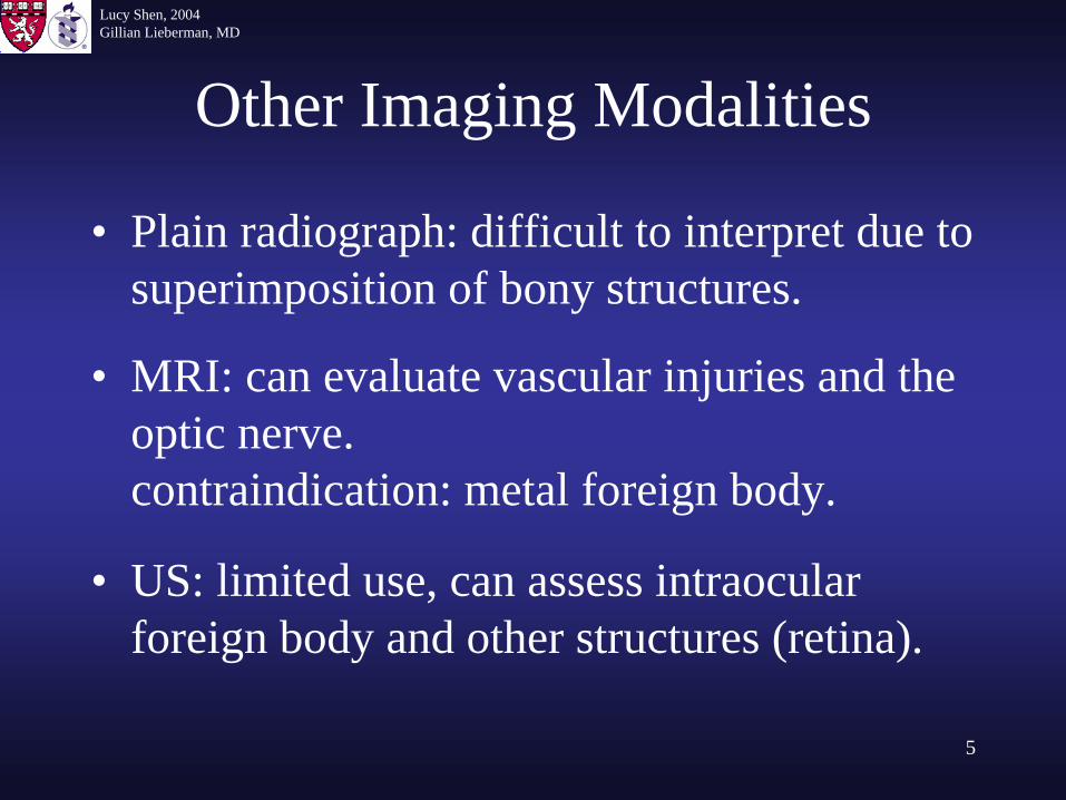
Other Imaging Modalities
• Plain radiograph: difficult to interpret due to superimposition of bony structures.
• MRI: can evaluate vascular injuries and the optic nerve.contraindication: metal foreign body.
• US: limited use, can assess intraocular foreign body and other structures (retina).
Lucy Shen, 2004Gillian Lieberman, MD
6
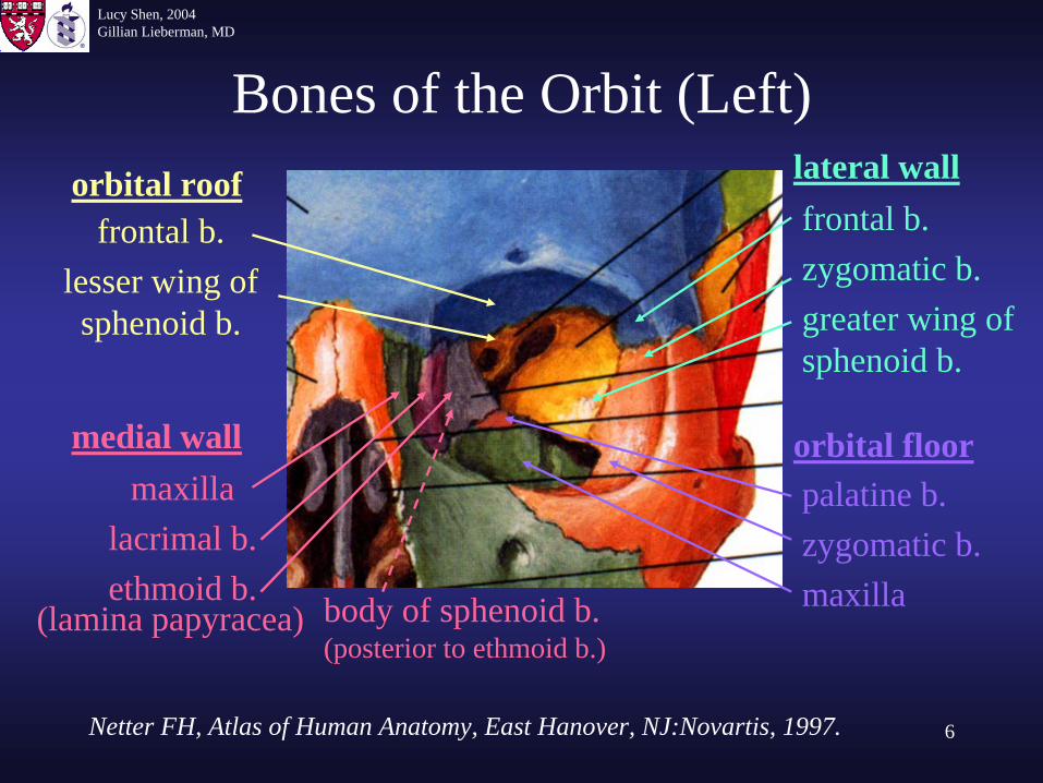
Bones of the Orbit (Left)
frontal b.lesser wing of sphenoid b.
orbital roof lateral wallfrontal b.zygomatic b.greater wing of sphenoid b.
orbital floorpalatine b.zygomatic b.maxilla
medial wallmaxilla
lacrimal b.ethmoid b.
(lamina papyracea) body of sphenoid b.(posterior to ethmoid b.)
Netter FH, Atlas of Human Anatomy, East Hanover, NJ:Novartis, 1997.
Lucy Shen, 2004Gillian Lieberman, MD
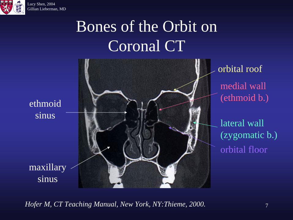
7
Bones of the Orbit on Coronal CT
orbital roof
medial wall (ethmoid b.)
orbital floor
lateral wall(zygomatic b.)
ethmoid sinus
maxillary sinus
Hofer M, CT Teaching Manual, New York, NY:Thieme, 2000.
Lucy Shen, 2004Gillian Lieberman, MD
8
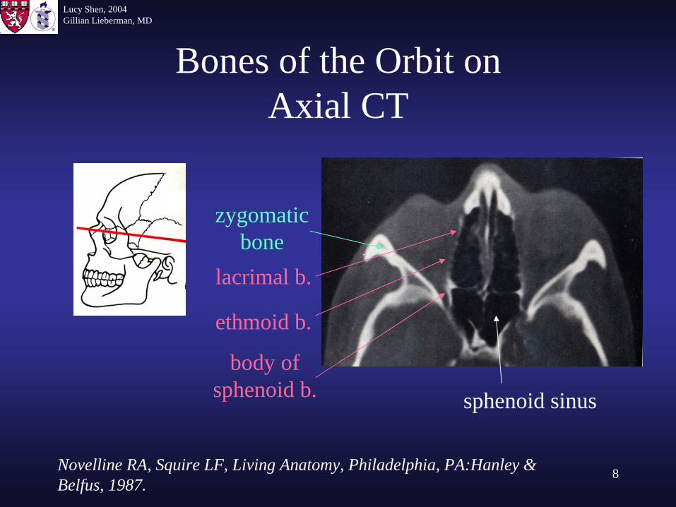
Bones of the Orbit on Axial CT
sphenoid sinus
body of sphenoid b.
zygomatic bone
lacrimal b.
ethmoid b.
Novelline RA, Squire LF, Living Anatomy, Philadelphia, PA:Hanley & Belfus, 1987.
Lucy Shen, 2004Gillian Lieberman, MD
9
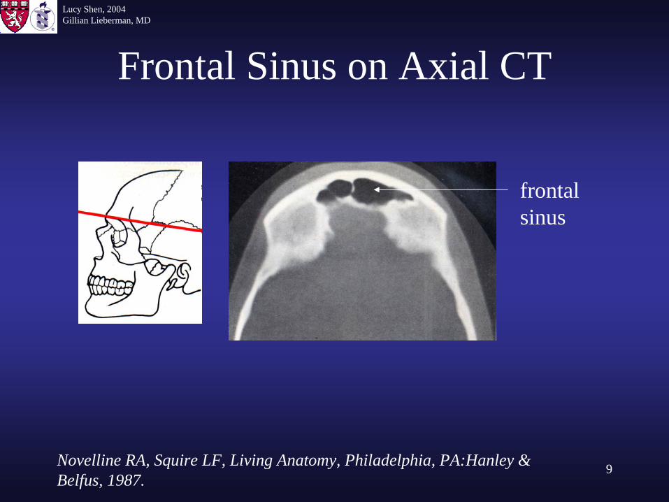
Frontal Sinus on Axial CT
frontal sinus
Novelline RA, Squire LF, Living Anatomy, Philadelphia, PA:Hanley & Belfus, 1987.
Lucy Shen, 2004Gillian Lieberman, MD
10
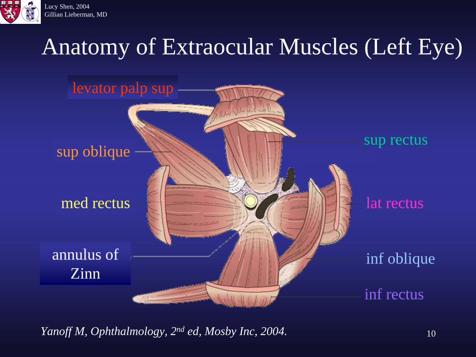
Anatomy of Extraocular Muscles (Left Eye)
Yanoff M, Ophthalmology, 2nd ed, Mosby Inc, 2004.
sup oblique
levator palp sup
med rectus
inf rectus
lat rectus
sup rectus
inf obliqueannulus of Zinn
Lucy Shen, 2004Gillian Lieberman, MD
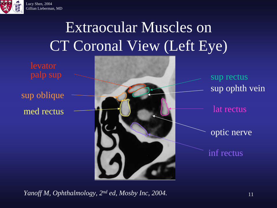
11
Extraocular Muscles on CT Coronal View (Left Eye)
optic nerve
sup ophth veinsup rectus
lat rectus
inf rectus
med rectus
sup oblique
levator palp sup
Yanoff M, Ophthalmology, 2nd ed, Mosby Inc, 2004.
Lucy Shen, 2004Gillian Lieberman, MD
12
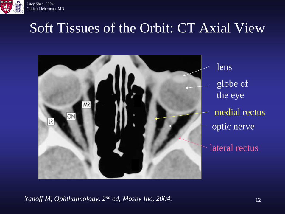
Soft Tissues of the Orbit: CT Axial View
lens
globe of the eye
optic nervemedial rectus
lateral rectus
Yanoff M, Ophthalmology, 2nd ed, Mosby Inc, 2004.
Lucy Shen, 2004Gillian Lieberman, MD
13
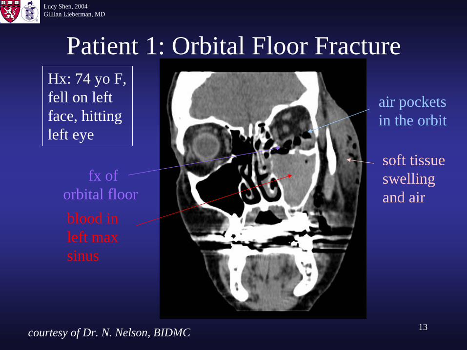
Patient 1: Orbital Floor FractureHx: 74 yo F, fell on left face, hitting left eye
air pockets in the orbit
soft tissue swelling and air
blood in left max sinus
fx of orbital floor
courtesy of Dr. N. Nelson, BIDMC
Lucy Shen, 2004Gillian Lieberman, MD
14
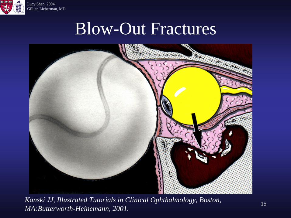
Blow-Out Fractures
• Patient 1 suffered a blow-out fracture.• Definition: blunt trauma to the orbit displacing the
fragmented bones outward. The globe can be pushed inward.
• Fracture often involves the medial wall or the orbital floor or both. Muscle entrapment may occur.
• The following slide illustrates a blow-out fracture.
Lucy Shen, 2004Gillian Lieberman, MD
15
Blow-Out Fractures
Kanski JJ, Illustrated Tutorials in Clinical Ophthalmology, Boston, MA:Butterworth-Heinemann, 2001.
Lucy Shen, 2004Gillian Lieberman, MD
16
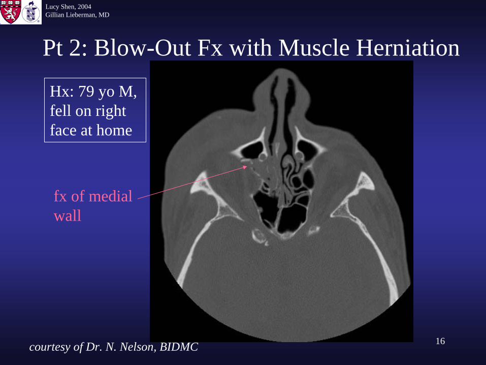
Pt 2: Blow-Out Fx with Muscle Herniation
Hx: 79 yo M, fell on right face at home
fx of medial wall
courtesy of Dr. N. Nelson, BIDMC
Lucy Shen, 2004Gillian Lieberman, MD
17
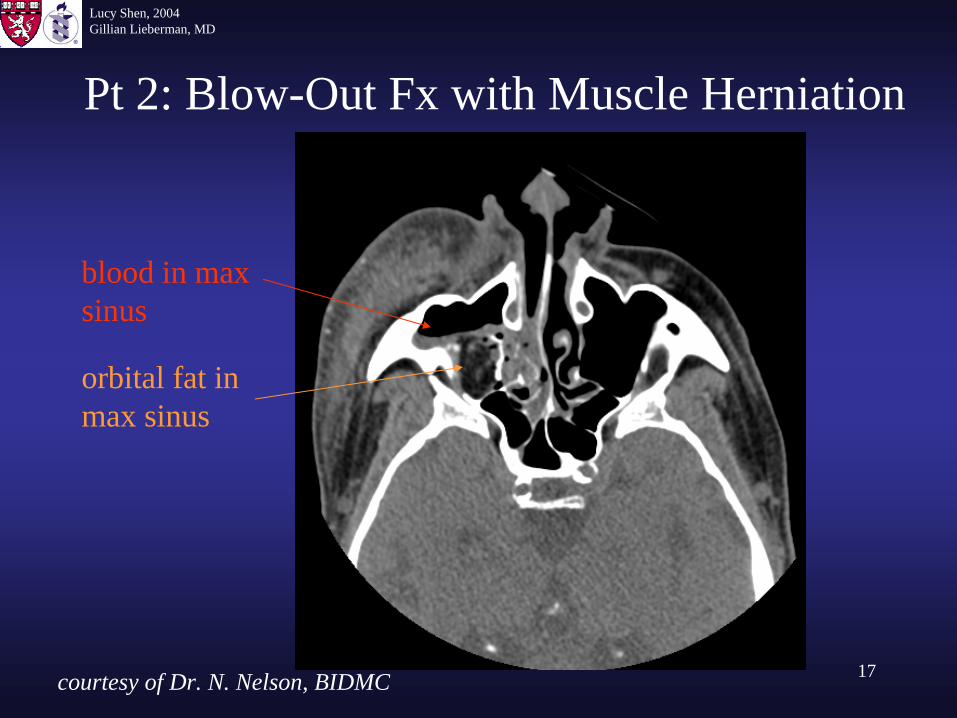
Pt 2: Blow-Out Fx with Muscle Herniation
blood in max sinus
orbital fat in max sinus
courtesy of Dr. N. Nelson, BIDMC
Lucy Shen, 2004Gillian Lieberman, MD
18
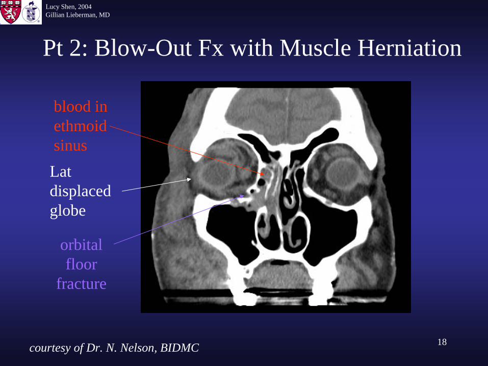
Pt 2: Blow-Out Fx with Muscle Herniation
blood in ethmoid sinus
orbital floor
fracture
Lat displaced globe
courtesy of Dr. N. Nelson, BIDMC
Lucy Shen, 2004Gillian Lieberman, MD
19
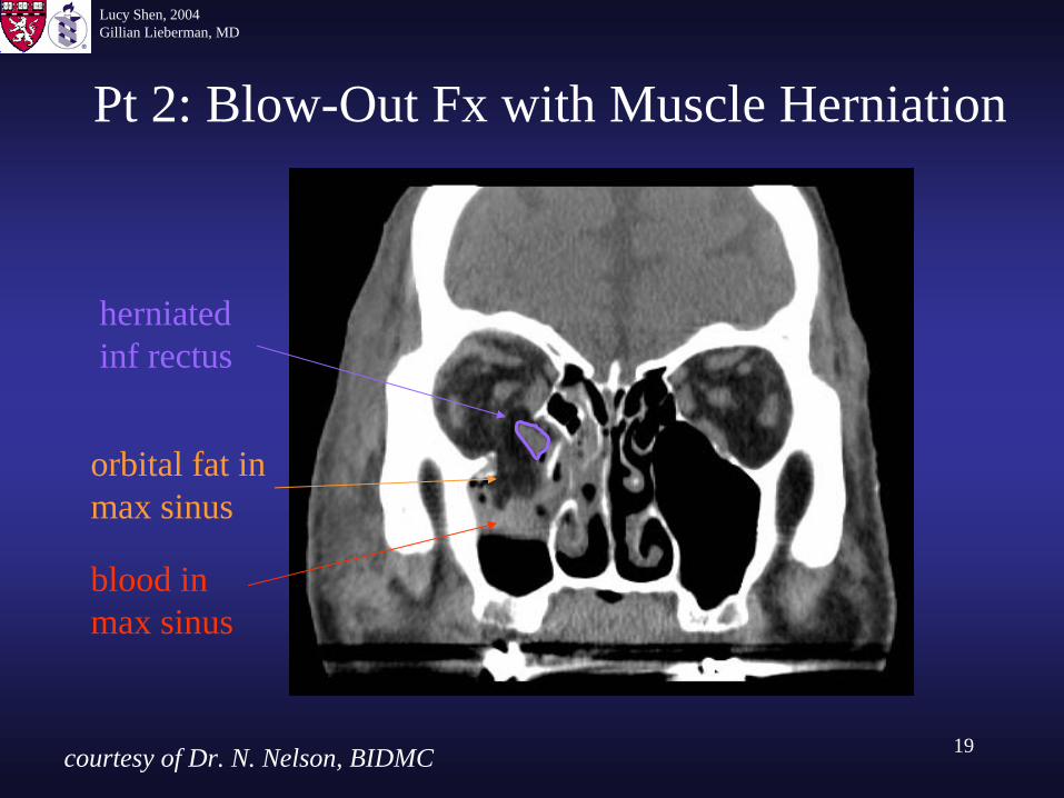
Pt 2: Blow-Out Fx with Muscle Herniation
blood in max sinus
orbital fat in max sinus
herniated inf rectus
courtesy of Dr. N. Nelson, BIDMC
Lucy Shen, 2004Gillian Lieberman, MD
20
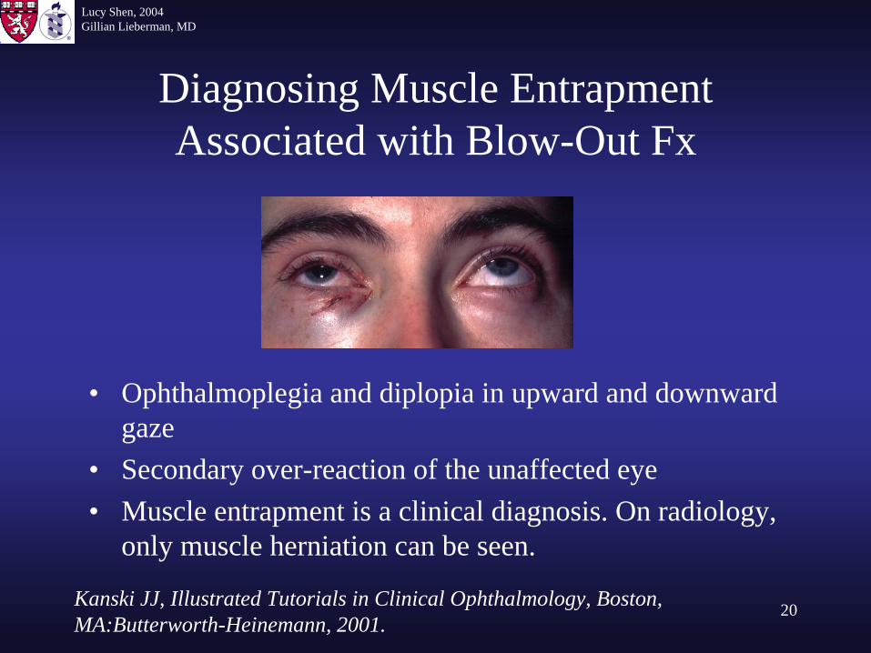
Diagnosing Muscle Entrapment Associated with Blow-Out Fx
• Ophthalmoplegia and diplopia in upward and downward gaze
• Secondary over-reaction of the unaffected eye• Muscle entrapment is a clinical diagnosis. On radiology,
only muscle herniation can be seen.
Kanski JJ, Illustrated Tutorials in Clinical Ophthalmology, Boston, MA:Butterworth-Heinemann, 2001.
Lucy Shen, 2004Gillian Lieberman, MD
21
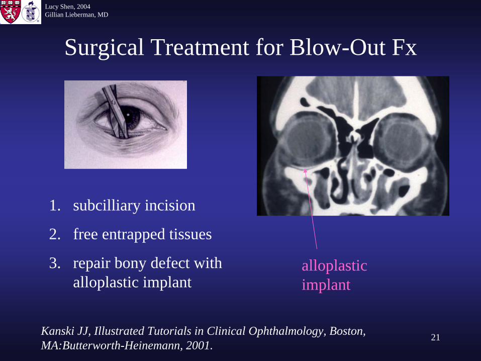
Surgical Treatment for Blow-Out Fx
1. subcilliary incision
2. free entrapped tissues
3. repair bony defect with alloplastic implant
alloplastic implant
Kanski JJ, Illustrated Tutorials in Clinical Ophthalmology, Boston, MA:Butterworth-Heinemann, 2001.
Lucy Shen, 2004Gillian Lieberman, MD
22
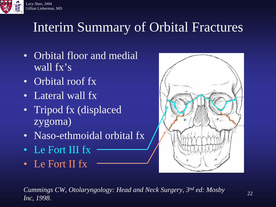
Interim Summary of Orbital Fractures
• Orbital floor and medial wall fx’s
• Orbital roof fx• Lateral wall fx• Tripod fx (displaced
zygoma)• Naso-ethmoidal orbital fx• Le Fort III fx• Le Fort II fx
Cummings CW, Otolaryngology: Head and Neck Surgery, 3nd ed: Mosby Inc, 1998.
Lucy Shen, 2004Gillian Lieberman, MD
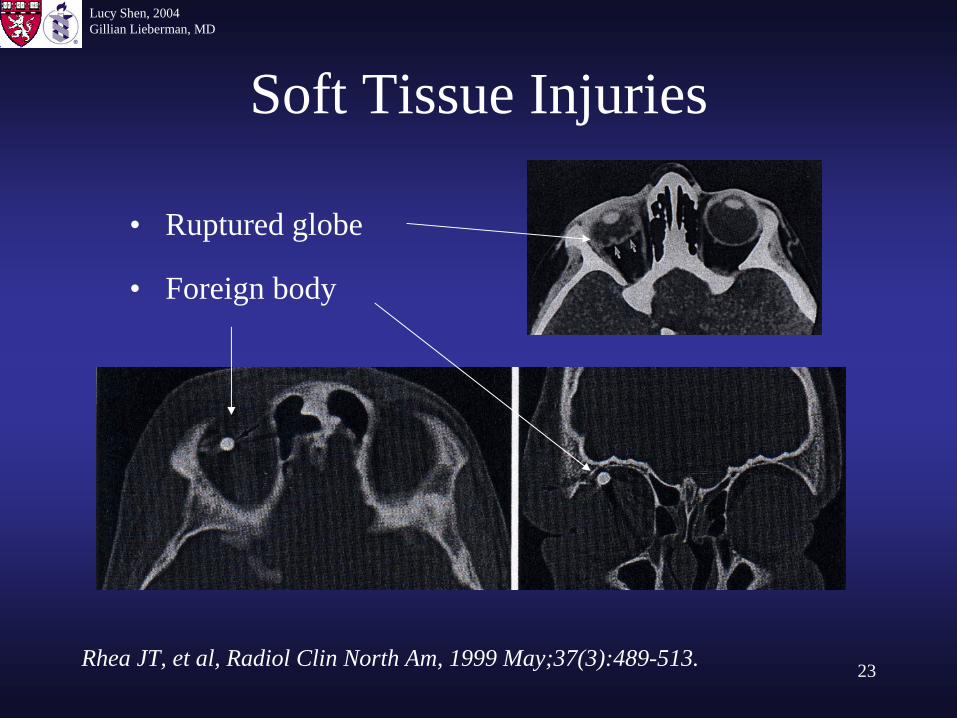
23
Soft Tissue Injuries
• Ruptured globe
• Foreign body
Rhea JT, et al, Radiol Clin North Am, 1999 May;37(3):489-513.
Lucy Shen, 2004Gillian Lieberman, MD
24
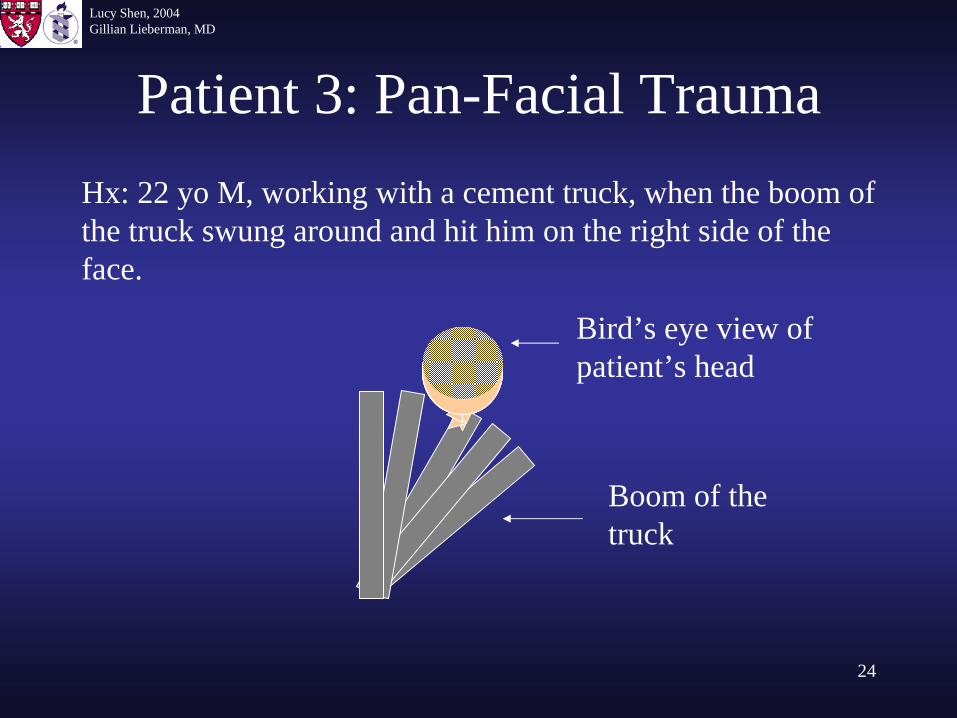
Patient 3: Pan-Facial TraumaHx: 22 yo M, working with a cement truck, when the boom of the truck swung around and hit him on the right side of the face.
Bird’s eye view of patient’s head
Boom of the truck
Lucy Shen, 2004Gillian Lieberman, MD
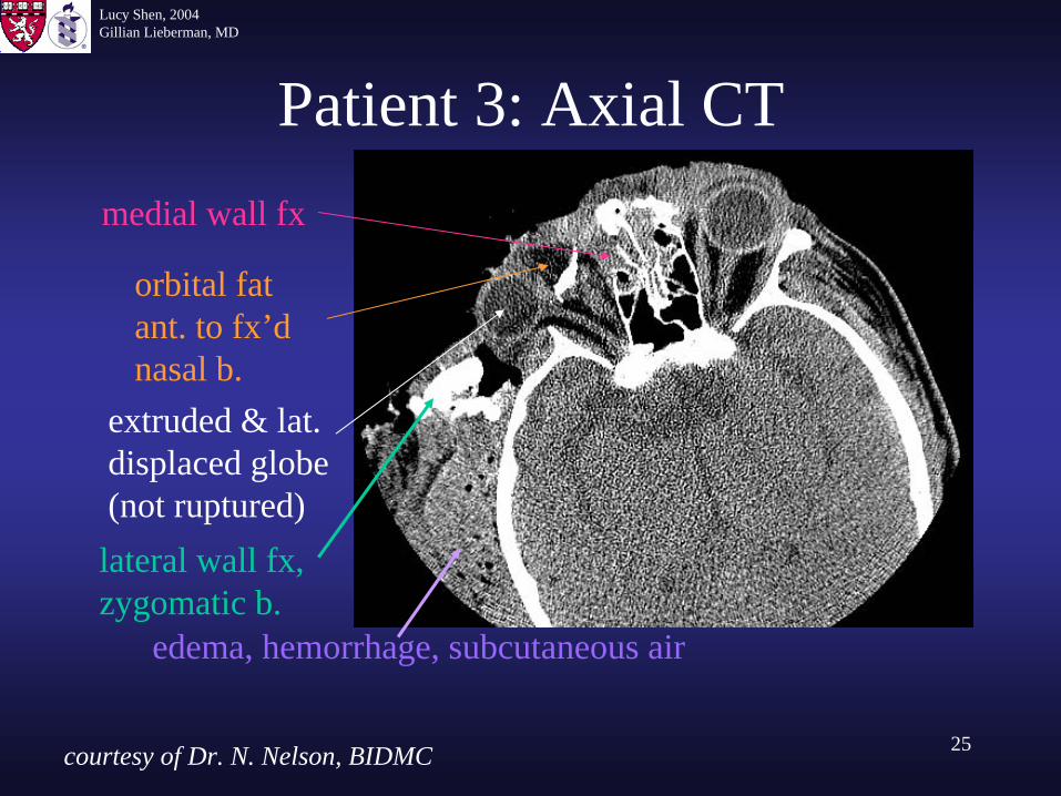
25
Patient 3: Axial CT
orbital fat ant. to fx’d nasal b.
extruded & lat. displaced globe (not ruptured)lateral wall fx, zygomatic b.
medial wall fx
edema, hemorrhage, subcutaneous air
courtesy of Dr. N. Nelson, BIDMC
Lucy Shen, 2004Gillian Lieberman, MD
26
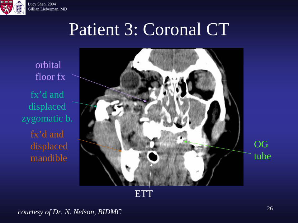
Patient 3: Coronal CT
fx’d and displaced mandible
OG tube
ETT
orbital floor fx
fx’d and displaced
zygomatic b.
courtesy of Dr. N. Nelson, BIDMC
Lucy Shen, 2004Gillian Lieberman, MD
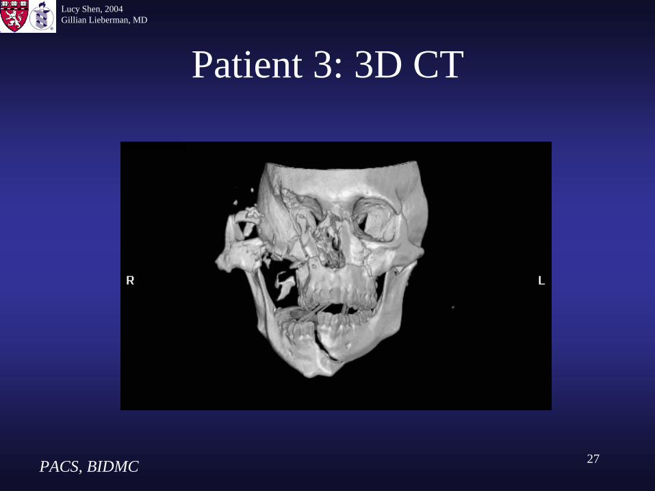
27
Patient 3: 3D CT
PACS, BIDMC
Lucy Shen, 2004Gillian Lieberman, MD
28
Patient 3: Findings on 3D CT
• Fractures of all 4 orbital walls• Orbital rim was shattered to > 8 pieces• Le Fort I fracture of the maxilla• Fractured and displaced mandible
Lucy Shen, 2004Gillian Lieberman, MD
29
Patient 3: Hospital Course
• Patient was hospitalized in the trauma ICU.• He underwent tracheostomy and extensive
facio-reconstructive surgeries.• The next 2 slides are from a CT taken after
his first major facial surgery.
Lucy Shen, 2004Gillian Lieberman, MD
30
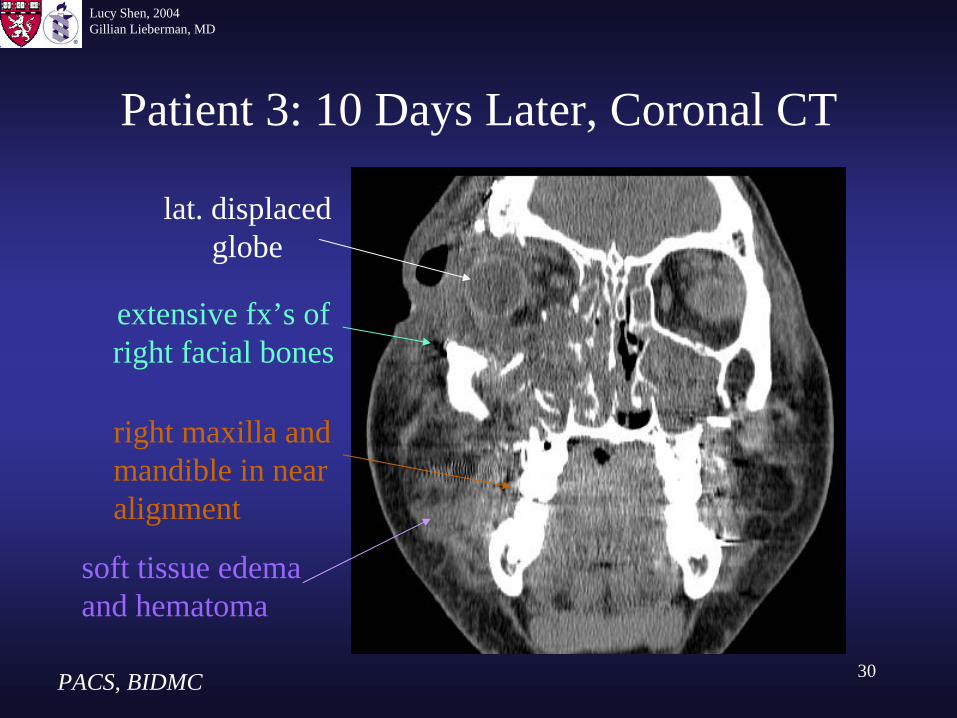
Patient 3: 10 Days Later, Coronal CT
lat. displaced globe
right maxilla and mandible in near alignment
extensive fx’s of right facial bones
soft tissue edema and hematoma
PACS, BIDMC
Lucy Shen, 2004Gillian Lieberman, MD
31
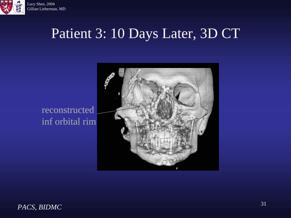
Patient 3: 10 Days Later, 3D CT
PACS, BIDMC
reconstructed inf orbital rim
Lucy Shen, 2004Gillian Lieberman, MD
32
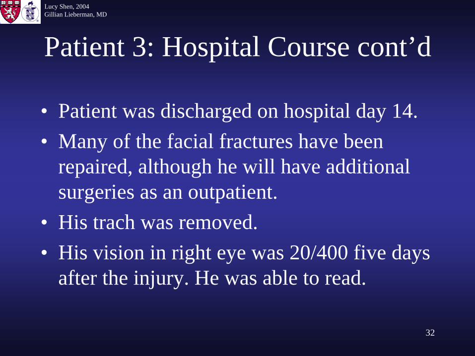
Patient 3: Hospital Course cont’d
• Patient was discharged on hospital day 14.• Many of the facial fractures have been
repaired, although he will have additional surgeries as an outpatient.
• His trach was removed.• His vision in right eye was 20/400 five days
after the injury. He was able to read.
Lucy Shen, 2004Gillian Lieberman, MD
33
Orbital Trauma: in Summary
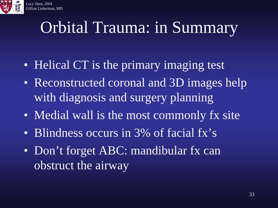
• Helical CT is the primary imaging test• Reconstructed coronal and 3D images help
with diagnosis and surgery planning• Medial wall is the most commonly fx site• Blindness occurs in 3% of facial fx’s• Don’t forget ABC: mandibular fx can
obstruct the airway
Lucy Shen, 2004Gillian Lieberman, MD
34
References
• Cummings CW, Otolaryngology: Head and Neck Surgery, 3nd ed: Mosby Inc, 1998. • Grainger RG, Allison A, Diagnostic Radiology: A Textbook of Medical Imaging, 4th ed:
Churchill Livingstone, Inc, 2001.• Hofer M, CT Teaching Manual, New York, NY:Thieme, 2000. • Kanski JJ, Illustrated Tutorials in Clinical Ophthalmology, Boston, MA:Butterworth-
Heinemann, 2001.• Mauriello JA, Lee HJ, Nguyen L, CT of soft tissue injury and orbital fractures.
Radiol Clin North Am. 1999 Jan;37(1):241-52.• Netter FH, Atlas of Human Anatomy, East Hanover, NJ:Novartis, 1997. • Novelline RA, Squire LF, Living Anatomy, Philadelphia, PA:Hanley & Belfus, 1987.• Rhea JT, Rao PM, Novelline RA, Helical CT and three-dimensional CT of facial and
orbital injury. Radiol Clin North Am, 1999 May;37(3):489-513. • Yanoff M, Ophthalmology, 2nd ed, Mosby Inc, 2004.
Lucy Shen, 2004Gillian Lieberman, MD
35
Acknowledgements
• Nicole Nelson, MD• Steve Reddy, MD• Loren Borud, MD• Larry Barbaras, our webmaster• Gillian Lieberman, MD• Pamela Lepkowski