
Orbital Lymphoma
29
Ranbir Singh Sandhu Gillian Lieberman, MD 1 Orbital Lymphoma Ranbir Singh Sandhu, University College London Medical School Gillian Lieberman MD, BIDMC
Transcript of Orbital Lymphoma
Ranbir Singh Sandhu
Gillian Lieberman, MD
1
Orbital Lymphoma
Ranbir Singh Sandhu,University College London Medical School
Gillian Lieberman MD, BIDMC
Ranbir Singh Sandhu
Gillian Lieberman, MD
2
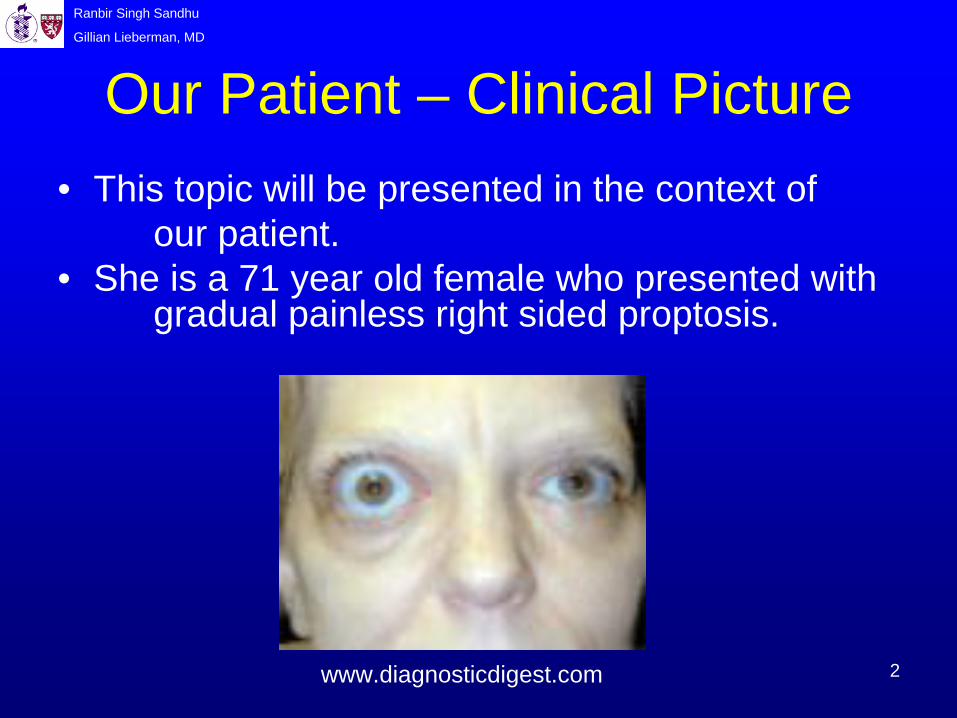
Our Patient – Clinical Picture• This topic will be presented in the context of
our patient.• She is a 71 year old female who presented with
gradual painless right sided proptosis.
www.diagnosticdigest.com

Ranbir Singh Sandhu
Gillian Lieberman, MD
3
Imaging Options for the OrbitsTo image the orbit, we can use:
• Plain Film • Its main use is screening for metallic foreign bodies.
• Ultrasound• It is used to characterize abnormalities (e.g., masses).
• CT • For further characterization of abnormalities and bone detail (e.g.,
abnormal bone mineralization).
• MRI • It is chosen for its enhanced soft tissue contrast.
• Angiogram • It identifies aberrant blood vessels such as those created by
tumors (not routinely used).
Ranbir Singh Sandhu
Gillian Lieberman, MD
4
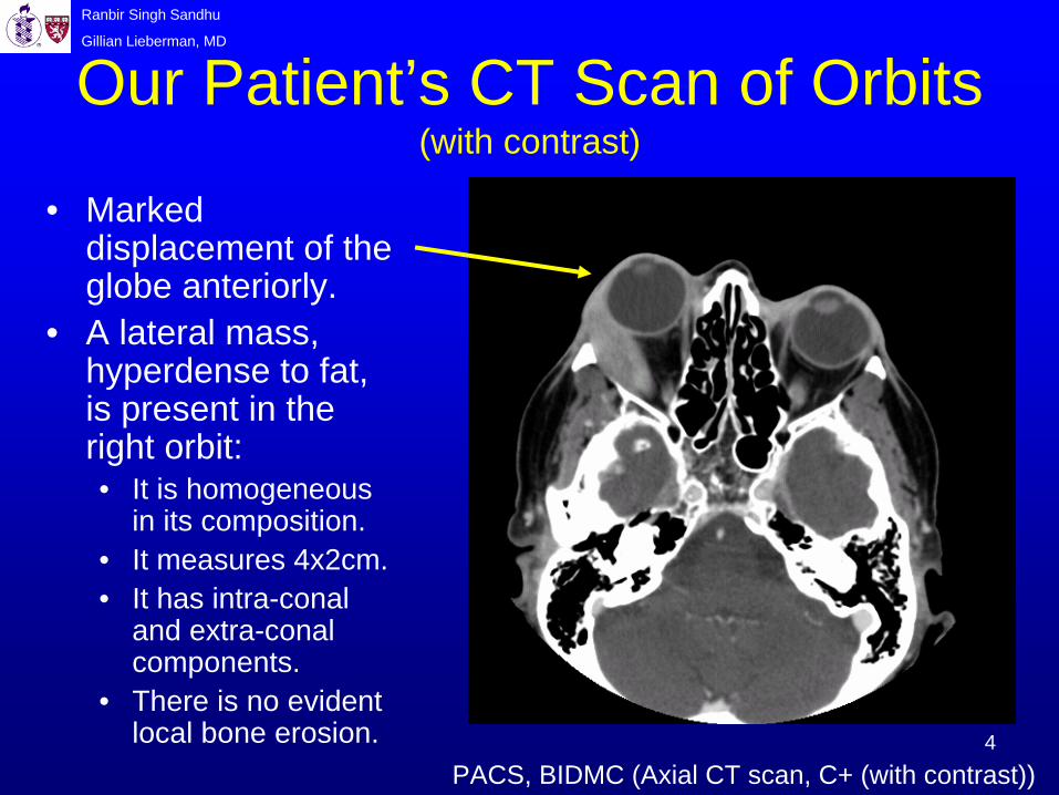
Our Patient’s CT Scan of Orbits (with contrast)
PACS, BIDMC (Axial CT scan, C+ (with contrast))
• Marked displacement of the globe anteriorly.
• A lateral mass, hyperdense to fat, is present in the right orbit:• It is homogeneous
in its composition.• It measures 4x2cm.• It has intra-conal
and extra-conal components.
• There is no evident local bone erosion.
Ranbir Singh Sandhu
Gillian Lieberman, MD
5
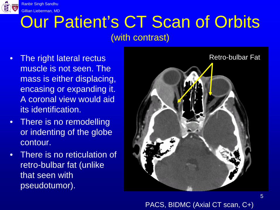
Our Patient’s CT Scan of Orbits (with contrast)
• The right lateral rectus muscle is not seen. The mass is either displacing, encasing or expanding it. A coronal view would aid its identification.
• There is no remodelling or indenting of the globe contour.
• There is no reticulation of retro-bulbar fat (unlike that seen with pseudotumor).
Retro-bulbar Fat
PACS, BIDMC (Axial CT scan, C+)

Ranbir Singh Sandhu
Gillian Lieberman, MD
6
Our Patient’s CT Scan of Orbits (with contrast)
• This is the level of the superior aspect of the orbit.
• There is still no evident bone erosion.
• Right lacrimal gland involvement is likely since it is not visualized in the right orbit unlike in the left orbit.
Left Lacrimal Gland
PACS, BIDMC (Axial CT scan, C+)
Ranbir Singh Sandhu
Gillian Lieberman, MD
7
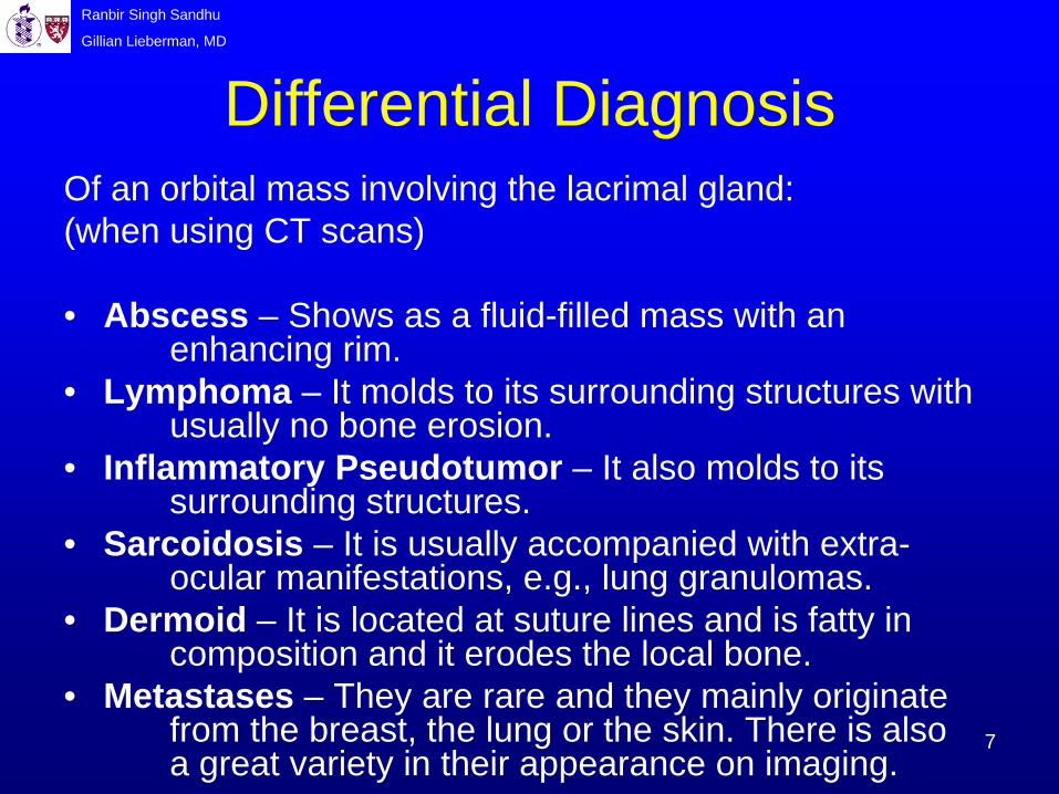
Differential DiagnosisOf an orbital mass involving the lacrimal gland: (when using CT scans)
• Abscess – Shows as a fluid-filled mass with an enhancing rim.
• Lymphoma – It molds to its surrounding structures with usually no bone erosion.
• Inflammatory Pseudotumor – It also molds to its surrounding structures.
• Sarcoidosis – It is usually accompanied with extra- ocular manifestations, e.g., lung granulomas.
• Dermoid – It is located at suture lines and is fatty in composition and it erodes the local bone.
• Metastases – They are rare and they mainly originate from the breast, the lung or the skin. There is also a great variety in their appearance on imaging.

Ranbir Singh Sandhu
Gillian Lieberman, MD
8
Evaluating the Differentials Using the patient’s CT Scan of Orbits
• Abscess: • This is unlikely as the mass on the CT scan is
homogeneous in its appearance and it has no enhancing rim.
• Lymphoma:• This is possible as the patient’s mass is encasing the
lateral rectus muscle and it is not eroding the local bone.
• Inflammatory Pseudotumor:• This is also possible for the same reasons as
lymphoma but the patient’s proptosis is painless and there is no reticulation of retro- bulbar fat (these are usual for pseudotumor).

Ranbir Singh Sandhu
Gillian Lieberman, MD
9
Evaluating the Differentials Using the CT Scan of Orbits
• Sarcoidosis:• It is an unlikely possibility so plain films could be
checked for any signs of lung pathology. • Dermoid:
• This is unlikely as the mass is not located near the suture lines nor is it fatty in its composition nor is it eroding bone.
• Metastases:• It is a possibility but they are rare. Imaging of the
breasts, lungs and skin could be done to check for any signs of tumor presence.

Ranbir Singh Sandhu
Gillian Lieberman, MD
10
Our Patient’s Past Medical History
• Our patient has a past medical history of widespread systemic lymphoma accompanied with left orbit involvement.
• Her Pathology results from her pleural effusion came back as:
• Grade I Follicular Center Cell Lymphoma (this is a type of Non-Hodgkin’s Lymphoma)
• She then went on to have 6 cycles of chemotherapy (cytoxan, vincristine & prednisone) as well as left orbit irradiation which created a left sided cataract which has since been replaced with an intra-ocular lens.

Ranbir Singh Sandhu
Gillian Lieberman, MD
11
Our Patient’s Body CT Scan (With Contrast) Before Treatment
Widespread Pleural Effusion
Enlarged Axillary Lymph Node
Mediastinal Lymphadenopathy
PACS, BIDMC (Axial CT scan, C+)
Ranbir Singh Sandhu
Gillian Lieberman, MD
12
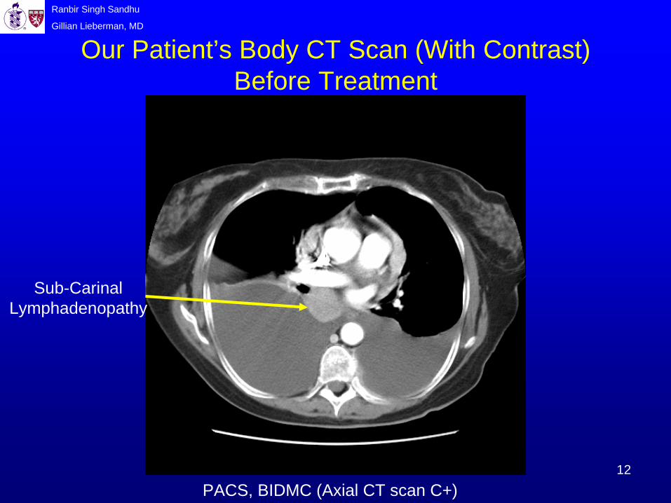
Our Patient’s Body CT Scan (With Contrast) Before Treatment
Sub-Carinal Lymphadenopathy
PACS, BIDMC (Axial CT scan C+)
Ranbir Singh Sandhu
Gillian Lieberman, MD
13
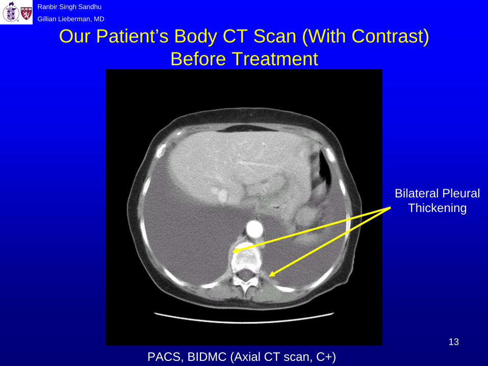
Our Patient’s Body CT Scan (With Contrast) Before Treatment
Bilateral Pleural Thickening
PACS, BIDMC (Axial CT scan, C+)
Ranbir Singh Sandhu
Gillian Lieberman, MD
14
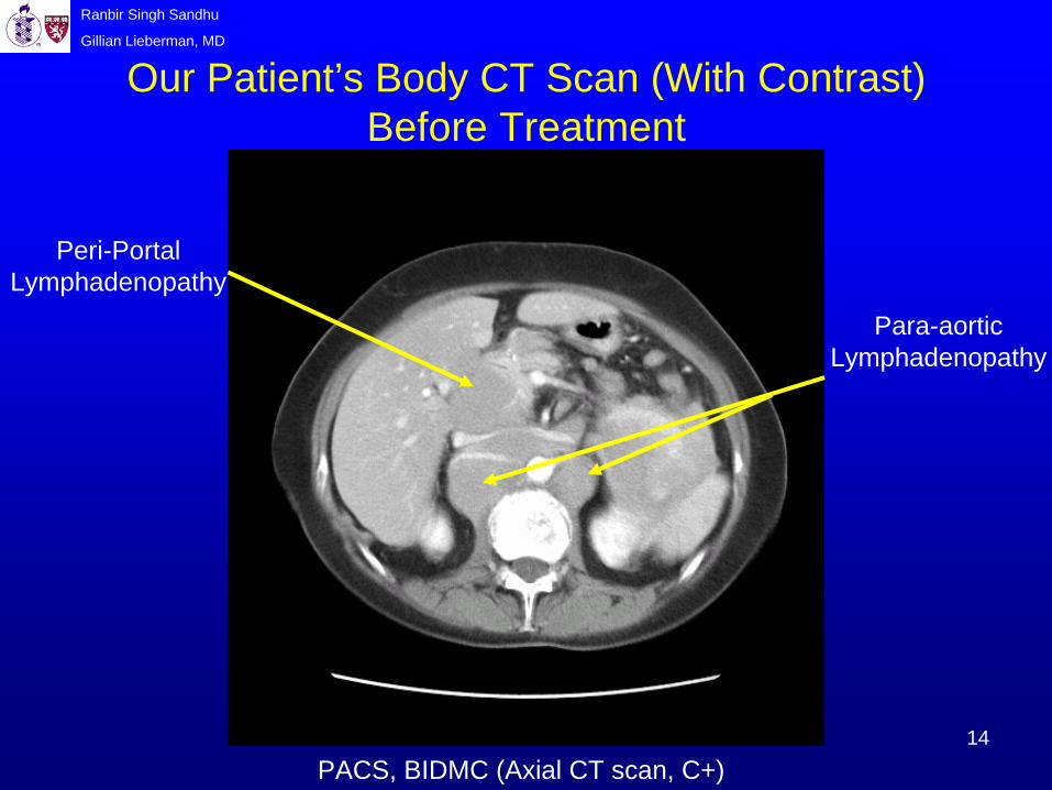
Our Patient’s Body CT Scan (With Contrast) Before Treatment
Peri-Portal Lymphadenopathy
Para-aortic Lymphadenopathy
PACS, BIDMC (Axial CT scan, C+)
Ranbir Singh Sandhu
Gillian Lieberman, MD
15
Course Of Our Patient• Given our patient’s past medical history of lymphoma
with left orbit involvement, it was decided as highly likely that the right orbital mass was most likely to be a recurrence of her lymphoma.
• A biopsy was not taken after considering its risk of complications.
• To treat her right orbital lymphoma she went on to have 3 further cycles of chemotherapy (fludarabine and cyclophosphamide).
• Following treatment, her right orbital lymphoma went into complete remission!
• She is now on ‘Rituximab’ maintainance therapy and is now symptom-free enjoying 3 rounds of 18-hole golf a week!

Ranbir Singh Sandhu
Gillian Lieberman, MD
16
Our Patient’s CT Scan of Orbits After Treatment (with contrast)
Ocular Lens Replacement Of
Catarct
• There is no evidence of any remaining tumor in the right or left orbits
• There is no evidence of any bone erosion in the right or left orbits.
• The right globe is no longer being displaced.
• The lateral rectus muscle can be clearly seen in both orbits in contrast to its incasement before treatment.
• An intra-ocular lens replacement can be seen in the left eye.
Right Lateral Rectus Muscle
PACS, BIDMC (Axial CT scan, C+)

Ranbir Singh Sandhu
Gillian Lieberman, MD
17
• No tumor is visible in the superior aspect of the orbit.
• The right lacrimal gland is no longer involved in any pathology.
PACS, BIDMC (Axial CT scan, C+)
Our Patient’s CT Scan of Orbits After Treatment (with contrast)
Ranbir Singh Sandhu
Gillian Lieberman, MD
18
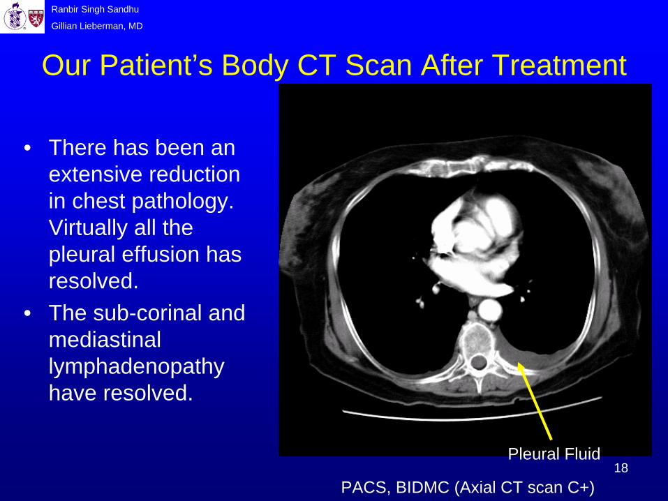
Our Patient’s Body CT Scan After Treatment
• There has been an extensive reduction in chest pathology. Virtually all the pleural effusion has resolved.
• The sub-corinal and mediastinal lymphadenopathy have resolved.
Pleural Fluid
PACS, BIDMC (Axial CT scan C+)
Ranbir Singh Sandhu
Gillian Lieberman, MD
19
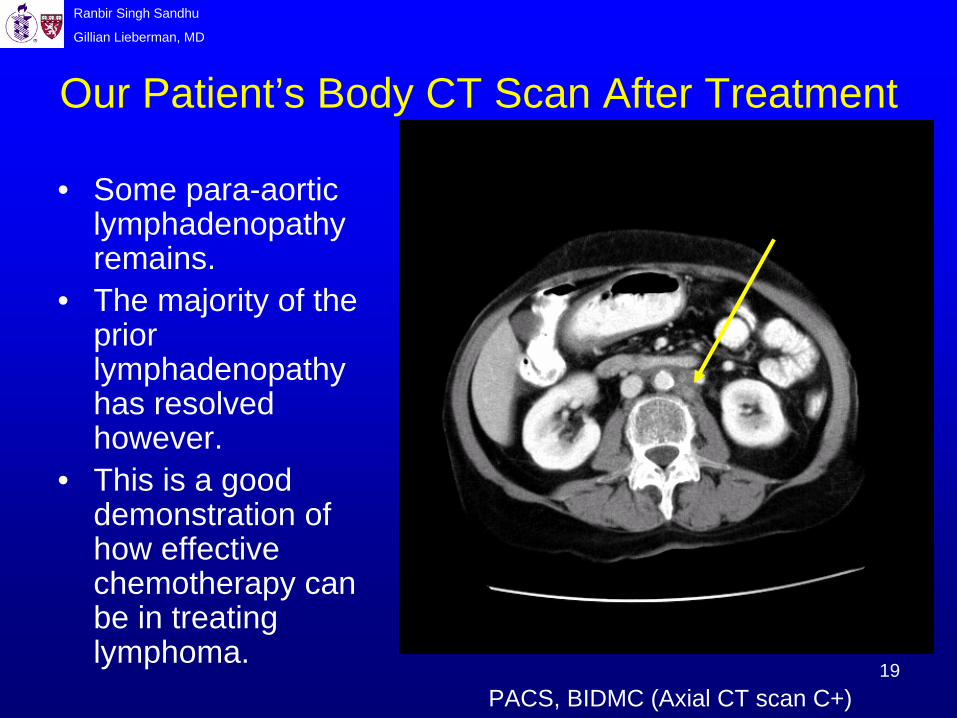
Our Patient’s Body CT Scan After Treatment
• Some para-aortic lymphadenopathy remains.
• The majority of the prior lymphadenopathy has resolved however.
• This is a good demonstration of how effective chemotherapy can be in treating lymphoma.
PACS, BIDMC (Axial CT scan C+)

Ranbir Singh Sandhu
Gillian Lieberman, MD
20
Notes On Orbital Lymphoma• Incidence:
• 75% of patients with orbital involvement have systemic lymphoma1, therefore image other sites, e.g., the neck, chest and abdomen, for any lymphoma.
• The incidence increases with advancing age2.• There is no sex predilection2.
• Classifications:• Subconjunctival involving• Lacrimal gland involving • Orbital involving (usually presents with proptosis)• Eyelid involving (usually presents with ptosis)
• (our patient had both lacrimal gland and orbital involving)
Ranbir Singh Sandhu
Gillian Lieberman, MD
21
Notes On Orbital Lymphoma• Location:
• Intra-conal (within the cone created by the extra ocular muscles, EOMs).
• Extra-conal (outside the cone of EOMs).• Orbital lymphomas are mostly located in the anterior
and superior aspects of the orbit.
• Clinical Symptoms:• Insidious onset• Diplopia (double vision)• Proptosis (bulging of the eyeball out of the socket) • Painless presentation (pain is more common with
pseudotumor)
Ranbir Singh Sandhu
Gillian Lieberman, MD
22
Notes On Orbital Lymphoma• Imaging Characteristics of Orbital Lymphomas:
• Usually no osseous destruction• except rarely for some malignant tumors • instead it molds to surrounding structures
• Orbital lymphomas are:• Hyperdense relative to fat on CT3. • Hypointense on T1-weighted MRI3.• Isointense to extra ocular muscle on T1 & T2-
weighted MRI3.• Orbital lymphomas are Gallium and FDG avid.
Ranbir Singh Sandhu
Gillian Lieberman, MD
23
Notes On Orbital Lymphoma• Treatment:
• Orbital lymphoma responds well to conventional chemotherapy (using radiation if an adjunct is required but note that its propensity to create cataracts)
• Miscellaneous Facts: • Orbital lymphoma types range from benign lymphoid
hyperplasia to malignant lymphoma confirmed by a biopsy.
• Imaging cannot differentiate well between orbital lymphoma and inflammatory pseudotumor however empirical steroid treatment will often be employed followed by a biopsy if the mass does not resolve.
Ranbir Singh Sandhu
Gillian Lieberman, MD
24
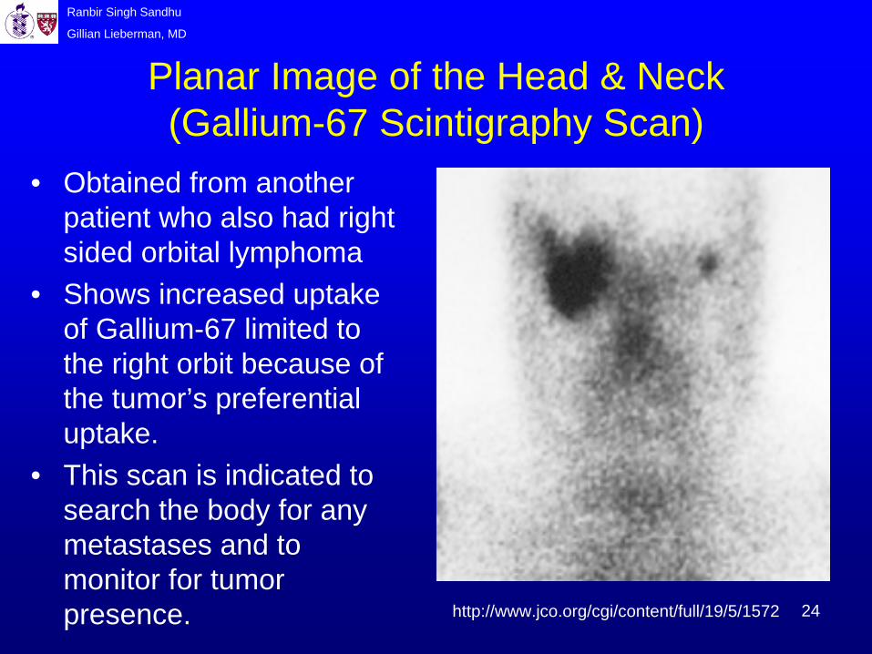
Planar Image of the Head & Neck (Gallium-67 Scintigraphy Scan)
http://www.jco.org/cgi/content/full/19/5/1572
• Obtained from another patient who also had right sided orbital lymphoma
• Shows increased uptake of Gallium-67 limited to the right orbit because of the tumor’s preferential uptake.
• This scan is indicated to search the body for any metastases and to monitor for tumor presence.
Ranbir Singh Sandhu
Gillian Lieberman, MD
25
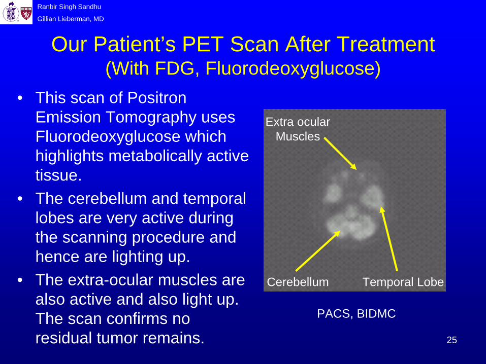
Our Patient’s PET Scan After Treatment (With FDG, Fluorodeoxyglucose)
PACS, BIDMC
• This scan of Positron Emission Tomography uses Fluorodeoxyglucose which highlights metabolically active tissue.
• The cerebellum and temporal lobes are very active during the scanning procedure and hence are lighting up.
• The extra-ocular muscles are also active and also light up. The scan confirms no residual tumor remains.
Extra ocular Muscles
Cerebellum Temporal Lobe
Ranbir Singh Sandhu
Gillian Lieberman, MD
26
Summary• On a CT scan, orbital lymphoma is seen to mold to its
surrounding structures with usually no bone destruction.
• 75% of patients with lymphoma in the orbits will also have lymphoma at other sites so these must be imaged following presentation (e.g., the neck/chest/abdomen).
• Imaging cannot differentiate well between orbital lymphoma and inflammatory pseudotumor however empirical steroid treatment will often be employed.
• Orbital lymphomas are very responsive to conventional chemotherapy treatment, which is shown by our patient who is symptom free and continues to lead a happy life.
Ranbir Singh Sandhu
Gillian Lieberman, MD
27
References• Abner A., Lange R., Gauvin G. (2001) Unusual sites of
malignancy: Case 2 Orbital Lymphoma. Journal of Clinical Oncology, Vol 19, Issue 5 (March), 2001: 1572-1573
• Bhatia S., Paulino A.C., Buatti J.M., Mayr N.A., Wen B.C. (2002) Curative Radiotherapy for Primary Orbital Lymphoma. International Journal Of Radiation Oncology Biological Physics, 54(3): 818-23
• Curtin H.D., Som P.M. Head and Neck Imaging, 4th ed. 2003. Chapter 8, 9
• Das Narla L., Newman B., Spottswood S.S., Narla S., Kolli R. (2003) Inflammatory Pseudotumour. Radiographics, 23: 719-729
• Flanders A.E. (1987) Orbital Lymphoma Role of CT and MRI. Radiologic Clinics of North America, 25: 601-612.
Ranbir Singh Sandhu
Gillian Lieberman, MD
28
References• Fung C.Y., Tarbell N.J., Lucarelli M.J., Goldberg S., Linggood
R.M., Harris N.L., Ferry J.A. (2003) Ocular adnexal lymphoma: clinical behaviour of distinct World Health Organisation classification subtypes. International Journal of Radiation OncologyBiological Physics, 57(5) 1382-91.
• 3Hosten N. & Bornfeld N. Imaging of the Globe and Orbit, Thieme Press: 1998
• 2Moslehi R, Divesa S, Schairer C, Fraumeni J. (2006) Rapidly Increasing Incidence of Ocular Non-Hodgkin Lymphoma. Journal of the National Cancer Institute, Vol 98, No. 13 936-39.
• Noyek A. Head and Neck Radiology, J.B. Lippincott Company Press: 1991
• www.uhrad.com/mriarc/mri049.htm• 1http://brighamrad.harvard.edu/Cases/bwh/hcache/392/full.html
Ranbir Singh Sandhu
Gillian Lieberman, MD
29
Acknowledgements
Gillian Lieberman, MDHugh Curtin, MD
Douglas Teich, MDSanjay Shetty, MD
Vaibhav Khasgiwala, MDJason Handwerker, MD
Pamela Lepkowski


![PRIMARY NON-HODGKIN LYMPHOMA OF THE ORBIT ......the patients, presentation is with orbital mass [2]. Common signs at presentation are: palpable mass, pain, exophtalmia, ptosis, dyplopia,](https://static.fdocuments.us/doc/165x107/614396f46b2ee0265c0224b6/primary-non-hodgkin-lymphoma-of-the-orbit-the-patients-presentation-is.jpg)
