Oral Premalignancy - jssuni.edu.in · Enlarged and hyperchromatic cells are visible at this low...
72
Oral Premalignancy
Transcript of Oral Premalignancy - jssuni.edu.in · Enlarged and hyperchromatic cells are visible at this low...
Oral Premalignancy
Premalignant lesion is a morphologically altered
tissue in which cancer is more likely to occur, than its
apparently normal counter parts.
Premalignant condition is a generalized state or
condition associated with significantly increased risk for
cancer development.
Various oral mucosal lesions, particularly red lesions
(erythroplakias) and some white lesions (leukoplakias),
have a
potential for malignant change
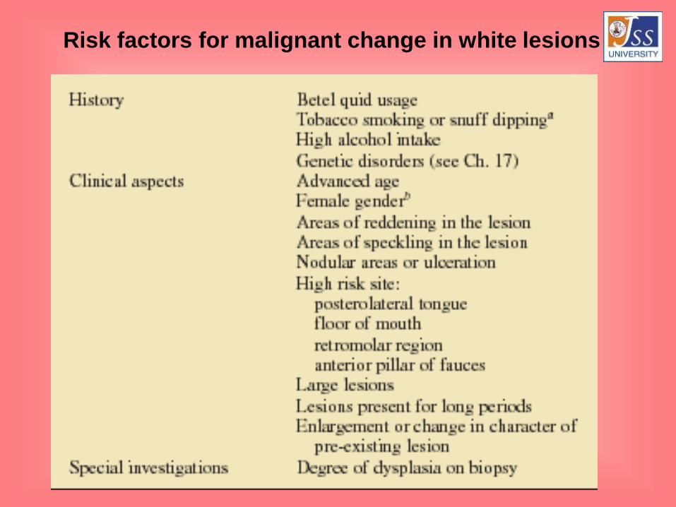
Risk factors for malignant change in white lesions
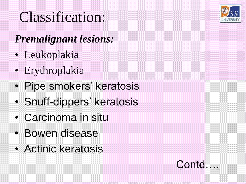
Classification:
Premalignant lesions:
• Leukoplakia
• Erythroplakia
• Pipe smokers’ keratosis
• Snuff-dippers’ keratosis
• Carcinoma in situ
• Bowen disease
• Actinic keratosis
Contd….
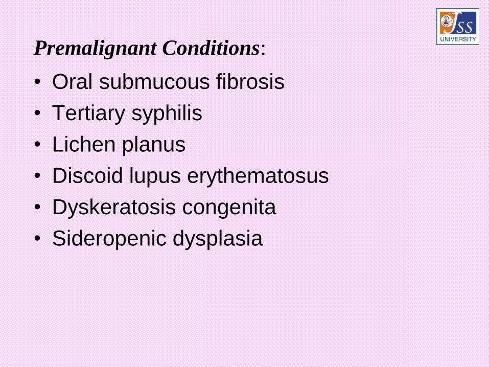
Premalignant Conditions:
• Oral submucous fibrosis
• Tertiary syphilis
• Lichen planus
• Discoid lupus erythematosus
• Dyskeratosis congenita
• Sideropenic dysplasia
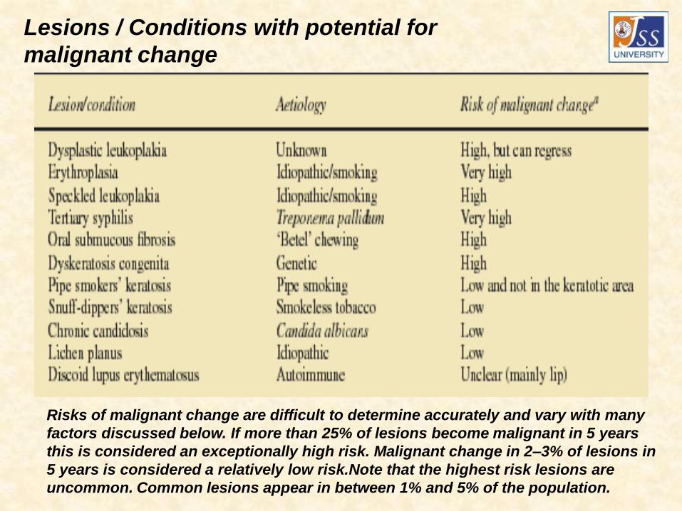
Lesions / Conditions with potential for
malignant change
Risks of malignant change are difficult to determine accurately and vary with many
factors discussed below. If more than 25% of lesions become malignant in 5 years
this is considered an exceptionally high risk. Malignant change in 2–3% of lesions in
5 years is considered a relatively low risk.Note that the highest risk lesions are
uncommon. Common lesions appear in between 1% and 5% of the population.
The best predictor of the potential for malignant transformation
is the degree of dysplasia seen histologically. For this reason,
and because a few lesions will already be malignant, biopsy of
red and white patches is mandatory. The term dysplasia (literally,
abnormal growth) is given to the cytological abnormalities seen
in both malignant and premalignant cells. Premalignancy is
distinguished from malignancy only by the latter’s invasiveness
and release of metastases.
Epithelial dysplasia: histological
features
• Drop-shaped rete ridges
• Nuclear hyperchromatism
• Nuclear pleomorphism
• Altered nuclear/cytoplasmic ratio
• Excess mitotic activity
• Loss of polarity of cells
• Deep cell keratinisation
• Disordered or loss of differentiation
• Loss of intercellular adherence
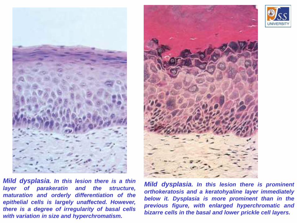
Mild dysplasia. In this lesion there is a thin
layer of parakeratin and the structure,
maturation and orderly differentiation of the
epithelial cells is largely unaffected. However,
there is a degree of irregularity of basal cells
with variation in size and hyperchromatism.
Mild dysplasia. In this lesion there is prominent
orthokeratosis and a keratohyaline layer immediately
below it. Dysplasia is more prominent than in the
previous figure, with enlarged hyperchromatic and
bizarre cells in the basal and lower prickle cell layers.
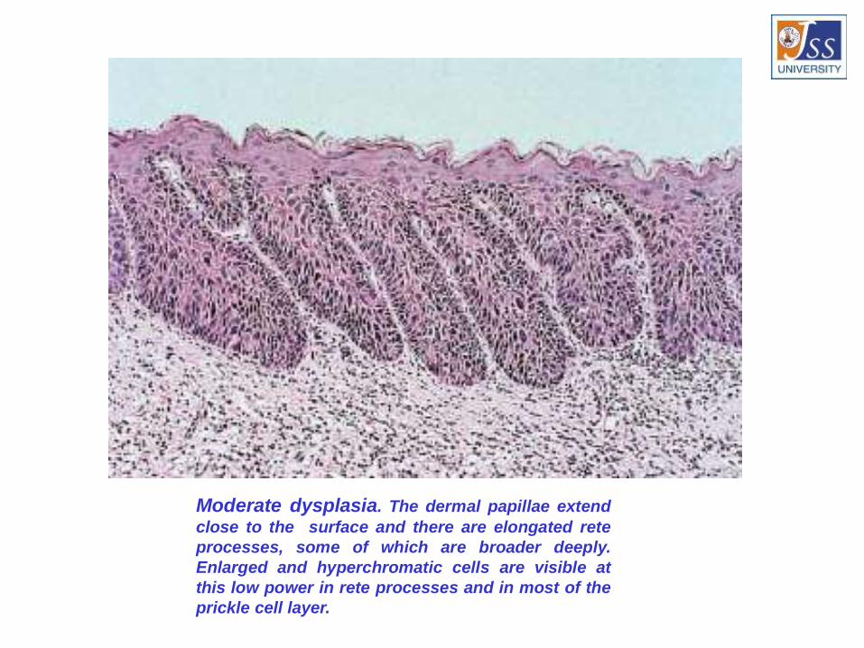
Moderate dysplasia. The dermal papillae extend
close to the surface and there are elongated rete
processes, some of which are broader deeply.
Enlarged and hyperchromatic cells are visible at
this low power in rete processes and in most of the
prickle cell layer.
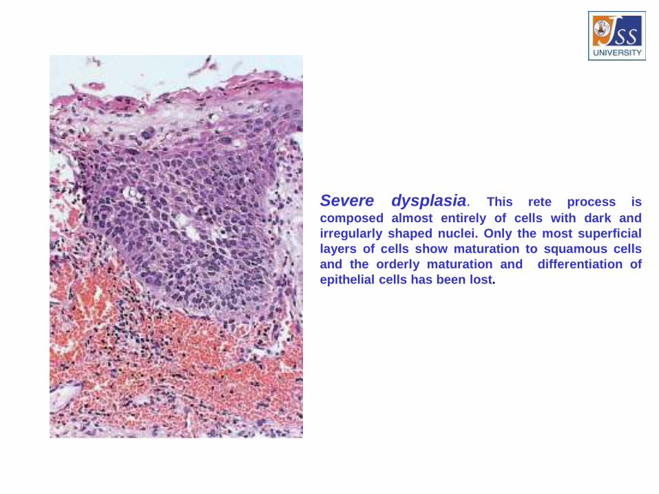
Severe dysplasia. This rete process is
composed almost entirely of cells with dark and
irregularly shaped nuclei. Only the most superficial
layers of cells show maturation to squamous cells
and the orderly maturation and differentiation of
epithelial cells has been lost.
LEUKOPLAKIA
( Leuko-white , Plakia-patch)
Definition: A white hyperkeratotic ,non scrapable
patch in the oral cavity which cannot be
characterized as any other clinical entity and is
always associated with tobacco intake. Definition
does not carry any histological connotation
with it.
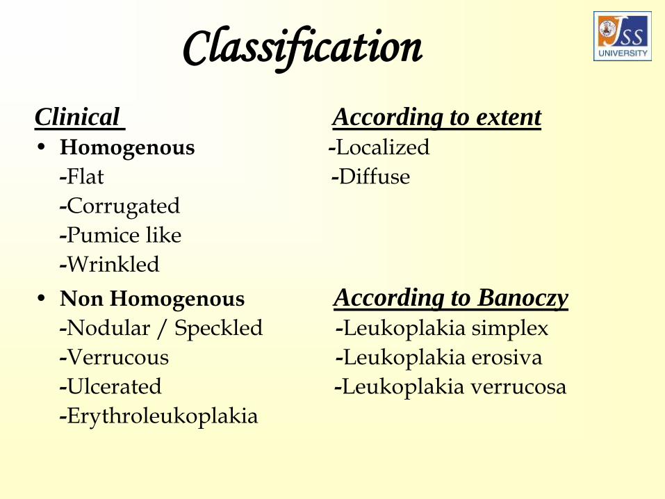
Classification
Clinical According to extent
• Homogenous -Localized
-Flat -Diffuse
-Corrugated
-Pumice like
-Wrinkled
• Non Homogenous According to Banoczy
-Nodular / Speckled -Leukoplakia simplex
-Verrucous -Leukoplakia erosiva
-Ulcerated -Leukoplakia verrucosa
-Erythroleukoplakia
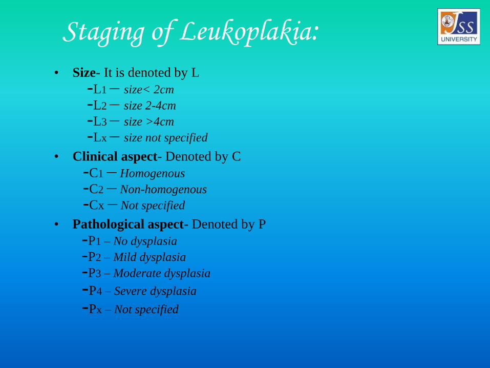
Staging of Leukoplakia:• Size- It is denoted by L
-L1 – size< 2cm
-L2 – size 2-4cm
-L3 – size >4cm
-Lx – size not specified
• Clinical aspect- Denoted by C
-C1 – Homogenous
-C2 – Non-homogenous
-Cx – Not specified
• Pathological aspect- Denoted by P
-P1 – No dysplasia
-P2 – Mild dysplasia
-P3 – Moderate dysplasia
-P4 – Severe dysplasia
-Px – Not specified
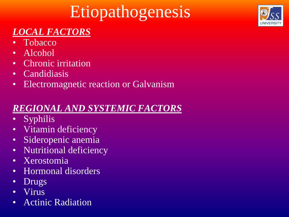
EtiopathogenesisLOCAL FACTORS• Tobacco• Alcohol• Chronic irritation• Candidiasis• Electromagnetic reaction or Galvanism
REGIONAL AND SYSTEMIC FACTORS• Syphilis• Vitamin deficiency• Sideropenic anemia• Nutritional deficiency• Xerostomia• Hormonal disorders• Drugs• Virus• Actinic Radiation

Clinical features
• Sex and distribution
• Common sites
• Appearance
• Surface
• Color
• Symptoms
• Sharp staging
• Ebbing tide type
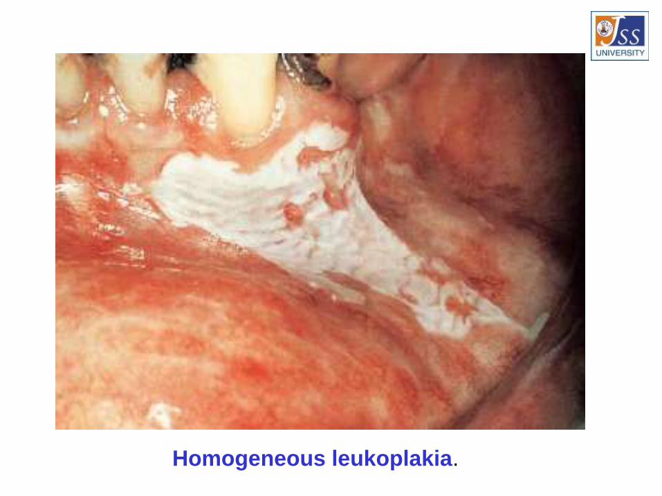
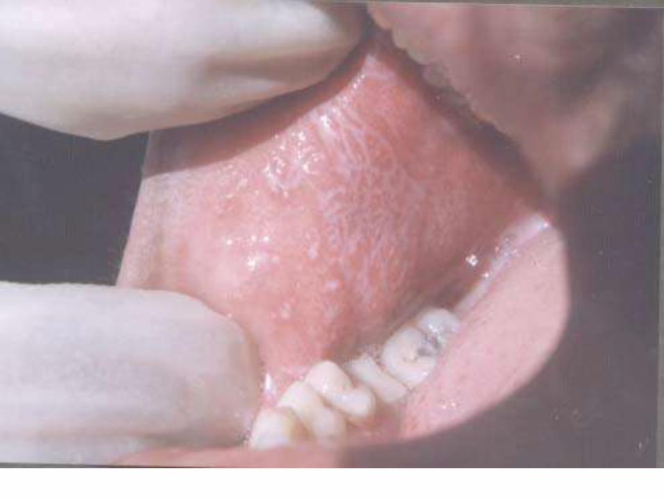
Homogeneous leukoplakia.
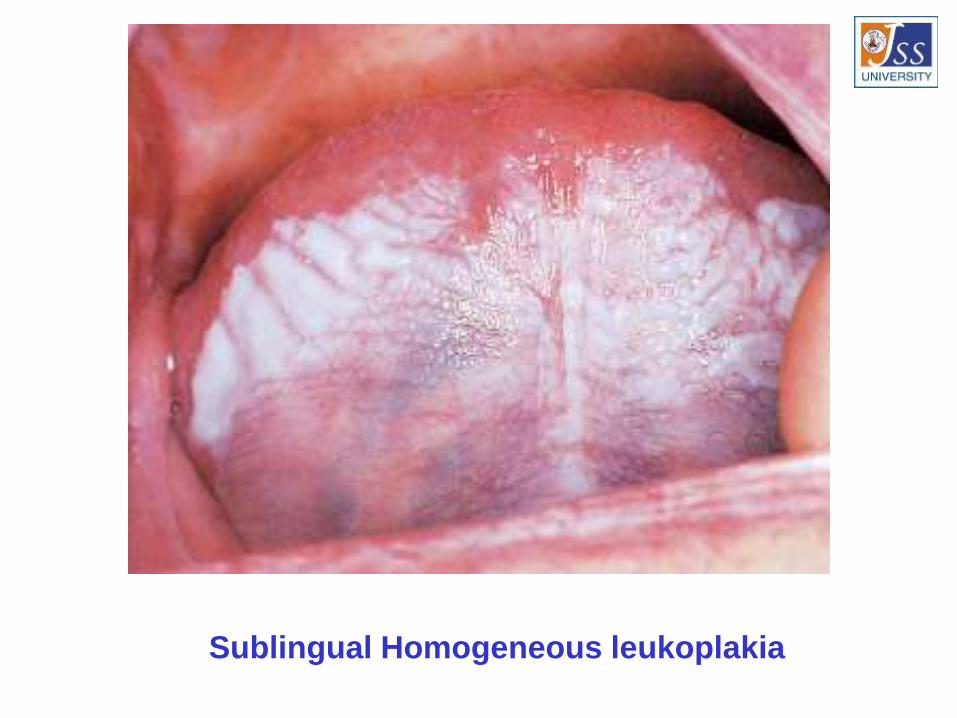
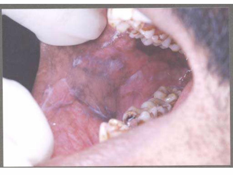
Sublingual Homogeneous leukoplakia
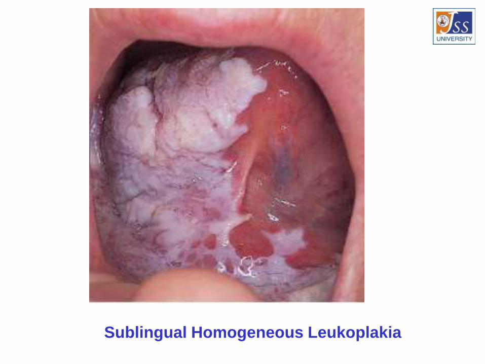
Sublingual Homogeneous Leukoplakia
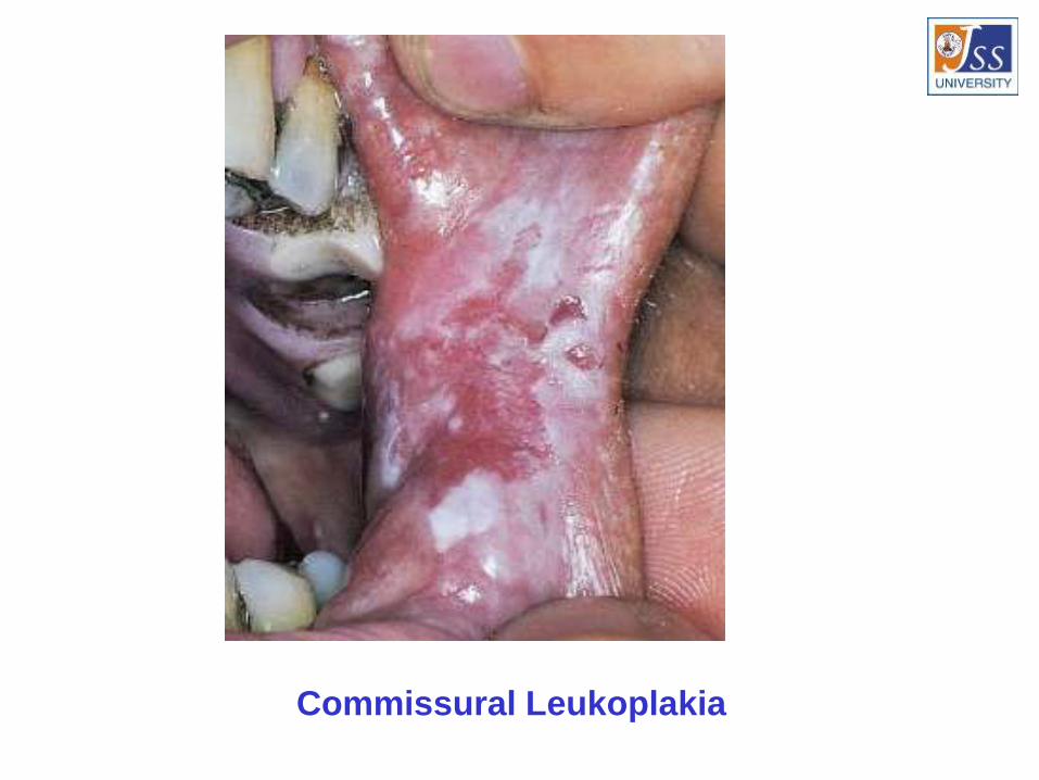
Commissural Leukoplakia
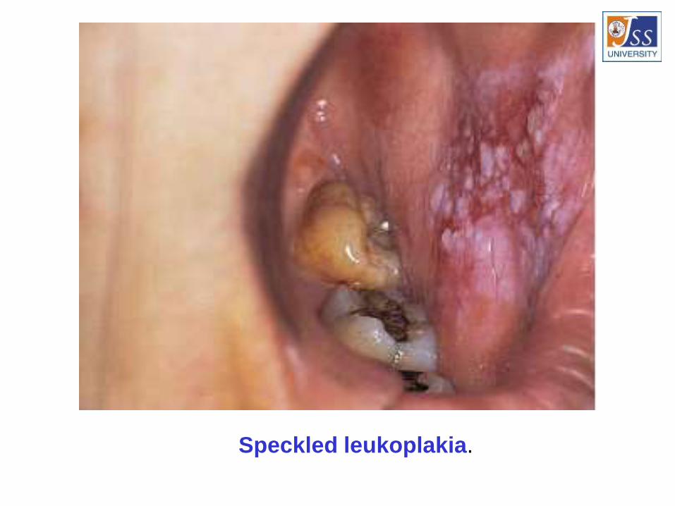
Speckled leukoplakia.
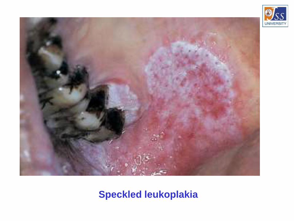
Speckled leukoplakia
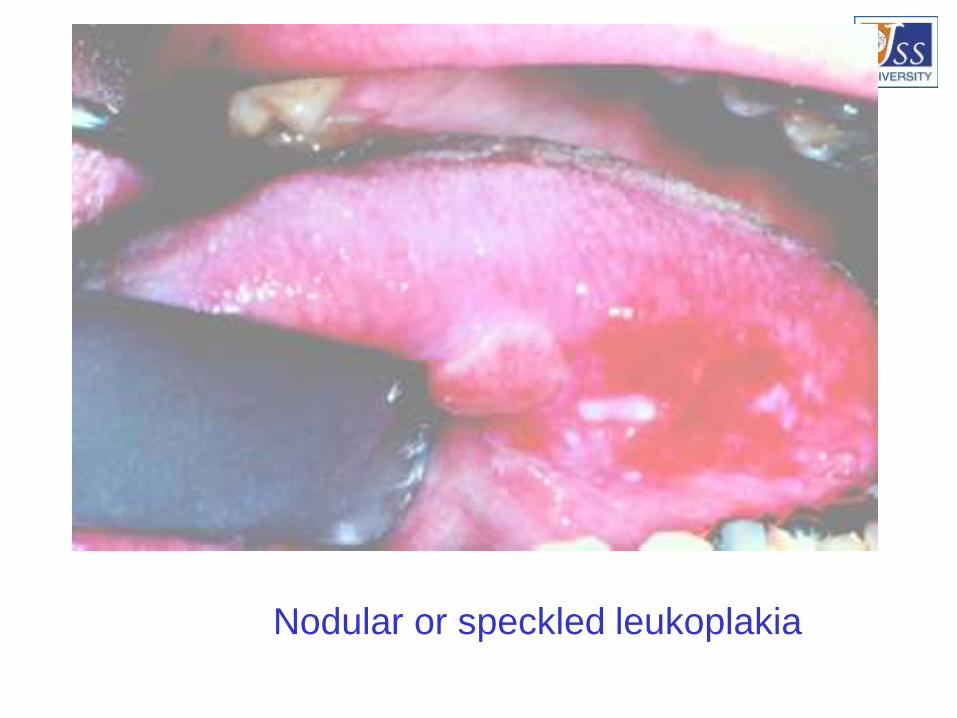
Nodular or speckled leukoplakia
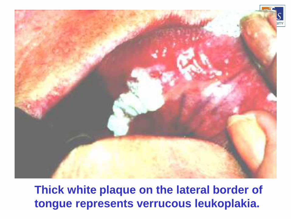
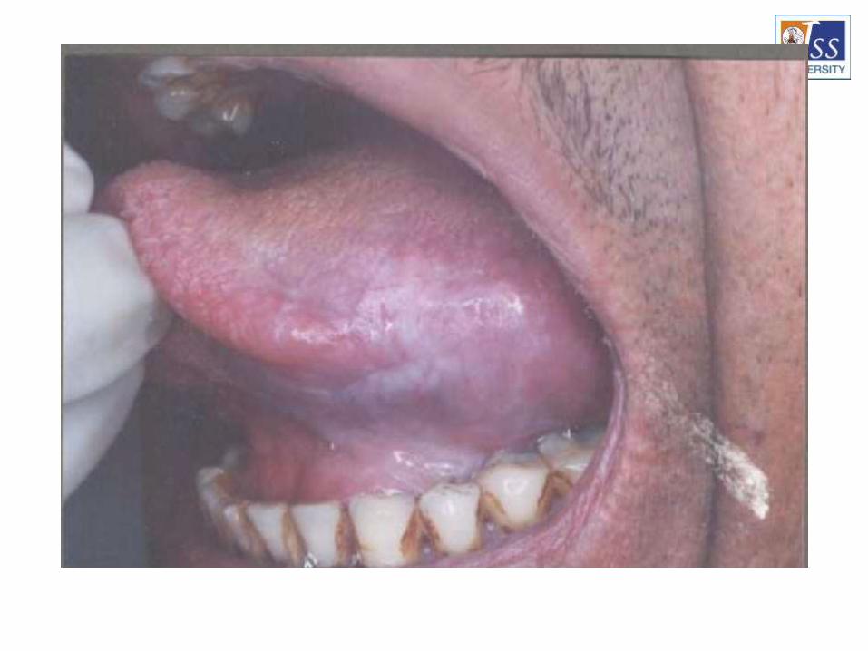
Thick white plaque on the lateral border of
tongue represents verrucous leukoplakia.
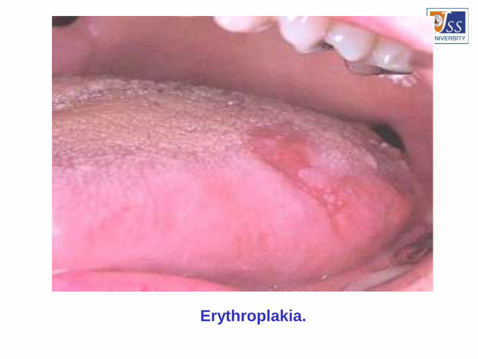
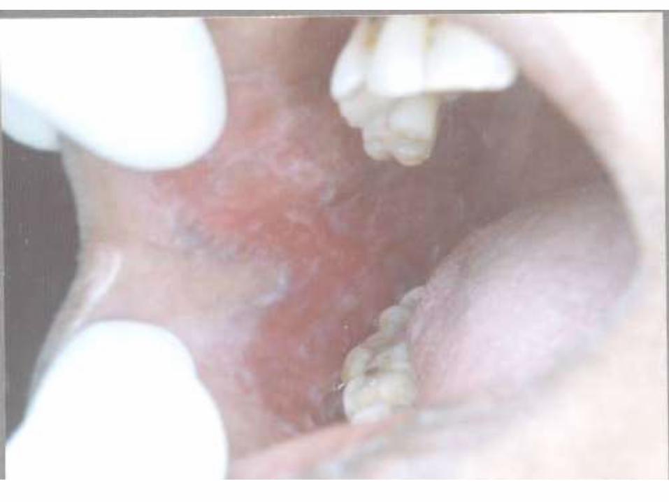
Erythroplakia.
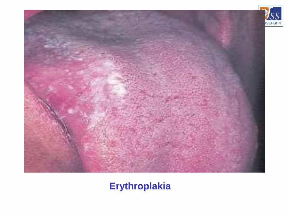
Erythroplakia
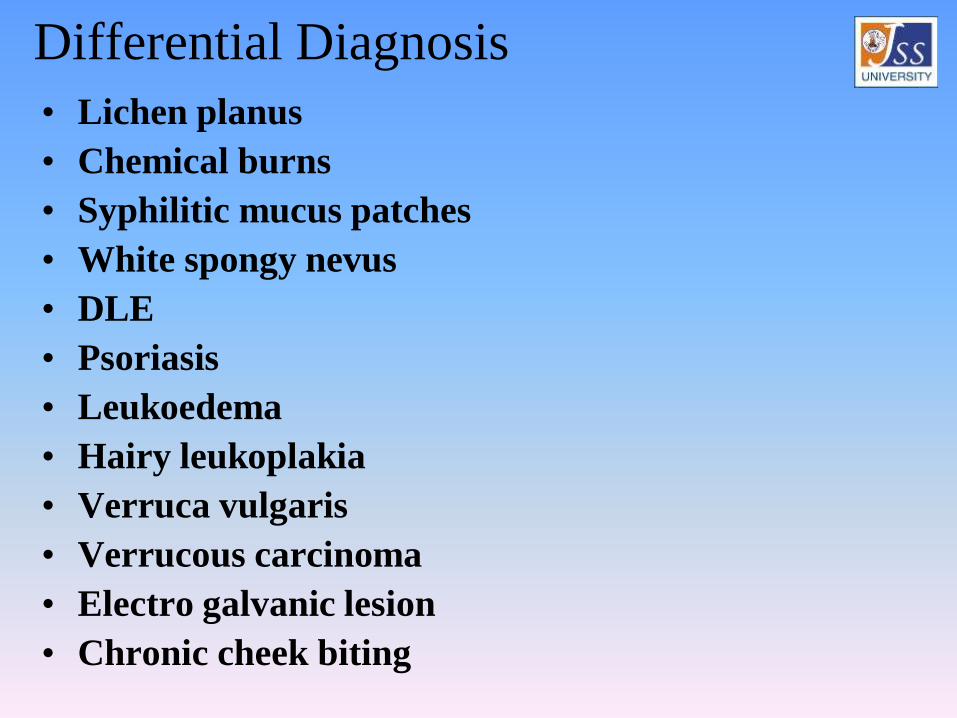
Differential Diagnosis
• Lichen planus
• Chemical burns
• Syphilitic mucus patches
• White spongy nevus
• DLE
• Psoriasis
• Leukoedema
• Hairy leukoplakia
• Verruca vulgaris
• Verrucous carcinoma
• Electro galvanic lesion
• Chronic cheek biting
Management:
• Limitation of etiological factor
• Conservative treatment
-Vitamin therapy (Vit.A 75000-3,00,000 IU )
-Vit.A +Vit.E
-13-cis-retinoic acid
-Antioxidant therapy
-Lycopene 2-8mg/ day for 2 months
-Beta-carotene
-Vit.A Palmitate
-Vit.B complex
-Antimycotic Tt. (Nystatin 5 lac IU bid +20% borax or 1% gentian violet)
-Panthenol lingual tab
-Estrogen
• Surgical Management
-Conventional surgery-Excision-Mucosal flap
-Cryosurgery
-Fulguration (electrosurgery or electrocautry)
-LASER-Biopsy-Laser peel-Ablation-Surface vaporization
• Miscellaneous
-Radiation therapy
-Topical chemotherapy (Bleomycin,Azathiprine etc)
LICHEN PLANUS
• Descirbed in 1869 by Erasmus Wilson.
• Various mucosal surfaces involved.
• Common inflammatory disease of skin with
characteristic violaceous, polygonal,
pruritic papules.
• It also involves mucosa ,nails & hair
Definition
Relatively common dermatological
disorder occurring on skin and oral
mucus membrane and refers to lace like
pattern produced by symbolic algae and
fungal colonies on the surface of rocks in
nature (lichens).
Types:• Reticular
• Papular
• Atrophic
• Erosive
• Bullous
• Annular
• Actinic
• Linear
• Hypertrophic
Etiology:
• Immunologicali)Cell mediated immune response
ii)Autoimmunity
iii)Immunodeficiency
• Genetic factors
• Infections
• Drugs and chemicals
• Psychogenic factor
• Habits
Clinical Features:
• Age & Sex: 35-55 Yrs ; F > M
• Incidence & Prevalence : 0.9 % - 1.2 %
• Site : Skin , oral & other mucus membranes. About 50% of
skin cases have oral lesions, 25% only oral lesions
• Oral and genital mucosal lesions: ‘Vaginogingivalsyndrome’
• Symptoms: Intense pruritis (skin),Burning mouth
• Signs: Characteristic Six ‘P’; Planar, polygonal, purple,
pruritic, papules & plaques.
Oral lichen planus
• Sites: Buccal mucosa (84%)
• Burning mouth
• Wickham’s striae
• Typical lacy, reticular patterns, rings over the
buccal mucosa.
• Associated hyper melanosis
• Usually bilateral.
• Stress induced

Treatment:• Medicinal therapy
-Steroids-Antifungal agents-Vit.-A-Cyclosporine-Dapsone
Surgical Therapy
Psychotherapy
PUVA Therapy
Symptomatic treatment
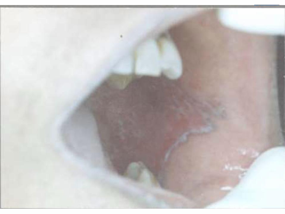
Oral Submucous Fibrosis
• Synonyms:Asian sideropenic dysphagia, oral
mucosal disease, oral fibrosis, OSMF, oral soft
tissue disease.

Symptoms of OSF
• Progressive inability to open the mouth (trismus) due to oral
fibrosis and scarring
• Oral pain and burning sensation upon consumption of spicy
foodstuffs
• Other symptoms
-- Change of gustatory sensation
- Hearing loss due to stenosis of the eustachian tubes
- Dryness of the mouth
- Nasal tonality to the voice
- Dysphagia to solids (if the esophagus is involved)
- Impaired mouth movements (eg, eating, whistling,
blowing, sucking)
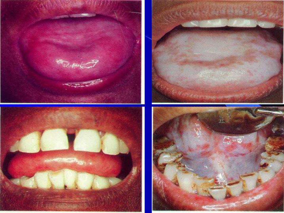
Staging of OSF
• Stage 1: Stomatitis includes erythematous mucosa,
vesicles, mucosal ulcers, melanotic mucosal pigmentation,
and mucosal petechia.
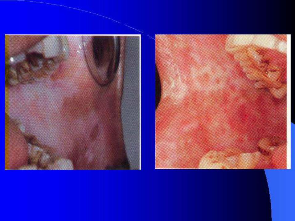
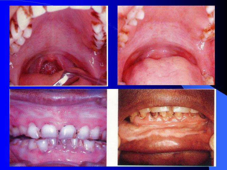
• Stage 2: Fibrosis occurs in ruptured vesicles and ulcers
when they heal, which is the hallmark of this stage.
Early lesions demonstrate blanching of the oral mucosa.
• Stage 3: Sequelae of OSF
Speech and hearing deficits
Leukoplakia
Lab Studies:
•No specific diagnostic tests are available for OSF.
Some OSF studies report the following laboratory
findings:
•Decreased hemoglobin levels
•Decreased iron levels
•Decreased protein levels
•Increased erythrocyte sedimentation rate (ESR)
•Decreased vitamin B complex levels
Other Tests:
•Cytologic smears may be performed.
Treatment• Medical Care
– Steroids
– Placental extracts
– Hyaluronidase
– IFN-g
– Antioxidants
Surgical Care:
•Simple excision of the fibrous bands
•Split-thickness skin grafting following bilateral
temporalis myotomy or coronoidectomy
•Nasolabial flaps and lingual pedicle flaps
Patient Education
• Discontinue the habit
• Eliminate tobacco
• Avoid spicy foodstuffs
• Eat a complete and healthy diet
• Maintain proper oral hygiene
• Schedule regular oral examinations
Special Concerns
• An unhealing ulcer in the lesion
• Lesion undergoing red changes
(erythroplakia)
• A burning sensation in the mouth
• An exophytic mass
• A lump in the neck
• Difficulty in chewing, swallowing, or
speaking