Oral Microbiology and Immunology, 23(23): 1-6...
28
Posted at the Institutional Resources for Unique Collection and Academic Archives at Tokyo Dental College, Available from http://ir.tdc.ac.jp/ Title Stimulation of Fusobacterium nucleatum biofilm formation by Porphyromonas gingivalis Author(s) Alternative Saito, Y; Fujii, R; Nakagawa, KI; Kuramitsu, HK; Okuda, K; Ishihara, K Journal Oral Microbiology and Immunology, 23(23): 1-6 URL http://hdl.handle.net/10130/821 Right This is the pre-peer reviewed version of the following article: Oral Microbiol Immunol. 2008 Feb;23(1):1-6., which has been published in final form at http://www3.interscience.wiley.com/journal/11940648 7/abstract.
Transcript of Oral Microbiology and Immunology, 23(23): 1-6...

Posted at the Institutional Resources for Unique Collection and Academic Archives at Tokyo Dental College,
Available from http://ir.tdc.ac.jp/
TitleStimulation of Fusobacterium nucleatum biofilm
formation by Porphyromonas gingivalis
Author(s)
Alternative
Saito, Y; Fujii, R; Nakagawa, KI; Kuramitsu, HK;
Okuda, K; Ishihara, K
Journal Oral Microbiology and Immunology, 23(23): 1-6
URL http://hdl.handle.net/10130/821
Right
This is the pre-peer reviewed version of the
following article: Oral
Microbiol Immunol. 2008 Feb;23(1):1-6., which has
been published in
final form at
http://www3.interscience.wiley.com/journal/11940648
7/abstract.

1
Stimulation of Fusobacterium nucleatum biofilm formation by
Porphyromonas gingivalis
Yuriko Saito1, Rie Fujii1, Kan-Ichi Nakagawa1, Howard K. Kuramitsu2, Katsuji Okuda3 and
Kazuyuki Ishihara3, 4*
1Department of Endodontics, Pulp and Periapical Biology, 3Deparment of Microbiology
and 4Oral Health Science Center, Tokyo Dental College
2Department of Oral Biology, States University of New York at Buffalo
Key words: Biofilm, Synergistic effect, Fusobacterium nucleatum, Porphyromonas
gingivalis, Periapical periodontitis
Running title: P. gingivalis- activate biofilm formation by F. nucleatum
*Corresponding author
Kazuyuki Ishihara
1-2-2 Masago, Mihama-ku
Chiba 261-8502, Japan
Tel:+81-43-270-3742, Fax:+81-43-270-3744
e-mail: [email protected]

2
Abstract
Introduction: Bacterial infection is a major cause of periapical periodontitis. Eradication
of these microorganisms from apical lesions is essential to the success of endodontic
treatment. The aim of this study was to clarify the molecular interaction between
Fusobacterium nucleatum, Porphyromonas gingivalis and other microorganisms associated
with periapical periodontitis.
Methods: Microorganisms isolated from periapical lesions were inoculated into type-I
collagen-coated polystyrene microtiter plates and maintained at 37 oC under anaerobic
conditions for 2 days, after which, the quantity of organized biofilm on the plates was
evaluated by crystal violet staining. Growth enhancement via soluble factor was evaluated
by separated coculture using a 0.45 µm membrane filter.
Results: Fusobacterium nucleatum exhibited strong adherence to type-1 collagen-coated
polystyrene microplates. Biofilm formation by F. nucleatum was significantly enhanced
by Porphyromonas gingivalis. Enhancement of F. nucleatum biofilm formation was
complemented by compartmentalized coculture with P. gingivalis. Enhancement of
biofilm formation by P. gingivalis was only slightly reduced by inactivation of its AI-2
producing gene luxS.
Conclusion: The results suggest that P. gingivalis enhances biofilm formation by F.
nucleatum by releasing diffusible signaling molecules other than AI-2.
3
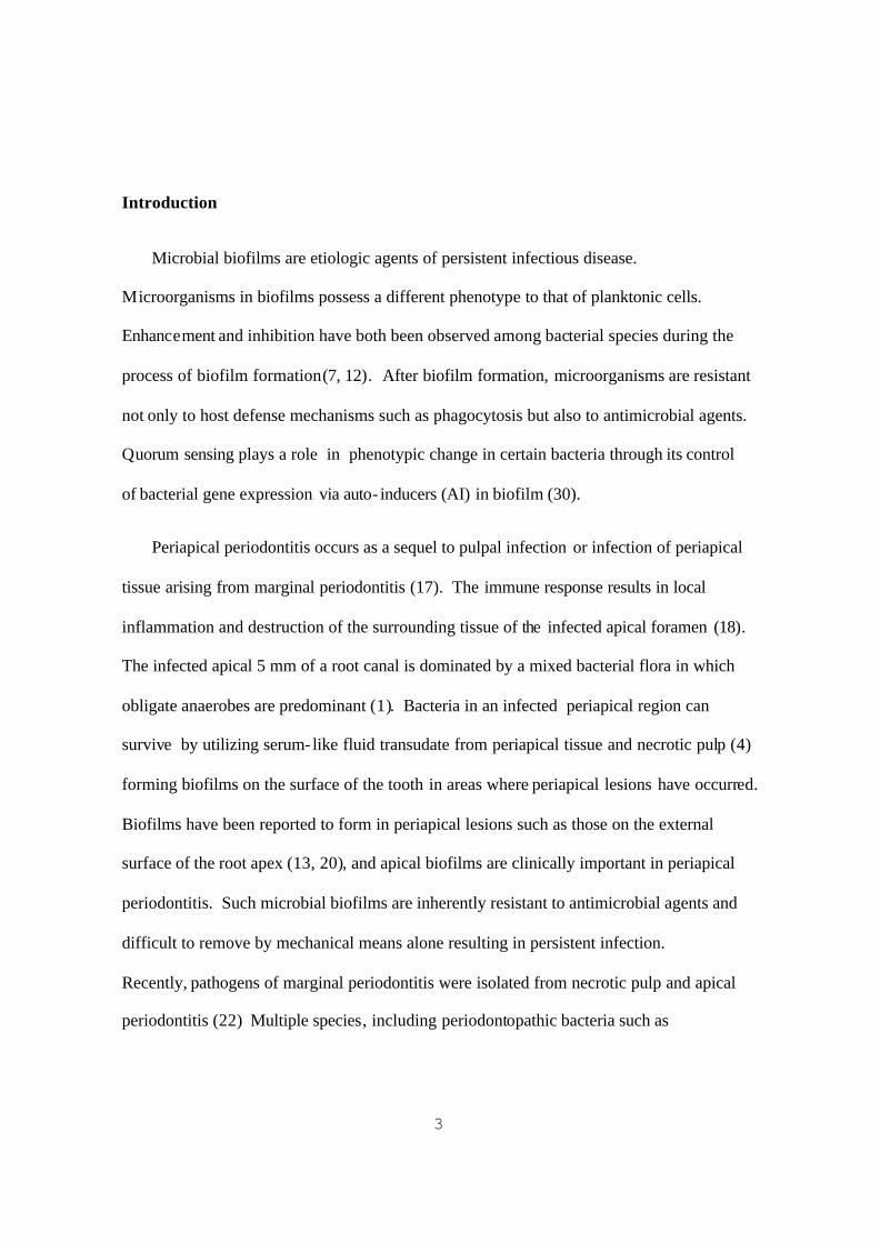
Introduction
Microbial biofilms are etiologic agents of persistent infectious disease.
Microorganisms in biofilms possess a different phenotype to that of planktonic cells.
Enhancement and inhibition have both been observed among bacterial species during the
process of biofilm formation (7, 12). After biofilm formation, microorganisms are resistant
not only to host defense mechanisms such as phagocytosis but also to antimicrobial agents.
Quorum sensing plays a role in phenotypic change in certain bacteria through its control
of bacterial gene expression via auto- inducers (AI) in biofilm (30).
Periapical periodontitis occurs as a sequel to pulpal infection or infection of periapical
tissue arising from marginal periodontitis (17). The immune response results in local
inflammation and destruction of the surrounding tissue of the infected apical foramen (18).
The infected apical 5 mm of a root canal is dominated by a mixed bacterial flora in which
obligate anaerobes are predominant (1). Bacteria in an infected periapical region can
survive by utilizing serum- like fluid transudate from periapical tissue and necrotic pulp (4)
forming biofilms on the surface of the tooth in areas where periapical lesions have occurred.
Biofilms have been reported to form in periapical lesions such as those on the external
surface of the root apex (13, 20), and apical biofilms are clinically important in periapical
periodontitis. Such microbial biofilms are inherently resistant to antimicrobial agents and
difficult to remove by mechanical means alone resulting in persistent infection.
Recently, pathogens of marginal periodontitis were isolated from necrotic pulp and apical
periodontitis (22) Multiple species, including periodontopathic bacteria such as

4
Fusobacterium nucleatum and Porphyromonas gingivalis have been detected in biofilms
associated with periapical periodontitis lesions (19). Therefore, it is important to clarify the
nature of interbacterial communication among these biofilm- forming bacteria.
The role of intercellular signaling molecules such as AI-2 in biofilm formation
resulting in periapical periodontitis lesions remains to be clarified. In this stud y, we
investigated synergistic effects in the process of biofilm formation and communication
among bacteria , focusing particularly on F. nucleatum and P. gingivalis.
Materials and methods
Culture conditions and bacterial strains
All procedures used in this study were approved by the Review Board of the Tokyo
Dental College Ethical Committee. Written informed consent was obtained from each
subject before sample collection. Twenty clinical strains isolated aseptically from apical
periodontitis lesions in 23 patients were used in this study. The apices of the teeth were
aseptically isolated during apicoectomy. Root apices were placed in reduced transport
media (25) containing glass beads, and microorganisms on the surface of the teeth were
dispersed with a vortex mixer for 30 min. The microorganisms were then serially diluted
from 10-1 to 10-5 and inoculated onto Tryptic soy agar (Becton Dickinson Microbio logy
System, Cockeysville, MD) containing 5 µg/ml hemin, 0.5 µg/ml menadione and 10%
horse blood (blood agar plate). The isolated strains were identified by 16S rDNA
sequencing using the Full Gene 16S rDNA Bacterial Identification Kit (Applied
Biosystems, Foster City, CA). These strains were maintained on blood agar plates.

5
Evaluation of biofilm-forming ability
Biofilm formation on the root canal apex or on periodontal tissues is affected by the
ability of the bacteria to adhere to surface- located type-I collagen. Biofilm- forming
activities of a total of 20 isolated strains were quantified according to the method of
Takahashi et al (26) with minor modifications. Briefly, the microorganisms were
inoculated into TSB cons isting of Tryptic soy broth (TSB; Becton Dickinson)
supplemented with 5 µg/ml hemin and 0.5 µg/ml menadione , and precultured anaerobically
at 37oC for 2 days. Fifty-µl cultures were then inoculated into collagen type I-coated 96-
well flat-bottom microplates (Asahi Techno glass, Funabashi, Japan) containing 150 µl of
the same medium, and cultured anaerobically at 37oC for 2 days. The culture medium was
then removed from each well and 50 µl of 0.1% (wt/vol) crystal violet (CV) solution was
added. After 15 min, the wells were rinsed twice with 350 µl distilled water and air-dried.
The CV remaining in the biofilm was solubilized and extracted with 200 µl of 99% ethanol.
Biofilm mass was evaluated at an optical density of 595 nm using a microplate reader
(BIO-RAD, Herucules, CA).
Evaluation of synergistic effects in multi-species biofilms
Biofilm formation by coculture of F. nucleatum TDC100 with partner strains was also
evaluated. P. gingivalis ATCC33277, FDC381 and two Gram-positive strains
(Streptococcus sanguinis TDC15, Staphylococcus epidermidis TDC78) were used as
partner strains. To evaluate the effect of AI-2 on biofilm formation, a P. gingivalis luxS-

6
deficient mutant, CW221 (32), constructed from P. gingivalis FDC381 strain was used.
Each microorganism was inoculated into TSB and precultured over-night anaerobically.
Bacterial cells from 300 µl precultured F. nucleatum TDC100 and 300 µl precultured
partner strain were inoculated into a collagen type I-coated 12-well flat-bottom microplate
(IWAKI, Funabasi, Japan) containing 1200 µl of the same medium and cultured
anaerobically at 37 oC for 2 days. In the case of F. nucleatum alone, 600 µl precultured F.
nucleatum was inoculated. Biofilm mass was measured as described above.
To investigate the induction of signaling between species by diffusible bacterial
mediators, a two-compartment separated culture system was used according to the method
of Yoshida et al. (32). Five hundred µ l TSB was placed in each well of a type-I collagen-
coated polystyrene 12-well plate (IWAKI), which was then designated the lower well. Two
hundred fifty µl overnight culture of each strain was then inoculated into each lower well.
An insert (Transwell, Corning, Corning, NY) was then placed in each well, and designated
the upper well. Next, 500 µ l TSB was placed in each upper well. Finally, 250 µl overnight
culture partner strain was inoculated into each upper well. The organisms were cocultured
physically separated by a porous membrane (pore size, 0.4 µm; Falcon cell culture insert;
BD Labware, Lincoln Park, N.J.). After incubation at 37 oC for 2 days, the inner-well insert
was removed and biofilm mass in the lower well was measured as described above.
Biofilm formation by the species in the two-compartment system was quantitated according
to the following formula: [Biofilm mass of the cocultured species evaluated by crystal
violet] /[Biofilm mass of the species alone evaluated by crystal violet].

7
Effects of AI-2 on biofilm formation
F. nucleatum TDC100 and P. gingivalis FDC381 or CW221, which lacks luxS (32),
were cocultured, and biofilm formation was evaluated as described above. F. nucleatum
TDC 100 was inoculated into the lower compartment and P. gingivalis FDC381 or CW221
were inoculated into the upper compartments and incubated as described above. After
incubation, the mass of biofilm formed by F. nucleatum was assayed as described above.
Statistical analysis
Two-group comparisons were performed using the student t-test. In comparing data
from more than three groups, evaluation was carried out using an analysis of variance and
the Newman-Keuls multiple-comparison test.
Results
Biofilm-forming activity assay
The biofilm- forming activities of 20 strains from among 74 isolates are shown in
Table 1. Among these species, F. nucleatum and P. acnes were frequently isolated together
with other species. These microorganisms showed strong adherence to type-I collagen. As
F. nucleatum TDC100 showed the strongest adherence activity, further investigation of the
effects of other members of apical periodontal lesions on F. nucleatum biofilm formation
was performed using mainly this.strain.
Synergistic effect on biofilm formation by coculture

8
The effects of coculture on F. nucleatum biofilm formation are summarized in Fig 1.
Biofilm formation by coculture with S. epidermidis TDC78, P. gingivalis FDC 381 and
ATCC33277 was 1.8, 3.1 and 2.8 times greater (p<0.001), respectively, than that by F.
nucleatum TDC100 alone. Similar enhancement was also observed in F. nucleatum TDC
845. However, coculture with S. sanguinis TDC15 resulted in almost the same level of
biofilm formation as that by F. nucleatum TDC100 alone (data not shown).
Evaluation of involvement of intercellular signaling molecules on biofilm formation
Effects of complementation by P. gingivalis strains on the enhancement of biofilm
formation of microorganisms isolated from periapical periodontitis lesions were evaluated
by a two-compartment system. P. gingivalis ATCC33277 enhanced biofilm formation by
Veillonella atypica TDC 96, C. recuts and F. nucleatum TDC100 (Table 2). The
enhancement is especially significantly elavated for the combination of P. gingivalis ATCC
33277 and F. nucleatum TDC100. For combination of the gram positive bacteria, only S.
epidermidis TDC78 enhanced at 1.75 times of the growth of S. sanguinis biofilms (data not
shown, p<0.05).
The effects of coculture with other species on enhancement of biofilm formation by F.
nucleatum TDC100 using the two-compartment system are shown in Table 3. When each
strain was inoculated into the upper well and F. nucleatum TDC100 was inoculated into the
lower well, all strains in Table 3, except S. sanguinis TDC15, significantly enhanced
formation of F. nucleatum TDC100 biofilms (p<.0.001), and the activity of P. gingivalis

9
was statistically higher than that of S. epidermidis. This enhancement of biofilm formation
was also detected with F. nucleatum TDC845 (Table 3).
When F. nucleatum TDC100 was inoculated into the upper wells with each partner
strain in the lower wells, biofilm formation by P. gingivalis ATCC33277 and FDC 381 was
1.5 and 1.6 times higher, respectively, than that of each strain alone, as shown in Table 3.
On the other hand, coculture with S. sanguinis TDC15 yielded lower biofilm formation
than that obtained with S. sanguinis TDC15 alone (data not shown).
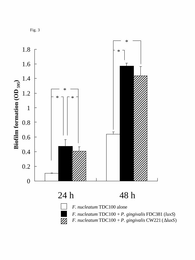
Evaluation of effects of AI-2 on biofilm formation
To determine the effects of AI-2 from P. gingivalis on biofilm formation by F.
nucleatum TDC100, biofilm formation of F. nucleatum TDC100 at 24 h and 48 h was
evaluated using P. gingivalis FDC381 or its luxS-deficient mutant CW221. As shown in Fig.
2, enhancement of biofilm formation by coculture of F. nucleatum TDC100 with P.
gingivalis wild type or luxS mutant CW221 was higher than that by F. nucleatum TDC100,
P. gingivalis FDC381 or CW221 alone (p<0.001). The results from the two-compartment
system are shown in Fig. 3. Enhancement of biofilm formation by F. nucleatum TDC100
with P. gingivalis FDC381 was only slightly higher than that with P. gingivalis CW221
when the partner strains were separated by membrane filters. However, this difference was
statistically significant at 24 h (p<0.001).
Discussion

10
We have found that many bacterial species isolated from surgical materials obtained
from patients with periapical periodontitis lesions formed biofilms on collagen-coated
polystyrene plates. Most of these strains are frequently isolated from apica l periodontitis
(19, 24). Among them, biofilm- forming ability is strongest in F. nucleatum TDC100. F.
nucleatum is also frequently isolated from lesions of periapical periodontitis (24, 27). In
lesions of apical periodontitis, the surfaces of the dentin and periodontal tissue contain
type-I collagen. The ability of F. nucleatum to bind to type-I collagen noted in the present
study agrees with the prevalence of F. nucleatum in periapical lesions.
In the present study, we demonstrated the enhancement of biofilm formation by F.
nucleatum, V. atypica and S. epidermidis by P. gingivalis, and of F. nucleatum by S.
epidermidis. These synergistic effects suggest that P. gingivalis enhances subsequent
colonization and biofilm formation by F. nucleatum, C. rectus, S. epidermidis and V.
atypica. Yamada et al. (31) reported that P. gingivalis strongly enhanced biofilm formation
by T. denticola in vitro. These species have often been isolated from abscesses in human
dento-alveolar lesions (19, 23, 29). In vitro stud ies have shown that F. nucleatum exhibited
the ability to coaggregate with Gram-positive cocci such as S. sanguinis,
Peptostreptococcus micros, as well as with P. gingivalis (8, 10, 11). F. nucleatum initially
adheres to early colonizers, including Gram-positive cocci, and enhances adherence of
periodontopathic bacteria such as P. gingivalis and T. denticola in periodontal lesions (9).
These reports suggest that F. nucleatum plays an important role in biofilm formation via its
strong adherence activity. Noguchi et al. (19) detected F. nucleatum with Tannerella
forsythia and P. gingivalis in extraradicular biofilms from clinical specimens. Taken

11
together with the results of the present study, this suggests that such synergistic effects may
play an important role in biofilm formation.
The synergistic effect between P. gingivalis and F. nucleatum was strongest among
the species isolated from the periapical periodontitis lesions used in this study. A
synergistic effect on pathogenicity between P. gingivalis and F. nucleatum was also
reported using a murine model (3). However, the synergistic effect between these two
microorganisms was reported only for growth support of P. gingivalis by F. nucleatum
under oxygenated and carbon-dioxide-depleted environments (2). The results of the present
study showed that P. gingivalis enhanced growth of F. nucleatum, providing direct
evidence of periodontal bacteria exerting a synergistic effect on biofilm formation by F.
nucleatum.
The slight reduction in biofilm formation by F. nucleatum TDC 100 with P.
gingivalis CW221 compared with the wild-type strain suggests that AI-2 is likely to be only
marginally involved in the enhancement of biofilm formation. Autoinducers were reported
to mediate changes in gene expression in microorganisms within biofilms (16). Several
oral microorganisms have been reported to produce AI-2 (5, 6, 32) The involvement of AI-
2 in biofilm formation in oral microorganisms in vitro has also been reported (21, 32).
Yoshida et al. (32) reported that biofilm formation by the luxS mutant of Streptococcus
mutans was complemented by Streptococcus gordonii, Streptococcus sobrinus or P.
gingivalis 381, but not by the P. gingivalis luxS-deficient mutant CW221. McNab et al.
(15) also suggested that S. gordonii produced an AI-2-like signaling molecule that

12
regulated various aspects of carbohydrate metabolism in some microorganisms.
Furthermore, LuxS-dependent intercellular communication is essential for biofilm
formation between P. gingivalis and S. gordonii. In the present study, enhanced biofilm
formation was observed with both P. gingivalis FDC381 and its luxS mutant CW 221, with
little difference between the two. This suggests that molecules other than AI-2 are involved
in the enhancement of biofilm formation by F. nucleatum. Loo et al. (14) showed that
several genes of S. gordonii, including those coding for signaling molecules, were involved
in biofilm formation. It has also been suggested that single-species biofilm formation is not
affected by inactivation of luxS (15, 28). Rickard et al. (21) reported that the optimal
concentration of 4,5-Dihydroxy-2,3-pentanedione (DPD), a product of the LuxS enzyme in
biofilm formation by Actinomyces naeslundii and Streptococcus oralis, was 100- fold lower
than the detection limit of the commonly utilized AI-2 assay. F. nucleatum also produces
AI-2 (5). Therefore, production of sufficient amounts of AI-2 by F. nucleatum to form
multispecies biofilms may have masked the potentially stimulatory effects of AI-2 secreted
by P. gingivalis. In addition, SDS-PAGE analysis of F. nucleatum TDC100 revealed that
some proteins, including an approximately 35 kDa protein, were predominantly expressed
in the separated cocultures with P. gingivalis FDC381 and CW221 strains, but not in F.
nucleatum TDC100 alone (data not shown). Further analysis will be required to clarify
how induction is initiated and the role of the 35 kDa protein in biofilm formation by F.
nucleatum.
13
Taken together, these results suggest that P. gingivalis secretes a molecule other than
AI-2 to enhance biofilm formation by F. nucleatum TDC 100, and that synergistic effects
on biofilm formation are an important factor in polymicrobial tooth apical infections.
Acknowledgements
This study was partia lly supported by Grant 16591837 from the Ministry of
Education, Science, Sport, Culture and Technology of Japan, as well as Grant 7A from the
Oral Health Science Center of Tokyo Dental College. We would like to thank Mr. Jeremy
Williams for editing this manuscript.

14
REFERENCES
1. Baumgartner JC, Falkler WA, Jr. Bacteria in the apical 5 mm of infected root canals. J
Endod 1991: 17: 380-383.
2. Diaz PI, Zilm PS, Rogers AH. Fusobacterium nucleatum supports the growth of
Porphyromonas gingivalis in oxygenated and carbon-dioxide-depleted environments.
Microbiology 2002: 148: 467-472.
3. Feuille F, Ebersole JL, Kesavalu, L, Stepfen, MJ, Holt, SC. Mixed infection with
Porphyromonas gingivalis and Fusobacterium nucleatum in a murine lesion model:
potential synergistic effects on virulence. Infect Immune 1996: 64: 2095-2100.
4. Figdor D, Davies JK, Sundqvist G. Starvation survival, growth and recovery of
Enterococcus faecalis in human serum. Oral Microbiol Immunol 2003: 18: 234-239.
5. Fong KP, Chung WO, Lamont RJ, Demuth DR. Intra- and interspecies regulation of
gene expression by Actinobacillus actinomycetemcomitans LuxS. Infect Immun 2001:
69: 7625-7634.
6. Frias J, Olle E, Alsina M. Periodontal pathogens produce quorum sensing signal
molecules. Infect Immun 2001: 69: 3431-3434.
7. Grenier D. Antagonistic effect of oral bacteria towards Treponema denticola. J Clin
Microbiol 1996: 34: 1249-1252.

15
8. Kaufman J, DiRienzo JM. Isolation of a corncob (coaggregation) receptor polypeptide
from Fusobacterium nucleatum . Infect Immun 1989: 57: 331-337.
9. Kolenbrander PE. Oral microbial communities: biofilms, interactions, and genetic
systems. Annu Rev Microbiol 2000: 54: 413-437.
10. Kolenbrander PE, Andersen RN. Inhibition of coaggregation between Fusobacterium
nucleatum and Porphyromonas (Bacteroides) gingivalis by lactose and related sugars.
Infect Immun 1989: 57: 3204-3209.
11. Kremer BH, van Steenbergen TJ. Peptostreptococcus micros coaggregates with
Fusobacterium nucleatum and non-encapsulated Porphyromonas gingivalis. FEMS
Microbiol Lett 2000: 182: 57-62.
12. Kuramitsu HK, Chen W, Ikegami A. Biofilm formation by the periodontopathic
bacteria Treponema denticola and Porphyromonas gingivalis. J Periodontol 2005: 76:
2047-2051.
13. Leonardo MR, Rossi MA, Silva LA, Ito IY, Bonifacio KC. EM evaluation of bacterial
biofilm and microorganisms on the apical external root surface of human teeth. J
Endod 2002: 28: 815-818.
14. Loo CY, Corliss DA, Ganeshkumar N. Streptococcus gordonii biofilm formation:
identification of genes that code for biofilm phenotypes. J Bacteriol 2000: 182: 1374-
1382.

16
15. McNab R, Ford SK, El-Sabaeny A, Barbieri B, Cook GS, Lamont RJ. LuxS-based
signaling in Streptococcus gordonii : autoinducer 2 controls carbohydrate metabolism
and biofilm formation with Porphyromonas gingivalis. J Bacteriol 2003: 185: 274-284.
16. Miller MB, Bassler BL. Quorum sensing in bacteria. Annu Rev Microbiol 2001: 55:
165-199.
17. Nair PNR. Pathogenesis of apical periodontitis and the causes of endodontic failures.
Crit Rev Oral Biol Med 2004: 15: 348-381.
18. Nair PNR. Pathobiology of primary apical periodontitis. In: Cohen, S and Hargreaves,
KM, ed. Pathways of the Pulp. St. Louis: Mosby, 2006:
19. Noguchi N, Noiri Y, Narimatsu M, Ebisu S. Identification and localization of
extraradicular biofilm- forming bacteria associated with refractory endodontic
pathogens. Appl Environ Microbiol 2005: 71: 8738-8743.
20. Noiri Y, Ehara A, Kawahara T, Takemura N, Ebisu S. Participation of bacterial
biofilms in refractory and chronic periapical periodontitis. J Endod 2002: 28: 679-683.
21. Rickard AH, Palmer RJ, Jr., Blehert DS, Campagna SR, Semmelhack MF, Egland PG,
et al. Autoinducer 2: a concentration-dependent signal for mutualistic bacterial biofilm
growth. Mol Microbiol 2006: 60: 1446-56.
22. Rocas IN, Siqueira JF, Jr., Santos KR, Coelho AM, De Janeiro R. "Red complex"
(Bacteroides forsythus, Porphyromonas gingivalis, and Treponema denticola ) in

17
endodontic infections: A molecular approach. Oral Surg Oral Med Oral Pathol Oral
Radiol Endod 2001: 91: 468-471.
23. Sklavounos A, Legakis NJ, Ioannidou H, Patrikiou A. Anaerobic bacteria in
dentoalveolar abscesses. Int J Oral Maxillofac Surg 1986: 15: 288-291.
24. Sunde PT, Tronstad L, Eribe ER, Lind PO, Olsen I. Assessment of periradicular
microbiota by DNA-DNA hybridization. Endod Dent Traumatol 2000: 16: 191-196.
25. Syed SA, Loesche WJ. Survival of human dental plaque flora in various transport
media. Appl Microbiol 1972: 24: 638-644.
26. Takahashi N, Ishihara K, Kato T, Okuda K. Susceptibility of Actinobacillus
actinomycetemcomitans to six antibiotics decreases as biofilm matures. J Antimicrob
Chemother 2007: 59: 59-65.
27. Vianna ME, Horz HP, Gomes BP, Conrads G. Microarrays complement culture
methods for identification of bacteria in endodontic infections. Oral Microbiol
Immunol 2005: 20: 253-258.
28. Wen ZT, Burne RA. Functional genomics approach to identifying genes required for
biofilm development by Streptococcus mutans. Appl Environ Microbiol 2002: 68:
1196-1203.
29. Williams BL, McCann GF, Schoenknecht FD. Bacteriology of dental abscesses of
endodontic origin. J Clin Microbiol 1983: 18: 770-774.

18
30. Xavier KB, Bassler BL. Interference with AI-2-mediated bacterial cell-cell
communication. Nature 2005: 437: 750-753.
31. Yamada M, Ikegami A, Kuramitsu HK. Synergistic biofilm formation by Treponema
denticola and Porphyromonas gingivalis. FEMS Microbiol Lett 2005: 250: 271-277.
32. Yoshida A, Ansai T, Takehara T, Kuramitsu HK. LuxS-based signaling affects
Streptococcus mutans biofilm formation. Appl Environ Microbiol 2005: 71: 2372-2380.

19
Table 1. Biofilm- forming activity on type-I collagen-coated polystyrene plates of 20 strains
isolated from refractory apical periodontitis lesions.
Species Biofilm formation
(OD595)*
Propionibacterium acnes TDC 18 0.642 + 0.048
Propionibacterium acnes TDC 58 0.307 + 0.025
Propionibacterium acnes TDC 95 0.567 + 0.130
Propionibacterium acnes TDC 103 0.549 + 0.059
Propionibacterium acnes TDC 121 0.457 + 0.082
Pseudomonas aeruginosa TDC 612 0.285 + 0.148
Pseudomonas aeruginosa TDC 66 0.491 + 0.070
Pseudomonas aeruginosa TDC 72 0.350 + 0.016
Fusobacterium nucleatum TDC 845 0.424 + 0.060
Fusobacterium nucleatum TDC 100 0.667 + 0.083
Klebsiella pneumoniae TDC 116 0.557 + 0.163
Klebsiella pneumoniae TDC 120 0.602 + 0.156

20
Staphylococcus epidermidis TDC 78 0.474 + 0.045
Staphylococcus epidermidis TDC 86 0.551 + 0.023
Staphylococcus hominis TDC 54 0.402 + 0.075
pasteuri pasteuri TDC 563 0.391 + 0.025
Campylobacter rectus TDC 67 0.364 + 0.070
Veillonella atypica TDC 96 0.237 + 0.067
Streptococcus sanguinis TDC15 0.357 + 0.028
Actinomyces naeslundii genotype 2 TDC 107 0.658 + 0.171
*Biofilm formation was quantified according to method of Takahashi et al. (26).
21
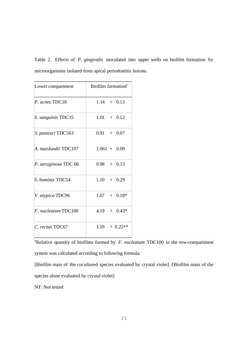
Table 2. Effects of P. gingivalis inoculated into upper wells on biofilm formation by
microorganisms isolated from apical periodontitis lesions.
Lower compartment Biofilm formationa
P. acnes TDC18 1.14 + 0.13
S. sanguinis TDC15 1.01 + 0.12
S. pasteuri TDC563 0.91 + 0.07
A. naeslundii TDC107 1.061 + 0.09
P. aeruginosa TDC 66 0.98 + 0.13
S. hominis TDC54 1.10 + 0.29
V. atypica TDC96 1.67 + 0.18*
F. nucleatum TDC100 4.19 + 0.43*
C. rectus TDC67 1.19 + 0.25**
aRelative quantity of biofilms formed by F. nucleatum TDC100 in the tow-compartment
system was calculated according to following formula:
[Biofilm mass of the cocultured species evaluated by crystal violet] /[Biofilm mass of the
species alone evaluated by crystal violet]
NT: Not tested

22
Data are representative of three independent runs of each experiment (n=18) *p<0.001,
**p<0.05 compared with the species alone by student t-test (p<0.001)

23
Table 3. Effects of partner strains inoculated into upper wells on biofilm formation by F.
nucleatum TDC100 and TDC845
Upper compartment
Biofilm formationa by
F. nucleatum TDC100
(OD595)
Biofilm formationa by
F. nucleatum TDC845
(OD595)
None 1.00 + 0.11
(0.116 + 0.013)
1.00 + 0.054
(0.312 + 0.017)
S. epidermidis TDC78 1.85 + 0.57* NT
P. gingivalis FDC381 4.15 + 0.46* 2.47 + 0.06*
P. gingivalisATCC33277 4.19 + 0.43* 2.56 + 0.12*
aRelative quantity of biofilms formed by F. nucleatum TDC100 was calculated according to
following formula:
[Biofilm mass of cocultured F. nucleatum TDC100 evaluated by crystal violet] /[Biofilm
mass of F. nucleatum TDC100 alone evaluated by crystal violet]
NT: Not tested
Data are representative of three independent runs of each exper iment (n=18) *Statistically
higher than F. nucleatum alone by student t-test (p<0.001)

24
Figure legends
Fig. 1. Biofilm formation by coculture of F. nucleatum TDC100 and TDC845 with partner
strains. Pairs of microorganisms were cultured either alone or cocultured together on type-I
collagen-coated microtiter plates. After 48 h cultivation, mass of organized biofilms was
evaluated by staining with crystal violet. Error bars indicate standard deviations. Data are
representative of three independent runs of each experiment (n=18) *p<0.001 compared
with culture of each strain alone by analysis of variance and Newman-Keuls multiple-
comparison test.
Fig. 2. Effects of P. gingivalis wild type and luxS-deficient mutant on biofilm formation by
F. nucleatum TDC100. P. gingivalis strain and F. nucleatum were cultured either alone
alone or cocultured together on type-I collagen-coated microtiter plates. After 48 h
cultivation, mass of organized biofilms was evaluated by staining with crystal violet. Error
bars indicate standard deviations. Data are representative of three independent runs of each
experiment (n=18) *p<0.001 compared with culture of each strain alone by analysis of
variance and Newman-Keuls multiple-comparison test.
Fig. 3 Effects of P. gingivalis wild type and luxS-deficient mutant inoculated into upper
wells on biofilm formation by compartmentalized F. nucleatum TDC100. Error bars
indicate standard deviations. Data are representative of three independent runs of each
experiment (n=18) *p<0.001 compared with F. nucleatum TDC100 monoculture by
student t-test.

1
Fig.1.
0
0.2
0.4
0.6
0.8
1
1.2
1.4
**
*
F. nucleatum TDC100 aloneS. epidermidis TDC78 alone S. epidermidis TDC78+ F. nucleatum TDC100P. gingivalis FDC381 alone P. gingivalis FDC381 + F. nucleatum TDC100P. gingivalis ATCC33277 alone P. gingivalis ATCC33277 + F. nucleatum TDC100F. nucleatum TDC845 alone P. gingivalis FDC381 + F. nucleatum TDC845
Bio
film
for
mat
ion
(OD
595)
*

2
Fig. 2
0
0.2
0.4
0.6
0.8
1
1.2
F. nucleatum TDC100 alone P. gingivalis FDC381 (luxS) aloneP. gingivalis FDC381 (luxS) + F. nucleatum TDC100
P. gingivalis CW221 (∆luxS) alone
P. gingivalis CW221 (∆luxS) + F. nucleatum TDC100
* *
Bio
film
for
mat
ion
(OD
595)
3
.
0
0.2
0.4
0.6
0.8
1
1.2
1.4
1.6
1.8
24 h 48 h
Fig. 3
* *
F. nucleatum TDC100 alone F. nucleatum TDC100 + P. gingivalis FDC381 (luxS)F. nucleatum TDC100 + P. gingivalis CW221 (∆luxS)
Bio
film
form
atio
n (O
D59
5)
*
*
*