Oral Health Care in Children – A Preventive Perspective
32
2 Oral Health Care in Children – A Preventive Perspective Agim Begzati 1 , Kastriot Meqa 2 , Mehmedali Azemi 3 , Ajtene Begzati 1 , Teuta Kutllovci 1 , Blerta Xhemajli 1 and Merita Berisha 4 1 Department of Pedodontics and Preventive Dentistry, School of Dentistry, Medical Faculty, University of Prishtna, Prishtina, 2 Department of Periodontology and Oral Medicine, School of Dentistry, Medical Faculty,University of Prishtina, Prishtina, 3 Department of Paediatric, Medical Faculty, University of Prishtina, Prishtina, 4 National Institute of Public Health of Kosovo, Department of Social Medicine, Medical Faculty, University of Prishtina, Prishtina Republic of Kosovo 1. Introduction Health has been described by the World Health Organization (WHO) as follows:”health comprise complete physical and social well-being and is not merely the absence of disease” (World Health Organization 1946). According to World Health Organization, oral health is the overall health of teeth and tooth- supporting tissues, and oral soft tissues, with the aim of fulfilling physiological functioning of the masticatory organ for chewing, phonation and esthetics. The US the Department of Health defined the health as oral “standard of health of the oral and related tissues which enables an individual to eat, speak and socialize without active disease, discomfort or embarrassment and which contributes to general well-being” (US Department of Health, 2000). Oral health is integral to general health and should not be considered in isolation. Oral disease has detrimental effects on an individual’s physical and psychological well- being and it reduces quality of life. Oral health is not only important to person’s appearance and sense of well-being, but also to person’s overall health. Dental caries is the most common cause of the disturbances of normal functions in the oral cavity, respectively it is a lack of preventive and curative measures. Gingivitis represents another serious problem for oral health. Data have shown a high prevalence of gingivitis among children. Gum disease is an inflammation of the gums, which may also affect the bone supporting the teeth, and may be followed with periodontitis. The role of dental plaque, respectively of the periopathogenic bacteria, has been considered as the most important factor in occurrence of caries and gum diseases. Plaque is a sticky colorless film of bacteria (biofilm) that constantly builds up, thickens and hardens on the teeth. If it is not removed by daily brushing and flossing, this plaque may harden into tartar and may contribute to inflammation and infections in the gums. Plaque is
Transcript of Oral Health Care in Children – A Preventive Perspective
2
Oral Health Care in Children – A Preventive Perspective
Agim Begzati1, Kastriot Meqa2, Mehmedali Azemi3, Ajtene Begzati1, Teuta Kutllovci1, Blerta Xhemajli1 and Merita Berisha4
1Department of Pedodontics and Preventive Dentistry, School of Dentistry, Medical Faculty, University of Prishtna, Prishtina,
2Department of Periodontology and Oral Medicine, School of Dentistry, Medical Faculty,University of Prishtina, Prishtina,
3Department of Paediatric, Medical Faculty, University of Prishtina, Prishtina, 4National Institute of Public Health of Kosovo, Department of Social Medicine,
Medical Faculty, University of Prishtina, Prishtina Republic of Kosovo
1. Introduction
Health has been described by the World Health Organization (WHO) as follows:”health comprise complete physical and social well-being and is not merely the absence of disease” (World Health Organization 1946). According to World Health Organization, oral health is the overall health of teeth and tooth-supporting tissues, and oral soft tissues, with the aim of fulfilling physiological functioning of the masticatory organ for chewing, phonation and esthetics. The US the Department of Health defined the health as oral “standard of health of the oral and related tissues which enables an individual to eat, speak and socialize without active disease, discomfort or embarrassment and which contributes to general well-being” (US Department of Health, 2000). Oral health is integral to general health and should not be considered in isolation. Oral disease has detrimental effects on an individual’s physical and psychological well-being and it reduces quality of life. Oral health is not only important to person’s appearance and sense of well-being, but also to person’s overall health. Dental caries is the most common cause of the disturbances of normal functions in the oral cavity, respectively it is a lack of preventive and curative measures. Gingivitis represents another serious problem for oral health. Data have shown a high prevalence of gingivitis among children. Gum disease is an inflammation of the gums, which may also affect the bone supporting the teeth, and may be followed with periodontitis. The role of dental plaque, respectively of the periopathogenic bacteria, has been considered as the most important factor in occurrence of caries and gum diseases. Plaque is a sticky colorless film of bacteria (biofilm) that constantly builds up, thickens and hardens on the teeth. If it is not removed by daily brushing and flossing, this plaque may harden into tartar and may contribute to inflammation and infections in the gums. Plaque is

Oral Health Care – Pediatric, Research, Epidemiology and Clinical Practices
20
an important prerequisite in aethiology of caries because acid is generated within its substance to such an extent that enamel may be demineralised. Dietary sugars diffuse rapidly through plaque where they are converted to acids by bacterial metabolism. Mutans streptococci are now considered to be the major cariogenic bacteria species involved in the caries process. (Soames & Southam 1999; Norman & Franklin 1999). Oral diseases may contribute in many serious conditions, such as heart disease and stroke, pneumonia and other respiratory diseases, diabetes. Untreated cavities can also be painful and lead to serious infections. Currently, studies have been examining whether there is a link between poor oral health and heart disease and between poor oral health and women delivering pre-term, low birth rate babies. Caries and tooth supporting structures’ diseases (gingivitis and periodontitis), as the most spreading diseases worldwide, do not disturb only the dental and oral functions, but due to the complications and consequences of non-prevention or lack of treatment they may seriously endanger the systemic health and influence directly the living quality. Thus these diseases should be characterized not only as medical, but also as social problem. These diseases have been studied and discussed also by the public health fields, such as: Dental public health, Oral public health, Community public health, etc. Dental public health has been described by the American Board of Dental Health as the science and art of preventing and controlling dental disease and promoting dental health through organized community efforts (Winslow 1920). The terms public health and community health are used synonymously, and both refer to the “effort that is organized by society to protect, promote and restore the health and quality of life of people” (Block 2003). Since dental caries, as well as gingivitis and periodontitis, both have a high prevalence and etiological factor – the bacterial plaque, this chapter will be based on the explanation of the prevalence, ethiopathogenesis of the bacterial plaque and the role of the bacteria, favorizing factors for plaque accumulation (oral hygiene and feeding habits), and finally the role of the preventive measures from the educational perspective. The explanation of these objectives will be done through scientific examinations conducted from the subjects regarding the dental caries in general and early childhood caries in particular, as well as through oral health promotion in children and mothers.
2. Dental caries
2.1 Definition, etiology and risk factors Dental caries may be defined as a bacterial disease of the hard tissues of the teeth characterized by demineralization of the inorganic and destruction of the organic substance of tooth (Soames & Southam 1999). Dental caries is one of the most prevalent diseases in children worldwide. The Centers for Disease Control and Prevention reports that dental caries is perhaps the most prevalent infectious diseases in children. Dental caries is five times more common than asthma and seven times more common than hay fever in children (US Department of Health and Human Services 2000). Dental caries is a disease that affects all age groups, most commonly children. The general opinion regarding the etiology of dental caries nowadays is that it is a very complex multifactorial disease, presented with high prevalence in all age groups. It has already been established that dental caries is a chronic infectious process with a

Oral Health Care in Children – A Preventive Perspective
21
multifactorial etiology. Dietary factors, oral microorganisms that can produce acids from sugars, and host susceptibility all need to coexist for caries to develop (Konig & Navia 1995). Analyzing the etiology, prevalence, clinical specifics, consequences and complications, dental caries is estimated as a serious disease, which represents not only as a health problem, but also a great serious social and economic problem. Many studies, clinical, but mostly longitudinal epidemiological studies, have offered convincing facts on the multifactorial nature of this disease. The multitude of factors that influence in the dental caries occurrence, having in mind that they act together, not separately, contribute in the complexity of the pathogenesis of caries, making it more difficult to undertake efficient preventive measures. There are some important factors that comprise the etiological circles of the dental caries: host or the tooth, dental or bacterial plaque, substrate – carbohydrates and saliva, and altogether co-react with the time factor. These circles are the circulus viciosus of the dental caries development. The hard dental structures, initially the enamel, undergo the demineralization process, respectively the caries. The caries development in the enamel surface is equally dependent from the inner hard dental structure and from the intensity of the extrinsic factors’ action. Newest concept in the field of dentistry gives an explanation how dental caries is caused as a result of the disturbance of the “Caries Balance” (Featherstone 2004). This misbalance may be manifested in the beginning of demineralization or during the process of remineralization. The theory of “Caries Balance” defines dental caries as a disease of hard dental tissues, and the destruction of the enamel surface as a result of the disruption of the balance of demineralization and remineralization. The defect in the enamel surface is a result of the domination of the demineralization process and such process has progressive course directed towards pulpar space. Which process will dominate depends on the proportions of the factors that constitute “Caries Balance”, i.e. protetctive and pathological factors. Pathological factors that include: 1. cariogenic bacteria, 2. frequent ingestion of fermentable carbohydrates, and 3. salivary dysfunction drive the caries process towards demineralization. Protective factors that include: 1. salivary components, 2. fluoride and remineralization, and 3. antibacterial therapy drive the caries process towards remineralization. Effective caries managment revolves around these principles. In order to control dental caries, i.e. to prevent its occurrence or to start the remineralization process during initial stage, it is necessary that the proportions of these factors be kept in the direction of the protective factors. The level of risk for dental caries depends on the domination of the certain group of factors that participate in the “Caries Balance”. If there is a domination of the pathological factors, the risk for dental caries will be higher and the treatment needs will require larger restorative interventions, as well as other consequences. If there is a domination of protective factors, then the invasive restorative dentistry will have fewer burdens, and concentrate in minimal restorations of superficial caries. Biological factors tend to be similar within all cultures and populations, although habit/environmental factors tend to be influenced specifically by the culture in place.
Oral Health Care – Pediatric, Research, Epidemiology and Clinical Practices
22
2.2 The epidemiology of dental caries It has already been mentioned that dental caries is the mostly spread disease in the world. In a study carried out in Kosovo we have assessed the prevalence of dental caries in comparison with other countries. The data from this oral health assessment of children of Kosovo showed a very high caries experience in both the primary and permanent dentitions. Caries prevalence expressed via the DMFT index was very high. Epidemiological data (years 2002-2005) derived from our study showed a high prevalence of dental caries among children in Kosovo (89.2% among preschool children and 94.4% among school children). The mean dmft/DMFT index was 5.86 for preschool children (ages 2 to 6) and 4.86 for all school children (ages 7 to 14) (Begzati et al. 2011). The results from the same previous study show that dental health of these children in Kosovo is worse than that of children in other European countries. Specifically, the mean dmft of five-year-olds at preschools in Kosovo (8.1) was found to be higher than the same value of preschool children in USA (1.7) and in many other European countries (1991-1995), including Ireland (0.9), Spain (1.0), Denmark (1.3), Norway (1.4), Finland (1.4), Netherlands (1.7), United Kingdom (2.0), France (2.5), and Germany (2.5). Our results are only comparable to the rates in Belarus (7.4), Sarajevo, Bosnia (7.53) (ages 5-7) and Albania (8.5), (Marthaler 1995; Kobaslia 2000). The low treatment rate of children in Kosovo (<2%) indicates a high treatment need. Also, the mean DMFT (5.8) of school children in Kosovo (age 12) was higher in comparison with school children (age 12) of the following developed countries: Netherlands (1.1), Finland (1.2), Denmark (1.3), USA (1.4), United Kingdom (1.4), Sweden (1.5), Norway (2.1), Ireland (2.1), Germany (2.6) and Croatia (2.6) (16). The mean DMFT of Kosovo’s children (age 12) was similar to the mean values in Latvia (7.7), Poland (5.1) and a group of 12- to 14-year-olds in Sarajevo, Bosnia (7.18) (Marthaler 1995; Kobaslia 2000). As it was previously mentioned, the low treatment rate of the children in Kosovo is unfavorable and indicates a high treatment need.
2.3 Oral health assessment in school and preschool children – Epidemiological study In order to assess the oral health of preschool and school children, the dental examination was carried out (Begzati et al. 2011). The sample in this study consisted of two groups derived from a multi-site examination: preschool and school children. From a total of 3,793 examined children, there were 1,237 preschool children (aged 2 to 6 years old) and 2,556 school children (aged 7 to 14 years old). This was a cross-sectional study conducted in randomly selected locations in Kosovo. The sample size was calculated with a confidence level of 95% and a confidence interval of 2. The study was specifically based on the dmft/DMFT index, following the recommendations of the World Health Organization (WHO Oral Health Surveys 1997). Preschool children were examined at various kindergartens in different locations of Kosovo. The examinations were done under natural light, using a dental mirror and a probe. It was performed by five dentists from the Prishtina University Dental Clinics, mainly from the Preventive Dentistry Department. The Study Group for Oral Health Promotion conducted the study, and the examiners received relevant training in advance. Diagnostic criteria were calibrated (Hunt 1986), with an inter-examiner reliability of kappa = 0.92 based on the examination of 30 children of different ages. For the caries assessments, all tooth surfaces were examined. Every defect in the tooth was tested with a probe, and every visual change in the enamel transparency in the early phases of demineralization was defined as a carious
Oral Health Care in Children – A Preventive Perspective
23
lesion. Decayed, filled and extracted/missing (due to caries) teeth were recorded in a modified WHO Oral Health Assessment Form. DMFT (for permanent dentition) and dmft (for primary dentition) describe the number, or the prevalence, of caries in an individual. DMFT and dmft are methods to numerically express the caries experience and are obtained by calculating the number of decayed (D), missing (M), and filled (F) teeth (T).
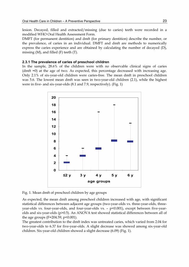
2.3.1 The prevalence of caries of preschool children In the sample, 28.6% of the children were with no observable clinical signs of caries (dmft =0) at the age of two. As expected, this percentage decreased with increasing age. Only 2.1% of six-year-old children were caries-free. The mean dmft in preschool children was 5.6. The lowest mean dmft was seen in two-year-old children (2.1), while the highest were in five- and six-year-olds (8.1 and 7.9, respectively). (Fig. 1)
Fig. 1. Mean dmft of preschool children by age groups
As expected, the mean dmft among preschool children increased with age, with significant statistical differences between adjacent age groups (two-year-olds vs. three-year-olds, three-year-olds vs. four-year-olds, and four-year-olds vs. – p<0.001), except between five-year-olds and six-year-olds (p>0.5). An ANOVA test showed statistical differences between all of the age groups (F=204.59, p<0.001). The greatest contribution to the dmft index was untreated caries, which varied from 2.04 for two-year-olds to 6.37 for five-year-olds. A slight decrease was showed among six-year-old children. Six-year-old children showed a slight decrease (6.09) (Fig. 1).
0
2
4
6
8
10
12
14
16
18
20
≤2 y 3 y 4 y 5 y 6 y
age groups
Oral Health Care – Pediatric, Research, Epidemiology and Clinical Practices
24
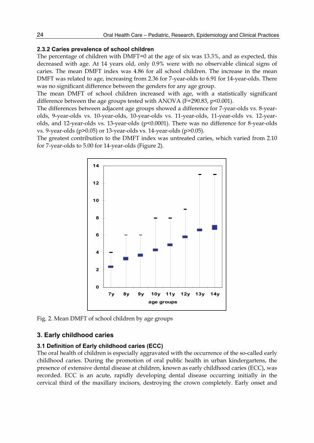
2.3.2 Caries prevalence of school children The percentage of children with DMFT=0 at the age of six was 13.3%, and as expected, this decreased with age. At 14 years old, only 0.9% were with no observable clinical signs of caries. The mean DMFT index was 4.86 for all school children. The increase in the mean DMFT was related to age, increasing from 2.36 for 7-year-olds to 6.91 for 14-year-olds. There was no significant difference between the genders for any age group. The mean DMFT of school children increased with age, with a statistically significant difference between the age groups tested with ANOVA (F=290.83, p<0.001). The differences between adjacent age groups showed a difference for 7-year-olds vs. 8-year-olds, 9-year-olds vs. 10-year-olds, 10-year-olds vs. 11-year-olds, 11-year-olds vs. 12-year-olds, and 12-year-olds vs. 13-year-olds (p<0.0001). There was no difference for 8-year-olds vs. 9-year-olds (p>0.05) or 13-year-olds vs. 14-year-olds (p>0.05). The greatest contribution to the DMFT index was untreated caries, which varied from 2.10 for 7-year-olds to 5.00 for 14-year-olds (Figure 2).
Fig. 2. Mean DMFT of school children by age groups
3. Early childhood caries
3.1 Definition of Early childhood caries (ECC) The oral health of children is especially aggravated with the occurrence of the so-called early childhood caries. During the promotion of oral public health in urban kindergartens, the presence of extensive dental disease at children, known as early childhood caries (ECC), was recorded. ECC is an acute, rapidly developing dental disease occurring initially in the cervical third of the maxillary incisors, destroying the crown completely. Early onset and
0
2
4
6
8
10
12
14
7y 8y 9y 10y 11y 12y 13y 14y
age groups
Oral Health Care in Children – A Preventive Perspective
25
rampant clinical progression makes ECC a serious public health problem. Due to varying clinical, etiological, localization, and course features, this pathology is found under different names such as labial caries (LC), caries of incisors, nursing bottle mouth, rampant caries (RC), nursing bottle caries (NBC), nursing caries, baby bottle tooth decay (BBTD), early childhood caries (ECC), rampant infant and early childhood dental decay, and severe early childhood caries (SECC) (James 1957; Goose 1967; Fass 1962; Winter et al.1966; Derkson & Ponti 1982; Ripa 1988; Arkin 1986; Bruered et al. 1989; Kaste & Gift 1995; Tinanoff et al. 1998; Horowitz 1998; Drury et al. 1999). According to Davis, the definition of this pathology has always been complex and “difficult to be described, but when it is seen, you know what it’s about” (Davis 1998). Up to now there have been many proposals for definition and diagnostic criteria, described in detail by Ismail & Sohn 1999. The preferred and most commonly used term today is early childhood caries (ECC), proposed by the Centers for Disease Control and Prevention (CDC) (Kaste et al. 1996). Numerous biological, psychosocial, and behavioral risk factors are involved in the etiology of ECC, supporting the multifactorial character of the disease (Wyne 1999; Seow 1998). Based on this concept, dental caries can be defined as demineralization of tooth tissue consequent to a dental infection that is dependent on frequent exposure to fermentable carbohydrates and is influenced by saliva and fluoride and other trace elements (Drury et al. 1999). Dietary habits are also deeply implicated in the development of ECC, despite the fact that it is considered an infectious disease (Lopez 2000). Consumption of sweets with high concentrations of glucose, saccharine, or fructose, especially if taken in processed juices (Newbrun 1982), and their prolonged intake play an important role in caries development in children with ECC (Wendt 1991). To evaluate the prevalence of ECC and various caries risk factors such as quantity of cariogenic Streptococcus mutans colonies, oral hygiene, sweets preference, bottle feeding in preschool children, and fluoride use, we have conducted a study at our preschool children (Begzati et al. 2010). In this study we have included 1,008 children of both sexes, from 1 to 6 years of age, from 9 kindergartens of Prishtina, capital of Kosovo. The sample was random, representing 80% of all kindergarten children. The sample size was calculated with a confidence level of 95% and a confidence interval of 2.
3.2 Dental examination and diagnostic criteria of ECC ECC was defined as “initial occurrence of caries in cervical region of at least two maxillary incisors.” Using a careful lift-the-lip examination, the presence or absence of ECC was recorded depending on the presence of “noncavity caries/white spot lesions” or “cavity caries.” With the aim of studying the clinical and etiological aspects of ECC, a sub-sample of children with ECC was included for further analysis. The latter part of the examination, which included the clinical study of ECC development (according to ECC stages), determination of bacterial colony sampling, oral hygiene index (OHI), and filling out of the questionnaire, was conducted in the Pediatric Dentistry Clinic of the School of Dentistry. Children with ECC were examined using the light of the dental unit, with dental mirror and probe. All examinations were carried out by Prof Begzati, with intra-examiner reliability of kappa = 0.95 based on the examination of 15 children of different ages.

Oral Health Care – Pediatric, Research, Epidemiology and Clinical Practices
26
3.2.1 ECC prevalence The prevalence of ECC varies in different countries, which may depend on the diagnostic criteria. While in some developed countries having advanced programs for oral health protection, the prevalence of ECC is around 5% (Derkson & Ponti 1982; Ripa 1988; Kaste et al. 1996; Davenport 1990; Hinds & Gregory 1995). In some countries of Southeastern Europe (Kosovo’s neighbors), this prevalence reaches 20% (Bosnia) and 14% (Macedonia) (Huseinbegović 2001, Apostolova et al. 2003). Much higher ECC prevalence has been reported for such places as Quchan, Iran (59%) (Mazhari et al. 2007) and Alaska (66.8%) (Kelly & Bruerd 1987). At American Indian children the prevalence is 41.8% [23]. Similarly, in North American populations, the prevalence at high-risk children ranges from 11% to 72% (Berkowitz 2003). In our study, from the total 1,008 examined children aged 1-6 years, the caries prevalence expressed in terms of the caries index per person, or dmft > 0, was 86.31%, with a mean dmft of 5.8. The prevalence of ECC was 17.36%, or 175 out of 1,008 examined children. The sub-sample of children with diagnosed ECC consisted of 150 children out of 175 invited for further analysis. Twenty-five children of this group from different kindergartens didn’t show up in the Department. The mean age of children with ECC was 3.8 ± 1.2 years. The mean dmft in children with ECC was 11 ± 3.6. There was no statistical difference of ECC prevalence between genders (t test = 1.81, P = 0.07). As expected, the lowest mean dmft score was found at age 2 (6.47 ± 2.13), with an age-related increase in dmft of 12.8 at age 6 (Table 1). In comparing the mean dmft in ECC children with respect to age, there was a significant statistical difference between age 2 and ages 4, 5, and 6. (One-Way ANOVA test F = 16, P < 0.001).
Age (N) dmft ± SD
2 (22) 5.5 ± 2.1
3 (42) 9.7 ± 3.4
4 (38) 12.8 ± 2.6
5 (36) 12.9 ± 2.7
6 (12) 12.8 ± 1.3
Total (150) 11 ± 3.6
Table 1. Mean dmft in children with ECC
3.2.2 Clinical course of ECC In order to explain the clinical course of ECC, we propose the following stages in the occurrence and progression of carious lesions in ECC: ECCi (initial stage)-white spot lesion or initial defect in enamel of cervix. ECCc (circular stage)-lesion in the dentin and circular distribution of this lesion proximally. ECCd (destructive stage)-destruction of more than half the crown without affecting the incisal edge. ECCr (radix relicta stage)-total destruction of the crown. The development of ECC on the maxillary incisors (at least 2) from its initial stage was monitored after a reexamination 1 year later (Table 2).

Oral Health Care in Children – A Preventive Perspective
27
Baseline Reexamination N 27 25
Mean dmft ± SD 5.1 ± 1.8 8.8 ± 0.7
ECC stages
Initial stage (N, %) (27) 100% (0) 0% Circular stage (N, %) / (7) 28% Destructive stage (N, %) / (5) 20% Radix relicta stage (N, %) / (9) 36%
Extraction (N, %) / (4) 16%
Table 2. ECC progression from initial stage at 1-year follow-up
The clinical course /ECC stages were not equally distributed. The most commonly present stage was that of radix relicta (41.7%), while the stage that appeared least frequently was the initial stage (15.4%), or 27 out of 150 children with ECC. There was a significant difference between the stages of ECC (P < 0.0001). Twenty-five of the 27 children with ECC in the initial stage were reexamined 1 year after the baseline examination (2 children did not appear for reexamination due to address change). The 1-year reexamination showed that the initial stage had advanced to the circular stage in 28% of children, destructive stage in 20%, radix relicta stage in 36%, and having been extracted due to ECC in 16% of children. Mean age of subjects with initial stage of ECC was 2 ± 0.7. Mean dmft on reexamination showed an increase from 5.1 to 8.8 (P < 0.001).
3.2.3 Clinical consequences of ECC Scientific research suggests that the development of ECC occurs in 3 stages. The first stage is characterized by a primary infection of the oral cavity with ECC. The second stage is the proliferation of these organisms to pathogenic levels as a consequence of frequent and prolonged exposure to cariogenic substrates. Finally, a rapid demineralization and cavitation of the enamel occurs, resulting in rampant dental caries (Berkowitz 2003). A 1-year follow-up of ECC development from the initial stage, representing decay at the enamel level and its progression to more destructive stages, shows even development in all affected teeth. It is quite an acute development, because in 2/3 of the children, the ECC has progressed to more complicated stages destructive and radix relicta stages. Within 1 year, the dmft values have increased to 3.7. Consecutively, these children commonly experience pain from pulpitis, gangrene, and apical periodontitis. Also, these conditions are often followed by abscesses and cellulitis, sometimes with phlegmona, seriously endangering the child’s general health. De Grauwe, in describing the progression of ECC, has noticed that the development of caries from the enamel to the dentin level can occur within 6 months (De Grauwe 2004). The rapid development of ECC and its clinical appearance, especially in primary incisors, identifies it in its initial stages as a risk factor for future caries in the primary and permanent dentitions (Al-Shalan et al. 1997). Children with congenital heart anomalies are frequent patients in our departments, some of them exhibiting severe ECC. There is strong evidence that untreated dental disease is an important etiological factor in the pathogenesis of infective endocarditis, a condition that still carries a high risk of mortality (Child 1996).

Oral Health Care – Pediatric, Research, Epidemiology and Clinical Practices
28
3.3 Risk factors of caries in general and of ECC in particular 3.3.1 Contagious nature of ECC There are many different types of microorganisms inhabiting the oral cavity, whose existence is maintained through ecological mechanism. This mechanism includes: saliva, crevicular gingival fluid, antimicrobial components of these fluids, intermicrobial synergism and antagonism, food, tooth, etc. The presence of microorganisms in the dental plaque depends on the presence of cariogenic bacteria in saliva. Their amount in saliva depends on the secretion level, enzyme presence, as well as on mechanisms with synergistic or antagonistic action. The microorganisms initially present in saliva and afterwards adhering to the tooth surface cannot express their cariogenic action separately or in small amounts. Their cariogenic effect is higher as their affinity to create bacterial colonies increases. Of the great interest in the cariogenesis process are only two bacterial genera: mutant streptococci and lactobacills (Norman & Franklin 1999). A very important role in occurrence of ECC is attributed to the bacterium Streptococcus mutans-called “the window of infection” (Caufield et al. 1993) in that it is responsible for the primary oral infection in the first phase of ECC (Berkowitz 1980; Berkowitz et al. 1996). Mother is regarded as the so called “window of infection” of S. mutans transmission to the newborn. As the data from the literature show, the role of S mutans in the etiology of ECC, especially in the initial phase, is very crucial (Berkowitz 1980, Caufield et al. 1993). These data also demonstrate the high prevalence of this bacterium in preschool children. S. mutans is found at the earliest ages, with the prevalence of 53% in 6- to 12-month-old children (Milgrom et al. 2000), 60% in 15-month-olds (Karn et al. 1988), 67% in 18-month-old Swedes (Hallonsten et al. 1995), and 94.7% in 3- to 4-year-old Chinese (Li et al. 1994). Almost all preschool urban Icelandic children were found to carry S mutans (Holbrook 1993). According to the studies of Ge and Caufield, all S-ECC children were S mutanspositive (Ge et al. 2008). Borutta 2002, found that in 80% of children (3 years old) diagnosed with caries, the presence of S. mutans was demonstrated, while higher counts of this bacterium were found in children with ECC. The high prevalence of S. mutans was also demonstrated in our study: 98% of preschool children. Expressed in colony-forming units (CFU/mL saliva), 93% of the ECC children in our study had a high S. mutans counts (CFU > 105). Higher salivary counts of S. mutans have been correlated with high dmft values (11.5) in our study. This significant correlation between high dmft or caries experience and high S. mutans counts has been demonstrated in other studies (Köhler et al. 1988; Köhler et al. 1995; Twetman & Frostner 1991; Maciel et al. 2001).
3.3.2 S. mutans prevalence in children with ECC In this study the presence of S. mutans was determined by using the CRT bacteria test (Ivoclar Vivadent, Liechtenstein) on the saliva previously stimulated by chewing paraffin. Bacterial counts were recorded as colony-forming units per milliliter (CFU/mL) of saliva. The number of bacterial colonies was graded as follows: Class 0 and Class 1 (CFU < 105/mL saliva), and Class 2 and Class 3 (CFU ≥ 105/mL saliva), according to the manufacturers’ scoring-card (Ivoclar-Vivadent, Lichtenstein). In younger subjects, with less saliva collected, the modified spatula method was used. The results showed that only a small number of children (2%) with ECC exhibited the absence of S. mutans (Class 0). In other words, S. mutans prevalence in children with ECC

Oral Health Care in Children – A Preventive Perspective
29
was 98%. The lowest class (Class 1) was recorded in 5% of the children with ECC, while classes that represent higher risk for caries (Classes 2 and 3) were present in 34% and 59%, respectively, of children with ECC (Table 3).
S. mutans N % Mean dmft ± SD Ttest
class
Class 0 3 2% 3 ± 0
Class 1 8 5% 5.4 ± 2.0 P<0.001
Class 2 51 34% 9.1 ± 3.0
Class 3 88 59% 12.8 ± 2.5
values in CFU/mL saliva
< 105(0 and 1) 11 7% 4.7 ± 1.1 T=5.5 P<0.001 ≥ 10 5(2 and 3) 139 93% 11.5 ± 3.2
Total 150 100% 11 ±3.6
Table 3. S. mutans distribution in children with ECC
Only 11 children with ECC exhibited a low level of S. mutans colonies (CFU < 105), with the mean dmft of this group being 4.7. The groups with higher CFU of S. mutans (Classes 2 and 3), representing 93% of the children, had a mean dmft of 11.5 ±3. Comparing the mean dmft of children with ECC by S. mutans classes of CFU showed a significant difference between Class 1 and Classes 2 and 3 (t = 5.5, P < 0.001). Considerable epidemiological studies have established a positive correlation between early childhood caries and S. mutans (Matee et al. 1992; Li et al. 2000; Berkowitz, 1996; Beighton et al. 2004).
3.3.3 Dietary habits in children with ECC In a study, parents and kindergarten teachers were asked to fill out a questionnaire about child’s dietary habits, including questions regarding frequency of sweets consumption throughout the day, as well as the type of sweets. Parents answered questions about bottle feeding: first use, duration, manner, and fluid content. They were also asked if they put their children to sleep with a bottle.
Sweets consumption in children with ECC We found that the frequency of sweets consumption in approximately 93% of the children was 1–3 or more times a day. Sweets consumption between meals and during kindergarten hours was common. There was a statistical correlation between daily sweets consumption and dmft in children with ECC (F = 7.26, P < 0.001). In our study, the frequency of sweets consumption of children with ECC was very high. It is of great concern that kindergartens as educational institutions do not have a more serious approach to a healthy diet and reduction of food containing sugar. On the contrary, at least once a day, sweet food (jam, chocolate, cream, biscuits, or cake) is served to children. Also, serving of this food is very common between meals. The literature shows the high caries values in children who have frequently used sweets (Holbrook et al. 1989), it also shows a high consumption of sweets between meals (Ölmez et al. 2003).
Oral Health Care – Pediatric, Research, Epidemiology and Clinical Practices
30
Bottle feeding in children with ECC Another important factor in the etiology of ECC is bottle feeding, which is accompanied by high salivary counts of S. mutans. The relationship between bottle usage and salivary counts of S. mutans has been reported (Mohan et al. 1998). In our children, the duration of bottle feeding with sweetened milk or juice is very long, wherein nearly 4/5 of children are bottle fed from 1 to 3 and more years. Most of the children with ECC represent subjects who are bottle fed up to age 2 (48%) and 3 and up (39%). Of the children with ECC, 6% were not bottle fed and 7% were bottle fed up to age 1. Comparing the dmft of children with ECC with regard to duration of bottle feeding shows statistical correlation (F = 20.83, P < 0.001). Another harmful practice is putting children to sleep with a juice-filled bottle, which is practiced in 2/3 of children with ECC, although Johnsen (1982) has reported that 78% of parents of children with ECC had attempted to substitute water for a cariogenic liquid (e.g., apple juice, formula) in the bedtime nursing bottle. A review of the literature from the etiological point of view of ECC shows that “the use of a bottle at night” is not the only cause of ECC (Plat & Cebazas 2000).
3.3.4 Tooth brushing and the OHI in children with ECC Regarding tooth brushing parents and kindergarten teachers were asked to fill out a questionnaire about the: frequency, parents’ participation during brushing, how it was controlled, and use of fluoride-containing toothpaste and fluoride tablets. The OHI was determined by using the Plaque Test (Ivoclar Vivadent) according to the Greene-Vermilion index. There is a high level of negligence in the oral hygiene of our children. More than half do not brush their teeth at all, exhibiting a very high OHI (1.52). No child recorded OHI-1. Although a dmft of 13 was found in children with OHI-3, no significant difference was found when comparing the dmft with respect to OHI (F = 2.52, P = 0.085). The importance of the primary dentition of oral health promotion must be focused on the education of mothers to motivate their children for oral hygiene. Unfortunately, we found “bad conviction” of mothers regarding primary teeth that they will be replaced, thus neglecting the care for children’s teeth. Data from the literature show that cooperation of mothers is very important in overcoming the belief that the deciduous dentition can be neglected (Rasmund & Tracy 2003). Regarding the frequency of tooth brushing, around 52% of the children did not brush at all, but there was no statistical difference in dmft in terms of frequency of brushing (F = 2.10, P = 0.106). Oral hygiene habits established at the age of 1 can be maintained throughout early childhood (Wendet 1995).
3.3.5 Fluoride use From the answers of mothers regarding fluoride use, we ascertained a considerable lack of knowledge about the benefits of this agent in maintaining healthy tooth structure. This information gap can be inferred from their answers. When asked, “Do you give fluoride tablets to your child?” their answers were stated as if they have been asked about some medication: “I give those tablets to my child as needed.” The absence of fluoride in
Oral Health Care in Children – A Preventive Perspective
31
Kosovo’s municipal drinking water may highly influence caries prevalence rates in children. Nutritional counseling, fluoride therapy, and oral hygiene may be required to prevent development of carious lesions in children. In the case of high-risk patients such as children with ECC with a predominance of high salivary counts of S. mutans, the use of either the antibacterial rinse chlorhexidine gluconate or the oral health care gel chlorhexidine has been suggested (Featherstone 2004).
3.3.6 Social factors The oral health promotion and preventive measures are also influenced by social and economical factors. Statistical data from our country such as: large families (with average size of 6.5 members), high unemployment rate (in 2008 it marked 45.4%, for female 56.4%), high birth rate (16%) and the lowest economical growth in the region (Statistical Office of Kosovo 2008), represent some of the aggravating factors when dealing with the health issues of the population, including oral health issues. Given the complexity of factors associated with ECC, it is unfortunate that most of the interest has only been from dental organizations. The critical change needed to accomplish the necessary research for the prevention of ECC should be expanding our network by involving other health professionals, community leaders, national organizations serving children, and political leaders (Ismail 1998).
3.4 Preventive measures for ECC Early childhood caries (ECC) is a health problem with biological, social and behavioral determinants. Intervention treatment does not resolve this problem. It is difficult, sometimes impossible and expensive. The only safest way is prevention of this complex pathology. European Academy of Pediatric Dentistry (2008) has recommended general strategies for ECC prevention: • Oral health assessments with counseling at regularly scheduled visits during the first
year of life are an important strategy to prevent ECC. • Children’s teeth should be brushed daily with a smear of fluoride toothpaste as soon as
they erupt. • Professional applications of fluoride varnish are recommended at least twice yearly in
groups or individuals at risk. • Parents of infants and toddlers should be encouraged to reduce behaviours that
promote the early transmission of mutans streptococci. Based on these recommendations, we will describe detailed preventive measures: primary prevention – prenatal and postnatal care; and secondary prevention – parents’ and dental professionals’ role.
3.4.1 Primary prevention • It should begin during prenatal period and it consists of pregnant woman’s needs’
fulfillment with necessary and healthy products; • Proper quality of food for the newborn during the enamel maturation phase; • Fluoridation of newly-erupted teeth; • Antimicrobial therapy with chlorhexidine.
Oral Health Care – Pediatric, Research, Epidemiology and Clinical Practices
32
3.4.2 Secondary prevention • Mothers’ education on recognizing the first signs of ECC using “lift-the-lip” technique.
The aim of this measure is early detection of the so-called “white spot”. • Parents should be encouraged to avoid bad feeding habits of their children and give
effort for proper feeding: • breast-feeding of the baby; • the use of cup instead of the bottle as early as possible; • not sleeping with bottle in mouth; • avoid the use of fabricated juices or soda; • the use of natural, a little sweetened, juice or tea, or just water; • avoid the discontinuation of bottle use by the method “bottle is gone”; • reduce the liquid in the bottle, gradually by night, • reduce sweets as much as possible; • no sweets between meals; • daily tooth brushing, at least twice a day, obligatory before going to bed.
• Necessary consultations with the dentist - Professional educational activities targeting primary health care providers (pediatricians, internists, family physicians, obstetricians, mid-level medical practitioners: • early identification of disease, • fluoride supplements as appropriate, • healthful feeding practices, • snacking behaviors that promote good oral health, and • referral to the dentist by 12 months of age.
4. Gingivitis and periodontitis
The periodontium, also known as "the supporting structures of the teeth", comprises a developmental, biologic, and functional unit, which undergoes certain changes with age and is subjected to morphologic changes related to functional alterations. The attachment of the tooth to the bone tissue of the jaws and maintenance of the oral cavity masticatory mucosa surface’s integrity are the main functions of the periodontium. The periodontium (περι = around, οδονσ = tooth) comprises the following tissues 1) the gingiva, 2) the periodontal ligament, 3) the root cementum, and 4) the alveolar bone.
4.1 Normal anatomy 4.1.1 Gingiva In the coronal direction the coral pink gingiva terminates in the free gingival margin, which has a scalloped outline. In the apical direction the gingiva is continuous with the loose, darker red alveolar mucosa (lining mucosa) from which the gingiva is separated by a, usually, easily recognizable borderline called either the mucogingival junction (arrows) or the mucogingival line. Since the hard palate and the maxillary alveolar process are covered by the same type of masticatory mucosa, there is no mucogingival line present in the palate. Two parts of the gingiva can be differentiated: 1) the free gingiva and 2) the attached gingiva.
4.1.2 Periodontal ligament The soft, richly vascular and cellular connective tissue which joins the root cementum with socket wall and surrounds the roots of the teeth is the periodontal ligament. The periodontal
Oral Health Care in Children – A Preventive Perspective
33
ligament is situated in the space between the roots of the teeth and the lamina dura or the alveolar bone proper. The alveolar bone surrounds the tooth to a level approximately 1mm apical to the cemento-enamel junction. The portions of the principal fibers of the periodontal ligament which are embedded in the root cementum and in the alveolar bone proper are called Sharpey's fibers.
4.1.3 Root cementum The cementum is a special mineralized tissue covering the root surfaces. It has many features in common with bone tissue. It contains no blood or lymph vessels, has no innervation, does not undergo physiologic resorption or remodeling, but is characterized by continuing deposition throughout life. It attaches the periodontal ligament fibers to the root and contributes to the process of repair after damage to the root surface. Different forms of cementum have been described, such as acellular, extrinsic fiber cementum; cellular, mixed stratified cementum; and cellular, intrinsic fiber cementum.
4.1.4 Alveolar bone The alveolar process is defined as the parts of the maxilla and the mandible that form and support the sockets of the teeth. Together with the root cementum and the periodontal membrane, the alveolar bone constitutes the attachment apparatus of the teeth, the main function of which is to distribute and resorb forces generated by, for example, mastication and other tooth contacts.
4.2 Classification of periodontal diseases The classification of periodontal diseases (Flemmig 1999) was revised in a workshop (AAP classification of periodontal diseases) that took place in 1999. This classification includes eight main categories: - Gingival diseases - Chronic periodontitis - Aggressive periodontitis - Periodontitis as a manifestation of systemic diseases - Necrotizing periodontal diseases - Abscesses of the periodontium - Periodontitis associated with endodontic lesions - Developmental or acquired deformities and conditions.
4.3 Prevalence of periodontal diseases Inflammatory alterations of gingiva are found among the majority of children in the primary dentition. The prevalence of gingivitis reaches its maximum at around 11 years among girls, whereas among boys 2 years later (Massler et al. 1952) Studies from 24 European countries from 1982 to 1992 (Sheiham et al. 2002) using Community Periodontal Index of Treatment Needs (CPITN) showed these results: - the mean prevalence of shallow pockets (CPITN-3) is 36% in West Europe and 45% in
East Europe - the mean prevalence of deep pockets (CPITN-4) is 9% in West Europe and 23% in East
Europe

Oral Health Care – Pediatric, Research, Epidemiology and Clinical Practices
34
- the average of periodontitis affected sextants was 0.2-2.4 for CPITN-3 and 0-0.8 for CPITN-4.
The Third National Health and Nutrition Examination Survey (NHANES III) as the largest population-based study during 1988-1994 showed that periodontitis affected at least 35% of US population between 30 and 90 years of age, with mild form in 22% and advanced form in 13%. The severe forms of periodontitis affected more men than women, and more African-Americans and Hispanics than Caucasians. Tooth loss may be the definitive consequence of destructive periodontal disease. Teeth lost due to periodontal disease’s consequences are evidently not in agreement with registration in epidemiological surveys. The prevalence and the severity of the disease may, thus, be underestimated. The so-called histologically healthy gingiva is possible only under experimental conditions and is attained by prolonged and overall nonexistence of microbial plaque. Thus, the more appropriate term used to express the health of gingiva is clinically normal gingiva, which almost always is followed by a polymorphonuclear granulocyte and lymphocyte infiltrate. The form of periodontal disease that affects the primary dentition, the condition formerly called prepubertal periodontitis, has been reported to appear in both a generalized and a localized form (Page et al. 1983).
4.4 Risk factors for periodontitis There is an abundance of both empirical evidence and substantial theoretical justification for accepting the widespread belief that many diseases have more than one cause, i.e. that they are of multifactorial etiology (Kleinbaum et al. 1982). In a relatively large number of cross-sectional studies, multiple risk putative risk factors for periodontal disease have been examined (Beck et al. 1990, Grossi et al. 1994, Horning et al. 1992, Ismali & Szpunar 1990).
4.4.1 Tobacco smoking The biological possibility of a connection between tobacco smoking and periodontitis was found on the potential effects of several tobacco-related substances, notably nicotine, carbon monoxide and hydrogen cyanide. Data from the NHANES III study (Tomar & Asma 2000) suggested that 42% of periodontitis cases in the USA can be attributed to current smoking, and 11% to former smoking.
4.4.2 Diabetes mellitus Diabetes as a risk factor for periodontitis has been addressed and debated for several years (Genco & Loe 1993), but recently, a number of biological mechanisms have been identified, by which the disease may contribute to impaired periodontal conditions. Examiners showed that diabetics were three times more likely to suffer from periodontal disease than non-diabetics (Emrich et al. 1991).
4.5 Periodontal disease and risk for systemic disease Besides the risk from periodontitis deriving from the systemic diseases, it has been suggested that periodontal diseases may represent a possible risk factor for systemic diseases. Emerging facts show that periodontal diseases, in fact, have systemic consequences.

Oral Health Care in Children – A Preventive Perspective
35
DeStefano et al (1993) in a prospective group of 9760 subjects found a nearly two-fold higher risk of coronary heart disease for patients with periodontal infection. But other studies have failed to document this association, indicating that this association is more complex and conditional (Hujoel et al. 2000). Another systemic consequence that may be attributed to the periodontal disease is preterm low birth-weight. It has been hypothesized that women with preterm labor may be indirectly affected through remote infections, one of which is periodontal disease (McGregor et al. 1988). In a pioneer study conducted by Offenbacher et al. (1996) with 124 subjects it was found that periodontitis, defined as 60% and more sites with attachment loss of 3 and more millimeters, conferred 7.9 fold risk for preterm low birth weight cases. A study in Kosovo with 200 parturients has acquired results of lower odd ratio (more than 3 fold), but significantly higher with women with periododntitis compared with women with no periodontitis (Meqa 2007).
5. Oral Health promotion
Good health is a major resource for social, economic and personal development. Political, economic, social, cultural, environmental, behavioral and biological factors can enhance or impair health. Health promotion action aims at making these conditions conducive to health. Health promotion therefore goes beyond health care. It puts health on the agenda of policy makers in all sectors and at all levels, directing them to be aware of the health consequences of their decisions and to accept their responsibilities for health. An important role in the oral health promotion activities, besides dental professionals, belongs to the kindergarten and school teachers, as well as mothers at home. Surely, mother’s role is very important and decisive in child’s education, not only from the aspect of oral health. Health care for children cannot be designed without understanding their vulnerability and essential link to parents. Health professionals, educators, and researchers must partner with parents in all activities related to children’s health, from clinical care to community programs to policy planning. Parental oral disease, attitudes, and past experiences with dental care have a direct impact on their own and their developing child’s oral health. Disease transmission, the practice of oral home care, and development of healthy attitudes towards oral health are all impacted by family factors. (Wendy & Mouradian 2001).
5.1 Mother’s knowledge regarding oral health care It is of the greatest interest to maintain mother’s oral health during pregnancy. In our studies we have found that oral health problems among pregnant women are as common as among children. Oral health should be an integral part of prenatal care. Improving oral health during pregnancy not only enhances the overall health of women, but also contributes to improving the oral health of their children. Because of the complexity of factors related to dental caries, the role of oral health education is believed to be very important, especially for mothers. In order to explain the mother’s importance, respectively her knowledge related to oral health promotion, we have conducted a study in our country. Mothers who accompanied their children to the University Dentistry School of Kosovo, Pedodontics Department, were interviewed. The aim of our study was to determine the caries experience and parental
Oral Health Care – Pediatric, Research, Epidemiology and Clinical Practices
36
knowledge regarding primary preventive measures for dental caries in children, mainly from the educational perspective of mothers in Kosovo. Data related to the following information was collected: social (mother’s age, economic status, mother’s level of education, employment, number of children in the family), dental experiences (dental visits, reasons of the visits), oral hygiene (daily frequency, knowledge of tooth brushing techniques, tooth brushing duration), feeding habits (sugar consumption, bottle feeding), fluoride and antimicrobial agents use, knowledge on their role in dental health, and pregnancy (prenatal control, premature birth). There is no doubt of the importance of the maternal role in maintaining good oral health among their children. The mother’s level of education is quite important, especially in our country, where women do not have a sufficient level of education. According to the official data, nearly half of all women completed only their primary education (44.9%) while only 7.2% finished high school or obtained a university degree. Unfortunately, even in this century, illiteracy exists in Kosovo, with the level ranging from 2% to 4%. The number of illiterate women is almost three times higher than that of men. Demographic data are characterized by a high birth rate (15.7%), equivalent to an average of three children per mother. Social data show low employment rates among women. The unemployment rate is approximately 62% for women and 35% for men. Even though female presence is very high in trade and services such as health or education, oral health education has been mostly neglected (Kosovo Statistical Office 2008). The results of our examination have shown that maternal effort, as well as their knowledge of oral health protection for their children, is largely inadequate. This inadequacy also has been reflected in the dental status of the children, with a mean dmft 6.3. The highest mean dmft was recorded among children whose mothers finished primary and secondary school while a lower mean dmft (4.5) was recorded among children whose mothers finished high school or obtained a university degree. Although mothers with higher educational levels had more knowledge, as evidenced by the lower mean dmft, it was still high according to WHO standards. When the dmft index structure was analyzed, 78% of the teeth were decayed, 16% were extracted, and only 6% were filled. The children’s dental health status was even worse given the high prevalence of ECC (above 21%), with a mean dmft of 11 for these cases. The dental experiences of our children, for nearly two-thirds of all respondents, unfortunately involved visits to the dentist only when the pain appeared. Our examination was based on maternal levels of knowledge regarding oral health (over 600 interviewed mothers), especially since they play crucial roles in educating their children. However, we made an attempt to raise the attention of not only mothers, but also of health professionals regarding the importance of the early prevention of dental and oral diseases in children through pre- and postnatal health education.
5.2 Prenatal health education Health education ought to start during the prenatal period. It consists of regular OB/GYN visits and adequate access to necessary medical advice for both maternal and pediatric health. Unfortunately, only 8% of the mothers sought advice from their obstetrician regarding oral health protection measures for their future child. Nearly three-quarters of the mothers neglected their own oral health by not visiting the dentist during their pregnancies. Even though mothers reported occasional discomfort regarding their oral health, such as
Oral Health Care in Children – A Preventive Perspective
37
gingival bleeding or toothaches, their visits to the dentist were very rare, with only 21% seeking help from the dentist. Another factor driving maternal negligence towards their own oral health was the inadequate level of preparedness among healthcare personnel, as well as their hesitant attitudes. Pregnant mothers who neglect to address their oral health issues may face consequences in their general health status, as well as in oral health status, with frequent dental caries, erosions, and periodontal disease. According to the U.S. Department of Health and Human Services, improving oral health during pregnancy not only enhances the overall health of women, but also contributes to improved oral health for their children. Oral health should be an integral part of prenatal care. Pregnancy and early childhood are particularly important times to access oral health care because the consequences of poor oral health can have a lifelong impact (US Department of Health and Human Services, 2000) Dental problems such as caries, erosion, epulis, periodontal infection, loose teeth, and ill-fitting crowns, bridges, and dentures may have special significance during pregnancy (Casassimo 1996, Gajendra 2004). The bacteria responsible for periodontal disease are capable of producing a variety of chemical inflammatory mediators such as prostaglandins, interleukins and tumor necrosis factor that can directly affect the pregnant woman (Romero et al. 2004). Data from the literature have shown that oral health problems are common in pregnant women and in young children (Crall 2005). Untreated cavities in mothers may be associated with the risk of caries in children. Finally, untreated oral infections may become systemic problems during pregnancy and may even contribute to preterm birth (Lewit & Monheit 1992). Periodontal disease is caused by gram-negative anaerobic bacteria. Studies have suggested that periodontal infections may contribute to the birth of preterm/low birth weight babies (Geopfert et al. 2004). In our study, the prevalence of premature births is 9%, and it has been reported that children born prematurely have a higher prevalence of ECC. Improving the oral health of pregnant women prevents complications of dental diseases during pregnancy, has the potential to decrease early childhood caries and may reduce preterm and low birth weight deliveries. Assessment of oral health risks in infants and young children, along with anticipatory guidance, has the potential to prevent early childhood caries.
5.3 Postnatal health education 5.3.1 First dental visit Regarding the issue of parental effort on seeking professional advice from the dentist, the data from the literature do not show satisfactory levels in other countries. Even in the developed nations, there is a dominant perception that children do not need early dental visits because they will subsequently lose their baby teeth. For instance, Dr. Horowitz has stated that “it is pretty hard for the parent to believe that the child should be sent to the dentist after the first tooth has erupted, and maybe earlier. A mistake that I made by myself, relying in my dentist’s advice not sending my child to the dentist before he turns three. Later I realized that it was a mistake” Reagan continues to cite authors Horowitz and Hale, who think that an important attention should be paid to the first dental visit and suggest that “the dental community needs to step forward and encourage these early visits” (Regan 2002).
Oral Health Care – Pediatric, Research, Epidemiology and Clinical Practices
38
We found that 18% of the mothers had no idea when their children should visit a dentist, while more than half believed that their children should first visit a dentist when they are three years old. Unfortunately, a considerable portion of mothers (18%) believe that the first visit to the dentist should occur only when the child is in pain. While the common perception 30 years ago was that the first dental visit should occur at the age of three, today it is preferred that the first visit occur before the child’s first birthday, specifically between 6 and 12 months of age.
5.3.2 Mother’s knowledge about contagious nature of dental caries It is already understood that dental caries is a contagious disease. There are many ways of transmission from the mother’s mouth to the child’s mouth, such as by kissing or when mothers “clean” their children’s pacifiers or taste their children’s juice or food (Berkowitz 1983). This bad habit was also recorded in our study. When mothers were asked whether they cleaned their baby‘s pacifiers or tasted the fluid from their baby‘s bottle, 35% confirmed such behaviors. According to the results of our study, mothers were not aware of this bacterium and the ways of it’s transmission in the child’s mouth. Previously, it was believed that mutans streptococci were not present in a child’s mouth until the eruption of the first tooth. However, some recent examinations have proven that high S. mutans levels may be observed in some children before their first birthday (Plotzitza et al. 2002). S. mutans was also found in the dental plaque of 12-month-old babies (Habibian et al. 2002). Data from the literature have confirmed such a correlation; if the mother’s SM count is low, the child’s count is low as well, and vice versa (Köhler & Andreen 1984). Nevertheless, this hypothesis needs further clarification.
5.3.3 Mother’s knowledge regarding feeding habits Bottle feeding – It has been demonstrated that bottle feeding may contribute to dental caries, especially early childhood caries (ECC). The relationship between bottle usage and high salivary levels of the cariogenic bacteria S. mutans has been reported (Mohan et al. 1998). Surprisingly, even though a considerable number of mothers in our study were unemployed and did not send their children to kindergarten, they fed their children with bottles (65%). Unfortunately, maternal knowledge regarding the role of bottle feeding in the occurrence of ECC was generally low (12%). An unfavourable practice involves mothers’ putting their children to sleep with milk- or juice-filled bottles, which was practiced for 74% of the children. Other authors have also reported on this bad practice (Jonsen 1988). Several studies have reported that the majority of the U.S. preschool population take, or have taken, a bottle to bed (Kaste & Gift 1995). In one study of children in the U.S. Head Start programs, a surprisingly high proportion (69%) of the children without maxillary anterior caries reported taking a bottle to bed, while 86% of the children with maxillary anterior caries reported taking a bottle to bed (O’Sullivan & Tinanoff 1993). In another study, 90% of the children in a population with and without caries were bottle-fed between 12 and 18 months of age, although the prevalence of nursing caries was only 20% (Serwint et al. 1993). The importance of bottle feeding with the occurrence of caries (especially in early and rampant occurrence in very young children) is reflected in the terminology of this
Oral Health Care in Children – A Preventive Perspective
39
pathology. The term ‘baby bottle tooth decay’ was proposed by the Healthy Mothers - Healthy Babies Coalition as an alternative that would be more appropriate for patient acceptance and focus increased attention on the potential harms of using a nursing bottle (Arkin 1986). More recent evidence suggests that taking a bottle to bed may be a stronger predictor of frontal tooth ECC patterns than previously believed (Douglass et al. 2001). Sweets consumption – There was a high percentage of sweets consumption among our children. Unfortunately, even though over 70% of mothers are aware that sweets can damage the children’s teeth, they do not give efforts to reduce this habit. Nearly 90% of the children consumed sweets at least once a day. Another serious factor is that sweets were consumed in between meals among 2/3 of children. In developed countries, advanced preventive programs focused on reducing the consumption of sweets. Data from the literature indicate that only 21% and 45% of children in Finland and Denmark, respectively, consumed sweets once a week (Matilla et al. 2000). The consumption of sweets containing sucrose constituents may be considered an important factor in the occurrence of caries (Marthaler 1990). The association between the intake of sucrose and dental caries has been well established in numerous studies, conducted mainly in northern and western European countries and in North America (Rugg-Gunn 1993). On the other hand, authors have reported that with the correct implementation of preventive and educational measures, there was a reduction in the development of caries, despite continued consumption of sucrose containing foods (Marthaler 1990). Kosovar cuisine is similar to that in the Middle and Near East, especially regarding sweets, with relatively frequent and high amounts of starch. The mechanism by which the starch added to sucrose increases the cariogenic potential of foods could be that the presence of starch increases the retention time of the food in the mouth (Lingstrom et al. 1993). Additionally, there are some indications that starch can increase acid production from sucrose when both nutrients are present together (Glor et al. 1988). Studies in the literature have reported a correlation between the consumption of sweets and S. mutans colonies, especially associated with high counts. It is obvious that there could be a threshold for the number of oral S. mutans colonies that eventually allow fermentable carbohydrates to exert harmful effects on teeth (Garsia-Closas et al. 1997).
5.3.4 Mother knowledge regarding oral hygiene Whether the child will start tooth brushing at an early age depends on maternal habits. Cleansing of the baby teeth should be started by the time of eruption of the first primary tooth. A small piece of clean gauze or a small toothbrush can be used. The child imitates parental behaviours, including oral hygiene habits. In a study in England, Witlle (1988) reported that 60% of children started brushing their teeth from the age of one; presumably, their teeth were initially brushed by their parents. Another study in England reported that in 80% of cases, mothers brushed the teeth of their children (Holt RD et al. 1996). A study conducted in Bosnia, a country that is geographically close to Kosovo, concluded that only 3% of the parents assisted their children with their first tooth brushing efforts (Huseinbegović 2001). In our study, 38% of the mothers stated that their children did not brush their teeth at all. Over 90% had no knowledge regarding proper tooth brushing techniques. Mothers rarely

Oral Health Care – Pediatric, Research, Epidemiology and Clinical Practices
40
assisted their children during tooth brushing (5%). Studies in the literature show a strong relationship between the poor maternal oral health and the prevalence of caries in their children (Dye et al. 2011).
5.3.5 Fluoride and antimicrobial agent utilization Fluoride, the key agent in battling caries, works primarily by topical action: inhibition of demineralization and enhancement of re-mineralization. Fluoride incorporated during tooth development is insufficient to play significant role in caries protection. Fluoride is needed regularly throughout life to protect teeth against caries. It is now realized that the most important action mechanism of fluoride takes place on the enamel surface of the tooth. Fluoride inhibits the loss of minerals and promotes the re-mineralization process. The data from literature demonstrated the importance of fluoridation on the prevention of dental caries. Preschool children from Ireland showed a very low mean dmft (0.9), largely because their drinking water was regularly fluoridated (Marthaler et al. 1996). A study in Tennessee, USA, reported a mean dmft of 4.15 for the fluoridated communities and a mean dmft of 7.49 for the non-fluoridated communities (David et al. 2001). Recommendations for fluoride supplementation can be made based on the fluoride content of the water, the child’s age, and the child’s caries risk. Among all endogenous and exogenous methods, the most effective method is water fluoridation. The drinking water in Kosovo is insufficient and has a fluoride level of less than 0.3 mg/l; however, the optimal range is between 0.8 and 1.5 mg/l. In the absence of fluoridated water, fluoride tablets may be used. Weintraub (1999) claims that “before you think of any preventive measure, put fluoride salts on the teeth”. The systemic and topical use of fluoride is the most effective measure to prevent dental caries. Exposure to topical fluoride via fluoridated toothpaste twice a day is a major component of caries prevention therapy. Fluoride varnish may be applied with a soft brush, and reapplication is recommended every 3 to 6 months. Antimicrobial treatment of caries is based on the use of two well-known agents (fluoride and chlorhexidine) for achieving selective antimicrobial control of carious microflora. Fluoride and chlorhexidine have an antimicrobial action against M. Streptococci that is significantly higher than that which they have against other non-cariogenic bacterial species. A combination of fluoride varnish and chlorhexidine application is used to lower the mutans streptococci count. Another successful antibacterial therapy against cariogenic bacteria is treatment with a chlorhexidine gluconate rinse or gel. Mothers from our study had not used fluoride and any antimicrobial agents (e.g., chlorhexidine), nor did they have any knowledge regarding their utilization. The data from literature have confirmed the positive antibacterial role of chlorhexidine in S. mutans colonies’ destruction. Antimicrobial treatment strategy has become a real concern for many dental professionals. A multitude of clinical trials confirms the caries-inhibiting effect of chlorhexidine (Zhang at al. 2006).
6. Oral health strategies
The initiatives undertaken by WHO, have global impact on the national and international policy development, one of which is the WHO Global Oral Health Program. High prevalence of oral diseases, such as dental caries, is not only individual preoccupation, but also a serious public health problem. WHO is continuously developing

Oral Health Care in Children – A Preventive Perspective
41
strategies and objectives on prevention of oral disease through oral health promotion. Thus, it may be of a great interest for the readers to get familiar with these WHO strategies and objectives.
6.1 The objectives of the WHO Global Oral Health Program (ORH) (www.who.int/oral_health/strategies_2010) The objectives of the WHO Global Oral Health Program (ORH), one of the technical programs within the Department of Chronic Diseases and Health Promotion (CHP), have been reoriented according to the new strategy of disease prevention and promotion of health. Greater emphasis is put on developing global policies in oral health promotion and oral disease prevention, coordinated more effectively with other priority programs of CHP and other clusters and with external partners. Several principles form the basis for the carried out work. The WHO Oral Health Program works on building oral health policies towards effective control of risks to oral health, based on the common risk factors approach. The focus is on modifiable risk behaviors related to diet, nutrition, use of tobacco and excessive consumption of alcohol, and hygiene. Oral health is part of total health and essential to quality of life and WHO projects intend to translate the evidence into action programs. The Oral Health Program therefore gives priority to integration of oral health with general health programs before community or national levels. The WHO Oral Health Program works from the life-course perspective, currently the community programs for improved oral health of the elderly and of children is given high priority. The implementation of school oral health programs within the framework of the WHO Health Promoting Schools Initiative is supported and guidelines are developed. Oral health systems reorientation towards prevention and health promotion is recommended in light of the Ottawa Charter, the primary health care concept and the Jakarta Declaration on leading Health Promotion into the 21st Century. In addition, global goals for oral health by the year 2020 strive for development of quality of oral health systems. The Program works on application of evidence-based strategies in oral health promotion, prevention and treatment of oral diseases worldwide, health systems research and development. Emphasis is also given on prevention and care of oral mucosal lesions, including oral cancer and oral manifestations of HIV/AIDS, cranio-facial disorders, trauma and injuries.
6.2 Oral health within WHO strategic directions WHO's goals are to build healthy populations and communities and to combat ill health. Four strategic directions provide the broad framework for focusing WHO's technical work, which also have implications for the Oral Health Program. • Reducing oral disease burden and disability, especially in poor and marginalized
populations. • Promoting healthy lifestyles and reducing risk factors to oral health that arise from
environmental, economic, social and behavioral causes. • Developing oral health systems that equitably improve oral health outcomes, respond
to people's legitimate demands, and are financially fair. • Framing policies in oral health, based on integration of oral health into national and
community health programs, and promoting oral health as an effective dimension for development policy of society.

Oral Health Care – Pediatric, Research, Epidemiology and Clinical Practices
42
In accordance with WHO overall priorities, the Global Oral Health Program has adopted the following priorities and strategic orientations.
6.3 Strategies and approaches in oral disease prevention and health promotion Priority is given to diseases linked by common, preventable and lifestyle related risk factors (e.g. unhealthy diet, tobacco use), including oral health. High relative risk of oral disease relates to socio-cultural determinants such as poor living conditions; low education; lack of traditions, beliefs and culture in support of oral health. Communities and countries with inappropriate exposure to fluorides imply higher risk of dental caries and settings with poor access to safe water or sanitary facilities are environmental risk factors to oral health as well as general health.
6.4 Health promotion and oral health The WHO Oral Health Program applies the philosophy "think globally - act locally". The development of program for oral health promotion in targeted countries focuses on: • Identification of health determinants; mechanisms in place to improve capacity to
design and implement interventions that promote oral health. • Implementation of community-based demonstration projects for oral health promotion,
with special reference to poor and disadvantaged population groups. • Building capacity in planning and evaluation of national program for oral health
promotion and evaluation of oral health promotion interventions in operation. • Development of methods and tools to analyze the processes and outcomes of oral
health promotion interventions as part of national health program. • Establishment of networks and alliances to strengthen national and international
actions for oral health promotion. Emphasis is also given to the development of networks for exchange of experiences within the context of the WHO Mega Country Program.
7. Conclusion
Oral health is integral to general health and should not be considered in isolation. Oral diseases have detrimental effects on an individual’s physical and psychological well-being and reduce quality of life. The commonest disease is dental caries. Other important conditions are gum (periodontal) disease. Dental caries is one of the most prevalent diseases among children worldwide. Its lack of treatment and complications that may occur can endanger general health. The development of dental caries is a dynamic process. Caries progression or reversal is determined by the balance between protective and pathological factors in the mouth. The most important component in the treatment of the caries disease is prevention. Understanding the balance between pathological factors and protective factors is the key to successful prevention of caries. Analyzing the etiology, prevalence, clinical specifics, consequences and complications, caries in general and ECC in particular are estimated as serious diseases, which represent not only health problem, but also a great serious social and economic problem. A well-recognized association exists between socio-economic status and oral health, and trends suggest that disease is increasingly concentrated in the lower income groups. However, oral diseases are largely preventable. In underdeveloped countries, oral health consequences due to dental caries also represent a
Oral Health Care in Children – A Preventive Perspective
43
serious public health problem. The presence of dental caries, especially of ECC, may reflect on the oral health status of children in countries with insufficient health system and inefficient primary dentistry. Early Childhood Caries (ECC) is a public health problem with biological, social and behavioral determinants. The preventive activities must start at an early age. Home-care methods are more than necessary. Primary prevention must start in the prenatal stage to fulfill the needs of pregnancy. Parents should be encouraged to avoid bad feeding habits and to instruct and supervise their children in tooth brushing. Mothers should be instructed to use the lift-the-lip technique to spot the white-spot lesions as first signs of dental caries. Newly erupted teeth must be treated with fluoride agents, and, as needed, antimicrobial agents containing chlorhexidine and thymol. Further investigation is needed to assess the effectiveness of new intervention strategies beyond traditional measures that are not strictly dependent on access to dental professional providers. Education is the essential pillar of the preventive measures. The main role in this pillar plays mother’s education, respectively her knowledge regarding the oral health. Unfortunately, an overall preventive program in oral health promotion, including oral health education, is absent in many underdeveloped countries. There are some negative indicators that contribute to the low levels of oral health knowledge, such as the lack of curriculum subjects on oral health, the lack of training and seminars on the issue, insufficient print materials for educational purposes, the lack of information for pregnant women, and negligence by dentists regarding measures for mothers to educate their children. Although some mothers with higher educational levels were highly motivated to improve their children‘s oral health, much more needs to be accomplished. Institutional efforts towards maternal and pediatric oral health education must be implemented immediately. Permanent and sustained oral health promotion organized with the participation of the entire civil society, with the mandatory presence of key stakeholders in the areas of education and healthcare, represent one of the highest priorities. The WHO strategies and objectives implementation regarding oral health promotion should be understood in the right manner and should be implemented continuously.
8. References
AAP Workshop on the Classification of Periodontal Diseases 1999. In: Flemmig, T.M. (1999). Periodontitis. Ann Periodontol, Vol. 4, pp. 32-37.
Apostolova, D., Asprovsa, V. & Simovska N (2003). Circular caries-ECC-a problem at the earliest age. 8th Congress of the Balkan Stomatological Society, (Abstract Book) Tirana, 2003.
Arkin, E.B. (1986). The Healthy Mothers, Healthy Babies Coalition: four years of progress. Public Health Repository, Vol. 101, pp. 147-156.
Beck, J.D., Koch, G.G., Rozier, R.G. & Tudor, G.E. (1990). Prevalence and risk indicators for periodontal attachment loss in a population of older community-dwelling blacks and whites. Journal of Periodontology Vol. 61, pp. 521-528.
Beighton, D., Brailsford, S., Samaranayake, L.P., Brown, J.P., Ping, F.X., Grant-Mills, D., Harris, R., Lo, E.C., Naidoo, S., Ramos-Gomez, F., Soo, T.C., Burnside, G. & Pine, C.M. (2004). A multi-country comparison of caries-associated microflora in demographically diverse children. Community Dental Health, Vol. 21, pp. 96–101.
Oral Health Care – Pediatric, Research, Epidemiology and Clinical Practices
44
Begzati, A., Berisha, M. & Meqa, K. (2010). Early childhood caries in preschool children of Kosovo - a serious public health problem. BMC Public Health, Vol. 10, pp. 788.
Begzati, A., Meqa, K., Siegenthaler, D., Berisha, M. & Mautsch, W. (2011). Dental health evaluation of children in Kosovo. European Journal of Dentistry, Vol. 5, pp. 32-39.
Berkowitz. R.J., Turner, J. & Green, P. (1980). Primary oral infection of infants with Streptococcus mutans. Archives of Oral Biology, Vol. 25, pp. 221-224.
Berkowitz, R.J. (1996). Etiology of nursing caries; a microbiologic perspective. Journal of Public Health Dentistry, Vol. 56, No. 1, pp. 51-54.
Berkowitz, R.J. (2003). Causes, Treatment and Prevention of Early Childhood Caries: A Microbiologic Perspective. Journal of the Canadian Dental Association, Vol. 69, No. 5, pp. 304-307.
Block, L.E. (2003). Dental public health: An Overview. In Gluck GM, Morganstein WM eds: Jong’s Community Dental Health, 5th ed., St. Louis, Mosby.
Borutta, A., Kneist, S. & Eherler, D.P. (2002). Oral health and Occurrence of Salivary S. mutans in Small Children, Journal of Dental and Oral Medicine, Vol. 4, No. 3, Poster 128.
Bruered, B., Kinney, M.B. & Bothwell, E. (1989). Preventing baby bottle tooth decay in American Indian and Alaska native communities: a model for planning. Public Health Repository, Vol. 104, No. 6, pp. 631-640.
Casassimo, P. (1996). Bright Futures in Practice: Oral Health. Arlington, VA: National Center for Education in Maternal and Child Health.
Caufield, P.W., Cutter, G.R. & Dasanayake, A.P. (1993). Initial acquisition of mutans streptococci by infants: evidence for a discrete window of infectivity. Journal of Dental Research, Vol. 72, No. 1, pp. 37-45.
Centers for Disease Control and Prevention (CDCP), conference. Atlanta, GA, September 1994.
Child, J.S. (1996). Risks for and prevention of infective endocarditis. In: Child JS, ed. Cardiology Clinics—Diagnosis and Management of Infective Endocarditis. Philadelphia, Pa: WB Saunders Co, Vol. 14, pp. 327-343.
Constitution of the World Health Organization. Geneva, World Health Organization, 1946, p 3.
Crall, J.J. (2005). Opportunities for improving maternal and infant health through prenatal oral health care. In: McCormick MC, Siegal JE, editors. Prenatal Care. Cambridge: University Press pp. 261-270.
Davenport, E.S. (1990). Caries in preschool child: Aetiology. Journal of Dentistry, Vol. 18, pp. 300-303.
David, E.B., Rebecca, W.H., James, A.G., Jennifer, U.B. & William, W.W. (2001). Successful Implementation of Community Water Fluoridation via the Community Diagnosis Process. Journal of Public Health Dentistry, Vol. 61, No. 1, pp. 28-33.
Davis, G.N. (1998). Early childhood caries-a synopsis. Community Dentistry and Oral Epidemiology, Vol. 26, No. 1, pp. 106-116.
De Grauwe, A., Aps, J.K. & Martens, L.C. (2004). Early Childhood Caries (ECC): What’s in a name? European Journal of Pediatric Dentistry, Vol. 5, No. 2, pp. 62-70.
Derkson, G.D. & Ponti, P. (1999). Nursing bottle syndrome: prevalence end etiology in a non fluoridated city. Journal of the Canadian Dental Association, Vol. 6, pp. 389-393.
Oral Health Care in Children – A Preventive Perspective
45
DeStefano, F., Anda, R.F., Kahn, H.S., Williamson, D.F. & Russell, C.M. (1993). Dental disease and risk of coronary heart disease and mortality. British Medical Journal Vol. 306, pp. 688-691.
Douglass, J.M., Tinanoff, N., Tang, J.M.W. & Altman, D.S. (2001). Dental caries patterns and oral health behaviours in Arizona infants and toddlers. Community Dentistry and Oral Epidemiology, Vol. 29, pp. 14–22.
Drury, Th.F., Horowitz, A.M., Ismail, A.I., Maertens, M.P., Rozier, R.G. & Selwitz, R.H. (1999). Diagnosing and reporting Early Childhood Caries for Research Purposes. Journal of Public Health Dentistry, Vol. 59, pp. 192-197.
Dye, B.A., Vargas, C.M., Lee, J.J., Magder, L. & Tinanoff, N. (2011). Assessing the relationship between children’s oral health status and that of their mothers. Journal of American Dental Association, Vol. 142, No. 2, pp. 173-183.
Emrich, L.J., Shlossman, M. & Genco, R.J. (1991). Periodontal disease in non-insulin-dependent diabetes mellitus. Journal of Periodontology Vol. 62, pp. 123-131
Fass, E.N. (1962). Is bottle feeding of milk a factor in dental caries? Journal of Dentistry for Children, Vol. 29, pp. 245-251.
Featherstone, J.D.B. (2004). The Caries Balance: The Basis for Caries Management by Risk Assessment. Oral Health and Preventive Dentistry, Vol. 2, No 1, pp. 259-264.
Gajendra, S. & Kumar, J.V. (2004). Oral health and pregnancy: a review. New York State Dental Journal, Vol. 70, No. 1, pp. 40-44.
Garcia-Closas, R., GarcIa-Closas, M. & Serra-Majem, L. (1997). A cross-sectional study of dental caries, intake of confectionery and foods rich in starch and sugars, and salivary counts of streptococcus mutans in children in Spain. American Journal of Clinical Nutrition, Vol, 66, pp. 1257-1263.
Ge, Y., Caufield, P.W., Fisch, G.S. & Li, Y. (2008). Streptococcus mutans and Streptococcus sanguinis Colonization Correlated with Caries Experience in Children. Caries Research, Vol. 42, pp. 444–448.
Genco, R.J. & Loe, H. (1993). The role of systemic conditions and disorders in periodontal disease. Periodontology 2000 Vol. 2, pp. 98-116
Glor, E.B., Miller, C.H. & Spandau, D.F. (1988). Degradation of starch and its hydrolytic products by oral bacteria. Journal of Dental Research, Vol. 67, pp. 75-81.
Goepfert, A.R., Jeffcoat, M.K., Andrews, W.W., Faye-Petersen, O., Cliver, S.P. & Goldenberg, R.L. et al. (2004). Periodontal disease and upper genital tract inflammation in early spontaneous preterm birth. Obstetrics and Gynecology, Vol. 104, No. 4, pp. 777-783.
Goose, D.H. (1967). Infant Feeding and Caries of the Incisors: an Epidemiological Approach, Caries Research, Vol. 1, pp. 167-173.
Grossi, S.G., Zambon, J.J., Ho, A.W., Koch, G., Dunford, R.G., Machtei, E.E., Norderyd, O.M. & Genco, R.J. (1994). Assessment of risk for periodontal disease. I. Risk indicators for attachment loss. Journal of Periodontology Vol. 65, pp. 260-267
Guidelines on Prevention of Early Childhood Caries (2008). An EAPD Policy Document Approved by the EAPD Board November 2008.
Habibian, M., Beighton, D., Stefenson, R. & Lawson, M. (2002). Relationship between dietary behaviors, oral hygiene and mutans streptococci in dental plaque of group of infants in southern England. Archives of Oral Biology, Vol. 6, pp. 491-498.
Hallonsten, A.L., Wendt, L.K., Mejar, I., Birkhed, D., Hakansson, C., Lindwall, A.M., Edwardsson, S. & Koch, G. (1995). Dental Caries and prolonged breast-feeding in
Oral Health Care – Pediatric, Research, Epidemiology and Clinical Practices
46
18-month- old. Swedish children. International Journal of Pediatric Dentistry, Vol. 5, No. 3, pp. 149-155.
Hinds, K. & Gregory, J.R. (1995). National diet and nutritional survey: Children aged 1-1/2 to 4-1/2 years. Vol.2: Report of the dental survey. London: HMSO.
Holbrook, W.P., Kristinsson, M.J., Gunnarsdóttir, S. & Briem, B. (1989). Caries prevalence, Streptococcus mutans and sugar intake among 4-year-old urban children in Iceland. Community Dentistry and Oral Epidemiology, Vol. 17, No. 6, pp. 292-295.
Holbrook, W.P. (1993). Dental caries and cariogenic factors in pre-school urban Icelandic children. Caries Research, Vol. 27, No. 5, pp. 431-437.
Holt, R.D., Winter, G.B., Downer, M.C. & Bellis, W.J. (1996). Caries in pre-school children in Camden 1993/94. British Dental Journal, Vol. 181, pp. 405-410.
Horning, G.M., Hatch, C.L. & Cohen, M.E. (1992). Risk indicators for periodontitis in a military treatment population. Journal of Periodontology Vol. 63, pp. 297-302.
Horowitz, H.S. (1998). Research issues in early childhood caries. Community Dentistry and Oral Epidemiology, Vol. 26, No. 1, pp. 67-81.
Hujoel, PP., Drangsholt, M., Spiekerman, C. & DeRouen, T.A. (2000). Periodontal disease and coronary heart disease risk. JAMA Vol. 284, pp. 1406-1410.
Hunt, R.J. (1986). Percent agreement, Pearson’s correlation, and kappa as measures of inter-examiner reliability. Journal of Dental Research, Vol. 65, pp. 128-130.
Huseinbegović, A. (2001). Social and medical aspects of primary dentition caries in urban conditions. Master degree- Sarajevo.
Ismail, A.I. & Szpunar, S.M. (1990). Oral health status of Mexican-Americans with low and high acculturation status: findings from southwestern HHANES, 1982-84. Journal of Public Health Dentistry Vol. 50, pp. 24-31.
Ismail, A.I. (1998). Prevention of early childhood caries. Community Dentistry and Oral Epidemiology, Vol. 26, No. 1, pp. 49-61.
Ismail, A.I. & Sohn, W. (1999). A Systematic Review of Clinical Diagnostic Criteria of Early Childhood Caries. Journal of Public Health Dentistry, Vol. 59, No. 3, pp. 171-191.
James, P.M.C., Parfitt, G.J. & Falkner, F. (1957). A study of the aetiology of labial caries of the deciduous incisor teeth in small children. British Dental Journal, Vol. 103, No. 2, pp. 37–40.
Johnsen, D.C. (1982). Characteristics and backgrounds of children with “nursing caries.” Pediatric Dentistry, Vol. 4, No. 3, pp. 218–224.
Jonsen, D. (1988). Pediatric Dentistry, Total Patient Care Philadelphia, Lee & Febiger. Karn, T.A., O’Sullivan, D.M. & Tinannoff, N. (1988). Colonization of mutans streptococci in
8- to 15- month-old children. Journal of Public Health Dentistry, Vol. 58, No. 3, pp. 248-249.
Kaste, L.M. & Gift, H.C. (1995). Inappropriate infant bottle feeding. Status of the Healthy People 2000 Objective. Archives of Pediatric and Adolescence Medicine, Vol. 149, pp. 786-791.
Kaste, L.M., Selwitz, R.H., Oldakowski, R.J., Brunelle, J.A., Win, D.M. & Brown, L.J. (1996). Coronal Caries in the Primary and Permanent Dentition of Children and Adolescents 1-17 Years of Age: United States, 1988-1991, Journal of Dental Research, Vol. 75, Special No., pp. 631-641.
Kelly, M. & Bruerd, B. (1987). The prevalence of baby bottle tooth decay among two native American populations. Journal of Public Health Dentistry, Vol. 47, No. 2, pp. 94–97.

Oral Health Care in Children – A Preventive Perspective
47
Kleinbaum, D.G., Kupper, L.L. & Morgenstern, H. (1982). Epidemiologic Research. Principles and quantitative methods, 1st ed. New York: Van Nostrand Reinhold.
Kneist, S., Laurisch, L. & Heinrich-Weltzein, R. (1999). Der neue CRT-Mikrobiologischer hintergrund zum nachweis von S. mutans. Oralprophylaxe, Vol. 21, pp. 180-185.
Kobaslia, S., Maglaic, N. & Begovic, A. (2000). Caries prevalence of Sarajevo children. Acta Stomatologica Croatica, Vol. 34, pp. 83-85.
Konig, K.G. & Navia, E.M. (1995). Nutritional role of sugars in oral health. American Journal of Clinical Nutrition Vol. 62, Suppl. pp. 275S-83S.
Kosovo Statistical Office. (2008). Women and men in Kosovo. Available at http://esk.rks-gov.net/publikimet/cat_view/8-statistikat-e-popullsise.html, Accessed 20 July 2011.
Köhler, B. & Andreen, I. (1984). Influence of caries-preventive measures in mothers on cariogenic bacteria and caries experience in their children. Archives of Oral Biology, Vol. 39, pp. 907-911.
Köhler, B., Andreen, I. & Jonsson, B. (1988). The earlier the colonization by mutans streptococci, the higher the caries prevalence at 4 years of age. Oral Microbiology and Immunology, Vol. 3, pp. 14-17.
Köhler, B., Bjarnason, S., Care, R., Mackevica, I. & Rence, I. (1995). Mutans streptococci and dental caries prevalence in a group of Latvian preschool children. European Journal of Oral Sciences, Vol. 103, No. 4, pp. 264-266.
Lewit, E.M. & Monheit, A.C. (1992). Expenditures on Health Care for Children and Pregnant Women. Future Child, Vol. 2, No. 2, pp. 95-114.
Li, Y., Navia, J.M. & Caufield, P.W. (1994). Colonization by mutans streptococci in mouths of 3- and 4- year -old Chinese children with or without enamel hypoplasia. Archives of Oral Biology, Vol. 39. No. 12, pp. 1057-1062.
Li, Y., Wang, W. & Caufield, P.W. (2000). The fidelity of mutans streptococci transmission and caries status correlate with breast-feeding experience among chinese families. Caries Research, Vol. 34, pp. 123–132.
Lingstrom, P., Birkhed, D., Granfeldt, Y. & Björck, I. (1993). pH measurements of human dental plaque after consumption of starchy foods using the micro-touch and the sampling method. Caries Research, Vol. 27, pp. 394-401.
Lopez, L., Berkowitz, R.J., Moss, M.E. & Weinstein, P. (2000). Mutans streptotocci prevalence in Puerto Rican babies with cariogenic feeding behaviors. Pediatric Dentistry, Vol. 22, No. 4, pp. 299–301.
Maciel, S.M., Marcenes, W. & Sheiham, A. (2001). The relationship between sweetness preference, levels of salivary mutans streptococci and caries experience in Brazilian pre-school children. International Journal of Paediatric Dentistry, Vol. 11, pp. 123-130.
Marthaler, T.M. (1990). Changes in the prevalence of dental caries: how much can be attributed to changes in diet? Diet, nutrition and dental caries. Caries Research, Vol. 24, Suppl. l, pp. 3-15.
Marthaler, M., O’Mullane, M. & Vrbic, V. (1996). The prevalence of dental caries in Europe 1990-1995. Caries Research, Vol. 30, pp. 237-255.
Massler M, Cohen A, Shour I (1952). Epidemiology of gingivitis in children. J Am Dent Assoc , Vol. 45, pp. 319-324.

Oral Health Care – Pediatric, Research, Epidemiology and Clinical Practices
48
Matee, M.I., Mikx, F.H., Maselle, S.Y. & Van Palenstein-Helderman, W.H. (1992). Mutans streptococci and lactobacilli in breast-fed children with rampant caries. Caries Research, Vol. 26, pp. 183–187.
Matilla, M.L., Rautava, P., Sillanpaa, M. & Paunio, P. (2000). Caries in Five-year-old Children and Associations with Family-related Factors. Journal of Dental Research Vol. 79, No. 3, pp. 875-881.
Mazhari, F., Talebi, M. & Zoghi, M. (2007). Prevalence of Early Childhood Caries and its Risk Factors in 6-60 months old Children in Quchan, Dental Research Journal, Vol. 4, No. 2, pp. 96-101.
McGregor, J.A., French, J.I., Lawellin, D. & Todd, J.K. (1988). Preterm birth and infection: pathogenic possibilities. American Journal of Reproductive Immunology Vol. 16, pp. 123-132.
Meqa, K. (2007). Relationship between periodontal disease and preterm low birth weight. Masters degree, University of Prishtina Dentistry School.
Milgrom, P., Riedy, C.A., Weinstein, P., Tanner, A.C., Manibusan, L. & Bruss, J. (2000). Dental caries and its relationship to bacterial infection, hypoplasia, diet, and oral hygiene in 6- to 36-month-old children. Community Dentistry and Oral Epidemiology, Vol. 28, No. 4, pp. 295-306.
Ministry of Public Administration. Statistical Office of Kosovo. (http://esk.rks-gov.net/eng/index.php?option=com_docman&task=doc_download&gid=870&Itemid=8). Accessed on October 20th, 2010.
Mohan, A.M., O’Sallivan, D.M. & Tinanoff, N. (1998). The relationship between bottle usage/content, age and number of teeth with mutans streptococci colonization in 6-24 – month-old children. Community Dentistry and Oral Epidemiology Vol. 26, Suppl. 1, pp. 12-20.
Newbrun, E. (1982). Sugar and dental caries: a review of human studies. Science, Vol. 217, No. 4558, pp. 418–423.
Norman, O.H. & Franklin, G. (1999). Primary Preventive Dentistry. Appleton & Lange, Stamford, Connecticut.
Offenbacher, S., Katz, V., Fertik, G., Collins, J., Boyd, D., Maynor, G., McKaig, R. & Beck, J. (1996). Periodontal infection as a possible risk factor for preterm low birth weight. Journal of Periodontology Vol. 67, pp. 1103-1113.
O’Sullivan, D.M. & Tinanoff, N. (1993). Social and biological factors contributing to caries of the maxillary anterior teeth. Pediatr Dent Vol. 15, pp. 41-44.
Ölmez, S., Uzamis, M. & Erdem, G. (2003). Association between early childhood caries and clinical, microbiological, oral hygiene and dietary variables in rural Turkish children. The Turkish Journal of Pediatrics, Vol. 45, pp. 231-236.
Page, R.C., Bowen, T., Altman, L., Vandesteen, E., Ochs, H., Mackenzie, P., Osterberg, S., Engel, L.D. & Williams, B.L. (1983). Prepubertal periodontitis. I. Definition of a clinical disease entity. Journal of Periodontology Vol. 54, pp. 257-271.
Plat, L. & Cebazas, M.C. (2000). Early childhood dental caries. Building community system for young children. Los Angeles, CA: University of California-Los Angeles Center for Healthier Children, Families and Communities, Vol. 32, exec. summ. (4 pp.).
Plotziza, B., Kneist, S., Berger, J. & Hetzer, G. (2002). Occurrence of salivary S. mutans in one-year old children: Poster ORCA.
Oral Health Care in Children – A Preventive Perspective
49
Reagan, L. (2002). Big Bad Cavities: Breastfeeding Is Not the Cause. Mothering Vol. 113 (July/August).
Reisine, S. & Douglass, J.M. (1998). Psychosocial and behavioral issues in early childhood caries. Community Dentistry and Oral Epidemiolology, Vol. 26, No. 1, pp. 32-44.
Ripa, L.W. (1988). Nursing caries: a comprehensive review. Pediatric Dentistry, Vol. 10, pp. 268-282.
Romero, R., Chaiworapongsa, T., Kuivaniemi, H. & Tromp, G. (2004). Bacterial vaginosis, the inflammatory response and the risk of preterm birth: a role for genetic epidemiology in the prevention of preterm birth. American Journal of Obstetrics and Gynecology, Vol. 190, No. 6, pp. 1509-1519.
Rosamund, L.H. & Tracy, W. (2003). An oral health promotion program for an urban minority population of preschool children. Community Dentistry and Oral Epidemiolology, Vol. 31, No. 5, pp. 392-399.
Rugg-Gunn, A.J. (1993). Nutrition and dental health. Oxford, United Kingdom: Oxford University Press.
Seow, W.K. (1998). Biological mechanisms of early childhood caries. Community Dentistry and Oral Epidemiolology, Vol. 26, No. 1, pp. 8-27.
Serwint, J.R., Mungo, R., Negrete, V.F., Duggan, A.K. & Korsch, B.M. (1993). Child-rearing practices and nursing caries. Pediatrics, Vol. 92, pp. 233-237.
Sheiham, A. & Netuveli, G. (2002). Periodontal diseases in Europe. Periodontol 2000, Vol. 29, pp. 104-121.
Soames, J.V. & Southam, J.C. (1999). Oral Pathology. 3rd ed. Oxford. Tinanoff, N., Kaste, L.M. & Corbin, S.B. (1998). Early childhood caries: a positive beginning.
Community Dentistry and Oral Epidemiolology, Vol. 26, No. 1, pp. 117-119. Thakib, A., et al. (1997). Primary incisor decay before age 4 as a risk factor for future dental
caries. Paediatric Dentistry, Vol. 9, pp. 37-41. Tomar, S.L. & Asma, S. (2000). Smoking-attributable periodontitis in the United States:
findings from NHANES III. National Health and Nutrition Examination Survey. Journal of Periodontology Vol. 71, pp. 743-751.
Twetman, S. & Frostner, N. (1991). Salivary mutans streptococci and caries prevalence in 8-year-old Swedish schoolchildren. Swedish Dental Journal, Vol. 15, No. 3, pp. 145-151.
US Department of Health and Human Services. (2000). Oral Health in America: A Report of the Surgeon General. Rockville, MD: US Department of Health and Human Services, National Institute of Dental and Craniofacial Research, National Institutes of Health.
Weintraub, J.A. (1999). Prevention of early childhood caries: a public health perspective. Community Dentistry and Oral Epidemiolology, Vol. 26, Suppl. 1, pp. 62-66.
Wendt, L.K., Hallonstein, A.L. & Koch, G. (1991). Dental caries in one- and two-year-old children living in Sweden. Part I – A longitudinal study. Swedish Dental Journal, Vol. 15, No. 1, pp. 1–6.
Wendt, L.K. (1995). On oral health in infants and toddlers. Swedish Dental Journal, Vol. 19, Suppl., pp. 106:1-62.
Wendy, E. & Mouradian, M.D. (2001). The Face of a Child: Children’s Oral Health and Dental Education. Journal of Dental Education. Volume 65, No. 9, pp. 821-831.
Winslow, C.E. (1920). The untitled fields of public health. Modern Medicine Vol. 2, p. 183.
Oral Health Care – Pediatric, Research, Epidemiology and Clinical Practices
50
Winter, G.B., Hamilton, M.C. & James, P.M.C. (1966). Role of comforter as en etiological factor in rampant caries of deciduous dentition. Archives of Diseases in Children, Vol. 417, pp. 207-212.
Whittle, J.G. & Whittle, K.W. (1988). Household income in relation to dental health and dental health behaviors: the use of super profiles. Community Dental Health, Vol. 15, pp. 150-154.
World Health Organization (1997). Oral Health surveys. Basic Methods (4th ed.), World Health Organization, Geneva.
World Health Organization (2008). The World Oral Health Report 2003. Continuous improvement of oral health in the 21st century – the approach of the WHO Global Oral Health Programme, http://www.who.int/oral_health/media/ en/orh_report03_en.pdf. Accessed October 15th, 2010.
World Health Organization (2010). http://www.who.int/oral_health/strategies/ hp/en/index.html. Accessed October 15th, 2010.
Wyne, A.H. (1999). Early childhood caries: nomenclature and case definition. Community Dentistry and Oral Epidemiology, Vol. 7, pp. 313-315.
Zhang, Q., van't Hof, M.A. & Truin, G.J. (2006). Caries-inhibiting effect of chlorhexidine varnish in pits and fissures. Journal of Dental Research, Vol. 85, pp.469–472.