Oral glycotoxins are a modifiable cause of dementia and ... · Oral glycotoxins are a modifiable...
6
Oral glycotoxins are a modifiable cause of dementia and the metabolic syndrome in mice and humans Weijing Cai a , Jaime Uribarri b , Li Zhu a , Xue Chen a , Shobha Swamy a , Zhengshan Zhao a , Fabrizio Grosjean a,c , Calogera Simonaro d , George A. Kuchel c , Michal Schnaider-Beeri e , Mark Woodward f,g , Gary E. Striker a,b , and Helen Vlassara a,b,1 Department of a Geriatrics, Division of Experimental Diabetes, b Medicine, d Human Genetics, and e Psychiatry, The Icahn School of Medicine at Mount Sinai, New York, NY 10029; c Division of Nephrology, Department of Medicine, University of Pavia, 27100 Pavia, Italy; f UConn Center on Aging, University of Connecticut Health Center, CT 06030; and g Professorial Unit, Biostatistics, George Institute, Sydney, NSW 2050, Australia Edited* by Marc Feldmann, University of Oxford, London, United Kingdom, and approved January 24, 2014 (received for review September 12, 2013) Age-associated dementia and Alzheimer’s disease (AD) are cur- rently epidemic. Neither their cause nor connection to the meta- bolic syndrome (MS) is clear. Suppression of deacetylase survival factor sirtuin 1 (SIRT1), a key host defense, is a central feature of AD. Age-related MS and diabetes are also causally associated with suppressed SIRT1 partly due to oxidant glycotoxins [advanced gly- cation end products (AGEs)]. Changes in the modern diet include excessive nutrient-bound AGEs, such as neurotoxic methyl-glyoxal derivatives (MG). To determine whether dietary AGEs promote AD, we evaluated WT mice pair-fed three diets throughout life: low-AGE (MG - ), MG-supplemented low-AGE (MG + ), and regular (Reg) chow. Older MG + -fed mice, similar to old Reg controls, de- veloped MS, increased brain amyloid-β 42 , deposits of AGEs, gliosis, and cognitive deficits, accompanied by suppressed SIRT1, nicotin- amide phosphoribosyltransferase, AGE receptor 1, and PPARγ. These changes were not due to aging or caloric intake, as neither these changes nor the MS were present in age-matched, pair-fed MG - mice. The mouse data were enhanced by significant temporal cor- relations between high circulating AGEs and impaired cognition, as well as insulin sensitivity in older humans, in whom dietary and serum MG levels strongly and inversely associated with SIRT1 gene expression. The data identify a specific AGE (MG) as a modifiable risk factor for AD and MS, possibly acting via suppressed SIRT1 and other host defenses, to promote chronic oxidant stress and inflammation. Because SIRT1 deficiency in humans is both preventable and revers- ible by AGE reduction, a therapeutic strategy that includes AGE re- duction may offer a new strategy to combat the epidemics of AD and MS. neural | insulin resistance | obesity | nutrition | caloric restriction C ognitive dysfunction is currently one of the most prevalent and important polygenic age-related diseases (1–3). A link has been identified between dementia, the most frequent form of which is Alzheimer’s disease (AD), and the metabolic syndrome (MS) or diabetes type 2 (T2D) (1, 3), conditions largely related to environmental factors (4, 5). An emerging view suggests that there is a compromise in innate defense mechanisms preceding these conditions that is due to sustained elevation of oxidant stress (OS) (6). Modulation of the environment, i.e., caloric excess or caloric restriction (CR), can influence cognitive function (7, 8); how- ever, the calorie-sensitive pathway(s) involved are unknown. NAD + -dependent sirtuin 1 (SIRT1), an NAD + -dependent deace- tylase that positively regulates neuronal, immune, and endocrine responses, is down-regulated in aging-related diseases and is thought to contribute to cognitive decline (1, 9–11). Restora- tion of brain SIRT1 is widely implicated in the benefits of CR on the aging brain (7–9, 11). Glycotoxins or advanced glycation end products (AGEs) are a class of OS-promoting agents implicated in diabetes and aging, including brain injury due to AD and stroke (6, 12–14). Certain AGEs, such as the derivatives of methyl-glyoxal-imidazolone-H1 (MG-H1) amplify the proinflammatory properties of amyloid β 1–42 (Aβ) or tau protein (15–17). High MG levels in brain or the circu- lation are linked to cognitive decline in elderly subjects (15, 17, 18). Food-derived AGEs have emerged as contributors to chronic diseases due to their abundance in thermally altered nutrients (19, 20). AGE restriction in nutritionally and nutritionally bal- anced diets delayed metabolic and vascular diseases and ex- tended lifespan in mice (21, 22). The role of diet-derived AGEs in systemic AGE toxicity was confirmed in studies using a defined MG-supplemented low-AGE diet (MG + ). Old MG + mice, but not MG − mice, developed age-related MS and kidney and cardiac fibrosis, associated with inflammation and SIRT1 depletion in insulin-sensitive tissues (22). AGE restriction also improved in- sulin resistance and inflammation in humans (23, 24). We therefore postulated that oral AGEs, in addition to causing MS, might also predispose to dementia, and these both might be prevented by AGE restriction (22). The MG + /MG − mouse model provides an opportunity to explore the link of oral AGEs to these chronic conditions, free of either genetic or caloric manipulations. Herein, we show that cognitive dysfunction develops in par- allel with metabolic changes in old mice fed defined AGEs (MG + ) but not in AGE-restricted (MG − ) mice. These findings were supported by clinical findings, introducing previously Significance Suppression of NAD + -dependent sirtuin 1 (SIRT1) is linked to dementia or Alzheimer’s disease (AD) and the metabolic syn- drome (MS). Because advanced glycation end products (AGEs) promote MS and neurotoxicity, we conducted studies of C57BL6 mice fed isocaloric diets containing defined AGEs [methyl-glyoxal derivatives (MG)] to determine whether food AGEs promote AD and MS. MG + -fed, but not MG - -fed, mice developed brain SIRT1 deficiency, amyloid-β deposits, cognitive and motor deficits, and MS. These findings were validated in older healthy humans with high baseline circulating MG levels by a time-dependent decline in cognition and insulin sensitiv- ity. The data suggest that food-derived AGEs, an environ- mental factor, contribute to both AD and MS by causing chronic SIRT1 suppression. Importantly, reduction of food-de- rived AGEs is feasible and may provide an effective treatment strategy for both these epidemics. Author contributions: H.V. designed research; W.C., L.Z., X.C., S.S., Z.Z., F.G., C.S., G.A.K., M.S.-B., and H.V. performed research; W.C., J.U., M.W., G.E.S., and H.V. analyzed data; and W.C., J.U., G.E.S., and H.V. wrote the paper. The authors declare no conflict of interest. *This Direct Submission article had a prearranged editor. See Commentary on page 4743. 1 To whom correspondence should be addressed. E-mail: [email protected]. This article contains supporting information online at www.pnas.org/lookup/suppl/doi:10. 1073/pnas.1316013111/-/DCSupplemental. 4940–4945 | PNAS | April 1, 2014 | vol. 111 | no. 13 www.pnas.org/cgi/doi/10.1073/pnas.1316013111 Downloaded by guest on July 20, 2020
Transcript of Oral glycotoxins are a modifiable cause of dementia and ... · Oral glycotoxins are a modifiable...

Oral glycotoxins are a modifiable cause of dementiaand the metabolic syndrome in mice and humansWeijing Caia, Jaime Uribarrib, Li Zhua, Xue Chena, Shobha Swamya, Zhengshan Zhaoa, Fabrizio Grosjeana,c,Calogera Simonarod, George A. Kuchelc, Michal Schnaider-Beerie, Mark Woodwardf,g, Gary E. Strikera,b,and Helen Vlassaraa,b,1
Department of aGeriatrics, Division of Experimental Diabetes, bMedicine, dHuman Genetics, and ePsychiatry, The Icahn School of Medicine at Mount Sinai,New York, NY 10029; cDivision of Nephrology, Department of Medicine, University of Pavia, 27100 Pavia, Italy; fUConn Center on Aging, University ofConnecticut Health Center, CT 06030; and gProfessorial Unit, Biostatistics, George Institute, Sydney, NSW 2050, Australia
Edited* by Marc Feldmann, University of Oxford, London, United Kingdom, and approved January 24, 2014 (received for review September 12, 2013)
Age-associated dementia and Alzheimer’s disease (AD) are cur-rently epidemic. Neither their cause nor connection to the meta-bolic syndrome (MS) is clear. Suppression of deacetylase survivalfactor sirtuin 1 (SIRT1), a key host defense, is a central feature ofAD. Age-related MS and diabetes are also causally associated withsuppressed SIRT1 partly due to oxidant glycotoxins [advanced gly-cation end products (AGEs)]. Changes in the modern diet includeexcessive nutrient-bound AGEs, such as neurotoxic methyl-glyoxalderivatives (MG). To determine whether dietary AGEs promoteAD, we evaluated WT mice pair-fed three diets throughout life:low-AGE (MG−), MG-supplemented low-AGE (MG+), and regular(Reg) chow. Older MG+-fed mice, similar to old Reg controls, de-veloped MS, increased brain amyloid-β42, deposits of AGEs, gliosis,and cognitive deficits, accompanied by suppressed SIRT1, nicotin-amide phosphoribosyltransferase, AGE receptor 1, and PPARγ. Thesechanges were not due to aging or caloric intake, as neither thesechanges nor the MS were present in age-matched, pair-fed MG−
mice. The mouse data were enhanced by significant temporal cor-relations between high circulating AGEs and impaired cognition,as well as insulin sensitivity in older humans, in whom dietary andserum MG levels strongly and inversely associated with SIRT1 geneexpression. The data identify a specific AGE (MG) as a modifiable riskfactor for AD andMS, possibly acting via suppressed SIRT1 and otherhost defenses, to promote chronic oxidant stress and inflammation.Because SIRT1 deficiency in humans is both preventable and revers-ible by AGE reduction, a therapeutic strategy that includes AGE re-duction may offer a new strategy to combat the epidemics ofAD and MS.
neural | insulin resistance | obesity | nutrition | caloric restriction
Cognitive dysfunction is currently one of the most prevalentand important polygenic age-related diseases (1–3). A link
has been identified between dementia, the most frequent form ofwhich is Alzheimer’s disease (AD), and the metabolic syndrome(MS) or diabetes type 2 (T2D) (1, 3), conditions largely relatedto environmental factors (4, 5). An emerging view suggests thatthere is a compromise in innate defense mechanisms precedingthese conditions that is due to sustained elevation of oxidantstress (OS) (6).Modulation of the environment, i.e., caloric excess or caloric
restriction (CR), can influence cognitive function (7, 8); how-ever, the calorie-sensitive pathway(s) involved are unknown.NAD+-dependent sirtuin 1 (SIRT1), an NAD+-dependent deace-tylase that positively regulates neuronal, immune, and endocrineresponses, is down-regulated in aging-related diseases and isthought to contribute to cognitive decline (1, 9–11). Restora-tion of brain SIRT1 is widely implicated in the benefits of CR onthe aging brain (7–9, 11).Glycotoxins or advanced glycation end products (AGEs) are
a class of OS-promoting agents implicated in diabetes and aging,including brain injury due to AD and stroke (6, 12–14). CertainAGEs, such as the derivatives of methyl-glyoxal-imidazolone-H1
(MG-H1) amplify the proinflammatory properties of amyloid β1–42(Aβ) or tau protein (15–17). High MG levels in brain or the circu-lation are linked to cognitive decline in elderly subjects (15, 17, 18).Food-derived AGEs have emerged as contributors to chronic
diseases due to their abundance in thermally altered nutrients(19, 20). AGE restriction in nutritionally and nutritionally bal-anced diets delayed metabolic and vascular diseases and ex-tended lifespan in mice (21, 22). The role of diet-derived AGEsin systemic AGE toxicity was confirmed in studies using a definedMG-supplemented low-AGE diet (MG+). Old MG+ mice, but notMG− mice, developed age-related MS and kidney and cardiacfibrosis, associated with inflammation and SIRT1 depletion ininsulin-sensitive tissues (22). AGE restriction also improved in-sulin resistance and inflammation in humans (23, 24).We therefore postulated that oral AGEs, in addition to
causing MS, might also predispose to dementia, and these bothmight be prevented by AGE restriction (22). The MG+/MG−
mouse model provides an opportunity to explore the link oforal AGEs to these chronic conditions, free of either genetic orcaloric manipulations.Herein, we show that cognitive dysfunction develops in par-
allel with metabolic changes in old mice fed defined AGEs(MG+) but not in AGE-restricted (MG−) mice. These findingswere supported by clinical findings, introducing previously
Significance
Suppression of NAD+-dependent sirtuin 1 (SIRT1) is linked todementia or Alzheimer’s disease (AD) and the metabolic syn-drome (MS). Because advanced glycation end products (AGEs)promote MS and neurotoxicity, we conducted studies ofC57BL6 mice fed isocaloric diets containing defined AGEs[methyl-glyoxal derivatives (MG)] to determine whether foodAGEs promote AD and MS. MG+-fed, but not MG−-fed, micedeveloped brain SIRT1 deficiency, amyloid-β deposits, cognitiveand motor deficits, and MS. These findings were validated inolder healthy humans with high baseline circulating MG levelsby a time-dependent decline in cognition and insulin sensitiv-ity. The data suggest that food-derived AGEs, an environ-mental factor, contribute to both AD and MS by causingchronic SIRT1 suppression. Importantly, reduction of food-de-rived AGEs is feasible and may provide an effective treatmentstrategy for both these epidemics.
Author contributions: H.V. designed research; W.C., L.Z., X.C., S.S., Z.Z., F.G., C.S., G.A.K.,M.S.-B., and H.V. performed research; W.C., J.U., M.W., G.E.S., and H.V. analyzed data;and W.C., J.U., G.E.S., and H.V. wrote the paper.
The authors declare no conflict of interest.
*This Direct Submission article had a prearranged editor.
See Commentary on page 4743.1To whom correspondence should be addressed. E-mail: [email protected].
This article contains supporting information online at www.pnas.org/lookup/suppl/doi:10.1073/pnas.1316013111/-/DCSupplemental.
4940–4945 | PNAS | April 1, 2014 | vol. 111 | no. 13 www.pnas.org/cgi/doi/10.1073/pnas.1316013111
Dow
nloa
ded
by g
uest
on
July
20,
202
0

unidentified evidence of AGEs as a modifiable risk factor forboth AD and MS.
ResultsChronic Oral MG+ Promotes Systemic and Brain Changes in Old WTMice.MG+ (18 mo) mice fed an MG-supplemented diet had higherbody weight than pair-fed age-matched MG− mice fed a low-AGEdiet (Table 1), a finding attributed to the higher amount of AGE-modified visceral fat found only in MG+ (and Reg 24–26 mo) mice(22). Higher serum AGEs [serum eN-carboxymethyl-lysine (sCML)and sMG], plasma 8-isoprostanes, and lower adiponectin levelswere noted in MG+ and Reg mice (Table 1 and Fig. 1A), sug-gesting elevated OS in these two groups, but not in MG− mice(22). MG+ and Reg mice were also insulin resistant, based onhigher fasting insulin and leptin levels (Table 1) and on anabnormal i.p. glucose tolerance test (IGTT) (SI Materials andMethods) (22).Both brain protein- and lipid-associated AGE levels in Reg
and MG+ mice were higher than in MG− mice (Table 1 and Fig.1 B and C). Brain tissue from MG+ mice had reduced proteinlevels of SIRT1 and of the [NAD+/NADH]-regulating nicotin-amide phosphoribosyltransferase (NAMPT), relative to MG−
mice (Fig. 2 A and B), suggesting that exogenous AGEs induceparallel changes in brain and in the periphery (22). Brain AGE
receptor 1 (AGER1) and PPARγ levels were reduced, and re-ceptor for AGEs (RAGE) levels were enhanced in MG+, com-pared with MG−, brain tissue (Fig. 2 A and B) (22).
Oral MG+ Reduces ADAM10 Transcriptional Activity and Promotes AβAccumulation. A disintegrin and metalloproteinase binding protein10 (ADAM10) modulates amyloid precursor protein (APP) andsoluble APP-beta (APP, etc.) (sAPP-β) levels, limiting the ac-cumulation of Aβ1–42, and is regulated by SIRT1 (25). In thiscontext, ADAM10 mRNA and protein levels in MG+ and Regbrain were significantly lower than in MG− brain (Fig. 3 A and B,i and ii). Levels of total APP and sAPP-β, the product cleaved byβ-secretase, were similar in MG+ and Reg brains. In contrast,sAPP-β levels and the sAPP-β:APP ratio were lower in MG−
brains than in MG+ or Reg brains (Fig. 3C). Furthermore, thelevels of Aβ in the MG+ and Reg brains were significantly higherthan in the MG− brain (Fig. 3D).Morphometric analysis of hippocampal (HC) areas for anti–
GFAP-positive glia indicated significantly more cells and levels ofactivation in MG+ than in MG− HC (Fig. 4 A, B, and Inset). MG+
HC had prominent AGE-positive aggregates colocalizing withGFAP-positive cells in areas of dense glial populations (Fig. 4 Cand D, i and ii). In contrast, MG− HC sections displayed fewer
(% o
f RE
G)
sCM
LsM
G
**
-50 0 5025-25
P-C
ML
P-M
G
0
**
25-25
Brain Protein-AGEsSerum AGEs
L-C
ML
L-M
G
**
0 5025-25
Brain Lipid-AGEs
(% of REG)(% of REG)(% of REG)
A B C
Fig. 1. Oral MG+ leads to increased systemic and brain protein and lipid AGEs. Data are from 18-mo WT C57BL6 mice pair-fed MG+ or MG− diet and control(Reg) mice (24–26 mo, n = 8/group). (A) Serum CML and MG levels. (B) Brain protein CML and protein MG. (C) Brain lipid CML and lipid MG. Data are percent(mean ± SEM) above or below Reg controls (as shown in Table 1). *P < 0.05, MG+ vs. MG− mice.
MG- MG+A
NAMPT
β-actin
SIRT1
PPAR
RAGE
AGER1
00.40.8
SIRT1*
00.40.81.2
NAMPT*
00.51
1.5
AGER1
RAGEAU
00.51
1.5
*
*
0.51
1.5 PPAR*
MG- MG+
B
Fig. 2. Oral MG+ alters brain SIRT1, NAMPT, AGER1, RAGE, and PPARγ protein expression in MG+-fed mice brains. (A) Representative Western blots frombrain extracts of 18-mo C57BL6 mice fed an MG+ or MG− diet (n = 3–5) and (B) densitometry of A, shown as ratio (mean ± SEM) of target protein to β-actin.*P < 0.05 vs. MG− mice.
Cai et al. PNAS | April 1, 2014 | vol. 111 | no. 13 | 4941
MED
ICALSC
IENCE
SSE
ECO
MMEN
TARY
Dow
nloa
ded
by g
uest
on
July
20,
202
0

cells and no AGE-positive clusters (Fig. 4C). No cortical differ-ences were noted by specific nuclear protein staining.
Neocortical SIRT1 Expression Is Suppressed by Chronic MG+ Excess.Chronically elevated MG levels could directly or indirectly pre-dispose fetal neurons to injury. SIRT1 and NAMPT were sup-pressed in MG+ neuronal cells compared with cells from MG−
cells (Fig. S1 A–C). Reduced AGER1 levels (Fig. S1C) wereconsistent with higher intraneuronal RAGE, AGEs, and reactiveoxygen species (ROS) levels in Reg and MG+ neurons than inMG− neurons (Fig. S1 D and G). Moreover, ADAM10 wasmarkedly suppressed in MG+ neurons but not in MG− neurons(Fig. S1E).Prolonged ex vivo stimulation of Reg neurons with MG-BSA
(>72 h) resulted in a dose-dependent suppression of SIRT1,AGER1, and ADAM10 (Fig. S2 A and B), changes that wereassociated with increased ROS (Fig. S2C).
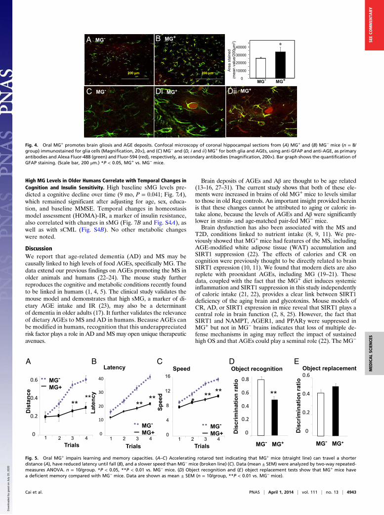
Chronic MG+ Impairs Learning andMemory.Basic motor coordinationand balance learning skills were first evaluated with the rotarodtest. MG− mice performed for a longer distance and at a higherspeed before falling from the rod compared with MG+mice (Fig. 5
A and B). MG+ mice showed a lower latency than MG− mice (Fig.5C). On testing object recognition, MG+ fed mice showed poorexploratory behavior with a lower discriminatory capacity betweena familiar and a novel object than MG− mice (Fig. 5D), whichspent ∼70% of the time exploring the new object. On testing ob-ject replacement, MG+-fed mice performed better, but this wasnot significant (Fig. 5E).
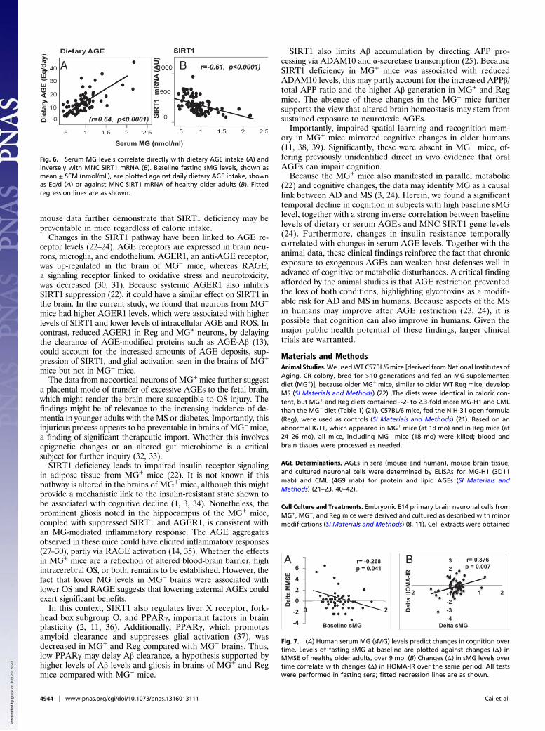
High MG Correlates with Dietary AGE Intake and SIRT1 Suppression inOlder Humans. At baseline, the cohort’s body mass index (BMI)and metabolic and biochemical parameters (n = 93, ≥60 y old,educated, 68% female) were within the range expected for theirage, as were calorie and dietary AGE intake (dAGE) (24, 26).Baseline cognitive function [by Mini Mental State Examination(MMSE)] was also normal (Table S1).Baseline sMG levels correlated positively with dAGE intake
(Fig. 6A) and inversely with mononuclear cell (MNC) SIRT1mRNA levels (Fig. 6B and Table S2). In addition, baselinedAGE and sMG levels both correlated with sCML, plasma8-isoprostanes, leptin, MNC TNFα protein, and RAGE mRNA,but inversely with SIRT1 mRNA and adiponectin levels (TableS2 and Fig. S3).
Table 1. Mice characteristics
Groups MG− MG+ Reg
Number/group 12 (6 F/6 M) 12 (6 F/6 M) 8 (4 F/4 M)Body weight (g) 30.8 ± 0.52 35.1 ± 1.8* 33.5 ± 1.5Brain weight (g) 0.48 ± 0.1 0.46 ± 0.05 0.48 ± 0.1Brain/body weight ratio 0.017 ± 0.004† 0.013 ± 0.001* 0.014 ± 0.003Food intake (g/d) 4.8 ± 0.8 4.9 ± 0.05 5 ± 0.7Food MG intake (nmol/d) 0.67 × 10⁴† 1.9 × 10⁴‡ 1.5 × 10⁴
Serum CML (sCML, U/mL) 21.9 ± 1.2§ 49.9 ± 1.1‡ 42.8 ± 1.5Serum MG (sMG, nmol/mL) 0.83 ± 0.2† 2.08 ± 0.29‡,{ 1.59 ± 0.2Fasting blood glucose (mg/dL) 82.6 ± 2.8 81.4 ± 4 83.1 ± 2.4Fasting insulin (nmol/L) 0.24 ± 0.02§ 0.41 ± 0.07* 0.45 ± 0.02Adiponectin (μg/mL) 13.7 ± 1.2† 7.8 ± 1.0‡ 8.2 ± 0.4Leptin (ng/mL) 10.3 ± 0.9† 22.7 ± 0.9‡ 18.0 ± 0.88-Isoprostane (pg/mL) 88 ± 5.5† 267 ± 32.4‡,{ 174 ± 22.3Brain protein CML (U/g brain) 100.3 ± 12.2 166.3 ± 19.4* 142.6 ± 10Brain protein MG (nmol/g brain) 63.4 ± 6.3 90.1 ± 11.05* 78.4 ± 6.5Brain lipid CML (U/g brain) 229.5 ± 15.5 352.5 ± 39.2* 290.4 ± 14.3Brain lipid MG (nmol/g brain) 6.3 ± 1.3 10.0 ± 0.6* 7.5 ± 0.5
Mice were WT C57BL6. MG− denotes 18-mo mice on low-AGE diet. MG+ denotes 18-mo mice on aMG-supplemented low-AGE diet. Reg denotes 24- to 26-mo control mice on standard NIH-31 open formula diet.Data are means ± SEM. *P < 0.05 and ‡P < 0.01 between MG+ and MG− mice; {P < 0.05 between MG+ and Regmice; †P < 0.05 and §P < 0.01 between MG− and Reg mice.
A B C DREG MG- MG+
actin
ADAM10
APP
sAPP
A -42mRNA
(% o
f REG
)
0 REG MG- MG+
50
100
150
200
250
*
0
50
100
150
ADAM10: i) protein
*
REG MG- MG+0
20
4060
80100
120
sAPP-β/APP Ratio
*
REG MG- MG+
200
REG MG- MG+
( of R
EG)
020406080
100120 *
ii) .
( of R
EG)
( of R
EG)
Fig. 3. Oral MG+ suppresses ADAM10 expression and increases Aβ in brain. Data are from 18-mo C57BL6 mice fed an MG+ or MG− diet and Reg mice (24–26mo, n = 8/group). (A) Western blots of brain extracts for ADAM10, APP, and soluble or sAPPβ (n = 3–5). β-actin is used as control. (B, i) Densitometry ofADAM10 protein data shown in A and (B, ii) of ADAM10 mRNA levels, by RT-PCR, shown as AU (n = 5/group). (C) Densitometry of sAPPβ and APP shown in Aand expressed as sAPPβ/APP ratio. (D) Aβ1–42 levels. Data in B–D are shown as percent (mean ± SEM) of Reg. *P < 0.05 MG+ vs. MG− mice.
4942 | www.pnas.org/cgi/doi/10.1073/pnas.1316013111 Cai et al.
Dow
nloa
ded
by g
uest
on
July
20,
202
0
High MG Levels in Older Humans Correlate with Temporal Changes inCognition and Insulin Sensitivity. High baseline sMG levels pre-dicted a cognitive decline over time (9 mo, P = 0.041; Fig. 7A),which remained significant after adjusting for age, sex, educa-tion, and baseline MMSE. Temporal changes in homeostasismodel assessment (HOMA)-IR, a marker of insulin resistance,also correlated with changes in sMG (Fig. 7B and Fig. S4A), aswell as with sCML (Fig. S4B). No other metabolic changeswere noted.
DiscussionWe report that age-related dementia (AD) and MS may becausally linked to high levels of food AGEs, specifically MG. Thedata extend our previous findings on AGEs promoting the MS inolder animals and humans (22–24). The mouse study furtherreproduces the cognitive and metabolic conditions recently foundto be linked in humans (1, 4, 5). The clinical study validates themouse model and demonstrates that high sMG, a marker of di-etary AGE intake and IR (23), may also be a determinantof dementia in older adults (17). It further validates the relevanceof dietary AGEs to MS and AD in humans. Because AGEs canbe modified in humans, recognition that this underappreciatedrisk factor plays a role in AD and MS may open unique therapeuticavenues.
Brain deposits of AGEs and Aβ are thought to be age related(13–16, 27–31). The current study shows that both of these ele-ments were increased in brains of old MG+ mice to levels similarto those in old Reg controls. An important insight provided hereinis that these changes cannot be attributed to aging or caloric in-take alone, because the levels of AGEs and Aβ were significantlylower in strain- and age-matched pair-fed MG− mice.Brain dysfunction has also been associated with the MS and
T2D, conditions linked to nutrient intake (8, 9, 11). We pre-viously showed that MG+ mice had features of the MS, includingAGE-modified white adipose tissue (WAT) accumulation andSIRT1 suppression (22). The effects of calories and CR oncognition were previously thought to be directly related to brainSIRT1 expression (10, 11). We found that modern diets are alsoreplete with prooxidant AGEs, including MG (19–21). Thesedata, coupled with the fact that the MG+ diet induces systemicinflammation and SIRT1 suppression in this study independentlyof caloric intake (21, 22), provides a clear link between SIRT1deficiency of the aging brain and glycotoxins. Mouse models ofCR, AD, or SIRT1 expression in mice reveal that SIRT1 plays acentral role in brain function (2, 8, 25). However, the fact thatSIRT1 and NAMPT, AGER1, and PPARγ were suppressed inMG+ but not in MG− brains indicates that loss of multiple de-fense mechanisms in aging may reflect the impact of sustainedhigh OS and that AGEs could play a seminal role (22). The MG−
MG-C MG+Di MG+Dii
MG-A
200 µm
B MG+
200 µm
0
10000
20000
30000
40000
MG- MG+
Are
a st
aine
d (m
ean
valu
e/20
0µm
2 ) ∗
Fig. 4. Oral MG+ promotes brain gliosis and AGE deposits. Confocal microscopy of coronal hippocampal sections from (A) MG+ and (B) MG− mice (n = 8/group) immunostained for glia cells (Magnification, 20×), and (C) MG− and (D, i and ii) MG+ for both glia and AGEs, using anti-GFAP and anti-AGE, as primaryantibodies and Alexa Fluor-488 (green) and Fluor-594 (red), respectively, as secondary antibodies (magnification, 200×). Bar graph shows the quantification ofGFAP staining. (Scale bar, 200 μm.) *P < 0.05, MG+ vs. MG− mice.
Object recognition Object replacement
Dis
crim
inat
ion
ratio
0
0.2
0.4
0.6
0
0.2
0.4
0.6
0.8
Dis
crim
inat
ion
ratio
MG- MG+MG- MG+
**
Trials
Dis
tanc
e
0
0.2
0.4
0.6
1 2 3 4
****
MG-
MG+
Trials
Spee
d
0
4
8
12
16
1 2 3 4
MG-
MG+
*****
Speed
Trials
Late
ncy
0
10
20
30
40
1 2 3 4
****
MG-
MG+
LatencyD EA C B
Fig. 5. Oral MG+ impairs learning and memory capacities. (A–C) Accelerating rotarod test indicating that MG+ mice (straight line) can travel a shorterdistance (A), have reduced latency until fall (B), and a slower speed than MG− mice (broken line) (C). Data (mean ± SEM) were analyzed by two-way repeated-measures ANOVA. n = 10/group. *P < 0.05, **P < 0.01 vs. MG− mice. (D) Object recognition and (E) object replacement tests show that MG+ mice havea deficient memory compared with MG− mice. Data are shown as mean ± SEM (n = 10/group, **P < 0.01 vs. MG− mice).
Cai et al. PNAS | April 1, 2014 | vol. 111 | no. 13 | 4943
MED
ICALSC
IENCE
SSE
ECO
MMEN
TARY
Dow
nloa
ded
by g
uest
on
July
20,
202
0
mouse data further demonstrate that SIRT1 deficiency may bepreventable in mice regardless of caloric intake.Changes in the SIRT1 pathway have been linked to AGE re-
ceptor levels (22–24). AGE receptors are expressed in brain neu-rons, microglia, and endothelium. AGER1, an anti-AGE receptor,was up-regulated in the brain of MG− mice, whereas RAGE,a signaling receptor linked to oxidative stress and neurotoxicity,was decreased (30, 31). Because systemic AGER1 also inhibitsSIRT1 suppression (22), it could have a similar effect on SIRT1 inthe brain. In the current study, we found that neurons from MG−
mice had higher AGER1 levels, which were associated with higherlevels of SIRT1 and lower levels of intracellular AGE and ROS. Incontrast, reduced AGER1 in Reg and MG+ neurons, by delayingthe clearance of AGE-modified proteins such as AGE-Aβ (13),could account for the increased amounts of AGE deposits, sup-pression of SIRT1, and glial activation seen in the brains of MG+
mice but not in MG− mice.The data from neocortical neurons of MG+ mice further suggest
a placental mode of transfer of excessive AGEs to the fetal brain,which might render the brain more susceptible to OS injury. Thefindings might be of relevance to the increasing incidence of de-mentia in younger adults with the MS or diabetes. Importantly, thisinjurious process appears to be preventable in brains of MG−mice,a finding of significant therapeutic import. Whether this involvesepigenetic changes or an altered gut microbiome is a criticalsubject for further inquiry (32, 33).SIRT1 deficiency leads to impaired insulin receptor signaling
in adipose tissue from MG+ mice (22). It is not known if thispathway is altered in the brains of MG+ mice, although this mightprovide a mechanistic link to the insulin-resistant state shown tobe associated with cognitive decline (1, 3, 34). Nonetheless, theprominent gliosis noted in the hippocampus of the MG+ mice,coupled with suppressed SIRT1 and AGER1, is consistent withan MG-mediated inflammatory response. The AGE aggregatesobserved in these mice could have elicited inflammatory responses(27–30), partly via RAGE activation (14, 35). Whether the effectsin MG+ mice are a reflection of altered blood-brain barrier, highintracerebral OS, or both, remains to be established. However, thefact that lower MG levels in MG− brains were associated withlower OS and RAGE suggests that lowering external AGEs couldexert significant benefits.In this context, SIRT1 also regulates liver X receptor, fork-
head box subgroup O, and PPARγ, important factors in brainplasticity (2, 11, 36). Additionally, PPARγ, which promotesamyloid clearance and suppresses glial activation (37), wasdecreased in MG+ and Reg compared with MG− brains. Thus,low PPARγ may delay Aβ clearance, a hypothesis supported byhigher levels of Aβ levels and gliosis in brains of MG+ and Regmice compared with MG− mice.
SIRT1 also limits Aβ accumulation by directing APP pro-cessing via ADAM10 and α-secretase transcription (25). BecauseSIRT1 deficiency in MG+ mice was associated with reducedADAM10 levels, this may partly account for the increased APPβ/total APP ratio and the higher Aβ generation in MG+ and Regmice. The absence of these changes in the MG− mice furthersupports the view that altered brain homeostasis may stem fromsustained exposure to neurotoxic AGEs.Importantly, impaired spatial learning and recognition mem-
ory in MG+ mice mirrored cognitive changes in older humans(11, 38, 39). Significantly, these were absent in MG− mice, of-fering previously unidentified direct in vivo evidence that oralAGEs can impair cognition.Because the MG+ mice also manifested in parallel metabolic
(22) and cognitive changes, the data may identify MG as a causallink between AD and MS (3, 24). Herein, we found a significanttemporal decline in cognition in subjects with high baseline sMGlevel, together with a strong inverse correlation between baselinelevels of dietary or serum AGEs and MNC SIRT1 gene levels(24). Furthermore, changes in insulin resistance temporallycorrelated with changes in serum AGE levels. Together with theanimal data, these clinical findings reinforce the fact that chronicexposure to exogenous AGEs can weaken host defenses well inadvance of cognitive or metabolic disturbances. A critical findingafforded by the animal studies is that AGE restriction preventedthe loss of both conditions, highlighting glycotoxins as a modifi-able risk for AD and MS in humans. Because aspects of the MSin humans may improve after AGE restriction (23, 24), it ispossible that cognition can also improve in humans. Given themajor public health potential of these findings, larger clinicaltrials are warranted.
Materials and MethodsAnimal Studies.WeusedWT C57BL/6mice [derived fromNational Institutes ofAging, CR colony, bred for >10 generations and fed an MG-supplementeddiet (MG+)], because older MG+ mice, similar to older WT Reg mice, developMS (SI Materials and Methods) (22). The diets were identical in caloric con-tent, but MG+ and Reg diets contained ∼2- to 2.3-fold more MG-H1 and CMLthan the MG− diet (Table 1) (21). C57BL/6 mice, fed the NIH-31 open formula(Reg), were used as controls (SI Materials and Methods) (21). Based on anabnormal IGTT, which appeared in MG+ mice (at 18 mo) and in Reg mice (at24–26 mo), all mice, including MG− mice (18 mo) were killed; blood andbrain tissues were processed as needed.
AGE Determinations. AGEs in sera (mouse and human), mouse brain tissue,and cultured neuronal cells were determined by ELISAs for MG-H1 (3D11mab) and CML (4G9 mab) for protein and lipid AGEs (SI Materials andMethods) (21–23, 40–42).
Cell Culture and Treatments. Embryonic E14 primary brain neuronal cells fromMG+, MG−, and Reg mice were derived and cultured as described with minormodifications (SI Materials and Methods) (8, 11). Cell extracts were obtained
A B
Die
tary
AG
E (E
q/da
y)
SIR
T1 m
RN
A (A
U)
Serum MG (nmol/ml)
(r=0.64, p<0.0001)
r=-0.61, p<0.0001)
Fig. 6. Serum MG levels correlate directly with dietary AGE intake (A) andinversely with MNC SIRT1 mRNA (B). Baseline fasting sMG levels, shown asmean ± SEM (nmol/mL), are plotted against daily dietary AGE intake, shownas Eq/d (A) or against MNC SIRT1 mRNA of healthy older adults (B). Fittedregression lines are as shown.
-4
-2
0
2
4
6
0 2
Del
ta M
MSE
Baseline sMG
r= 0.376p = 0.007
-4-3-2-10123
-2 -1 0 1 2
Del
ta H
OM
A- IR
r= -0.268p = 0.041
Delta sMG
BA
Fig. 7. (A) Human serum MG (sMG) levels predict changes in cognition overtime. Levels of fasting sMG at baseline are plotted against changes (Δ) inMMSE of healthy older adults, over 9 mo. (B) Changes (Δ) in sMG levels overtime correlate with changes (Δ) in HOMA-IR over the same period. All testswere performed in fasting sera; fitted regression lines are as shown.
4944 | www.pnas.org/cgi/doi/10.1073/pnas.1316013111 Cai et al.
Dow
nloa
ded
by g
uest
on
July
20,
202
0

immediately or after 24–72 h, as needed. Neuronal cells from Reg brain werechronically exposed to different doses of MG-BSA or BSA (72 h).
Immunohistochemistry. Anesthetized mice were transcardially perfused with4% (wt/vol) paraformaldehyde (PF) and the brain was removed, processedfor immunohistochemistry, and viewed by a confocal microscope (Zeiss LSM510 Meta) (SI Materials and Methods) (8, 36).
Brain Functional Testing. Motor coordination, balance, and motor learningwere tested on the accelerating rotarod device (Series 8; IITC Life Science) (SIMaterials and Methods) (38). Object recognition and placement memorytests were conducted, and data were video recorded and analyzed (SIMaterials and Methods) (39).
Human Studies. This study was an observational study in healthy adult(n = 93), ≥ 60-y-old, New York City residents who provided informedconsent. Participants were evaluated at baseline and 9 mo later for time-dependent changes in serum AGEs, markers of OS, and inflammation,insulin resistance, and cognition. Information was collected on medicalhistory, medications, and caloric and AGE intake. Exclusion criteria includedevidence of diabetes, cardiovascular or kidney disease, neuropsychiatric dis-ease, and cancer. Cognition was assessed by a Clinical Dementia Rating (CDR)scale and by a MMSE score, with normality defined as a CDR score of 0 (non-demented) and an MMSE score above the 10th percentile of age and educationnorms (17). All cognition tests were performed by trained research coordinators
at the Icahn School of Medicine Alzheimer’s Disease Research Center. After aninitial evaluation, participants underwent MMSE and provided a fasting bloodsample (at 0 and 9 mo). Plasma or sera was used for routine blood tests, andAGEs (sCML and sMG), adiponectin, leptin, insulin levels, and HOMA index andMNCs were used for gene assessment by RT-PCR (SI Materials and Methods)(23, 24).
Statistics. Animal data were expressed as means ± SEM, and differences weredetermined by Student t test. For comparisons among the three groups, one-way ANOVA with Bonferroni correction analysis was performed. The be-havioral data were analyzed using repeated-measures ANOVA with multiplecomparison tests performed with Bonferroni’s adjustment. Significance wasset at P < 0.05.
Human datawere analyzed for relationships between variables at baselineusing mean ± SD and quartiles, based on regression models and partialSpearman correlation coefficients and adjusting for age and sex. The tem-poral relationship between baseline sMG and Δ MMSE was explored usinggeneral linear regression models adjusting for baseline MMSE and years ofeducation, age, and sex. Also assessed were the correlations of Δ sMG,Δ sCML, and Δ HOMA-IR, using Spearman correlation coefficients, by Stata,version 11. Data with two-sided P < 0.05 were considered significant.
ACKNOWLEDGMENTS. This work was supported by National Institutes ofHealth Grants AG23188 (to H.V.) and M01-RR-00071 (to the GCRC of IcahnSchool of Medicine at Mount Sinai).
1. Janson J, et al. (2004) Increased risk of type 2 diabetes in Alzheimer disease. Diabetes53(2):474–481.
2. Guarente L, Franklin H (2011) Franklin H. Epstein Lecture: Sirtuins, aging, and medi-cine. N Engl J Med 364(23):2235–2244.
3. Talbot K, et al. (2012) Demonstrated brain insulin resistance in Alzheimer’s diseasepatients is associated with IGF-1 resistance, IRS-1 dysregulation, and cognitive decline.J Clin Invest 122(4):1316–1338.
4. Reddy VP, Zhu X, Perry G, Smith MA (2009) Oxidative stress in diabetes and Alzheimer’sdisease. J Alzheimers Dis 16(4):763–774.
5. Loy CT, Twigg SM (2009) Growth factors, AGEing, and the diabetes link in Alzheimer’sdisease. J Alzheimers Dis 16(4):823–831.
6. Vlassara H, Striker GE (2011) AGE restriction in diabetes mellitus: A paradigm shift.Nat Rev Endocrinol 7(9):526–539.
7. Srivastava S, Haigis MC (2011) Role of sirtuins and calorie restriction in neuro-protection: Implications in Alzheimer’s and Parkinson’s diseases. Curr Pharm Des17(31):3418–3433.
8. Qin W, et al. (2008) Regulation of forkhead transcription factor FoxO3a contributes tocalorie restriction-induced prevention of Alzheimer’s disease-type amyloid neuropa-thology and spatial memory deterioration. Ann N Y Acad Sci 1147:335–347.
9. Longo VD, Kennedy BK (2006) Sirtuins in aging and age-related disease. Cell 126(2):257–268.
10. Bonda DJ, et al. (2011) The sirtuin pathway in ageing and Alzheimer disease: Mech-anistic and therapeutic considerations. Lancet Neurol 10(3):275–279.
11. Fusco S, et al. (2012) A role for neuronal cAMP responsive-element binding (CREB)-1in brain responses to calorie restriction. Proc Natl Acad Sci USA 109(2):621–626.
12. Huebschmann AG, Regensteiner JG, Vlassara H, Reusch JE (2006) Diabetes and ad-vanced glycoxidation end products. Diabetes Care 29(6):1420–1432.
13. Vitek MP, et al. (1994) Advanced glycation end products contribute to amyloidosis inAlzheimer disease. Proc Natl Acad Sci USA 91(11):4766–4770.
14. Zimmerman GA, et al. (1995) Neurotoxicity of advanced glycation endproducts duringfocal stroke and neuroprotective effects of aminoguanidine. Proc Natl Acad Sci USA92(9):3744–3748.
15. Srikanth V, et al. (2013) Methylglyoxal, cognitive function and cerebral atrophy inolder people. J Gerontol A Biol Sci Med Sci 68(1):68–73.
16. Li XH, et al. (2012) Methylglyoxal induces tau hyperphosphorylation via promotingAGEs formation. Neuromolecular Med 14(4):338–348.
17. Beeri MS, et al. (2011) Serum concentration of an inflammatory glycotoxin, methyl-glyoxal, is associated with increased cognitive decline in elderly individuals. MechAgeing Dev 132(11-12):583–587.
18. Ahmed N, et al. (2005) Protein glycation, oxidation and nitration adduct residues andfree adducts of cerebrospinal fluid in Alzheimer’s disease and link to cognitiveimpairment. J Neurochem 92(2):255–263.
19. Koschinsky T, et al. (1997) Orally absorbed reactive glycation products (glycotoxins):An environmental risk factor in diabetic nephropathy. Proc Natl Acad Sci USA 94(12):6474–6479.
20. Birlouez-Aragon I, et al. (2010) A diet based on high-heat-treated foods pro-motes risk factors for diabetes mellitus and cardiovascular diseases. Am J ClinNutr 91(5):1220–1226.
21. Cai W, et al. (2008) Oral glycotoxins determine the effects of calorie restriction onoxidant stress, age-related diseases, and lifespan. Am J Pathol 173(2):327–336.
22. Cai W, et al. (2012) Oral advanced glycation endproducts (AGEs) promote insulinresistance and diabetes by depleting the antioxidant defenses AGE receptor-1 andsirtuin 1. Proc Natl Acad Sci USA 109(39):15888–15893.
23. Uribarri J, et al. (2011) Restriction of advanced glycation end products improves in-sulin resistance in human type 2 diabetes: Potential role of AGER1 and SIRT1. DiabetesCare 34(7):1610–1616.
24. Uribarri J, et al. (2013) Suppression of native defense mechanisms, SIRT1 and PPARγ, bydietary glycoxidants precedes disease in adult humans; relevance to lifestyle-engenderedchronic diseases. Amino Acids 46(2):301–309.
25. Donmez G, Wang D, Cohen DE, Guarente L (2010) SIRT1 suppresses β-amyloid pro-duction by activating the α-secretase gene ADAM10. Cell 142(2):320–332.
26. Vlassara H, et al. (2009) Protection against loss of innate defenses in adulthood by lowadvanced glycation end products (AGE) intake: Role of the antiinflammatory AGEreceptor-1. J Clin Endocrinol Metab 94(11):4483–4491.
27. Dickson DW, et al. (1996) Glycation and microglial reaction in lesions of Alzheimer’sdisease. Neurobiol Aging 17(5):733–743.
28. Schmidt AM, et al. (2009) The role of RAGE in amyloid-beta peptide-mediated pa-thology in Alzheimer’s disease. Curr Opin Investig Drugs 10(7):672–680.
29. Ko SY, Lin YP, Lin YS, Chang SS (2010) Advanced glycation end products enhanceamyloid precursor protein expression by inducing reactive oxygen species. Free RadicBiol Med 49(3):474–480.
30. Li JJ, Dickson D, Hof PR, Vlassara H (1998) Receptors for advanced glycosylationendproducts in human brain: Role in brain homeostasis. Mol Med 4(1):46–60.
31. Takuma K, et al. (2009) RAGE-mediated signaling contributes to intraneuronaltransport of amyloid-beta and neuronal dysfunction. Proc Natl Acad Sci USA 106(47):20021–20026.
32. Ford D, Ions LJ, Alatawi F, Wakeling LA (2011) The potential role of epigenetic re-sponses to diet in ageing. Proc Nutr Soc 70(3):374–384.
33. Marques SC, et al. (2012) Epigenetic regulation of BACE1 in Alzheimer’s disease pa-tients and in transgenic mice. Neuroscience 220:256–266.
34. Correia SC, et al. (2011) Insulin-resistant brain state: The culprit in sporadic Alzheimer’sdisease? Ageing Res Rev 10(2):264–273.
35. Deane R, et al. (2012) A multimodal RAGE-specific inhibitor reduces amyloidβ-mediated brain disorder in a mouse model of Alzheimer disease. J Clin Invest122(4):1377–1392.
36. Michán S, et al. (2010) SIRT1 is essential for normal cognitive function and synapticplasticity. J Neurosci 30(29):9695–9707.
37. Mandrekar-Colucci S, Karlo JC, Landreth GE (2012) Mechanisms underlying the rapidperoxisome proliferator-activated receptor-γ-mediated amyloid clearance and re-versal of cognitive deficits in a murine model of Alzheimer’s disease. J Neurosci32(30):10117–10128.
38. Monville C, Torres EM, Dunnett SB (2006) Comparison of incremental and accelerat-ing protocols of the rotarod test for the assessment of motor deficits in the 6-OHDAmodel. J Neurosci Methods 158(2):219–223.
39. Barker GR, Bird F, Alexander V, Warburton EC (2007) Recognition memory for objects,place, and temporal order: A disconnection analysis of the role of the medial pre-frontal cortex and perirhinal cortex. J Neurosci 27(11):2948–2957.
40. Uribarri J, et al. (2010) Advanced glycation end products in foods and a practicalguide to their reduction in the diet. J Am Diet Assoc 110(6):911–916, e12.
41. Bucala R, et al. (1994) Modification of low density lipoprotein by advanced glycationend products contributes to the dyslipidemia of diabetes and renal insufficiency. ProcNatl Acad Sci USA 91(20):9441–9445.
42. Fu MX, et al. (1996) The advanced glycation end product, Nepsilon-(carboxymethyl)lysine, is a product of both lipid peroxidation and glycoxidation reactions. J Biol Chem271(17):9982–9986.
Cai et al. PNAS | April 1, 2014 | vol. 111 | no. 13 | 4945
MED
ICALSC
IENCE
SSE
ECO
MMEN
TARY
Dow
nloa
ded
by g
uest
on
July
20,
202
0