Oral cavity
48
Oral Cavity and Associated Structures Junqueira: Chapters 15 & 16 TenCate: Chapters 11 & 12 Readings in Lab Manual Lips, Oral cavity (mucosa), Tongue & Salivary glands DEN 5120C January 29, 2007 Dr. Kelly Selman D2- 32 Vestibule Oral Cavity proper
-
Upload
swapnil-pakhale -
Category
Health & Medicine
-
view
21 -
download
5
Transcript of Oral cavity
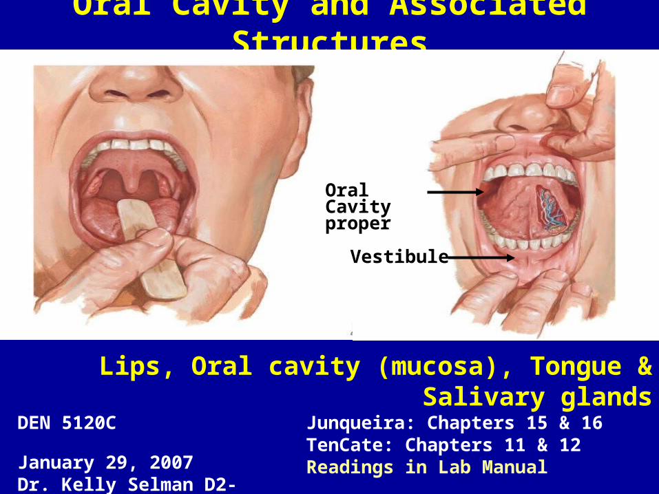
Oral Cavity and Associated Structures
Junqueira: Chapters 15 & 16TenCate: Chapters 11 & 12Readings in Lab Manual
Lips, Oral cavity (mucosa), Tongue & Salivary glands
DEN 5120C January 29, 2007Dr. Kelly Selman D2-32
Vestibule
Oral Cavityproper

bac
3 Regions of lip:a) Skinb) Red border (or Vermilion zone)c) Mucosa (wet)
Epithelium and underlying CT
Lip
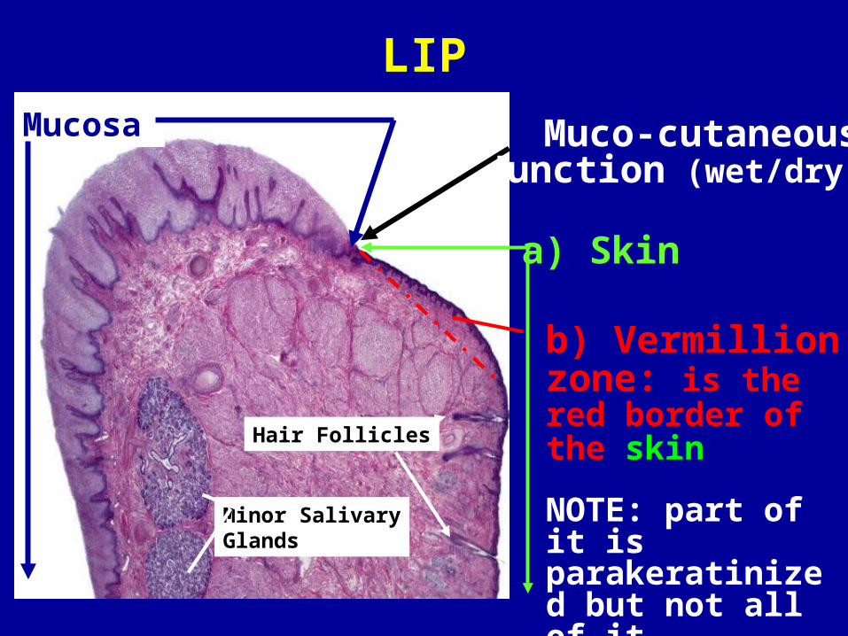
LIP
Minor SalivaryGlands
Mucosa
Hair Follicles
b) Vermillion zone: is the red border of the skin
NOTE: part of it is parakeratinized but not all of it
a) Skin
Muco-cutaneous Junction (wet/dry)

Skin
Hair follicle
Sebaceous gland (holocrine secretion)
Keratinized SSE(Epidermis)
Dermis

SSE-Non-keratinized SSE-Keratinized
BMBM
Surface LayerNK
K
Comparison & Review
Parakeratinized SSE has characteristics of both
TenCate 12-8

Muco-cutaneous junction
Mucosa (wet region)
Deep CT papillae withrich venous supply
Vermillion zone
Cutaneous region (skin)
*SSE keratinized*
VZ

Skin
Mucosa(Non-keratinized SSE)
Para-k
MC Junction
Parakeratinized region of skin
Skin (Keratinized SSE))
Mucocutaneous junction
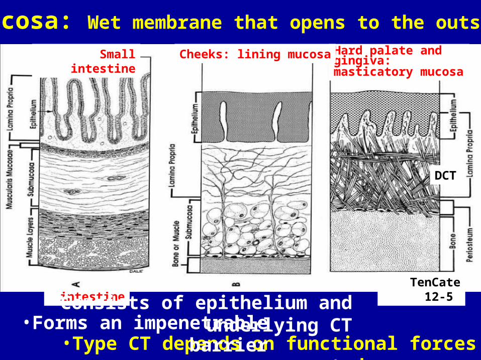
Mucosa: Wet membrane that opens to the outside
Small intestine
Hard palate and gingiva: masticatory mucosa
Small intestine Cheeks: lining mucosa
TenCate 12-5
•Type CT depends on functional forces exerted on mucosa
•Consists of epithelium and underlying CT•Forms an impenetrable barrier
DCT

Masticatory
Lining
Masticatory:•Hard palate• Gingiva
Lining:•Soft palate•Ventral tongue•Floor of mouth•Cheeks•Lips
Specialized:• Dorsal tongueSpecialized
TenCate 12-1; see Table 12-6
Oral mucosae: Lining, Masticatory & Specialized

Lining mucosa(Lips, cheeks, soft palate, floor of mouth)
Cheek (H&E)SSE
ubmucosa
Nonkeratinized
ucosa
•Flexible mucosa (many elastic fibers), overlies submucosa• Easy to inject into lamina propria (LCT), fluid spreads rapidly but also it is easy for infections to spread

Continuity of Gingival, Sulcular and Junctional epithelium
ep.
Sulcular ep. •The gingival epithelium ispart of a masticatory mucosa
•The Junctional Epitheliumattaches the gingiva to the tooth surface.
•The Junctional Epitheliumis is a circular arrangementof epithelial cells near the base of the gingival sulcus and is attached to both the tooth (enamel) and theunderlying CT
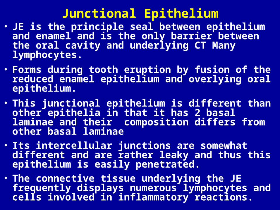
Junctional Epithelium• JE is the principle seal between epithelium and
enamel and is the only barrier between the oral cavity and underlying CT Many lymphocytes.
• Forms during tooth eruption by fusion of the reduced enamel epithelium and overlying oral epithelium.
• This junctional epithelium is different than other epithelia in that it has 2 basal laminae and their composition differs from other basal laminae
• Its intercellular junctions are somewhat different and are rather leaky and thus this epithelium is easily penetrated.
• The connective tissue underlying the JE frequently displays numerous lymphocytes and cells involved in inflammatory reactions.

Circumvallate PapillaFoliate P.
Fungiform P.
Filiform P.Body
Root
Lingual papillae, taste buds, lingual glands, lingual tonsils
Lingual tonsilPalatine tonsil
Sulcus terminalis
Taste & sensation
Tongue (Specialized
mucosa)
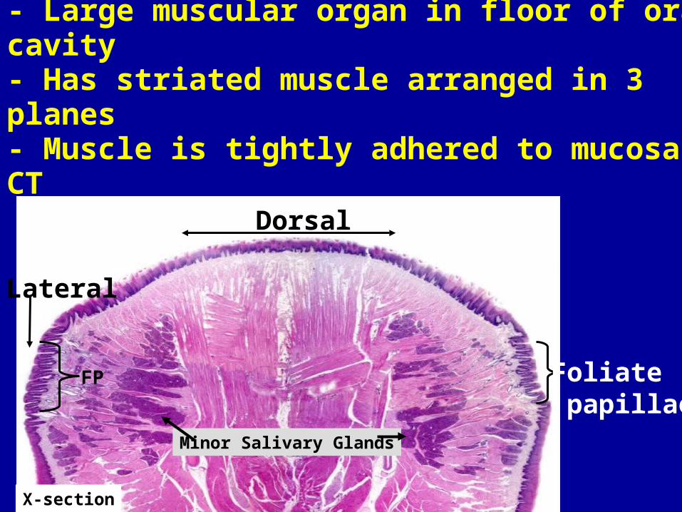
Tongue- Large muscular organ in floor of oral cavity- Has striated muscle arranged in 3 planes - Muscle is tightly adhered to mucosa by CT partitions
Dorsal
Foliate papillae
Lateral
Minor Salivary Glands
FP
X-section
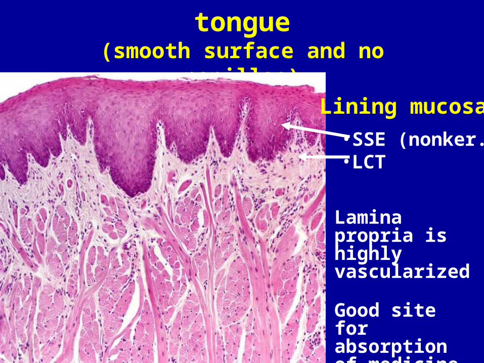
Ventral surface of tongue(smooth surface and no papillae)
Lining mucosa
•SSE (nonker.)•LCT
Lamina propria is highly vascularized
Good site for absorption of medicine
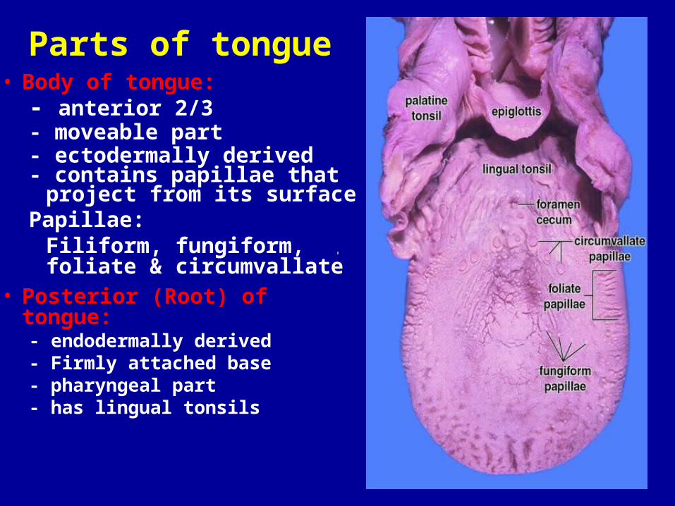
Parts of tongue• Body of tongue:
- anterior 2/3 - moveable part - ectodermally derived- contains papillae that
project from its surfacePapillae:
Filiform, fungiform, foliate & circumvallate
• Posterior (Root) of tongue:- endodermally derived- Firmly attached base - pharyngeal part- has lingual tonsils
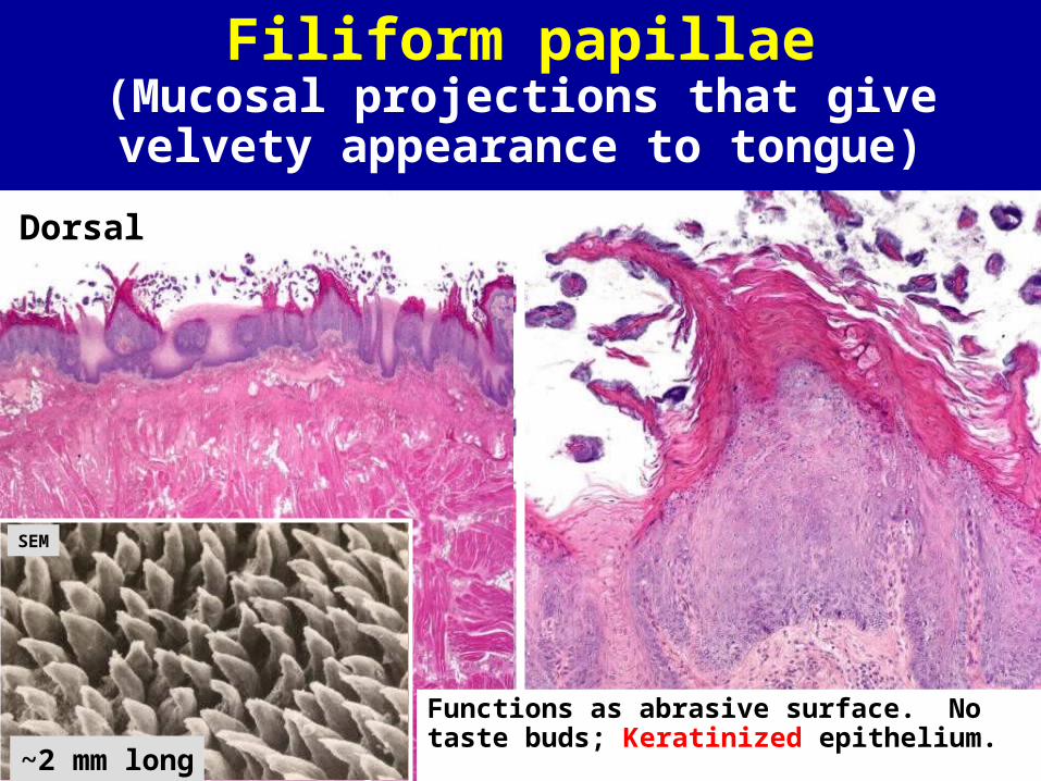
Filiform papillae(Mucosal projections that give velvety
appearance to tongue)
Functions as abrasive surface. Notaste buds; Keratinized epithelium. ~2 mm long
SEM
Dorsal
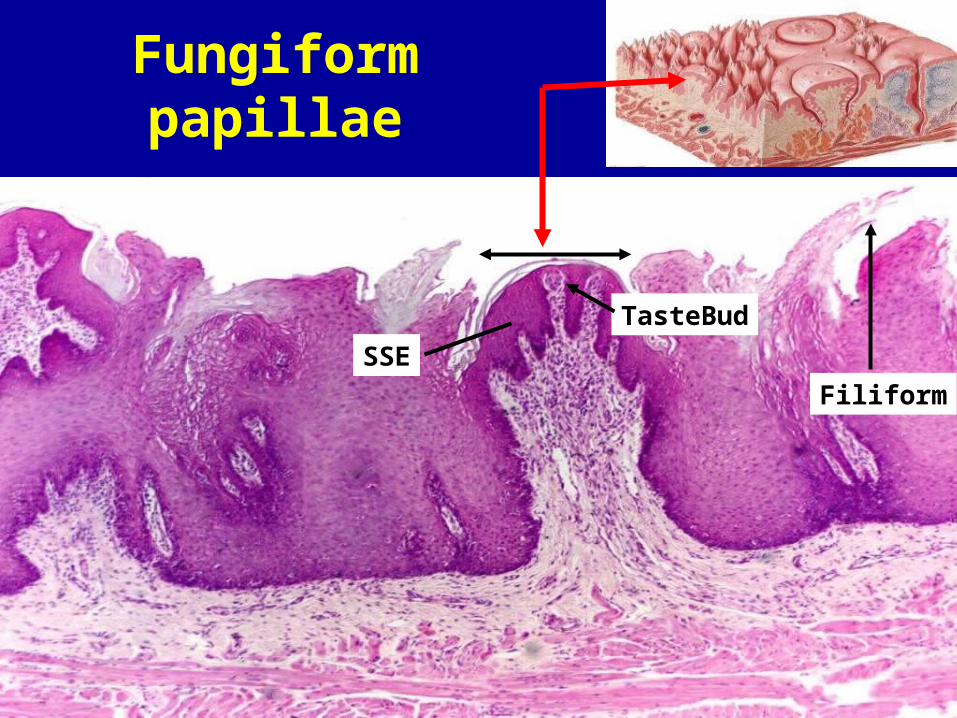
Fungiform papillae
SSE
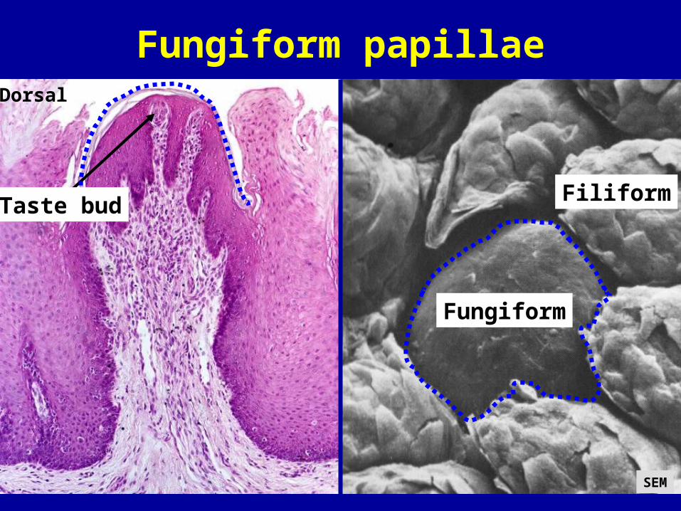
Filiform
TasteBud
Filiform
Fungiform
Taste bud
Dorsal
Fungiform papillae
SEM

Foliate papillae
•Location: form ridges on posterior lateral surface•Functional and noticeable papillae in the neonate that are reduced in size as one ages
Serous glands
Dorsal
TB
ducts
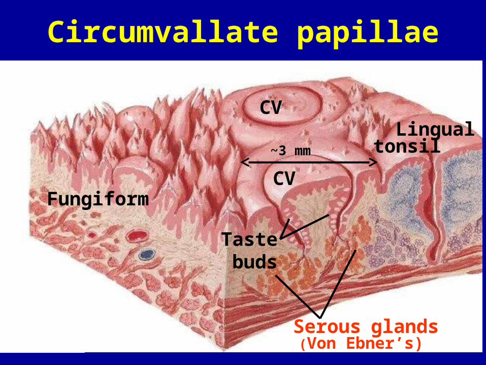
Circumvallate papillae
Serous glands(Von Ebner’s)
CV
CV
Taste
buds
Lingualtonsil
Fungiform
~3 mm
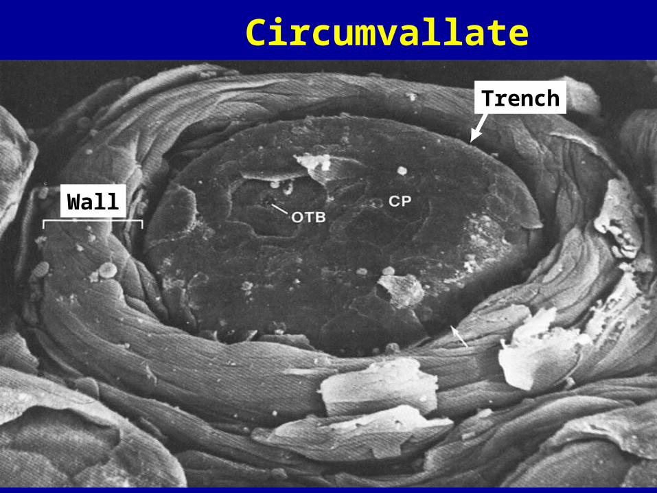
Circumvallate papilla
Wall
Trench
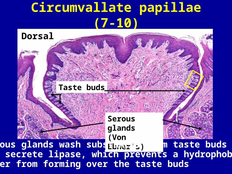
Circumvallate papillae (7-10)
Taste buds
Serous glands(Von Ebner’s)
Dorsal
Serous glands wash substances from taste buds and secrete lipase, which prevents a hydrophobic layer from forming over the taste buds
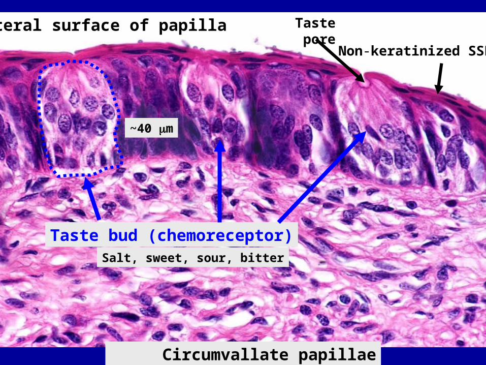
Circumvallate papillae (H&E)
Taste bud (chemoreceptor)Salt, sweet, sour, bitter
~40 m
Non-keratinized SSE
Taste poreLateral surface of papilla
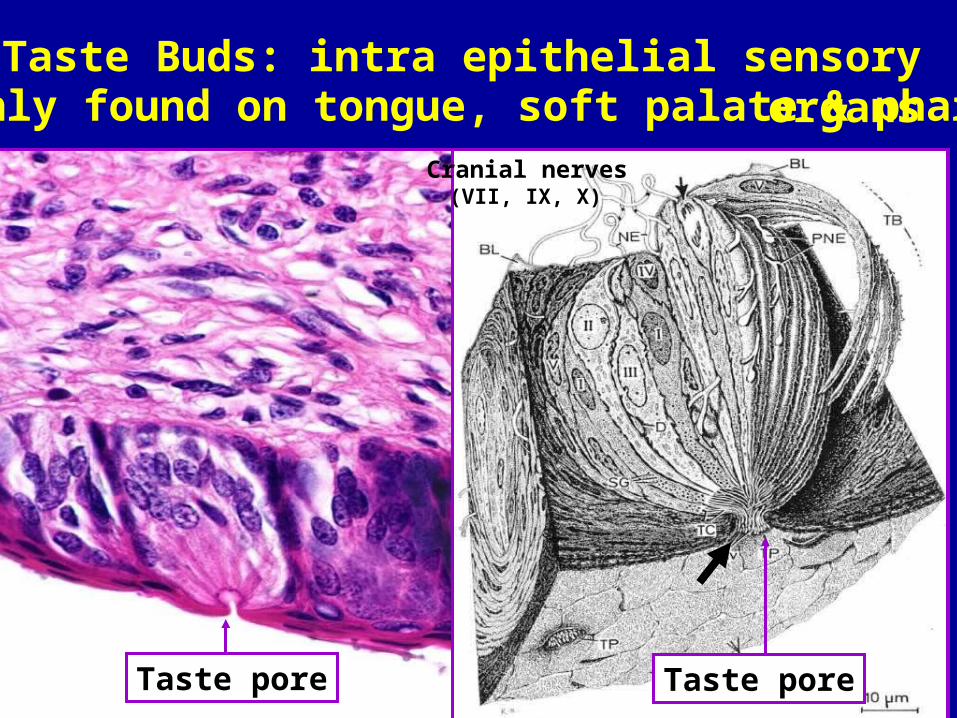
microvilli
Taste pore Taste pore
Cranial nerves(VII, IX, X)
Taste Buds: intra epithelial sensory organs(mainly found on tongue, soft palate & pharynx)
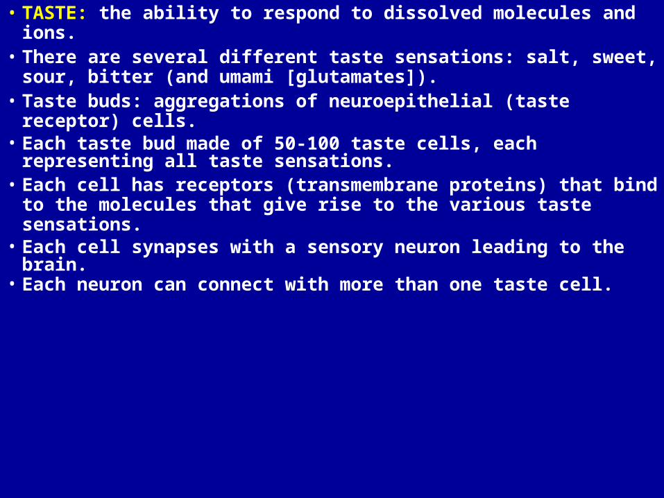
• TASTE: the ability to respond to dissolved molecules and ions.• There are several different taste sensations: salt, sweet, sour,
bitter (and umami [glutamates]).• Taste buds: aggregations of neuroepithelial (taste receptor) cells.• Each taste bud made of 50-100 taste cells, each representing all
taste sensations.• Each cell has receptors (transmembrane proteins) that bind to
the molecules that give rise to the various taste sensations.• Each cell synapses with a sensory neuron leading to the brain. • Each neuron can connect with more than one taste cell.

Summary Slide of Lingual Papillae
Filiform Fungiform
FoliateCircumvallate
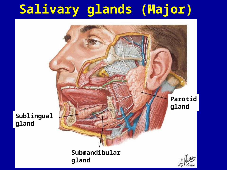
Salivary glands (Major)
Sublingual gland
Submandibulargland
Parotidgland
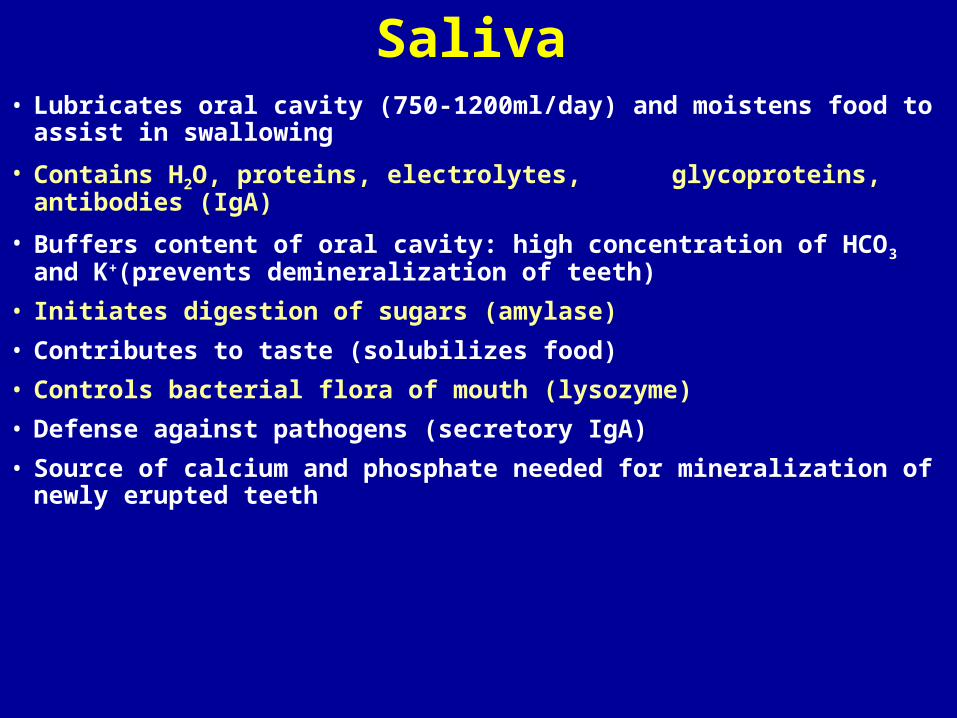
Saliva• Lubricates oral cavity (750-1200ml/day) and moistens food to assist in
swallowing
• Contains H2O, proteins, electrolytes, glycoproteins, antibodies (IgA)
• Buffers content of oral cavity: high concentration of HCO3 and K+
(prevents demineralization of teeth)
• Initiates digestion of sugars (amylase)
• Contributes to taste (solubilizes food)
• Controls bacterial flora of mouth (lysozyme)
• Defense against pathogens (secretory IgA)
• Source of calcium and phosphate needed for mineralization of newly erupted teeth
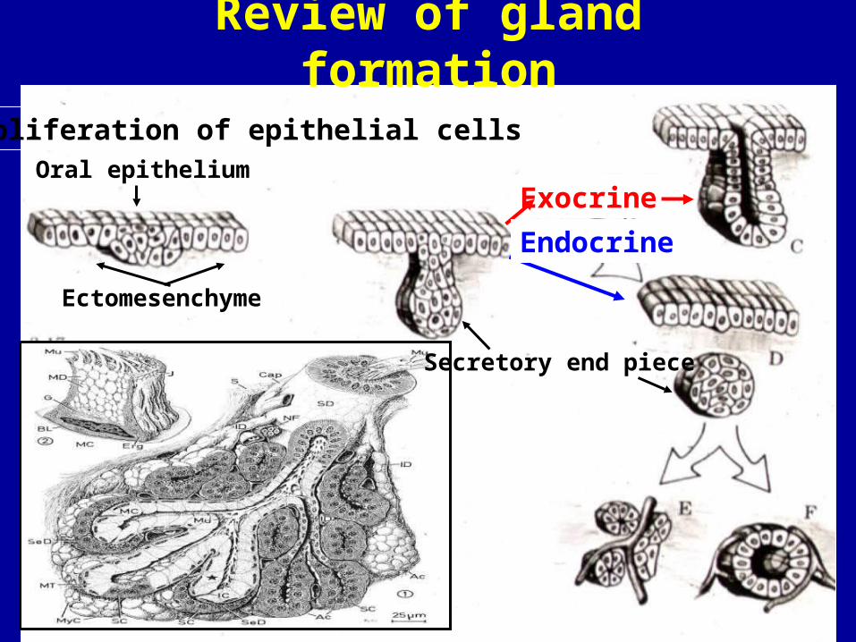
Exocrine
Proliferation of epithelial cellsOral epithelium
Ectomesenchyme
Secretory end piece
Endocrine
Review of gland formation
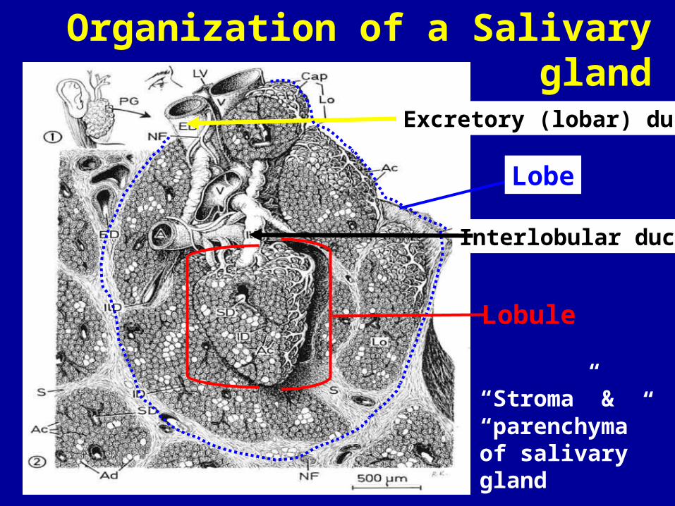
Interlobular duct
Lobule
“Stroma” & “parenchyma” of salivary gland
Lobe
Excretory (lobar) duct
Organization of a Salivary gland
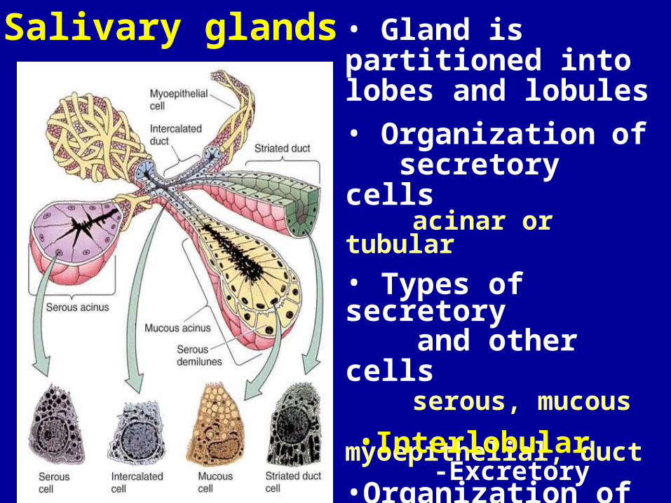
Salivary glands • Gland is partitioned into lobes and lobules
• Organization of secretory cells
acinar or tubular
• Types of secretory and other cells
serous, mucous myoepithelial, duct
•Organization of ducts•Intralobular:
-Intercalated -Striated
•Interlobular-Excretory
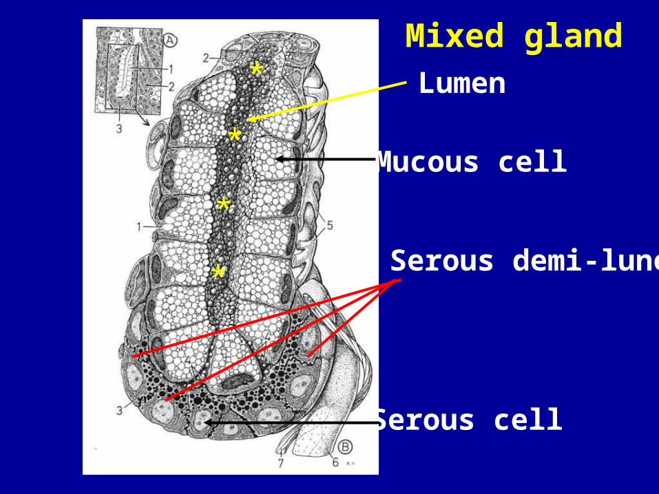
Mucous cell
Serous cell
Serous demi-lune
Lumen
*
*
*
*Mixed gland

Relationship of Secretory cell, Myoepithelial cell & Basement membrane
Secretory cell B
A
TenCate 11-19 TenCate 11-20
Myoepithelial cell
Secretory cell
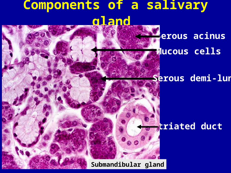
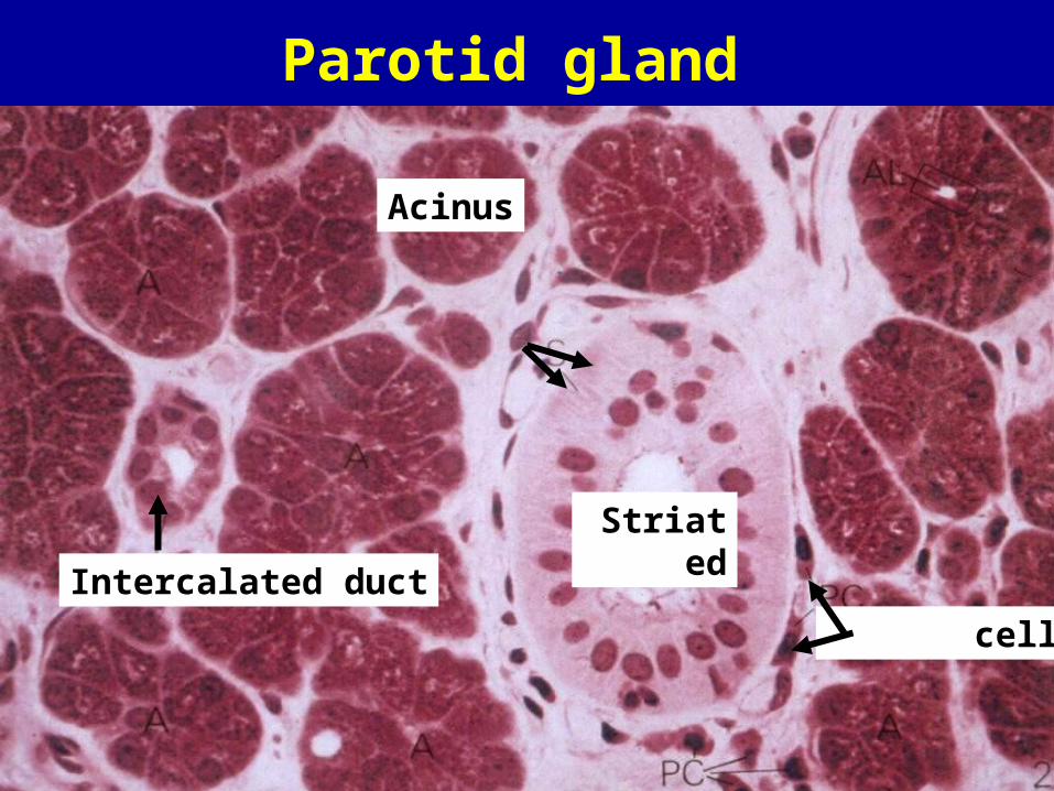
Components of a salivary gland
Striated duct
Serous acinus
Serous demi-lune
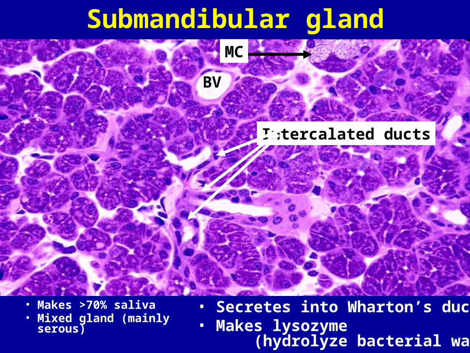
Submandibular gland
Mucous cells

Acinus and duct system of salivary gland
• Intercalated ducts and striated ducts are types of intralobular ducts (within lobule)• Excretory ducts are interlobular ducts (drain lobules and found between lobules)
(Intralobular)(Interlobular)
(Intralobular)
Outside of lobuleWithin lobule
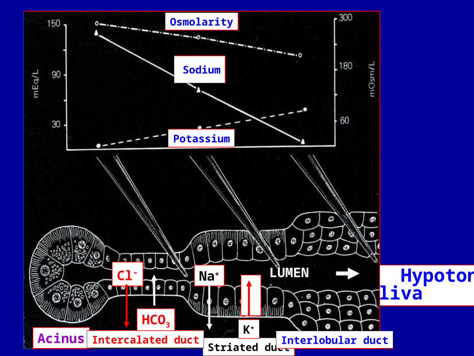
HypotonicSaliva
Sodium
Osmolarity
Potassium
Cl-
Striated duct
Na+
K+
Interlobular duct
HCO3
Acinus Intercalated duct
LUMEN

Intercalated ducts (Submandibular gland)
Acinus
Intercalated duct-striatedduct transition
Intercalated duct
SD
BV
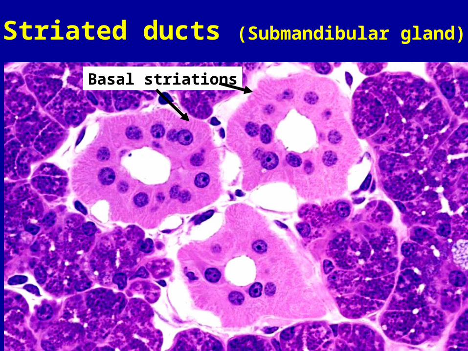
Striated ducts (Submandibular gland)
Basal striations

Striated ducts
TenCate Figs. 11-29 &11-31LUMEN LUMEN
BASAL SURFACE BASAL SURFACE
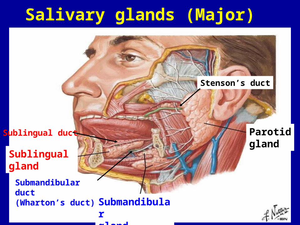
Sublingual gland
Submandibulargland
Parotidgland
Stenson’s duct
Submandibular duct(Wharton’s duct)
Sublingual duct
Salivary glands (Major)

•Largest gland but makes only ~25% of saliva•Secretes into Stenson’s duct•All serous cells•Typically has much adipose tissue •Synthesizes amylase and secretory IgA •Mumps: viral infection (inflammation & pain; facial nerve involved)
Parotid gland

Synthesis of Secretory IgAPrevents adhesion of bacteria and viruses to mucosa of oral cavity (“immunoexclusion”)
Parotid gland
(1)
(2)
(3)(4) (5)
Synthesized by conjoined activity of plasma cells and secretory cells of the parotid gland
Plasma cell
(made by secretory cell)
Secretory IgATranscytosis
of acinus
Parotid gland
Intercalated duct
Striated
Acinus
Plasma cells

Sublingual gland
*Submandibular *Gland (Whartons duct)
Parotidgland
Salivary glands (Major)
Submandibular gland
• Makes >70% saliva • Mixed gland (mainly serous)
• Secretes into Wharton’s duct• Makes lysozyme (hydrolyze bacterial walls)
Intercalated ducts
MC
BV
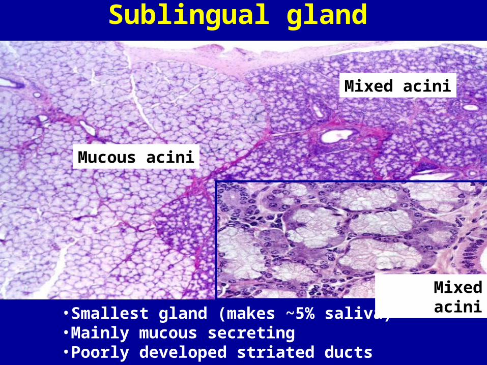
Sublingual gland
Mucous acini
Mixed acini
Mixed acini
•Smallest gland (makes ~5% saliva)•Mainly mucous secreting•Poorly developed striated ducts

UF
COLLEGE of DENTISTRY