[email protected] +27 87 15 111 47 Issue 1, 2014...
17
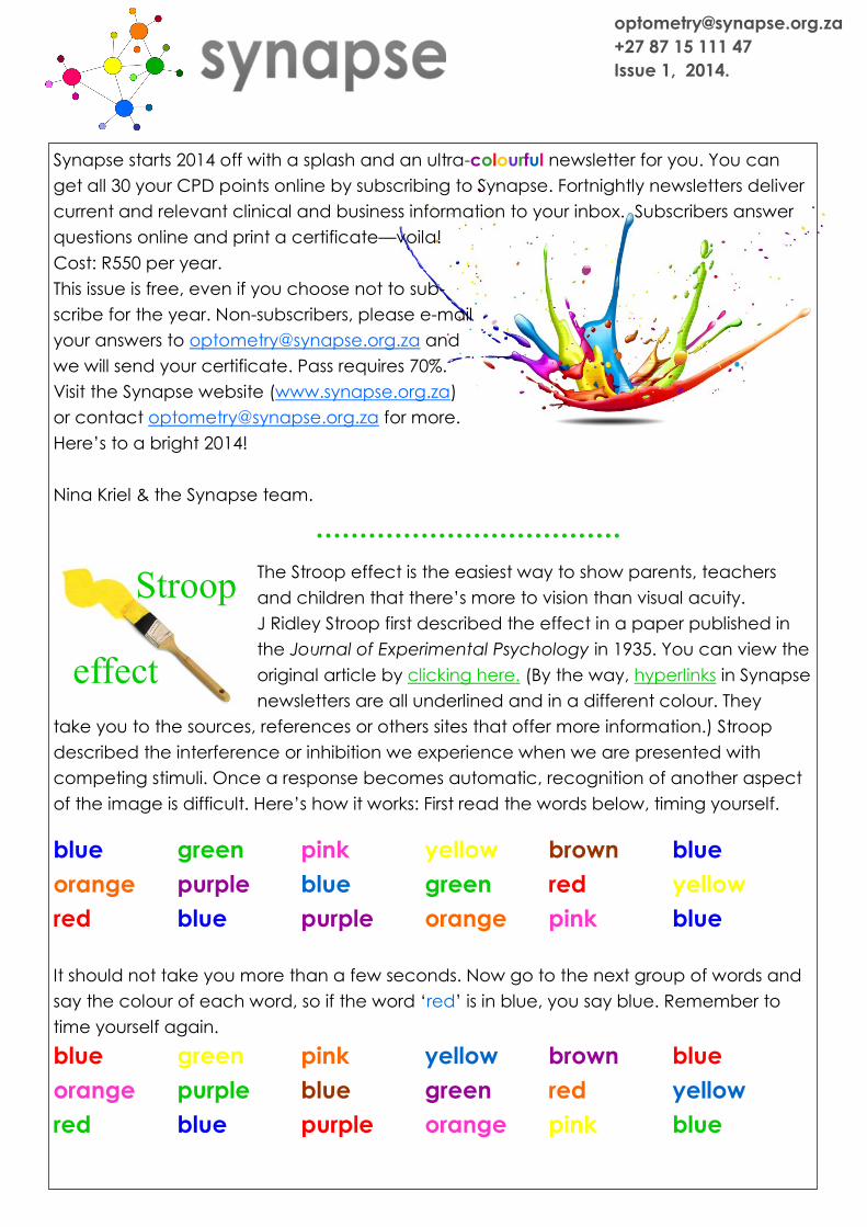
Synapse starts 2014 off with a splash and an ultra-colourful newsletter for you. You can get all 30 your CPD points online by subscribing to Synapse. Fortnightly newsletters deliver current and relevant clinical and business information to your inbox. Subscribers answer questions online and print a certificate—voila! Cost: R550 per year. This issue is free, even if you choose not to sub- scribe for the year. Non-subscribers, please e-mail your answers to [email protected] and we will send your certificate. Pass requires 70%. Visit the Synapse website (www.synapse.org.za ) or contact [email protected] for more. Here’s to a bright 2014! Nina Kriel & the Synapse team. The Stroop effect is the easiest way to show parents, teachers and children that there’s more to vision than visual acuity. J Ridley Stroop first described the effect in a paper published in the Journal of Experimental Psychology in 1935. You can view the original article by clicking here. (By the way, hyperlinks in Synapse newsletters are all underlined and in a different colour. They take you to the sources, references or others sites that offer more information.) Stroop described the interference or inhibition we experience when we are presented with competing stimuli. Once a response becomes automatic, recognition of another aspect of the image is difficult. Here’s how it works: First read the words below, timing yourself. blue green pink yellow brown blue orange purple blue green red yellow red blue purple orange pink blue It should not take you more than a few seconds. Now go to the next group of words and say the colour of each word, so if the word ‘red’ is in blue, you say blue. Remember to time yourself again. blue green pink yellow brown blue orange purple blue green red yellow red blue purple orange pink blue [email protected] +27 87 15 111 47 Issue 1, 2014. Stroop effect
Transcript of [email protected] +27 87 15 111 47 Issue 1, 2014...
Synapse starts 2014 off with a splash and an ultra-colourful newsletter for you. You can
get all 30 your CPD points online by subscribing to Synapse. Fortnightly newsletters deliver
current and relevant clinical and business information to your inbox. Subscribers answer
questions online and print a certificate—voila!
Cost: R550 per year.
This issue is free, even if you choose not to sub-
scribe for the year. Non-subscribers, please e-mail
your answers to [email protected] and
we will send your certificate. Pass requires 70%.
Visit the Synapse website (www.synapse.org.za)
or contact [email protected] for more.
Here’s to a bright 2014!
Nina Kriel & the Synapse team.
The Stroop effect is the easiest way to show parents, teachers
and children that there’s more to vision than visual acuity.
J Ridley Stroop first described the effect in a paper published in
the Journal of Experimental Psychology in 1935. You can view the
original article by clicking here. (By the way, hyperlinks in Synapse
newsletters are all underlined and in a different colour. They
take you to the sources, references or others sites that offer more information.) Stroop
described the interference or inhibition we experience when we are presented with
competing stimuli. Once a response becomes automatic, recognition of another aspect
of the image is difficult. Here’s how it works: First read the words below, timing yourself.
blue green pink yellow brown blue
orange purple blue green red yellow
red blue purple orange pink blue
It should not take you more than a few seconds. Now go to the next group of words and
say the colour of each word, so if the word ‘red’ is in blue, you say blue. Remember to
time yourself again.
blue green pink yellow brown blue
orange purple blue green red yellow
red blue purple orange pink blue
+27 87 15 111 47
Issue 1, 2014.
Stroop
effect

Stroop found that we take about 74% longer to complete the second task. Did you?
We also make more mistakes, some of which we don’t even notice. Parents and patients
will quickly realise how difficult it is to switch off their natural tendency to read the word.
In the image below, the first curve shows the students reading the words, the second
curve is when they name the colours of the text. You’ll notice that the first bell curve is
high and tight, the second has a flatter and wider distribution. You will remember from
developmental psychology days that a tight, tall curve is found when the skill that we are
measuring develops through maturation. If the skill requires learning and cognitive devel-
opment, the curve is flatter. The 2 results suggests that we need to learn to ignore the
(reading) clues and pay attention to the colour clues. Can we learn to select specific
clues? The test was repeated on week-
days for 2 weeks and there was indeed
an improvement in the times, but the
variability increased. That means that
some of us are better able to hone in
on one aspect of a image than others.
Some of us are better able to switch off
our first reaction, i.e. to read the word.
Here’s an interactive web-based version that allows you to compare yourself with other
participants in the end.
Colour run If your news year’s resolutions include more walking or run-
ning, why not register for a Colour Run? It’s just 5 km, at the
end of which everyone throws their coloured powder
in the air resulting in riotous colour everywhere.
Visit the website or like them on Facebook for 2014 dates.
The Colour of Pain (S Martinez-Conde)
Scientists have known for a long time that colour can be used to treat problems. Some
optometrists use tints divined by colorimetry to improve reading. Some use light therapy.
Some don’t believe that there is a scientific basis for either of these therapies.
Colour can influence our perception of pain. To reduce the pain from burns, cover the
affected (red) area so you are unable to look at it. Ideally, use a blue bandage.
Click image for video

A Frontiers in Human Neuroscience article reports that subjects had different perceptions
of pain depending on whether the injured area (the wrist) was in red, blue or green light.
Scientists used immersive virtual reality together with real heat applied to the wrists of ex-
perimental subjects. Participants saw their virtual arms get increasingly red, blue, or green
as the heat rose, and indicated, by pressing a button, when the sensation became pain-
ful. In an additional experimental condition, a gray dot close to the virtual arm became
red as the temperature increased, but the colour of the arm itself remained unaltered.
How the experiment was set up:
The participant saw the virtual environment
through a head mounted display. When
the participant felt the heat stimulation as
painful, she stopped the stimulation by
pressing a button with the other hand (A).
The participant’s arm resting on the table
matches the avatar’s posture (B).
First-person perspective view of the avatar
while the skin colour changed into blue
(C), red (D), or green (E). In the fourth
condition, the skin of the virtual arm did
not change colour but a gray spot on the
table turned into red (F).
The results showed that subjects experi-
enced pain earlier (that is, at lower physical
temperatures) when the arm was red than
when it was blue. Also, the experience of in-
creased pain was not associated to seeing
red per se, but it mattered whether the col-
our was on the body or not. A patch of red
near –but not on– the virtual arm resulted in
significantly less pain than that recorded with
the arm itself becoming red. Do you think patients are more likely to report pain when
they have a red eye than when the eye appears quiet?
More on pain illusions at Scientific American Mind
The grey illusion These two squares are
the same colour, but
we perceive them not
to be because of the
shadow. Click here to
see a video illustration. And here’s an-
other, an advert this time, just because
I can’t resist illusions!
X
X
Click to view

Our colour quest continues with a look at the red reflex and why the pupil can appear
white—a condition called leucocoria. This article comes from the NZ Optics site and is
reproduced with their kind permission. If you would like to subscribe, the contact form is
at http://www.nzoptics.co.nz/contact
Leucocoria (SP Shah & S Dai)
Leucocoria is derived from the Greek words ‘leuko’ for white and ‘koria’ for pupil. In more
obvious cases this may be seen on casual observation however, in all instances, the red
reflex of the eye is partially or wholly absent. This article describes methods to determine
the presence of leucocoria, outlines its aetiology and relevance in young children. It also
hopes to serve as a reminder to the reader that this important clinical sign must be spe-
cifically examined in every clinical consultation involving a young child.
The Red Reflex
The red reflex test uses transmission of light from an ophthalmoscope through all the
normally transparent parts of a subject’s eye, including the tear film, cornea, aqueous
humor, crystalline lens, and vitreous humor. This light reflects off the ocular fundus, is
transmitted back through the optical media and through the aperture of the ophthalmo-
scope, and is imaged in the eye of the examiner. The test is properly performed by
holding a direct ophthalmoscope close to the examiner’s eye with the ophthalmoscope
lens power set at ‘0’. In a darkened room, the ophthalmoscope light should then be
projected onto both eyes of the child simultaneously from approximately 45 cm away
(Bruckner reflex test). What needs to be considered is the quality and intensity, the size
of the pupil, the position and quality of the light reflex (a small (1mm) white corneal reflex
or the ‘Hirschberg reflex’). To be considered normal, a red reflex should be steady (no
nystagmus) emanate from both eyes and be symmetric in character. Dark spots in the
red reflex, a markedly diminished reflex, the presence of a white reflex, or asymmetry of
the reflexes are all indications of pathology. Note that there may be significant variation
in the red reflex in people of different racial or ethnic groups resulting from their differing
levels of pigmentation of the ocular fundus. Any factor that impedes or blocks this optical
pathway will result in an abnormality of the red reflex. An abnormal red reflex can result
from mucus or other foreign bodies in the tear film, corneal opacities, aqueous opacities,
iris abnormalities affecting the pupillary aperture (pupil), cataracts, vitreous opacities,
and retinal abnormalities including tumors or chorioretinal colobomata.
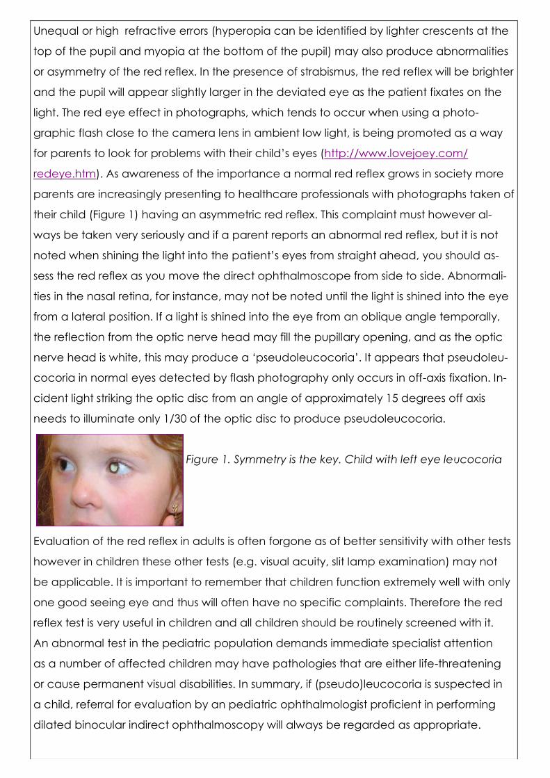
Unequal or high refractive errors (hyperopia can be identified by lighter crescents at the
top of the pupil and myopia at the bottom of the pupil) may also produce abnormalities
or asymmetry of the red reflex. In the presence of strabismus, the red reflex will be brighter
and the pupil will appear slightly larger in the deviated eye as the patient fixates on the
light. The red eye effect in photographs, which tends to occur when using a photo-
graphic flash close to the camera lens in ambient low light, is being promoted as a way
for parents to look for problems with their child’s eyes (http://www.lovejoey.com/
redeye.htm). As awareness of the importance a normal red reflex grows in society more
parents are increasingly presenting to healthcare professionals with photographs taken of
their child (Figure 1) having an asymmetric red reflex. This complaint must however al-
ways be taken very seriously and if a parent reports an abnormal red reflex, but it is not
noted when shining the light into the patient’s eyes from straight ahead, you should as-
sess the red reflex as you move the direct ophthalmoscope from side to side. Abnormali-
ties in the nasal retina, for instance, may not be noted until the light is shined into the eye
from a lateral position. If a light is shined into the eye from an oblique angle temporally,
the reflection from the optic nerve head may fill the pupillary opening, and as the optic
nerve head is white, this may produce a ‘pseudoleucocoria’. It appears that pseudoleu-
cocoria in normal eyes detected by flash photography only occurs in off-axis fixation. In-
cident light striking the optic disc from an angle of approximately 15 degrees off axis
needs to illuminate only 1/30 of the optic disc to produce pseudoleucocoria.
Figure 1. Symmetry is the key. Child with left eye leucocoria
Evaluation of the red reflex in adults is often forgone as of better sensitivity with other tests
however in children these other tests (e.g. visual acuity, slit lamp examination) may not
be applicable. It is important to remember that children function extremely well with only
one good seeing eye and thus will often have no specific complaints. Therefore the red
reflex test is very useful in children and all children should be routinely screened with it.
An abnormal test in the pediatric population demands immediate specialist attention
as a number of affected children may have pathologies that are either life-threatening
or cause permanent visual disabilities. In summary, if (pseudo)leucocoria is suspected in
a child, referral for evaluation by an pediatric ophthalmologist proficient in performing
dilated binocular indirect ophthalmoscopy will always be regarded as appropriate.

Aetiology
When leucocoria has been detected an extensive differential diagnosis must be consid-
ered. The most ominous cause is retinoblastoma and, as the most common intraocular
malignancy in childhood (affecting between 1/15,000 and 1/20,000 children), must
always be sought out and specifically excluded. In a study of over 1,500 retinoblastoma
patients with no family history of the disease, the presenting sign was leucocoria in 57%.
A posterior pole tumour as small
as 3-5mm in diameter has the ca-
pacity of cause leucocoria and
parents may refer to this as a
‘cat’s eye’. They may also only
notice it intermittently and only in
low lighting when the pupil dilates
naturally. The combination of
the near reflex and bright lighting
almost always defeats the repro-
ducibility and leaves parents
questioning their earlier observa-
tion. Intraocular retinoblastoma is
very treatable, but the mortality
for metastatic disease is high, thus
identification of tumours before
systemic spread is critical. Most
cases (>90%) are diagnosed prior
to the age of four years. The Inter-
national Intraocular Retinoblas-
toma Classification system (ABC
Classification—see table 1) is the
one most commonly used, with
classification stages based
according to the prognosis after
first-line chemotherapy and
adjuvant focal therapy.
Group A: small tumors away from foveola & disc
• Tumors <3 mm in greatest dimension
confined to the retina and…
• Located at least 3 mm from the foveola and
1.5 mm from the optic disc
Group B: all remaining tumors confined to the
retina
• All other tumors confined to the retina and
not in group A
• Subretinal fluid (without subretinal seeding)
< 3 mm from the base of the tumor
Group C: local subretinal fluid or vitreous seeding
• Subretinal fluid alone >3 mm and < 6 mm
from the tumor
• Vitreous or subretinal seeding < 3 mm from
the tumor
Group D: diffuse subretinal fluid or seeding
• Subretinal fluid > 6 mm from the tumor
• Vitreous or subretinal seeding > 3 mm from
the tumor
Group E: presence of any one or more of these
poor prognosis features
• More than 2/3 of the globe filled with tumor
• Tumor in the anterior segment or anterior to
the vitreous
• Tumor in or on the ciliary body
• Iris neovascularisation
• Neovascular glaucoma
• Opaque media from hemorrhage
• Tumor necrosis with aseptic orbital celullitis
• Phthisis bulbi
Table 1: The ABC Classification System.
For further classification systems and
more detail on retinoblastoma, click here.
In addition to leucocoria, the clinical features of retinoblastoma include strabismus, ocu-
lar pain from rubeosis iridis with secondary glaucoma, anterior displacement (dislocation)
of the ocular lens, heterochromia iridis, hyphaema, cellulitis, uveitis, endophthalmitis, and
pseudoinflammation and increase in the size of the ocular globe.
The most common cause of leucocoria worldwide is cataract. In a tertiary hospital setting
in Pakistan, 60% of the 39 patients under 10 years of age who presented with leucocoria
had congenital cataract (18% unilateral and 42% bilateral). Retinoblastoma was seen in
18% of children (11.2% unilateral and 7% bilateral). In another study, leucocoria was
caused by congenital cataract in 35%, followed by ocular malformations in 18%
(persistent hyperplastic primary vitreous, coloboma, disc anomaly, combined develop-
mental abnormalities), hereditary vitreoretinal disorders in 13%, retinopathy of prematurity
in 12%, trauma-associated diseases in 8% and retinoblastoma in 6%). Following in
frequency were inflammatory disorders (5%) and Coats’ disease (2%).
Remember, the retinoscope is a very useful instrument to pick up small lens opacities
which blacken the red reflex. The more posterior, central and sizable the lens opacity the
more likely it is to cause degradation of the retinal image. Generally, it is regarded that if
the disc is visible with a direct ophthalmoscope approximately 6/18 vision is attainable.
Unlike in the adult patient, the finding of a lens opacity in the paediatric population
constitutes an urgent referral for specialist care. On one hand, systemic problems are
more commonly detected in those bilaterally affected, but on the other, over half (51%)
of unilateral cataracts are associated with ocular abnormalities. This compares to 12% in
children bilaterally affected. Final visual acuties (VA) in infants identified and operated
with significant bilateral cataract are better than in those with unilateral cataract.
Long term studies of operated unilateral cataract show that approximately a third can
achieve a VA better than logMAR 0.6. Timing for congenital cataract is important and
the most treatment regimens are based on surgery within two months of birth combined
with prompt optical correction of the aphakia and aggressive occlusion therapy with
frequent follow-up.
There are a range of other causes of leucocoria and evaluation should follow in an
orderly progression. Just some of the features in the history and examination that can
be used to help identify the aetiology include; age of onset (for example if bilaterally
affected age when abnormal visual behaviour was first noticed), history of premature
birth could lead to diagnosis of retinopathy of prematurity causing retinal detachment,
general health questions are important (for example if determining a metabolic disorder
that may be causing the congenital cataract), family history is particularly important (a
family member who had removal of an eye at an early age could indicate retinoblas-
toma) laterality (strabismus is common in the unilaterally affected, so age when squint
noticed), the size of eye (leucocoria with microphthalmia may suggest persistent hyper-
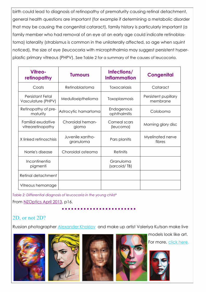
plastic primary vitreous (PHPV). See Table 2 for a summary of the causes of leucocoria.
Table 2: Differential diagnosis of leucocoria in the young child*
From NZOptics April 2013, p16.
2D, or not 2D?
Russian photographer Alexander Khoklov and make up artist Valeriya Kutsan make live
models look like art.
For more, click here.
Vitreo-
retinopathy Tumours
Infections/
Inflammation Congenital
Coats Retinoblastoma Toxocariasis Cataract
Persistant Fetal
Vasculature (PHPV) Medulloeipthelioma Toxoplasmosis
Persistent pupillary
membrane
Retinopathy of pre-
maturity Astrocytic hamartoma
Endogenous
ophthalmitis Coloboma
Familial exudative
vitreoretinopathy
Choroidal heman-
gioma
Corneal scars
(leucoma) Morning glory disc
X linked retinoschisis Juvenile xantho-
granuloma Pars planitis
Myelinated nerve
fibres
Norrie's disease Choroidal osteoma Retinitis
Incontinentia
pigmenti
Granuloma
(sarcoid/ TB)
Retinal detachment
Vitreous hemorrage
Practice management
This is the time of year when we typically plan for the future,
and may undertake to focus more on practice management.
It’s all good and well to plan sweeping changes, but we sel-
dom have the time to see it through with the increasing admin-
istrative demands in every sphere of practice. My recommen-
dation is to pick a few measures that are easily obtained from
your accounting or practice management software and focus
on your prescribing and dispensing to influence them. Get an
idea of where you are now, set your goal figures and then
decide how to get there. These figures may be useful:
1. Income
Income in this case is not what you’ve invoiced, but rather the
money banked or received from medical aids. You can meas-
ure it daily, weekly or monthly. Compare it with the previous
period (e.g. last month) to see trends and the previous year
(e.g. last January) to see growth. This is a more ‘real’ figure than
your turnover because mistakes in invoicing and discount
allowed are already reckoned in.
2. Average cost per visit.
Patients come in for our profes-
sional services—examinations,
CL follow-ups, vision therapy or further
investigation (fundus photos, visual fields etc) — and often get
spectacles, CLs or even accessories. What is the average
spend per patient? If you have more than 1 optometrist or dis-
pensing optician in the practice, you can calculate the aver-
age per provider. Differences within the practice may be due
to different patient profiles, but may also be due to different
prescribing patterns. It’s easy to become stuck in a rut and for-
get to suggest products or features that may benefit your pa-
tient such as ARC, near-support lenses, multiple pairs, photo-
chromic lenses or sunglasses. Some of us may be steering pa-
tients away from premium products and additional services out
of concern for costs, doing the patient a disservice.
Premium frames
Premium frame and
lens products require
a little extra explain-
ing. Expect added
attention to workman-
ship with techniques
and materials that are
more durable, designs
more detailed and
thoughtful, the fit
more ergonomic.
Like anything that is
made with a little
extra thought, care &
attention, a premium
frame promises to de-
liver fashion, comfort
and luxury. These
Etnia frames are
textured to resemble
wood and mother-of-
pearl, but in flattering,
modern colours.

Etnia’s children’s
frames are designed
to fit the child’s devel-
oping nose bridge
while delivering the
look they want.
The bigger, plastic
styles are all repre-
sented, with enough
colour to be adven-
turous, but not out-
landish. Try the jewel
toned matt turquoise,
blue & purple options
or the fiery reds and
oranges.
Etnia is stocked by
Optic Nerve. Contact
Jan on 082 327 2556 or
Make your clinical recommendation and explain that cheaper
alternatives do exist. Premium frame and lens products require
a little extra explaining. The materials, workmanship, techniques
and coatings are more durable, the designs more thoughtful
and ergonomic. Made with a little extra care and attention, a
premium frame promises the wearer fashion, comfort & luxury.
To get your average, simply take the number of appointments
you had in a day/ week/ month and divide it by the total turn-
over. If you are more likely to do several CL follow-ups (at no
extra cost) and your colleague does not, it will show in your
average. Extra exams are great if you have the time, and
spend that chair time educating your patient, but consider
whether you are charging enough for the initial visit to justify
the extra time. If you are consciously doing it as a practice
builder, or because you enjoy challenging contact lens fittings,
for example, that’s great but too many of us undervalue our
professional time.
The senior (oh, heck, let’s call me older) optometrist can com-
pare average per visit when a new associate joins the practice.
In the US, the average is about R3300 per visit, of which about
R500 to R800 is for the examination. Our margins on product are
comparatively higher in South Africa, while our examinations
are inexpensive. The US average for patients who get specta-
cles is around R5500.
3. Turnover per optometrist/ dispenser/ staff member
If the optometrist does not work full time, work out the equiva-
lent: A part time optometrist that works 1 day a week does 8
out of 40 hours in the week (or 45 hours if you work Saturdays)
and so should generate about 20% of the turnover. A rule of
thumb for staff is to have 1 staff member per R1M turnover. If
some of your staff are part time, calculate their equivalent as
above. A practice with an annual turnover of R1.5M should
manage on 1 full time and 1 part time staff member. This
depends on your mode of practice and how much of the
burden your practice assumes in terms of checking medical aid

benefits. If you can identify to your own satisfaction the reasons
why you need extra staff, or fewer staff members, it’s perfectly
acceptable. It’s more useful to understand the consequences
of your decisions than to match your figures to industry norms.
Hiring new staff? In the US about 46% of new
staff members are recruited by word-of-
mouth, followed by 28% via the internet,
11% by newspaper and 4% through recruiters.
These are guidelines only, but it can be very stressful when staff
have too much or too little to do. Both lead to poor service and
patient care.
4. Extras
We tend to examine, prescribe & dispense as if the medical aid
benefit is a limit. If the medical aid does not cover a glaucoma
assessment, do you still do pachymetry, tonometry, fundus
photographs and a visual field assessment? Do you charge
extra for that? Most of us do not. If we do that once a day, and
if we had charged a minimal amount—say R50—it works out to
R1000 per month that we are losing. That would pay off your
pachymeter!
To explain additional tests that incur further costs means we
have to explain things to patients, so appropriate billing ensures
we do the right battery of tests when necessary and educate
the patients about them. If we don’t charge for them, we’d
probably hurry through them, or not do them at all. The SAOA
will develop codes for additional professional investigations and
has already introduced the idea to some of the bigger medical
aids. Charging appropriate fees for the services you render is in
the best interest of the patient.
What’s in the mix?
The frames you stock
depend on the area
you practice in, the
medical aids you’ve
contracted with and
your patient demo-
graphic. The personal
taste and preferences
of your frame buyer(s)
definitely play a role.
MBA-CE suggests the
following mix for your
displays:
They also recommend
1/3 plastic and 2/3
metal, but I disagree.
It depends what your
patients are request-
ing, and I’m finding
plastic in higher de-
mand now.
Finally, your relation-
ship with your supplier
counts.
Is there back-up?
Do they carry
stock?
How is their service?
Unique product?

Vision related quality of life.
Measuring the impact of our interventions is important not only to clinicians, but also to
funders who generally seek the maximum benefit at the minimum price. Already we are
expected to motivate the supply of spectacles to children and contact lens wearers,
multifocals to emmetropic presbyopes and near support lenses to non-presbyopes. We
all know that there’s more to an Rx than the numbers, but how do we define it?
Researchers at the University College of London (UCL) are measuring the vision related
quality of life and functional vision of children and young people with visual impairment
in order to develop an age-appropriate patient reported outcome measure (PROM) for
epidemiological purposes. ‘In keeping with the increasing emphasis on PROMs for patient
-led assessment of impact of illness or disability and of health care, we are developing
questionnaires that children and young people with visual impairment can self-complete
and which can be used to measure the real-life impact of living with a visual disability.
These are intended to be complementary to the objective clinical measures (e.g. acuity)
in clinical settings, as well as to play part of policy in research, thus allowing children and
young people’s own perspectives of their visual loss to form part of healthcare provision
and decision-making.’ Currently completing the foundation research for 10-15 year olds,
the UCL has been awarded a 3 year grant by Fight for Sight and will develop instruments
for 6-9 year olds and 16-18 year olds as well.
Refractive services and spectacles are provided at no cost to Omani school children.
A recent study in the Omani Journal of Ophthalmology evaluated the impact of
compliance of spectacle wear on the vision related quality of life (VFQ) among 7th grad-
ers (12-13 years old) and 10th graders (15-16 years old).
Students were interviewed 1 year after having spectacles prescribed/ supplied, and clas-
sified as compliant if they still wore their spectacles (n=124), non-compliant if they did not
(n=124). The overall VFQ was found to be significantly higher among the compliant group
compared with the non-complaint group. The association of gender and the grade level
was not significant (P > 0.05) but the geographical location was significantly associated
to the difference of VFQ in both groups. I don’t have an explanation for this, except that
there areas may be more rural or urban and may have present different visual needs. The
VFQ related to the near work (RR = 1.3) and school related activities (RR = 1.3) was gener-
ally better in the compliant group. The article mentions that the low uptake of spectacle
wear is a concern: about 64% in primary and 80% in secondary school students. If cost is
not a deterrent, education may help to overcome the inevitable teasing and also for

parents and teachers to understand the importance of specta-
cles (e.g. for high hyperopia) that do not obviously improve VA.
Visual quality of life in children with refractive error was also
considered in a Spanish study reported in March 2013 in Eye
and Contact Lens. Subjects were slightly younger (6 -12 years
old) and all were myopic (-0.75D to –4.00D with <1.00DC.)
Children were prospectively assigned to either myopia control
with orthokeratology (OK) CLs or single vision spectacles and
re-assessed at 12– and 24 months to evaluate children's per-
ceptions in terms of overall vision, near vision, distance vision,
symptoms, appearance, satisfaction, activities, academic
performance, handling, and peer perceptions. The mean score
of all items was calculated as the overall score. Additionally,
parents/guardians were asked to rate their child's mode of
visual correction and their intention to continue treatment
after study completion.
Thirty-one children were fitted with OK lenses and 30 with SV
spectacles. The OK group rated overall vision, distance vision,
symptoms, appearance, satisfaction, activities, academic per-
formance, handling, peer perceptions, and the overall score
significantly better than children wearing SV spectacles
(all P<0.05). Near vision was better for the spectacle group and
obviously handling and care is not an issue. The results were
similar for the 12 month and 24 month follow up. Parents/
guardians of children wearing OK CLs rated visual correction
method and intention to continue treatment higher than par-
ents of children wearing SV spectacles (P≤0.01).
These studies confirm what we already know: Correction of
refractive error is a nuisance in spite of significantly improving
vision related quality of life. It’s important to consider health,
safety, lifestyle and maturity of the child when offering the
spectrum of refractive options to our pediatric patients and
their families.
Events & meetings March 2014
12-16 March
SECO
Atlanta, USA
16-17 March
Optometry Tomorrow
York, UK
May 2014
15-18 May
European Academy
of Optometry &
Optics
Warsaw, Poland
June 2014
6-9 June
British Contact Lens
Association Congress
Birmingham, UK
25-29 June
American Optometric
Association Congress
Philadelphia
USA
August 2014
13-15 August
SAOA Conference
and exhibition
Cape Town ICC
South Africa
Damien Hirst’s 2000 Mini at
Miami Design Week 2013.

Sobering statistics
StatsSA’s December/ January edition of Fieldworker reports that employment creation is
concentrated in smaller firms. Just over half of those who found jobs in 2012 did so at
small firms consisting of 0–9 employees. Most optometric practices fall within this sector.
According to StatsSA, the better educated employee is more likely to have a written
contract (98%), paid leave (89%), pension (79%), and medical aid (72%). Although South
African labour law does not require it, the HPCSA does require employed optometrists
and dispensing opticians to have a written contract. Also make sure that you personally
(not the practice or your employer) have professional indemnity cover. You, and not
your employer or the practice, will be held responsible in the event of a complaint. SAOA
members enjoy automatic professional indemnity cover in the practising categories.
Take it or ‘leave’ it SA has one of the highest absenteeism rates in the world during the festive season — up
to 20%, ranging from people who simply don’t show up for work, to those who take sick
leave or family responsibility leave
when they would generally not
have. Absenteeism in the public
sector is 3 times higher than in the
private sector and altogether it
costs SA over R12 billion per year.
The problem is experienced more
acutely by small enterprises (up to 9 employees) which don’t have the same redundancy
as bigger companies or government.
Employers: If you don’t close down, try to preserve morale with a staggered afternoon off
Employees: Labour law does allow for sick leave and family responsibility leave, but at a
time where the impact is acutely felt, your decision to take a personal day will not
ingratiate you with your employer and co-workers.
If you honestly do need to take sick leave at a peak time, discuss it with your employer,
produce supporting documentation even if it is not required and offer to make up
another time. Employers may conduct a return-to-work interview and document the
time and reasons for your absence in order to identify trends that are threatening to the
efficiency of the practice. Employers should keep accurate information on all leave
(contractual, general and sick– or family responsibility leave) taken, rather than making
unfounded decisions based on a vaguely remembered dodgy excuse last Christmas.
Absenteeism is an
employee’s intentional
or habitual absence
from the workplace.

The Basic Conditions of Employment Act, No 75 of 1997 (BCEA) regulates the leave of all
employees except
those who work less than 24 hours per month,
those who work as travelling salespersons, and
those who are not in senior managerial positions. (Senior managers (despite their job
descriptions) are those that can hire, fire and discipline).
Employees are entitled to 21 continuous days’ paid annual leave. Effectively, it’s 3 weeks’
leave. If they work only on a Monday, they get the equivalent i.e. 3 Mondays leave. The
purpose of the Act is to ensure that employees are given time to rest and be with their
families, not to place a monetary value on the leave. Leave must be taken no later than
6 months after the end of the leave cycle. It cannot be paid out by the employer unless
the employee is leaving the position.
Faan Coetzee of Cliffe, Hofmeyr Dekker says: ‘Employers are well advised to ensure that
leave is taken and that employees enforce their right to take leave.’ In a recent (Ludick v
Rural Maintenance (Pty) Ltd) judgment, the Labour Court ordered an employer to pay an
employee on termination of his contract all outstanding leave not taken. Employers
should note that leave not taken during the cycle is not automatically forfeited nor is any
right to payment in respect of that leave forfeited, at the very least for the last cycle.
Employers are expected to make a copy of the
Basic Conditions of Employment Act available to
all employees so that all are clear on their
rights and obligations. It can be
downloaded in full or is available in
a poster summary from most printers
and stationery suppliers.
At least we don’t typically have strike
action in our professions: South African
productivity is under significant pressure
due to rampant strike activity.
Family responsibility leave
of 3 days per year only when...
their child is born, or is sick (not
when the babysitter takes leave!)
at the death of a spouse/ life part-
ner/ parent/ adoptive parent/ grand
-parent, child/ adoptive child,
grandchild or sibling.
Employer may want proof
that leave was needed.
How do we learn the names of colours?
We’ve seen how complex a process it is to name colours. How do we learn to do it?
A Stanford language acquisition study investigates: For the experiment, the child is made
comfortable in the lab with some banter and then presented with 3 squares. He is asked
to pick the red one, then the blue one. The colours offered are not close or potentially
confusing (e.g. no red/ orange selections) - the point is not to trick or intimidate the child.
Although parents of participating children are generally confident that their children
know their colours, most 2 year olds fail this test, sometimes to the enormous distress and
concern of the parents who worry that this suggests colour vision problems. The study re-
ports that ‘[d]ivorced from context, most two and three-year olds might as well be color
blind; certainly they look that way when asked to correctly identify colors in a line-up, or
accurately use color words in novel contexts. What’s more, psychologists have found that
even after hours and hours of repeated training on color words, children’s performance
typically fails to noticeably improve, and children as old as 6 continue to make major
color naming errors.’ Considering that many 6 year old children can ride a bike and oper-
ate remote controls that are Greek to me, this seems incompatible. Why does it takes so
long for children to learn colours and when should we worry?
Firstly, colour is not universal. Some languages do not have different words for different
colours (e.g. in Sesotho, ‘-tala’ can be used for blue or green. In isiXhosa there is no word
for blue and speakers may borrow from Afrikaans (blou) or otherwise simply substitute
green (-hlaza.) The context usually makes the distinction clear. The Himba word ‘zoozu’
covers several dark colours including black, green, blue and purple while ‘serandu’
encompasses much of pink, purple and red. The problem, then, is not only learning
the word, but learning which particular (or general) hue goes with it.
Colour is everywhere, and that doesn’t help. If the child is to learn to distinguish between
a chicken and a bird, the context of farm (for chicken) or garden (for bird) may offer
helpful clues. Colour is not restricted to one place, so context is not helpful. This is
probably why children learn nouns before colours.
The way we speak doesn't help either. We tend to use colour words prenomi-
nally, i.e. before the nouns. When a child listens to speech, s/he is likely to hear
‘blue’ before ‘balloons’, and for a brief moment has nothing to connect the
blue to. Had we talked about ‘balloons that are blue’, the cognitive process
and language would be a better match. There’s a lot of fascinating language acquisition
theory if you follow the hyperlinks at Scientific American. There’s an older one on how
children contextually solve words they don’t know when they hear them in speech.

Questions: Issue 1 (2014)
2 CPD points have been applied for.
Please submit answers online or to [email protected], including your name & HPCSA number.
True or false?
1. The Stroop effect describes the tendency for an unchanging stimulus, steadily fixated
upon for some time, to fade and disappear with time.
2. When the distribution of a skill or ability is displayed on a bell curve, it shows the age at
which the skill is learnt.
3. Tolerance of pain is influenced by the wavelength of light shining on the area of trauma.
4. The brain’s ability to subconsciously compensate for unequal illumination ensures a more
accurate perception of colours and shades.
5. The Bruckner test is performed with a binocular indirect ophthalmoscope (BIO) from about
arm’s length.
6. A white pupil reflex is called leucocoria.
7. A Bruckner test will identify traumatic, but not neurological anisocoria.
8. Apparent anisocoria on Bruckner test (a brighter and larger red reflex in one eye) may be
due to strabismus of that eye.
9. A Bruckner test can identify refractive error, ametropia, strabismus & media opacities.
10. A photograph taken with flash from about 15 degrees of the visual axis can catch the
optic disc and cause pseudoleucocoria.
11. Although the most common cause of leucocoria is cataract, any child presenting with (or
parents reporting) leucocoria should be carefully worked up for retinoblastoma.
12. Onset of retinoblastoma peaks in the early teens.
13. VA of 6/60 should be attainable if the disc can be viewed by direct ophthalmoscopy.
14. Practice managers should aim to match his/ her practice figures to industry norms.
15. Children use context to confirm memorised words, especially nouns.
16. Practice income is the total of invoices created plus cash sales.
17. The Basic Conditions of Employment Act specifies that staff who work Monday to Friday
should effectively get 15 working days paid leave per year.
18. Affordability is the primary driver of compliance with spectacle wear in children.
19. Studies suggest that parents and children are more compliant in the long term with orthoK
lenses for myopia than single vision spectacles.
20. Joe’s aunt has died. His absence from work to
travel to and attend her funeral should be noted as
family responsibility leave.