Optogenetic investigation of neural circuits underlying...
16
To improve understanding of psychiatric and neurologi- cal disorders, it will be important to identify the under- lying neural circuits, to pinpoint the precise nature of the causally important aberrations in these circuits and to modulate circuit and behavioural dysfunction with precise and specific experimental interventions. However, such a deep, circuit-level understanding of neuropsychiatric disorders, or indeed even of normal CNS circuit function, has been challenging to achieve with traditional methods. The complexity of neural cir- cuitry has historically precluded the use of genetically and temporally precise manipulations to probe detailed mechanisms of function and dysfunction. Optogenetics 1,2 describes the now widespread use of microbial opsins 3 , or related tools 4 , that can be acti- vated by illumination to manipulate cells with high specificity and temporal precision 5–7 even within intact tissue or behaving animals 8–11 . Here, we briefly review how optogenetic approaches have been used to dissect neural circuits in animal models of symptoms that are relevant to fear, anxiety, depression, schizophrenia, addiction, social dysfunction, Parkinson’s disease and epilepsy. Successful probing of complex diseases in this way will depend on the validity of animal mod- els used to identify the crucial circuit elements and activity patterns that are involved in each cluster of symptoms, and the precision and efficiency of inter- ventions designed to selectively target these elements or patterns. Therefore, we also discuss new strategies for targeting opsins to specific cells or circuit ele- ments and principles for integrating optogenetics with electrophysiological, pharmacological and behavioural assessments. We also highlight the advantages and practical limitations of these approaches for the study of psychiatric and neurological disease. Technological advances in optogenetics The optogenetic toolbox includes a rapidly expand- ing array of available opsin variants that offer both distinct advantages and individual limitations in controlling cellular activity or signalling 3,12–21 . Other important components of the toolbox are light-delivery methods 6,9,22–28 , targeting strategies 16,29–31 and trans- genic rodent lines that increase the range of available specific cellular targets 32–34 . For example, the recent development of devices 35,36 and transgenic rat lines 37 that facilitate integration of optogenetic techniques with measures of neural activity have advanced the application of optogenetic tools to investigate the neu- ral bases of complex behaviours that are relevant to neuropsychiatric disease. Integration of optogenetics with mapping techniques. The recent integration of fMRI with optogenetic manip- ulation, now referred to as ofMRI, has not only vali- dated a previously assumed interpretation of the fMRI BOLD signal 38 (that increased neuronal activity in local excitatory neurons can causally trigger, rather than simply correlate with, an increase in the local BOLD signal) but has also shown that it is possible to assay the effects of precise optogenetic manipulations on global brain activity. Given that many neuropsychiatric 1 Department of Bioengineering, Stanford University, 318 Campus Drive, Clark Center, Stanford, California 94305-5444, USA. 2 Picower Institute of Learning and Memory, Department of Brain and Cognitive Sciences, Massachusetts Institute of Technology, Cambridge, Massachusetts 02139-4307, USA. 3 Department of Psychiatry, Stanford University, 401 Quarry Road, Stanford, California 94305-5717, USA. 4 CNC Program, Stanford University, Stanford, California 94305, USA. 5 Howard Hughes Medical Institute, Stanford University Medical School, Stanford, California 94305-5323, USA. e-mails: [email protected]; [email protected] doi:10.1038/nrn3171 Opsins Membrane-bound proteins that can incorporate small organic ‘retinal’ molecules to become a light receptor. Optogenetic investigation of neural circuits underlying brain disease in animal models Kay M. Tye 1,2 and Karl Deisseroth 1,3,4,5 Abstract | Optogenetic tools have provided a new way to establish causal relationships between brain activity and behaviour in health and disease. Although no animal model captures human disease precisely, behaviours that recapitulate disease symptoms may be elicited and modulated by optogenetic methods, including behaviours that are relevant to anxiety, fear, depression, addiction, autism and parkinsonism. The rapid proliferation of optogenetic reagents together with the swift advancement of strategies for implementation has created new opportunities for causal and precise dissection of the circuits underlying brain diseases in animal models. NEURAL CIRCUITS REVIEWS NATURE REVIEWS | NEUROSCIENCE VOLUME 13 | APRIL 2012 | 251 © 2012 Macmillan Publishers Limited. All rights reserved
Transcript of Optogenetic investigation of neural circuits underlying...
To improve understanding of psychiatric and neurologi-cal disorders, it will be important to identify the under-lying neural circuits, to pinpoint the precise nature of the causally important aberrations in these circuits and to modulate circuit and behavioural dysfunction with precise and specific experimental interventions. However, such a deep, circuit-level understanding of neuropsychiatric disorders, or indeed even of normal CNS circuit function, has been challenging to achieve with traditional methods. The complexity of neural cir-cuitry has historically precluded the use of genetically and temporally precise manipulations to probe detailed mechanisms of function and dysfunction.
Optogenetics1,2 describes the now widespread use of microbial opsins3, or related tools4, that can be acti-vated by illumination to manipulate cells with high specificity and temporal precision5–7 even within intact tissue or behaving animals8–11. Here, we briefly review how optogenetic approaches have been used to dissect neural circuits in animal models of symptoms that are relevant to fear, anxiety, depression, schizophrenia, addiction, social dysfunction, Parkinson’s disease and epilepsy. Successful probing of complex diseases in this way will depend on the validity of animal mod-els used to identify the crucial circuit elements and activity patterns that are involved in each cluster of symptoms, and the precision and efficiency of inter-ventions designed to selectively target these elements or patterns. Therefore, we also discuss new strategies for targeting opsins to specific cells or circuit ele-ments and principles for integrating optogenetics with
electrophysiological, pharmacological and behavioural assessments. We also highlight the advantages and practical limitations of these approaches for the study of psychiatric and neurological disease.
Technological advances in optogeneticsThe optogenetic toolbox includes a rapidly expand-ing array of available opsin variants that offer both distinct advantages and individual limitations in controlling cellular activity or signalling3,12–21. Other important components of the toolbox are light-delivery methods6,9,22–28, targeting strategies16,29–31 and trans-genic rodent lines that increase the range of available specific cellular targets32–34. For example, the recent development of devices35,36 and transgenic rat lines37 that facilitate integration of optogenetic techniques with measures of neural activity have advanced the application of optogenetic tools to investigate the neu-ral bases of complex behaviours that are relevant to neuropsychiatric disease.
Integration of optogenetics with mapping techniques. The recent integration of fMRI with optogenetic manip-ulation, now referred to as ofMRI, has not only vali-dated a previously assumed interpretation of the fMRI BOLD signal38 (that increased neuronal activity in local excitatory neurons can causally trigger, rather than simply correlate with, an increase in the local BOLD signal) but has also shown that it is possible to assay the effects of precise optogenetic manipulations on global brain activity. Given that many neuropsychiatric
1Department of Bioengineering, Stanford University, 318 Campus Drive, Clark Center, Stanford, California 94305-5444, USA.2Picower Institute of Learning and Memory, Department of Brain and Cognitive Sciences, Massachusetts Institute of Technology, Cambridge, Massachusetts 02139-4307, USA.3Department of Psychiatry, Stanford University, 401 Quarry Road, Stanford, California 94305-5717, USA.4CNC Program, Stanford University, Stanford, California 94305, USA. 5Howard Hughes Medical Institute, Stanford University Medical School, Stanford, California 94305-5323, USA.e-mails: [email protected]; [email protected]:10.1038/nrn3171
OpsinsMembrane-bound proteins that can incorporate small organic ‘retinal’ molecules to become a light receptor.
Optogenetic investigation of neural circuits underlying brain disease in animal modelsKay M. Tye1,2 and Karl Deisseroth1,3,4,5
Abstract | Optogenetic tools have provided a new way to establish causal relationships between brain activity and behaviour in health and disease. Although no animal model captures human disease precisely, behaviours that recapitulate disease symptoms may be elicited and modulated by optogenetic methods, including behaviours that are relevant to anxiety, fear, depression, addiction, autism and parkinsonism. The rapid proliferation of optogenetic reagents together with the swift advancement of strategies for implementation has created new opportunities for causal and precise dissection of the circuits underlying brain diseases in animal models.
N E U R A L C I R C U I T S
R E V I E W S
NATURE REVIEWS | NEUROSCIENCE VOLUME 13 | APRIL 2012 | 251
© 2012 Macmillan Publishers Limited. All rights reserved
fMRI(Functional magnetic resonance imaging). This method can use detection of blood oxygen levels as a proxy for neural activity, and offers a non-invasive method to globally assay brain activity in humans.
BOLD(Blood oxygen level dependent). The BOLD signal is one kind of signal that fMRI can use to assess neural activity.
ChannelrhodopsinA light-driven cation channel, found in algae, that can be used to depolarize cell membranes.
HalorhodopsinA light-driven chloride ion pump found in phylogenetically ancient archaea, known as halobacteria, that can be used to hyperpolarize cell membranes.
UP statesSub-threshold membrane depolarization states that have been observed to spontaneously occur in vivo in some neurons and that may serve to increase the intrinsic excitability of the neuron.
Cre recombinaseDNA recombinase that excises DNA sequences flanked by loxP sequences with the same orientation, or inverts sequences flanked by loxP sites with opposite orientation. It is effective in mammalian cells in vitro and in vivo.
VectorsVehicles used to transfer genetic material to a target cell.
diseases are likely to involve distributed perturbations, global approaches such as ofMRI may be crucial for identifying and mapping the downstream effects of cell-type or projection-specific manipulations (in an unbiased fashion).
Local, detailed circuit-mapping has also benefited greatly from optogenetics. Continuing a long-standing tradition of mapping neural circuitry in mammals with optical approaches39, and in certain cases using new classes of light delivery40, several elegant optoge-netic studies have already made substantial advances in detailed circuit mapping41–43. These studies have helped to clarify the role of specific cortical layers in the regulation of activity flow, as well as to delineate the detailed pattern of synaptic inputs arising from distinct cortical layers onto distinct subcellular loca-tions in neocortical principal cells. By providing a rich source of information that would have been difficult or impossible to obtain by other means, these studies may lay the groundwork for identifying circuit or con-nectivity phenotypes that can go awry in disease states.
New opsin variants. Earlier optogenetic tools, such as channelrhodopsin 2 (ChR2)5,13 — which enables action potential elicitation to be time-locked to light pulses — or halorhodopsin (NpHR)16,18,44–46 and pro-ton pumps16,19,21 — which enable hyperpolarization of membranes to inhibit the production of action potentials — are still useful. However, the expan-sion of the optogenetic toolbox (FIG. 1) now provides greater flexibility in experimental design and more powerful and refined manipulations. For example, engineered channelrhodopsin variants (including the ChETA family20,21 and ChIEF47) can be used to evoke ultra-fast firing frequencies (up to 200 Hz or more) in fast-spiking neurons.
Although the ability to elicit action potentials that are time-locked to light pulses is powerful, the syn-chrony and patterning of an experimentally delivered illumination pattern may not represent the physiologi-cal neural code. OptoXRs4 (opsin–receptor chimae-ras in which the intracellular loops of rhodopsin are replaced with intracellular loops from other G protein-coupled receptors such as adrenergic receptors) now allow light-activated initiation of specific G protein-coupled signalling cascades in targeted neurons within freely moving mammals. This can lead to altered excit-ability in a population of cells without dictating precise neural spike times. Alternatively, to increase excitability of neurons without dictating a specific pattern of firing, it is possible to apply a long-lasting and subthreshold membrane depolarization, as seen in cortical UP states. The step-function opsin (SFO)17 facilitates this kind of intervention by delivering a prolonged, bi-stable, subthreshold depolarization of membranes.
New opsins such as the stabilized step-function opsin (SSFO)48, which is a double mutant of ChR2 (Asp156Ala and Cys128Ser), are a substantial improve-ment on the previous17 single mutant SFO (for example, Cys128Ser) in that the stability of the step function-like depolarization is greater — on the order of 30 min17,21,48.
As SSFO also has an enhanced sensitivity to light, it enables the non-invasive light-induced activation of SSFO-expressing neurons up to 3 mm below the sur-face of the brain when an optical fibre is placed just above the brain surface48. Thus, the development of the SSFO may facilitate research in large brain regions or in large-brained animals.
Another group of noteworthy opsins is the red-shifted activation wavelength ChR1/VChR1 chimaera (C1V1) family, which includes variants that are sig-nificantly more potent48 than ChR2 and approximately fourfold more potent than the Volvox channelrho-dopsin 1 (VChR1), a red-shifted opsin developed previously14. Members of the C1V1 family and their associated variants have a peak activation wavelength of ~560 nm and can readily be activated by 590 nm light, thus increasing the feasibility of combinatorial excitation or depolarization of different populations of neurons at distinct experimental epochs or patterns21,48. These opsins have been used to investigate behaviours that are relevant to autism and schizophrenia48, and are promising candidates for the study of other diseases of the brain.
Use of transgenic rodents in optogenetics. Tools for conferring genetic specificity to opsin delivery are also improving. A large number of Cre recombinase-driver mouse lines have already been used in optogenetic research (reviewed in REF. 6). Although transgenic mouse lines have proven to be very useful in the study of cells, circuits and behaviours that are relevant to disease31,48–55, the more complex behavioural and elec-trophysiological assays available in rats could provide important additional insight. However, optogenetic research in rats has been hampered by a lack of genetic tools for targeting opsins to specific cell types that are implicated in disease. The recent development of two Cre recombinase-driver rat lines targeting tyrosine hydroxylase and choline acetyltransferase neurons, with cell-type-specific promoter or enhancer regions that were too large to package into most vectors37, has addressed this need and may facilitate research into animal models of psychiatric and neurological disease states. These, and other new transgenic animals, will set the stage for integrated studies combining in vivo electrophysiological and optogenetic studies during sophisticated behavioural assays.
Optogenetics in behavioural studiesOptogenetic approaches have already begun to alter the way in which traditional behavioural assays are used. In addition to cell-type-specific and projection- specific targeting strategies that allow unprecedented precision in manipulation, the temporal properties of optogenetics are reshaping the style of experimental design. For example, the immediate ‘reversibility’ of optogenetic manipulations, compared to traditional pharmacological techniques that require a lengthy wash-out period, allows multi-day tests to be con-densed into a single session. The elevated plus maze with optogenetics now allows within-subject
R E V I E W S
252 | APRIL 2012 | VOLUME 13 www.nature.com/reviews/neuro
© 2012 Macmillan Publishers Limited. All rights reserved
Nature Reviews | Neuroscience
VChR1
NpHR
ArcheBR
400
450
500
550
600
650
E123A
E123TChIEF T159C
ChRGR*
E123T/T159C H134R
Rh-CT
bPACBlaC
C1V1
Step function opsin (bi-stable depolarization)
Fast excitation
Fast inhibition
Biochemical modulation
WT
Peak
act
ivat
ion
λ (n
m)
a
b
1 ms 10 ms 100 ms 1 s 10 s 100 s 10,000 s1,000 s
1 min 30 minτoff
C1V1E162T
C1V1E122T/E162T
L132C*CatCH
ChR2C128T
Opto-β2Opto-α2
ChR2C128A
ChR2C128S
ChR2D156A
ChR2D156A/C128S
Hyperpolarizing
Red-shifted depolarizingBi-stable depolarizing
ChETA variants
Blue depolarizingBiochemical modulation
VChR1C123S
VChR1C123S/D151A
ChR NpHR BR/PR OptoXR
Na+CI– Ca2+ H+ K+
ATP ATP
cAMP
Bacterialcyclase
Gq Gs Gi
[InsP3]↑[DAG]↑
[cAMP]↑ [cAMP]↓
Conditioned place preference(CPP). A behavioural test in which an unconditioned stimulus is paired with one distinctive context and a neutral event is paired with a different context. Preference is determined by allowing the animal to move between the two contexts and measuring the amount of time spent in each context.
comparison of conditions (light on and light off) in a single behavioural session56. Similarly, paradigms such as conditioned place preference (CPP) were traditionally performed with a habituation day, a conditioning day
and a test day. As the traditional paradigm typically involved the animal being locked into one compart-ment of the chamber while receiving the treatment (such as a drug infusion) and then restricted in the
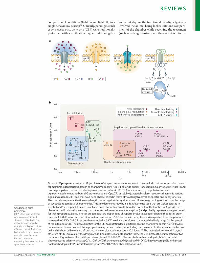
Figure 1 | Optogenetic tools. a | Major classes of single-component optogenetic tools include cation-permeable channels for membrane depolarization (such as channelrhodopsins (ChRs)), chloride pumps (for example, halorhodopsin (NpHR)) and proton pumps (such as bacteriorhodopsin or proteorhodopsin (BR/PR)) for membrane hyperpolarization, and light-activated membrane-bound G protein-coupled (OptoXR) or soluble (bacterial cyclase) receptors that mimic various signalling cascades. b | Tools that have been characterized in terms of wavelength activation spectra and decay kinetics. The chart shows peak activation wavelength plotted against decay kinetics and illustrates groupings of tools over the range of spectral and temporal characteristics. This also demonstrates why it is feasible to use tools that are well separated in spectral and/or temporal domains to achieve dual-channel control. It should be noted that the kinetics for OptoXR were characterized in vivo using an assay that measured a downstream readout (spiking) and probably represent an upper bound for these properties. Decay kinetics are temperature-dependent; all reported values except for channelrhodopsin-green receiver (ChRGR) were recorded at room temperature (an ~50% decrease in decay kinetics is expected if the temperature is increased to 37°C). ChRGR has only been studied at 34°C. We have therefore extrapolated the likely range for this protein at room temperature. The decay kinetics for the L132C mutation (calcium translocating channelrhodopsin (CatCH)) were not measured in neurons, and these properties may depend on factors including the presence of other channels in the host cell and the host cell tolerance of, and response to, elevated intracellular Ca2+ levels197. The recently determined198 crystal structure of ChR2 may allow the design of additional classes of optogenetic tools. The ‘/’ indicates the combination of two mutations. Figure is modified, with permission, from REF. 6 © (2011) Elsevier. Arch, archaerhodopsin; bPAC, bacterial photoactivated adenylyl cyclase; C1V1, ChR1/VChR1 chimaera; cAMP, cyclic AMP; DAG, diacylglycerol; eBR, enhanced bacteriorhodopsin; InsP
3, inositol trisphosphate; VChR1, Volvox channelrhodopsin 1.
R E V I E W S
NATURE REVIEWS | NEUROSCIENCE VOLUME 13 | APRIL 2012 | 253
© 2012 Macmillan Publishers Limited. All rights reserved

Nature Reviews | Neuroscience
Select opsin
Target cell type
Delivery of light
Set temporal parameters
Plan validation
Duty cycleThe time that a machine, system or light source spends in an active state as a fraction of the total time under consideration.
other compartment during the control treatment (such as saline infusion), the only behavioural ‘readout’ of the effect was on the test day. However, with the tem-poral agility of optogenetic techniques, the ‘condi-tioning day’ can now be performed with the animal moving freely and with light delivery only when the animal enters the conditioning chamber4. This ena-bles the experimenter to quantify a location preference for the animal during conditioning, thus enriching the data set by providing information about the time course over which a conditioned preference devel-ops. Furthermore, animals can be allowed to freely explore a chamber, and different light stimulation parameters (duty cycle, pulse frequency, pulse dura-tion and intensity) can be triggered depending on the animal’s location. This allows for a much finer measure of optimal stimulation parameters in comparison to different drug doses, which cannot be tested within a single session. This utility of the temporal resolution of optogenetics is particularly transformative in experi-ments that use temporally specific variables, such as discrete cues or tasks that have behaviours with critical time windows, such as a decision point in a T-maze.
However, as well as the new advantages that opto-genetic techniques offer, we are presented with several new limitations, caveats and considerations. One very important limitation to consider is the production of heat with illumination. When using high light powers
or duty cycles, the light that is emitted from the optical fibre may cause heating. Heating neurons may not only alter their activity in a nonspecific manner but may also be detrimental to cell health. Appropriate con-trols, as well as assessment of light source stability and performance, must be carefully and frequently exam-ined to ensure precise and reliable light output and interpretation of light effects6.
Another limitation of optogenetic tools is the poten-tial for toxicity at very high expression levels or long-term expression. As expression of a microbial opsin typically means the insertion of light-activated chan-nels or pumps into the cell membrane, there may be a maximal level of opsin expression that can be tolerated by a given cell. To determine whether opsin expres-sion has altered cell health, it is necessary to perform controls under the same conditions as the experimen-tal parameters. Cell health and all other performance parameters will vary with many parameters, including light intensity, virus titre, injection volume, vector, opsin, cell type, species and incubation time21. Finally, transient intracellular or extracellular ion balance changes (over seconds) may occur after modulated (or natural) activity patterns, and optogenetic experiments typically are designed with this in mind. It is important to consider the contribution of many parameters dur-ing the growth of this new field, and we recommend that experimenters empirically examine measures of interest in each new preparation.
In vivo optogenetic designDesigning in vivo optogenetic experiments for inves-tigation of the neural circuits underlying behaviour in both health and disease requires a unique set of consid-erations (BOX 1). In selecting an appropriate opsin gene for experiments, it is necessary to recognize the trade-offs that accompany each choice. For example, there is an inverse correlation between light sensitivity and off-kinetics21; volume of activation could be sacrificed in favour of greater precision of temporal control or vice versa. Thus, it is important to consider which proper-ties are most important for each experimental aim.
It is also important to determine the best targeting strategy (FIG. 2). One commonly used strategy is viral transduction, an approach that will initially limit opsin expression to the injection site. However, after some incubation time, viral transduction strategies enable ‘anterograde’ control capabilities6,7, meaning that the protein is expressed in local cell bodies and trafficked to the downstream terminals of those cell bodies; these projections can then be illuminated to control cells by virtue of their outgoing (efferent) connectivity. There are many promoters available for transducing viruses into wild-type animals6, and the most commonly used viral vectors are lentivirus and adeno-associated viruses. Other viruses that have potential utility include herpes simplex viruses (HSVs) or rabies viruses, which can infect axon terminals and present ‘retrograde’-like tar-geting opportunities for controlling afferents to a struc-ture; however, there may be more toxicity associated with these viruses.
Box 1 | Designing optogenetic experiments to study brain disease
There are at least five major steps in the design of optogenetic experiments to study behaviour in normal function or disease models (see the figure).
Select the opsin best suited to the experimental goals. There are trade-offs for different aspects of performance, such as peak photocurrent, kinetics, activation wavelength spectrum and light sensitivity.
Select targeting strategy or vector to express opsin in target cells. Many kinds of viral transduction can restrict cell-body expression of opsin to the injection site, and can be used in wild-type animals or Cre recombinase lines. Some transgenic lines constitutively express opsin.
Select light delivery method. Fibre optics are the most common method of light delivery to deep cell bodies or axon terminals. The numerical aperture, diameter and mode of the fibre will influence the spread of light. There are trade-offs for using acute or chronically implantable fibres, as acute fibres allow for pharmacological manipulations but are delicate and easy to break.
Choose appropriate temporal parameters. Duty cycle, pulse duration, frequency and epoch pattern are the key light-delivery parameters to select. Depending on the behavioural assay, exploring within-session light manipulations can maximize the utility of optogenetic tools.
Validate the experimental manipulation. To verify that the opsin, targeting strategy and illumination parameters are manipulating cells in the intended manner, confirmation using electrophysiology, immunohistochemistry or other measures is crucial for data interpretation.
R E V I E W S
254 | APRIL 2012 | VOLUME 13 www.nature.com/reviews/neuro
© 2012 Macmillan Publishers Limited. All rights reserved

Conditioned fear responsesA fear-associated stimulus (such as a shock-predictive tone) that may evoke conditioned responses such as freezing, fear-potentiated startle or increases in blood pressure, perspiration or heart rate.
For promoter sequences that are too large to package into a viral vector, Cre recombinase-driver lines used in conjunction with Cre-dependent opsin-expressing viral vectors offer an attractive alternative, as first demonstrated behaviourally in tyrosine hydroxylase (TH)::Cre mice31 and parvalbumin (PV)::Cre mice53,54. Many such targeting strategies exist29,33,57, and there are also transgenic mice that constitutively express ChR2 in certain types of cells32,33,58. Opsin expression in these mice is not subject to the variability that is associated with viral infection or the burden of a lengthy incu-bation time. These advantages may be particularly important for some applications; however, they are accompanied by the caveat that the illuminated region may contain not only local photosensitive targeted cell bodies but also photosensitive projections from cell bodies that are located elsewhere in the brain. Another recently developed strategy involves selectively infect-ing specific neuron types by using a viral vector that recognizes receptors on the exterior of the cell59,60.
Lastly, it is crucial to determine the most appropri-ate method for delivery of light into the brain. Optical fibres can be used either acutely (for example, a bare fibre that is itself a patch cable can be inserted using a guide cannula)9,10,25 or chronically (for example, an implantable optical fibre can be joined to a patch cable outside the brain)6,28. Acute optical fibres can deliver pharmacological agents to the same location as light56,61; however, because of the delicate nature of optical fibres, the risk of fibres breaking in the guide cannula is a substantial disadvantage. Although chroni-cally implantable fibres do not allow for integration with pharmacology at the same site, they do offer increased durability for multiple-day experiments6,28. Laser diodes are a widely used light source and are easy to couple to fibre optics6: light-emitting diodes (LEDs) can be used directly at the tissue (although there are associated problems owing to high local heat generation) or coupled to fibres in the same way as laser diodes (but with some light loss)22,26,62,63. Finally, once an opsin is expressed and light is delivered to the desired location, the light stimulation parameters should be considered. Possible issues include the effects of heating, light scattering and the physiological capac-ity of the targeted neurons. With so many variables, it is important to carefully validate — with imaging, physiology, or c-FOS staining — that neurons are being manipulated with the strength and specificity intended before interpreting any experimental results.
Circuitry of fear and anxiety disordersAnxiety disorders, which include generalized anxiety disorder, panic disorder, post-traumatic stress disorder (PTSD) and phobias, are the most common class of psychiatric diseases, with a lifetime prevalence of ~28% (REFS 64,65). Although anxiety disorders are common, the available treatments are inadequate in terms of efficacy and side effects66–68.
Anxiety is characterized by a sustained state of apprehension in the absence of an immediate threat65. By establishing methods for projection-specific
optogenetic targeting in rodent anxiety models, a spe-cific population of amygdala synapses has been identi-fied that can rapidly and reversibly modulate baseline anxiety levels in a freely moving mammal56. This study capitalized on the fact that microbial opsins can be expressed throughout the cell, including on the mem-branes of axons and axon terminals (a property that may be enhanced by the inclusion of specific target-ing sequences16), allowing axon depolarization to be generated simply by illumination of the axons them-selves. The study also used a bevelled light-delivery cannula to guide light selectively to axons projecting from one brain region to another. It was shown that selective optogenetic stimulation (with a channelrho-dopsin) of excitatory basolateral amygdala (BLA) cells with axons that project to the central nucleus of the amygdala (CeA) produced an anxiolytic effect. This phenotype was markedly different from the anxiogenic effect that was observed when BLA excitatory cell bod-ies were illuminated nonspecifically without regard to projection target56,69 (FIG. 3). Indeed, BLA projection neurons have many targets, including not only the centrolateral and centromedial nuclei of the amygdala but the nucleus accumbens (NAc), the prefrontal cortex, the bed nucleus of the stria terminalis and many other structures that could have fundamentally different (and even opposite) effects on anxiety. Thus, optogenetic projection targeting enabled resolution of this distinct endogenous pathway for anxiolysis in the amygdala. Moreover, selective illumination of these BLA–CeA axons expressing an enhanced version of the hyper-polarizing halorhodopsin, eNpHR3.0 (REF. 16), induced an anxiogenic effect, establishing that this cellular projection is capable of bidirectional modulation of behaviours that are relevant to anxiety56.
Panic attacks are briefer and more intense than anxi-ety episodes and are characterized by intense bursts of terror, apprehension and autonomic arousal, often with chronic consequences such as a debilitating fear of future attacks64,65,67. Interestingly, these can be triggered by isolated components (which are not threatening in isolation) of a context that was previously associ-ated with fear. Although it has long been thought that the lateral amygdala is a crucial region for processing fear70–74, it had not been demonstrated directly that activation of glutamatergic lateral amygdala neurons alone could induce a fear response. Optogenetic tools have now allowed researchers to selectively target the glutamatergic pyramidal neurons of the lateral amyg-dala, which also contains local GABAergic interneu-rons. Fear responses in mice were observed in response to light-induced activation of ChR2-expressing lateral amygdala neurons; moreover, pairing the presentation of a neutral stimulus (a tone) with lateral amygdala illumination led to the observation of conditioned fear responses to the tone alone69.
Fearful responses to specific stimuli or contexts can also include phobias, which can arise from exposure to harm or threat and represent the single largest sub-class of anxiety disorders, affecting up to 12% of the popu-lation67. Although many lesion and pharmacological
R E V I E W S
NATURE REVIEWS | NEUROSCIENCE VOLUME 13 | APRIL 2012 | 255
© 2012 Macmillan Publishers Limited. All rights reserved

a
Projectionc
f
b
e
Local somata
Recombinase- or promoter-dependent
Combinatorial local somata
Combinatorialprojection
d Projection termination
Nature Reviews | Neuroscience
A
BC A
BC A
BC
Laser
A
BC A
BC A
BC
Laser 1
Laser 2
A
BC A
BC A
BC
Laser
A
BC A
BC A
BC
Laser
A
BC A
BC A
BC
Laser 1
Laser 2
Light deliveryInjection site Viral expression
Virally encodedopsin
Single viral injection into B Opsin expression throughout B Illumination of B cell bodiesprojecting to A and C
Virally encodedrecombinase- or promoter-dependent opsin
Single viral injection intomixed population of neurons in B
Opsin expression only in neuronsexpressing recombinase orwith active promoter in B
Illumination of B cell bodies andmodulation of recombinase- or promoter-expressing cells
Virally encodedopsin
Single viral injection into B Opsin expression throughout B Illumination of B axons in A but not C. Corresponding cell bodies in B may be activated.
Mixture of virally encoded opsins
Mixed viral injection with spectrally separated opsins intomixed population of neurons in B
Opsin expression in B cell bodiesaccording to viral promoters or recombinase-dependent expression
Precisely temporally separable illumination of mixed neuronal populations in B with two colours of light
Viral injection of spectrally separated opsins into A and C
Opsin expression in A and C cellbodies according to viral promotersor recombinase activity
Illumination of mixed neuronalprojections in B activate independentaxons from A and C
A
BC A
BC A
BC
Laser
or
LaserVirally encodedlectin–recombinasefusion
Virally encoded recombinase-dependent opsin
Double viral injection into B and A.Recombinase expressed in A movestranscellularly to cells in B.
Opsin expression in B neuronsthat project to A (also achievablewith axon-transducing viruses)
Illumination of B cell bodiesor B axons in A withoutdirect modulation of C
Independentvirally encodedopsins
R E V I E W S
256 | APRIL 2012 | VOLUME 13 www.nature.com/reviews/neuro
© 2012 Macmillan Publishers Limited. All rights reserved

Figure 2 | Targeting strategies with optogenetic tools in vivo. a | Neuronal cell bodies can be directly stimulated by injecting a viral vector into the target region and implanting a local light-delivery device in the same region. b | Specific expression of the transgene in defined cell populations can be achieved by including cell-type-specific promoters within the viral vector or by injecting a recombinase-dependent virus into an animal that is engineered to express a recombinase (such as Cre) in particular cell types. c | The optogenetic tool can be targeted to axonal projections by injecting the virus at the location of neuronal cell bodies and delivering light to the target region harbouring opsin-expressing processes. d | In projection termination labelling, cells are targeted by virtue of their synaptic connectivity to the target region, probably excluding axons that are simply passing through the area. In the example shown, transcellular labelling is achieved using a recombinase-dependent system. The synaptic target site is injected with a virus expressing Cre that is fused to a transneuronal tracer (such as a lectin), and the cell body region is injected with a Cre-dependent virus. This results in cells that project to the Cre-injected area becoming light sensitive. Similar effects can be obtained using retrograde viruses (those that transduce the axon terminal), such as rabies or herpes simplex viruses (HSVs), although these approaches do not enable control over the postsynaptic cell type. e,f | Combinatorial manipulations at either neuronal somata (e) or projections (f) can be achieved with two different optogenetic tools that have well-separated activation spectra (responding to different wavelengths of light) and by using a light-delivery tool to merge multiple wavelengths of light. Figure is modified, with permission, from REF. 6 © (2011) Elsevier.
◀
studies have implicated the amygdala in the acquisi-tion and expression of conditioned fear 71,73–81, the intricate microcircuits in the amygdala have been difficult to causally dissect with traditional manipula-tions. Although optogenetic animal studies do not yet directly address panic disorder or phobias per se, by using optogenetic techniques to target protein kinase Cδ (PKCδ)-expressing neurons in a subnucleus of the lateral division of the CeA, researchers have recently found an inhibitory microcircuit in the CeA that gates the expression of conditioned fear82. Another study defined subpopulations of neurons in the CeA that are involved in the expression and generalization of con-ditioned fear83 (FIG. 3). These studies have highlighted the synergistic value of genetic and spatial targeting of optogenetic control by combining the focal injection of opsin-bearing viruses into the amygdala for spatial resolution with genetic targeting strategies (for exam-ple, targeting PKCδ cells within the amygdala) for cell type resolution. This approach has enabled researchers to determine the role of a particular cell type in the control of a symptom related to psychiatric disease in animal models.
PTSD is a class of pervasive anxiety disorder that can occur following a traumatic experience under specific conditions (for example, involving subjective helplessness) and is remarkably debilitating owing to its chronic influence on many aspects of social and occupational functioning and its resistance to treat-ment or extinction84–86. Indeed, an animal model of the processes that may be dysfunctional in PTSD is fear extinction, in which fear responses are gradually elimi-nated once the aversive stressor is no longer presented with the previously associated contextual stimulus86,87. An intriguing idea is that the fear associations in PTSD are pervasive and intractable to treatment because they are stored in several locations and circuits throughout the brain, with many independent and potent memory traces84,86,87. Taking this into
consideration, it is important to understand where long-term fear memories are stored and how they can be most effectively disrupted or reconfigured.
It has long been suggested that the hippocampus is important for the encoding of contextual fear88–91 but that long after consolidation (that is, in the ‘remote’ phase many weeks, months or years later) the memory is no longer stored in the hippocampus and is instead maintained in a distributed neocortical network such as in the anterior cingulate cortex92–96. However, in a recent study in which eNpHR3.0 was expressed in glu-tamatergic pyramidal neurons in the CA1 region of the hippocampus, it was shown that the hippocampus is indeed important for the expression of even remote fear memories. Neocortical networks are also important in this remote (long-term) phase and can be recruited to participate more heavily when the hippocampus is dys-functional97. In other words, even for remote fear mem-ories, the memory trace is stored in several locations that may be redundant, that may work especially hard to compensate for each other when needed and that therefore could require a number of simultaneous dis-tinct methods and approaches to resolve pathological forms of recall.
In summary, applying optogenetic tools to the study of fear and anxiety behaviours in rodents has yielded valuable and in many cases surprising results that would have been difficult or impossible to obtain with other methods. Fear conditioning is an excellent target for the early application of optogenetics to brain disease as it involves a simple, robust behavioural paradigm for which the underlying neural circuitry has been exten-sively characterized and is therefore ideal for validating novel techniques as well as advancing and refining our understanding of an emotional state that is perturbed in many diseases.
Circuitry of addictionDrug addiction is a chronic, relapsing condition charact-erized by compulsive drug seeking and substance use despite harmful consequences98–100. Addiction has been proposed to ‘hijack’ the brain’s natural reward sys-tem99–102; therefore, understanding the neural circuitry mediating reward processing may be crucial for under-standing the pathophysiology of addiction. Although it has long been known that the mesolimbic dopamine system is involved in reward processing103–107 and that the NAc is critically involved in both reward processing and other addiction-related behaviours100,101,105,108–110, the mechanistic processes and neural codes mediating these behaviours have been incompletely understood.
The motivation to understand these mechanistic processes spurred the development and application of new optogenetic targeting strategies. Promoters that could drive the expression of opsin genes specifically in dopamine neurons are too large to be packaged together with the microbial opsins into conventional viral vectors while maintaining functionality. However, as mentioned above, highly selective Cre-dependent viral vectors have been developed29 that enable opsins to be expressed exclusively in Cre+ cells. Notably, this
R E V I E W S
NATURE REVIEWS | NEUROSCIENCE VOLUME 13 | APRIL 2012 | 257
© 2012 Macmillan Publishers Limited. All rights reserved
BLA
CeL
CeM
LA
CeL
CeM
PAG
PKCδ–
PKCδ–
PKCδ+
PKCδ+
CeL
CeM
Freezing
BLA
CeL
CeM
?
On
On
On
Off
Off
Off
GABA-releasing terminals Glutamate-releasing terminals
Nature Reviews | Neuroscience
500 µm
a b e
c d
strategy was used to target ChR2 to dopamine neu-rons in the ventral tegmental area (VTA) to show that phasic, but not tonic, stimulation of VTA dopamine neurons at frequencies that reliably produced NAc dopamine transients was sufficient to support CPP31,
a paradigm that has been used to assay drug reward-related behaviours111,112. The use of optogenetics with CPP has enabled researchers to identify the functional contributions of distinct neural substrates in the NAc in reward-related behaviours. Distinct populations of
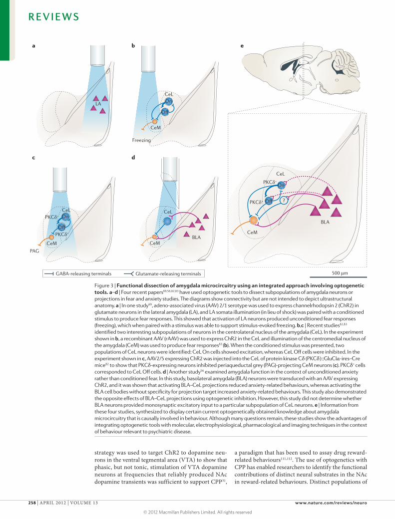
Figure 3 | Functional dissection of amygdala microcircuitry using an integrated approach involving optogenetic tools. a–d | Four recent papers69,56,82,83 have used optogenetic tools to dissect subpopulations of amygdala neurons or projections in fear and anxiety studies. The diagrams show connectivity but are not intended to depict ultrastructural anatomy. a | In one study69, adeno-associated virus (AAV) 2/1 serotype was used to express channelrhodopsin 2 (ChR2) in glutamate neurons in the lateral amygdala (LA), and LA somata illumination (in lieu of shock) was paired with a conditioned stimulus to produce fear responses. This showed that activation of LA neurons produced unconditioned fear responses (freezing), which when paired with a stimulus was able to support stimulus-evoked freezing. b,c | Recent studies82,83 identified two interesting subpopulations of neurons in the centrolateral nucleus of the amygdala (CeL). In the experiment shown in b, a recombinant AAV (rAAV) was used to express ChR2 in the CeL and illumination of the centromedial nucleus of the amygdala (CeM) was used to produce fear responses83 (b). When the conditioned stimulus was presented, two populations of CeL neurons were identified: CeL On cells showed excitation, whereas CeL Off cells were inhibited. In the experiment shown in c, AAV2/5 expressing ChR2 was injected into the CeL of protein kinase Cδ (PKCδ)::GluClα-ires–Cre mice82 to show that PKCδ-expressing neurons inhibited periaqueductal grey (PAG)-projecting CeM neurons (c). PKCδ+ cells corresponded to CeL Off cells. d | Another study56 examined amygdala function in the context of unconditioned anxiety rather than conditioned fear. In this study, basolateral amygdala (BLA) neurons were transduced with an AAV expressing ChR2, and it was shown that activating BLA–CeL projections reduced anxiety-related behaviours, whereas activating the BLA cell bodies without specificity for projection target increased anxiety-related behaviours. This study also demonstrated the opposite effects of BLA–CeL projections using optogenetic inhibition. However, this study did not determine whether BLA neurons provided monosynaptic excitatory input to a particular subpopulation of CeL neurons. e | Information from these four studies, synthesized to display certain current optogenetically obtained knowledge about amygdala microcircuitry that is causally involved in behaviour. Although many questions remain, these studies show the advantages of integrating optogenetic tools with molecular, electrophysiological, pharmacological and imaging techniques in the context of behaviour relevant to psychiatric disease.
R E V I E W S
258 | APRIL 2012 | VOLUME 13 www.nature.com/reviews/neuro
© 2012 Macmillan Publishers Limited. All rights reserved

Nature Reviews | Neuroscience
NAc VTA
BLA
3
2
1
2
4
MSND2R
MSND1R
ChAT
neurons in the NAc that express different dopamine receptor subtypes differentially modulate cocaine place preference; activating D1-expressing neurons enhances cocaine CPP, whereas activating D2-expressing neu-rons suppresses cocaine CPP113. In related work that also combined optogenetics with the use of CPP, cholinergic neurons in the NAc were shown to be cru-cially involved in modulating cocaine reward-related
place preference50, and acute optogenetic activation of defined G protein-coupled receptor pathways in the NAc supported CPP4 (FIG. 4).
To better understand the circuits underlying reward-seeking behaviour, a series of studies have examined whether optogenetic activation of specific brain areas and neural pathways can serve as ‘rewards’ to be sought in their own right. These studies draw on a long his-tory of experiments that showed that animals would perform an operant response (usually a lever press) to deliver small amounts of electrical current to specific brain regions114, a behaviour that is termed intracranial self-stimulation (ICSS). Although such experiments served as a powerful demonstration that reward could be triggered entirely within the CNS, the heterogene-ous neural populations activated by this technique precluded definitive knowledge about which cell types and neural projections could sustain ICSS behaviour. Two recent studies used optogenetics to establish causal roles for specific cell types in ICSS. Selectively activating excitatory projections from the BLA to the NAc (defined by a projection-specific targeting strategy) produced robust self-stimulation, whereas stimulation of the same cell bodies did not61. Moreover, this self-stimulation of BLA–NAc projections was dependent on D1 dopamine receptor (D1R) signalling61 (FIG. 4). Optogenetic self-stimulation of dopaminergic neu-rons in the VTA has also been demonstrated37,52 (FIG. 4). Together, these findings provide a greater understand-ing of the precise activity patterns in defined cells or projections that underlie processes that are thought to become pathological in states of substance dependence or abuse.
We anticipate that optogenetic tools will be lever-aged to test ideas that are based on indirect evidence. For example, GABAergic neurons in the VTA are emerging as a new target for addiction research because opioids act to reduce GABAergic suppression of VTA dopamine neurons115. With the availability of trans-genic mouse lines selectively expressing Cre, direct manipulation of VTA GABA neurons can be tested in assays of hedonic and reward-seeking behaviours. Relative to fear-conditioning, reward-related behav-iours may be more sensitive to motivational state. However, previous in vivo electrophysiological record-ings performed throughout the corticolimbic system during the acquisition116–119, maintenance106,108,110,120–122, extinction123,124 and reinstatement123,125 of reward-related behaviours have identified a rich landscape of neural correlates, and have set the stage for optogenetic interrogation in the pursuit of causality.
The strong in vivo electrophysiological tradition in this field is also supported by studies that used intracranial pharmacological manipulations to iden-tify circuits relevant to addiction and reward-seeking behaviours. Thus, looking towards the further integra-tion of optogenetics with high-speed readouts for neu-ral activity and neurochemical signals126,127, the field of reward-related behaviour represents a fertile proving ground for technological synergy set against a back-drop of sophisticated behavioural theory.
Figure 4 | Optogenetic dissection of limbic circuits in the context of reward-seeking behaviour. A schematic diagram (top panel), showing key neural projections involved in reward, and an expanded view of intra-accumbens microcircuitry (bottom panel; numbers indicate independent findings). The diagram shows connectivity but is not intended to depict ultrastructural anatomy. Cholinergic interneurons expressing choline acetyltransferase (ChAT) in the nucleus accumbens (NAc) modulate the activity of medium spiny neurons (MSNs) and modulate the ability of the animal to develop cocaine-conditioned place preference (1)50. Mice will readily work (nose poke) to receive illumination of channelrhodopsin 2 (ChR2)-expressing basolateral amygdala (BLA) axon terminals in the NAc (2). This self-stimulation behaviour is D1 dopamine receptor (D1R)-dependent, and does not occur when ChR2-expressing prefrontal cortex (PFC) axon terminals in the NAc are illuminated61. It has been shown that activation of dopaminergic neurons in the ventral tegmental area (VTA) can support operant responding in both rats and mice52, and in rats, illumination of tyrosine hydroxylase and ChR2-expressing axon terminals in the NAc also supports operant responding (3)37. The activation of D1R- or D2R-expressing NAc neurons shows differential effects on cocaine-conditioned place preference (4)113.
R E V I E W S
NATURE REVIEWS | NEUROSCIENCE VOLUME 13 | APRIL 2012 | 259
© 2012 Macmillan Publishers Limited. All rights reserved

Deep brain stimulation(DBS). Continuous therapeutic electric stimulation of subcortical areas at high frequencies (~130 Hz) using chronically implanted electrodes.
Circuitry of depressionAlthough up to 13% of the population will experience clinically relevant major depression at some point dur-ing life64,128, the pathophysiological underpinnings of depression and other mood disorders are poorly under-stood. Currently available antidepressant medications are often ineffective and even when effective must gen-erally be taken for 4–6 weeks to show improvements in patients129–132. Brain stimulation treatments that are milder and more targeted than electroconvulsive therapy are also being actively explored; for example, high-frequency electrical stimulation of the region near the subgenual cingulate gyrus may improve depres-sive symptoms in treatment-resistant patients133,134. Although this breakthrough finding sparked hope of an improved understanding of the neural substrates of depression, the mechanisms of this phenomenon are still poorly understood; for example, it is not clear whether high-frequency electrical stimulation exerts antidepressant effects by directly influencing axons passing through this area or local cell bodies, or whether the net effect that is causal in symptom remedi-ation is increasing, decreasing or otherwise influencing local cortical neural electrical activity.
In an attempt to better understand the underlying mechanisms of deep brain stimulation (DBS) treatments for depression better, optogenetic techniques have been used to target cell bodies in the prefrontal cor-tex of mice. Mice with a depression-related phenotype showed an antidepressant-like response to illumination of medial prefrontal cortical neurons expressing ChR2 (REF. 135). Although this study used a more targeted method than electrical stimulation, limiting activation to local cell bodies without activating fibres of passage, new optogenetic tools allow even greater specificity. For example, because the prefrontal cortex projects to many regions, including the dorsal raphe, the NAc, amygdala and other structures proposed to be involved in mood regulation, it will be particularly useful to examine these targets in a pathway-specific manner using projection targeting approaches. In addition, the use of cell-type-specific promoters or Cre-dependent targeting strategies would allow increased cellular, as well as regional, specificity.
Depression has been linked to many neural path-ways, including corticolimbic134, dorsal raphe136–138, hip-pocampal139–142, amygdalar143–146,65–68, striatal147–149 and mesolimbic dopamine130,150–152 circuits. Furthermore, depression has been linked to distinct neuromodula-tory systems and receptors, including serotonin153,154, noradrenaline155 and dopamine130,150–152,155,156. Most cur-rently available antidepressants modulate monoamines, globally modulating synaptic neurotransmission using serotonin, dopamine and/or noradrenaline. Given the diverse distribution and functionality of these recep-tors, increasing the specificity of drug targets could dramatically improve drug efficacy or reduce side effects157. Future development and implementation of optogenetic tools may inspire drug development with enhanced precision to provide more efficacious anti-depressants with fewer side effects.
An intrinsic obstacle in studying the neural circuits underlying depression is that the constellation of symp-toms that define clinical depression is not as readily modelled in animals as other diseases. For example, how can motivation be assessed independently from locomotion, and how can despair and helplessness be reliably assayed in animals? In contrast to addiction research in which the disease is defined by the behav-iour, in depression the disease state is mainly defined by the subjective experience of the patient. This is a major challenge with which depression researchers continue to wrestle to advance our understanding of this severely debilitating and common psychiatric disease. It may be effective to focus on the symptoms that have measur-able correlates in animals (such as assays for changes in motivated behaviours in the absence of gross locomo-tor effects, and hedonic behaviour changes). Despite the practical challenges in using optogenetic techniques to dissect the neural underpinnings of depression and other mood disorders, the immense potential benefit to the broader community of this reverse translational approach warrants a proliferation of work in this field.
Circuitry of autism and schizophreniaAutism and schizophrenia also present major experi-mental challenges for identifying neural circuits altered in states of disease because of the heteroge neity of symptoms that each nominally unitary condition encapsulates. Patients with autism and schizophrenia display diverse and variable arrays of symptoms that make the search for a single cause a daunting and pos-sibly unrealistic goal158–161. At the surface level, these diseases share certain signs and symptoms, such as social dysfunction, although the experienced physician will notice that the social behaviours appear to be quite different, even in dysfunction, in these two diseases.
Schizophrenia symptoms include disorganized speech and behaviour, as well as hallucinations, delu-sions and negative symptoms such as impaired social function162,163. This debilitating disease has been cor-related with perturbations in the balance of excitation and inhibition and with altered oscillations of cortical neural networks164, both of which may in theory lead to a breakdown in the transmission or processing of neural information. Although animal models of schizo-phrenia are still being optimized, one of the few reliable micro-anatomical abnormalities in psychiatric disease is the reduced number and functionality of neocortical parvalbumin neurons165,166, and therefore optogenetic approaches to the circuitry in question have included targeting these neurons in PV::Cre mouse lines53,54. These studies have revealed the roles of parvalbumin neurons in modulating gamma rhythms and informa-tion processing in neocortex that may help to lay the groundwork for a deeper understanding of information processing deficits in schizophrenia53,54.
Autism spectrum disease is characterized by impair-ment in social behaviour and communication as well as stereotyped, repetitive movements159,160,167. Autism is co-morbid with neurological disorders such as epilepsy168,169, and is also linked to anxiety and mood
R E V I E W S
260 | APRIL 2012 | VOLUME 13 www.nature.com/reviews/neuro
© 2012 Macmillan Publishers Limited. All rights reserved

disorders158,170. The complexity and variability of symp-toms in autism have made it particularly difficult to study, but one theory has been that imbalance in cel-lular excitation and inhibition (excitation–inhibition imbalance) may be causative171–173. Optogenetic tools offered the ability to empirically test this idea by ena-bling the induction of excitation–inhibition imbalance. The expression of a SSFO, which produces subthreshold membrane potential changes that last many minutes, in glutamatergic prefrontal cortical neurons allowed the level of cortical excitation to be elevated48. This resulted in an increase in rhythmic activity in the gamma band (30–80 Hz) — a trait linked to autism174 — and also vir-tually abolished unconditioned social behaviour48. To determine whether this effect was related to an imbal-ance in the activity of excitatory and inhibitory cells, a novel red-shifted opsin developed for the purpose of combinatorial excitation was used to partially rescue the phenotype by simultaneously increasing the drive of both excitatory and inhibitory cells48.
An imbalance in excitation and inhibition may also contribute to other psychiatric disorders, such as anxi-ety and depression56, and may give rise to interesting candidate endophenotypes in other circuits, such as rhythmic oscillations (for example, gamma oscilla-tions) and altered activity percolation through the circuit. Indeed, given the highly overlapping circuits that are involved in psychiatric disorders such as anxi-ety, depression, addiction, schizophrenia and autism, it is possible that excitation and inhibition imbalances throughout the brain could contribute to the high rate of co-morbidity among these disease states156,158,162,175–178, and optogenetic tools now offer the capability to test these fascinating and potentially unifying theories.
Circuitry of neurological disordersOptogenetic approaches have been applied to funda-mental research questions in a variety of neurological disorders such as Parkinson’s disease51,55, epilepsy168,169, blindness due to neuronal loss179,180, failure of respira-tion181,182 and neuropsychiatric sleep disorders25. Our basic understanding of motor circuitry has also been enhanced by optogenetic investigations; for example, it had long been suggested that the basal ganglia regu-late movement through the balance of two pathways, the ‘direct’ pathway, which promotes locomotion, and the ‘indirect’ pathway, which inhibits locomo-tion183–185. Selective targeting of each of these pathways was made possible by Cre-dependent optogenetic targeting of D1R- or D2R-expressing cells51, thus pro-viding the first direct empirical confirmation of this long-standing theory.
Parkinson’s disease, a neurodegenerative disease, is characterized by rigidity, tremor, postural instability and slow movement186,187. Although DBS in the subgen-ual cingulate cortex has been used in humans to treat depression133, DBS in the subthalamic nucleus (STN), a part of the basal ganglia circuit, and in other targets has shown remarkable therapeutic effects for treatment of the motor impairments associated with Parkinson’s disease188–190. However, the cellular mechanisms of this
therapy have been unclear and highly controversial. Selective optogenetic control of afferent fibres (but not selective control of local somata) in the STN was shown to have profound therapeutic effects on motor behav-iours in a hemi-parkinsonian rat model103 (FIG. 5). These data suggested a model for DBS treatment in which white matter tracts or axonal pathways are the most effective direct target of control. The reasons for the efficacy of control by this means could include the fact that recruitment of large or asymmetric neural struc-tures with a point source of energy such as an electrode or an optical fibre is most efficient if spatially local-ized incoming axon tracts are the direct initial target. This concept carries direct implications for electrode contact positioning in DBS for depression as well as Parkinson’s disease. Alternatively (or additionally), it may be that the flow of activity between brain regions that can be targeted at the level of axon tracts (rather than neural coding by somata per se within a region) is the most functionally relevant endophenotype for neuropsychiatric disease symptomatology.
Pathway-specific optogenetic approaches in com-bination with whole-cell recordings in a corticotha-lamic circuit linked to epilepsy revealed a pathological generation of aberrant oscillations relevant to seizure activity169. Optogenetic activation of neurons in the retrotrapezoid nucleus–parafacial respiratory group induced active expiration and regulated the rhythm of respiration181. It may be worth noting that application of optogenetic approaches can also further the study of stem cell-based interventions for Parkinson’s disease191; optogenetic targeting of grafted stem-cell-derived dopamine neurons has shown that opsin-expressing grafted cells can be functionally integrated into net-works of wild-type mouse striatum168. The uncondi-tioned modulation of feeding behaviour was shown to be under the control of two functionally opposing subpopulations of neurons in the hypothalamus57. An elegant examination of aggressive behaviour demon-strated that attacks could be evoked with optogenetic activation of a specific subset of ventromedial hypo-thalamic neurons192. Lastly, restoration of vision by optogenetic activation of cones was shown to rescue the blindness associated with retinitis pigmentosa179,180.
ConclusionWe have provided here a brief summary of some recent investigations relevant to the study of neu-ropsychiatric disease that used optogenetic tools. In these cases, the precision of optogenetics has pro-vided major experimental leverage193 and has led to insights into neural circuit function and dysfunction. Although the impact of these recent investigations has already been substantial, there remains much work to do; for example, the application of optogenetics to non-human primates is still in its infancy 24,27,194,195, and many disease states and symptom clusters remain unexplored. We anticipate several major areas of optogenetic tool advancement in the future, from tool development to scientific application, mainly driven by demand in the field.
R E V I E W S
NATURE REVIEWS | NEUROSCIENCE VOLUME 13 | APRIL 2012 | 261
© 2012 Macmillan Publishers Limited. All rights reserved

Nature Reviews | Neuroscience
STN cells upon afferent activation
STN
Striatal illumination in D1R::Cre or D2R::Cre mice
SNr
Striatum
M1
D2
GP
–1 0 1 2Time (s)
–1 0 1 2Time (s)
D2D1
Hz
Hz
12
0
20
0
HFS
4 s50 µV
LFS
4 s50 µV
0.5 s50 µV
5 ms50 µV
GABA-releasing terminals
Glutamate-releasing terminals
D1
With respect to developments in opsin engineer-ing, despite the rapidly proliferating library of opsins, there remain areas that require increased attention. For example18, we anticipate the engineering of blue-shifted hyperpolarizing opsins with narrower activation wave-length spectra to allow for enhanced combinatorial neu-ronal inhibition experiments21. Another much needed development is a hyperpolarizing SFO to allow sustained inhibition of neurons without requiring constant illumi-nation, which can cause heating and may be impractical for chronic inhibition experiments. In addition to light-sensitive pumps and channels, we anticipate the contin-ued expansion of the OptoXR family4, as light-sensitive domains are being added to an increasing number of receptor and even intracellular signalling proteins so that optogenetics will expand to the study of cell signalling in addition to the study of neural activity.
The development of improved targeting strate-gies on both a cellular and subcellular level is just as important as the development of new opsins and opsin
variants. Improved or expanded recombinase strategies are well within reach, and would facilitate circuit-level investigations. We have steadily improved the expres-sion of opsins at the membrane, but further exploration in this area may produce targeting strategies that allow selective opsin expression in subcellular compartments such as dendrites, soma or axon terminals, thereby increasing the level of precision196. Currently, we are able to selectively express opsins in cells with a given genetic phenotype or marker, but methods to selec-tively exclude opsin expression in cells with a given genetic identity will also be useful.
In addition, methods of light delivery will require continual improvement. The optical fibre provides a small zone of illumination and, rather than opsin expression, the illumination pattern may often be limit-ing. Many experiments may call for broader volumes of illumination (to facilitate achieving behavioural effects from optogenetic manipulations in larger species such as monkeys). We also anticipate continued improvement
Figure 5 | Functional mapping of basal ganglia circuitry using optogenetics in the context of Parkinson’s disease. A schematic diagram (top panel) shows key neural projections that are involved in parkinsonian behaviour and treatment. Data in the bottom left panel are from a study that used a constitutively expressing channelrhodopsin 2 (ChR2) mouse line (Thy1::ChR2) to identify a mechanistic explanation for the therapeutic effects of deep brain stimulation (DBS) 55. By illuminating and recording in the subthalamic nucleus (STN), this paper showed that afferent fibres entering the STN, rather than local cell bodies themselves, are likely to be the direct target of DBS in the correction of parkinsonian motor activity. High-frequency stimulation (HFS) of the afferent fibres into STN potently silenced the structure as shown and reversibly abolished the parkinsonian symptoms. By contrast, low-frequency stimulation (LFS) of the afferents simply added spikes on top of endogenous spikes and worsened parkinsonian symptoms. Data in the bottom right panel are from a study that used a Cre-dependent adeno-associated virus (AAV) to selectively express ChR2 in either D1 dopamine receptor (D1R)::Cre or D2R::Cre mice to examine the differential contributions of the direct and indirect pathways with respect to motor output. Activation of D1R-expressing neurons silenced local basal ganglia activity and increased ambulation, whereas activation of D2R-expressing neurons increased this activity and enhanced immobile or bradykinetic (slow) behaviour51. Black bars indicate the duration of illumination. The bottom left panel is reproduced, with permission, from REF. 55 © (2009) American Association for the Advancement of Science. The bottom right panel is reproduced, with permission, from REF. 51 © (2010) Macmillan Publishers Ltd. All rights reserved. GP, globus pallidus; M1, primary motor cortex; SNr, substantia nigra pars reticulata.
R E V I E W S
262 | APRIL 2012 | VOLUME 13 www.nature.com/reviews/neuro
© 2012 Macmillan Publishers Limited. All rights reserved

1. Deisseroth, K. Optogenetics. Nature Methods 8, 26–29 (2011).
2. Deisseroth, K. et al. Next-generation optical technologies for illuminating genetically targeted brain circuits. J. Neurosci. 26, 10380–10386 (2006).
3. Zhang, F. et al. The microbial opsin family of optogenetic tools. Cell 147, 1446–1457 (2011).
4. Airan, R. D., Thompson, K. R., Fenno, L. E., Bernstein, H. & Deisseroth, K. Temporally precise in vivo control of intracellular signalling. Nature 458, 1025–1029 (2009).This study developed the OptoXRs (photosensitive G protein-coupled receptors based on vertebrate opsin genes) for mammalian in vivo use and showed that light-activated intracellular signalling cascades could support conditioned place preference.
5. Boyden, E. S., Zhang, F., Bamberg, E., Nagel, G. & Deisseroth, K. Millisecond-timescale, genetically targeted optical control of neural activity. Nature Neurosci. 8, 1263–1268 (2005).The initial demonstration of single-component optogenetics using a microbial opsin gene (in this case, a channelrhodopsin).
6. Yizhar, O., Fenno, L. E., Davidson, T. J., Mogri, M. & Deisseroth, K. Optogenetics in neural systems. Neuron 71, 9–34 (2011).A comprehensive practical overview describing the implementation of optogenetic tools in neural systems.
7. Fenno, L., Yizhar, O. & Deisseroth, K. The development and application of optogenetics. Annu. Rev. Neurosci. 34, 389–412 (2010).
8. Carter, M. E. & de Lecea, L. Optogenetic investigation of neural circuits in vivo. Trends Mol. Med. 17, 197–206 (2011).
9. Aravanis, A. M. et al. An optical neural interface: in vivo control of rodent motor cortex with integrated fiberoptic and optogenetic technology. J. Neural Eng. 4, S143–S156 (2007).
10. Kravitz, A. V. & Kreitzer, A. C. Optogenetic manipulation of neural circuitry in vivo. Curr. Opin. Neurobiol. 21, 433–439 (2011).
11. Lima, S. Q. & Miesenböck, G. Remote control of behavior through genetically targeted photostimulation of neurons. Cell 121, 141–152 (2005).
12. Nagel, G. et al. Channelrhodopsin-1: a light-gated proton channel in green algae. Science 296, 2395–2398 (2002).
13. Nagel, G. et al. Channelrhodopsin-2, a directly light-gated cation-selective membrane channel. Proc. Natl Acad. Sci. USA 100, 13940 –13945 (2003).
14. Zhang, F. et al. Red-shifted optogenetic excitation: a tool for fast neural control derived from Volvox carteri. Nature Neurosci. 11, 631–633 (2008).
15. Berndt, A. et al. High-efficiency channelrhodopsins for fast neuronal stimulation at low light levels. Proc. Natl Acad. Sci. USA 108, 7595–7600 (2011).
16. Gradinaru, V. et al. Molecular and cellular approaches for diversifying and extending optogenetics. Cell 141, 154–165 (2010).
17. Berndt, A., Yizhar, O., Gunaydin, L. A., Hegemann, P. & Deisseroth, K. Bi-stable neural state switches. Nature Neurosci. 12, 229–234 (2009).
18. Zhang, F. et al. Multimodal fast optical interrogation of neural circuitry. Nature 446, 633–639 (2007).The first behavioural loss-of-function using optogenetics; achieved here with the inhibitory optogenetic tool NpHR.
19. Chow, B. Y. et al. High-performance genetically targetable optical neural silencing by light-driven proton pumps. Nature 463, 98–102 (2010).
20. Gunaydin, L. A. et al. Ultrafast optogenetic control. Nature Neurosci. 13, 387–392 (2010).
21. Mattis, J. et al. Principles for applying optogenetic tools derived from direct comparative analysis of microbial opsins. Nature Methods 9, 159–172 (2011).An empirical comparison of most existing microbial opsin-derived optogenetic tools under matched conditions, including the development of several new opsin variants.
22. Iwai, Y., Honda, S., Ozeki, H., Hashimoto, M. & Hirase, H. A simple head-mountable LED device for chronic stimulation of optogenetic molecules in freely moving mice. Neurosci. Res. 70, 124–127 (2011).
23. Schneider, M. B., Gradinaru, V., Zhang, F. & Deisseroth, K. Controlling neuronal activity. Am. J. Psychiatry 165, 562 (2008).
24. Diester, I. et al. An optogenetic toolbox designed for primates. Nature Neurosci. 14, 387–397 (2011).
25. Adamantidis, A. R., Zhang, F., Aravanis, A. M., Deisseroth, K. & de Lecea, L. Neural substrates of awakening probed with optogenetic control of hypocretin neurons. Nature 450, 420–424 (2007).The first application of a microbial opsin to show a behavioural change in a mammal (gain of function, using a channelrhodopsin).
26. Grossman, N. et al. Multi-site optical excitation using ChR2 and micro-LED array. J. Neural Eng. 7, 016004 (2010).
27. Bernstein, J. G. et al. Prosthetic systems for therapeutic optical activation and silencing of genetically-targeted neurons. Proc. Soc. Photo Opt. Instrum. Eng. 6854, 68540H (2008).
28. Sparta, D. R. et al. Construction of implantable optical fibers for long-term optogenetic manipulation of neural circuits. Nature Protoc. 7, 68512–68523 (2011).
29. Atasoy, D., Aponte, Y., Su, H. H. & Sternson, S. M. A FLEX switch targets Channelrhodopsin-2 to multiple cell types for imaging and long-range circuit mapping. J. Neurosci. 28, 7025–7030 (2008).
30. Kuhlman, S. J. & Huang, Z. J. High-resolution labeling and functional manipulation of specific neuron types in mouse brain by Cre-activated viral gene expression. PLoS ONE 3, e2005 (2008).
31. Tsai, H.-C. et al. Phasic firing in dopaminergic neurons is sufficient for behavioral conditioning. Science 324, 1080–1084 (2009).The initial behavioural application of the double-floxed inverted open reading frame tool enabling selective opsin expression in neurons positive for Cre recombinase. This is used in the TH::Cre mouse to demonstrate the hedonic properties of phasic dopamine neuron firing.
32. Arenkiel, B. R. et al. In vivo light-induced activation of neural circuitry in transgenic mice expressing Channelrhodopsin-2. Neuron 54, 205–218 (2007).
33. Zhao, S. et al. Cell type-specific channelrhodopsin-2 transgenic mice for optogenetic dissection of neural circuitry function. Nature Methods 8, 745–752 (2011).
34. Taniguchi, H. et al. A resource of cre driver lines for genetic targeting of GABAergic neurons in cerebral cortex. Neuron 71, 995–1013 (2011).
35. Anikeeva, P. et al. Optetrode: a multichannel readout for optogenetic control in freely moving mice. Nature Neurosci. 15, 163–170 (2012).
36. Wang, J. et al. Integrated device for combined optical neuromodulation and electrical recording for chronic in vivo applications. J. Neural Eng. 9, 016001 (2012).
37. Witten, I. B. et al. Recombinase-driver rat lines: tools, techniques, and optogenetic application to dopamine-mediated reinforcement. Neuron 72, 721–733 (2011).
38. Lee, J. H. et al. Global and local fMRI signals driven by neurons defined optogenetically by type and wiring. Nature 465, 788–792 (2010).
39. Peterlin, Z. A., Kozloski, J., Mao, B.-Q., Tsiola, A. & Yuste, R. Optical probing of neuronal circuits with calcium indicators. Proc. Natl Acad. Sci. USA 97, 3619–3624 (2000).
40. Shoham, S. Optogenetics meets optical wavefront shaping. Nature Methods 7, 798–799 (2010).
41. Petreanu, L., Huber, D., Sobczyk, A. & Svoboda, K. Channelrhodopsin-2-assisted circuit mapping of long-range callosal projections. Nature Neurosci. 10, 663–668 (2007).
42. Petreanu, L., Mao, T., Sternson, S. M. & Svoboda, K. The subcellular organization of neocortical excitatory connections. Nature 457, 1142–1145 (2009).
43. Adesnik, H. & Scanziani, M. Lateral competition for cortical space by layer-specific horizontal circuits. Nature 464, 1155–1160 (2010).
44. Han, X. & Boyden, E. S. Multiple-color optical activation, silencing, and desynchronization of neural activity, with single-spike temporal resolution. PLoS ONE 2, e299 (2007).
45. Zhao, S. et al. Improved expression of halorhodopsin for light-induced silencing of neuronal activity. Brain Cell Biol. 36, 141–154 (2008).
46. Gradinaru, V., Thompson, K. R. & Deisseroth, K. eNpHR: a Natronomonas halorhodopsin enhanced for optogenetic applications. Brain Cell Biol. 36, 129–139 (2008).
47. Lin, J. Y., Lin, M. Z., Steinbach, P. & Tsien, R. Y. Characterization of engineered channelrhodopsin variants with improved properties and kinetics. Biophys. J. 96, 1803–1814 (2009).
48. Yizhar, O. et al. Neocortical excitation/inhibition balance in information processing and social dysfunction. Nature 477, 171–178 (2011).The introduction of two new opsin variants, C1V1 and SSFO, for combinatorial optogenetic excitation in vivo. The study showed the importance of altered excitation/inhibition ratios to social behaviour and cognition.
49. Berglund, K., Kuner, T., Feng, G. & Augustine, G. J. Imaging synaptic inhibition with the genetically encoded chloride indicator clomeleon. Cold Spring Harb. Protoc. 2011, 1492–1497 (2011).
50. Witten, I. B. et al. Cholinergic interneurons control local circuit activity and cocaine conditioning. Science 330, 1677–1681 (2010).The first behavioural loss-of-function in mammals achieved using optogenetics. This study involved the introduction of an inhibitory opsin into cholinergic interneurons using the double-floxed inverted open reading frame tool in a choline acetyltransferase (ChAT)::Cre mouse to demonstrate the role of cholinergic neurons in mediating cocaine-conditioned place preference.
51. Kravitz, A. V. et al. Regulation of parkinsonian motor behaviours by optogenetic control of basal ganglia circuitry. Nature 466, 622–626 (2010).This study used optogenetics to provide empirical evidence of the long-proposed effects of the direct and indirect pathways in the striatum on movement.
in the speed and precision of single-cell activation through refinement of the ability to visualize and selec-tively illuminate individual cells to allow the playback of specific neuronal population codes. The creation of a method to simultaneously perform optogenetic activa-tion and achieve calcium imaging at a single-cell or even dendritic-spine resolution in a freely moving mammal would also be an enormous advance for the field.
It is intriguing to speculate that a very specific (and now testable) neural circuit dysfunction (such as an imbalance in excitation and inhibition) could be caus-ally involved in multiple psychiatric diseases including
anxiety, depression, addiction, schizophrenia and autism. Variation in precise symptomatology from dis-ease to disease could be more closely linked to variation in the role of the affected circuit than to fundamentally distinct principles of the pathologically altered neural activity propagation. In particular, given the high rate of co-morbidity among the various mental illnesses and the shared symptomatology between individual diseases, identification of such simplifying themes and unifying theories by optogenetic or other means is one of the most pressing needs and exciting avenues of research into neurological and psychiatric disease.
R E V I E W S
NATURE REVIEWS | NEUROSCIENCE VOLUME 13 | APRIL 2012 | 263
© 2012 Macmillan Publishers Limited. All rights reserved
52. Adamantidis, A. R. et al. Optogenetic interrogation of dopaminergic modulation of the multiple phases of reward-seeking behavior. J. Neurosci. 31, 10829–10835 (2011).
53. Sohal, V. S., Zhang, F., Yizhar, O. & Deisseroth, K. Parvalbumin neurons and gamma rhythms enhance cortical circuit performance. Nature 459, 698–702 (2009).Together with reference 54, this study used optogenetic tools to link parvalbumin cells to gamma rhythms, information transmission and processing in the neocortex.
54. Cardin, J. A. et al. Driving fast-spiking cells induces γ rhythm and controls sensory responses. Nature 459, 663–667 (2009).
55. Gradinaru, V., Mogri, M., Thompson, K. R., Henderson, J. M. & Deisseroth, K. Optical deconstruction of parkinsonian neural circuitry. Science 324, 354–359 (2009).This study provided a mechanistic explanation for how DBS alleviates parkinsonian symptoms using optogenetic techniques in animal models.
56. Tye, K. M. et al. Amygdala circuitry mediating reversible and bidirectional control of anxiety. Nature 471, 358–362 (2011).The first optogenetic projection-specific targeting study of behaviour, showing that acute activation of neuron axon terminals can produce an opposite effect on anxiety to that produced by activating neuron somata. It was also the first study to show axon-terminal specific inhibition using optogenetics in vivo.
57. Aponte, Y., Atasoy, D. & Sternson, S. M. AGRP neurons are sufficient to orchestrate feeding behavior rapidly and without training. Nature Neurosci. 14, 351–355 (2011).
58. Hägglund, M., Borgius, L., Dougherty, K. J. & Kiehn, O. Activation of groups of excitatory neurons in the mammalian spinal cord or hindbrain evokes locomotion. Nature Neurosci. 13, 246–252 (2010).
59. Choi, J., Young, J. A. T. & Callaway, E. M. Selective viral vector transduction of ErbB4 expressing cortical interneurons in vivo with a viral receptor-ligand bridge protein. Proc. Natl Acad. Sci. USA 107, 16703–16708 (2010).
60. Li, F., Ryu, B. Y., Krueger, R. L., Heldt, S. A. & Albritton, L. M. Targeted entry via somatostatin receptors using a novel modified retrovirus glycoprotein that delivers genes at levels comparable to wild type viral glycoproteins. J. Virol. 86, 373–381 (2011).
61. Stuber, G. D. et al. Excitatory transmission from the amygdala to nucleus accumbens facilitates reward seeking. Nature 475, 377–380 (2011).This study showed that amygdalar projections to the NAc, but not cortical projections, mediate ICSS.
62. Zorzos, A. N., Boyden, E. S. & Fonstad, C. G. Multiwaveguide implantable probe for light delivery to sets of distributed brain targets. Opt. Lett. 35, 4133–4135 (2010).
63. Wentz, C. T. et al. A wirelessly powered and controlled device for optical neural control of freely-behaving animals. J. Neural Eng. 8, 046021 (2011).
64. Kessler, R. C. et al. Lifetime prevalence and age-of-onset distributions of DSM-IV disorders in the National Comorbidity Survey Replication. Arch. Gen. Psychiatry 62, 593–602 (2005).
65. Lieb, R. Anxiety disorders: clinical presentation and epidemiology. Handb. Exp. Pharmacol. 405–432 (2005).
66. Baldwin, D., Woods, R., Lawson, R. & Taylor, D. Efficacy of drug treatments for generalised anxiety disorder: systematic review and meta-analysis. BMJ 342, d1199 (2011).
67. Beck, A. T., Emery, G. & Greenberg, R. L. Anxiety Disorders and Phobias: A Cognitive Perspective (Basic Books, 2005).
68. Woods, J. H., Katz, J. L. & Winger, G. Benzodiazepines: use, abuse, and consequences. Pharmacol. Rev. 44, 151–347 (1992).
69. Johansen, J. P. et al. Optical activation of lateral amygdala pyramidal cells instructs associative fear learning. Proc. Natl Acad. Sci. USA 107, 12692–12697 (2010).
70. Adolphs, R., Tranel, D., Damasio, H. & Damasio, A. Fear and the human amygdala. J. Neurosci. 15, 5879–5891 (1995).
71. Davis, M. The role of the amygdala in fear and anxiety. Annu. Rev. Neurosci. 15, 353–375 (1992).
72. LeDoux, J. E. Emotion: clues from the brain. Annu. Rev. Psychology 46, 209–235 (1995).
73. Fanselow, M. S. & Gale, G. D. The amygdala, fear, and memory. Ann. NY Acad. Sci. 985, 125–134 (2003).
74. Maren, S. Synaptic mechanisms of associative memory in the amygdala. Neuron 47, 783–786 (2005).
75. Jüngling, K. et al. Neuropeptide S-mediated control of fear expression and extinction: role of intercalated GABAergic neurons in the amygdala. Neuron 59, 298–310 (2008).
76. LeDoux, J., Cicchetti, P., Xagoraris, A. & Romanski, L. The lateral amygdaloid nucleus: sensory interface of the amygdala in fear conditioning. J. Neurosci. 10, 1062–1069 (1990).
77. Miserendino, M. J. D., Sananes, C. B., Melia, K. R. & Davis, M. Blocking of acquisition but not expression of conditioned fear-potentiated startle by NMDA antagonists in the amygdala. Nature 345, 716–718 (1990).
78. Paré, D., Quirk, G. J. & Ledoux, J. E. New vistas on amygdala networks in conditioned fear. J. Neurophysiol. 92, 1–9 (2004).
79. Rodrigues, S. M., Schafe, G. E. & LeDoux, J. E. Intra-amygdala blockade of the NR2B subunit of the NMDA receptor disrupts the acquisition but not the expression of fear conditioning. J. Neurosci. 21, 6889–6896 (2001).
80. Wilensky, A. E., Schafe, G. E., Kristensen, M. P. & LeDoux, J. E. Rethinking the fear circuit: the central nucleus of the amygdala is required for the acquisition, consolidation, and expression of pavlovian fear conditioning. J. Neurosci. 26, 12387–12396 (2006).
81. Han, J.-H. et al. Selective erasure of a fear memory. Science 323, 1492–1496 (2009).
82. Haubensak, W. et al. Genetic dissection of an amygdala microcircuit that gates conditioned fear. Nature 468, 270–276 (2010).Together with reference 83, this study identified a new population of neurons, and identified functional specificity in PKCδ-expressing neurons in a multisynaptic circuit in the context of conditioned fear expression.
83. Ciocchi, S. et al. Encoding of conditioned fear in central amygdala inhibitory circuits. Nature 468, 277–282 (2010).
84. Yehuda, R. Post-traumatic stress disorder. N. Engl. J. Med. 346, 108–114 (2002).
85. Charney, D. S., Deutch, A. Y., Krystal, J. H., Southwick, S. M. & Davis, M. Psychobiologic mechanisms of posttraumatic stress disorder. Arch. Gen. Psychiatry 50, 295–305 (1993).
86. Rauch, S. L., Shin, L. M. & Phelps, E. A. Neurocircuitry models of posttraumatic stress disorder and extinction: human neuroimaging research–past, present, and future. Biol. Psychiatry 60, 376–382 (2006).
87. Milad, M. R., Rauch, S. L., Pitman, R. K. & Quirk, G. J. Fear extinction in rats: implications for human brain imaging and anxiety disorders. Biol. Psychology 73, 61–71 (2006).
88. Rudy, J. W. & O’Reilly, R. C. Contextual fear conditioning, conjunctive representations, pattern completion, and the hippocampus. Behav. Neurosci. 113, 867–880 (1999).
89. Fanselow, M. S. Contextual fear, gestalt memories, and the hippocampus. Behav. Brain Res. 110, 73–81 (2000).
90. Phillips, R. & LeDoux, J. Differential contribution of amygdala and hippocampus to cued and contextual fear conditioning. Behav. Neurosci. 106, 274–285 (1992).
91. Kim, J. J., Rison, R. A. & Fanselow, M. S. Effects of amygdala, hippocampus, and periaqueductal gray lesions on short-and long-term contextual fear. Behav. Neurosci. 107, 1093–1098 (1993).
92. McGaugh, J. L. Memory—a century of consolidation. Science 287, 248–251 (2000).
93. Frankland, P. W. et al. Consolidation of CS and US representations in associative fear conditioning. Hippocampus 14, 557–569 (2004).
94. Frankland, P. W. & Bontempi, B. The organization of recent and remote memories. Nature Rev. Neuroscience 6, 119–130 (2005).
95. Wiltgen, B. J., Brown, R. A. M., Talton, L. E. & Silva, A. J. New circuits for old memories: the role of the neocortex in consolidation. Neuron 44, 101–108 (2004).
96. Lee, J. L. C., Everitt, B. J. & Thomas, K. L. Independent cellular processes for hippocampal memory consolidation and reconsolidation. Science 304, 839–843 (2004).
97. Goshen, I. et al. Dynamics of retrieval strategies for remote memories. Cell 147, 678–689 (2011).This study showed that remote memory recall is dependent on hippocampal activity, using fast-acting optogenetic methods.
98. Koob, G. F. Drugs of abuse: anatomy, pharmacology and function of reward pathways. Trends Pharmacol. Sci. 13, 177–184 (1992).
99. Wise, R. A. Drug-activation of brain reward pathways. Drug Alcohol Depend. 51, 13–22 (1998).
100. Everitt, B. J. & Robbins, T. W. Neural systems of reinforcement for drug addiction: from actions to habits to compulsion. Nature Neurosci. 8, 1481 (2005).
101. Robinson, T. E. & Berridge, K. C. The neural basis of drug craving: an incentive-sensitization theory of addiction. Brain Res. Rev. 18, 247–291 (1993).
102. Kelley, A. E. & Berridge, K. C. The neuroscience of natural rewards: relevance to addictive drugs. J. Neurosci. 22, 3306–3311 (2002).
103. Schultz, W., Dayan, P. & Montague, P. R. A neural substrate of prediction and reward. Science 275, 1593–1599 (1997).
104. Spanagel, R. & Weiss, F. The dopamine hypothesis of reward: past and current status. Trends Neurosci. 22, 521–527 (1999).
105. Berridge, K. C. & Robinson, T. E. What is the role of dopamine in reward: hedonic impact, reward learning, or incentive salience? Brain Res. Rev. 28, 309–369 (1998).
106. Ikemoto, S. & Panksepp, J. The role of nucleus accumbens dopamine in motivated behavior: a unifying interpretation with special reference to reward-seeking. Brain Res. Rev. 31, 6–41 (1999).
107. Kalivas, P. W. & Stewart, J. Dopamine transmission in the initiation and expression of drug-and stress-induced sensitization of motor activity. Brain Res. Rev. 16, 223–244 (1991).
108. Ambroggi, F., Ishikawa, A., Fields, H. L. & Nicola, S. M. Basolateral amygdala neurons facilitate reward-seeking behavior by exciting nucleus accumbens neurons. Neuron 59, 648–661 (2008).
109. Cardinal, R. N., Parkinson, J. A., Hall, J. & Everitt, B. J. Emotion and motivation: the role of the amygdala, ventral striatum, and prefrontal cortex. Neurosci. Biobehav. Rev. 26, 321–352 (2002).
110. Nicola, S. M., Yun, I. A., Wakabayashi, K. T. & Fields, H. L. Firing of nucleus accumbens neurons during the consummatory phase of a discriminative stimulus task depends on previous reward predictive cues. J. Neurophysiol. 91, 1866–1882 (2004).
111. Carr, G. D., Fibiger, H. C. & Phillips, A. G. Conditioned place preference as a measure of drug reward. in The Neuropharmacological Basis of Reward (Topics in Experimental Psychopharmacology) Vol. 1 (eds Liebman, J. M. & Cooper, S. J.) 264–319 (Oxford Univ. Press, 1989).
112. Tzschentke, T. M. Measuring reward with the conditioned place preference paradigm: a comprehensive review of drug effects, recent progress and new issues. Prog. Neurobiol. 56, 613–672 (1998).
113. Lobo, M. K. et al. Cell type-specific loss of BDNF signaling mimics optogenetic control of cocaine reward. Science 330, 385–390 (2010).This study showed that D1 and D2 neurons in the NAc have distinctly different effects on cocaine-conditioned place preference.
114. Olds, J. & Milner, P. Positive reinforcement produced by electrical stimulation of septal area and other regions of rat brain. J. Comp. Physiol. Psychol. 47, 419–427 (1954).
115. Lüscher, C. & Malenka, R. C. Drug-evoked synaptic plasticity in addiction: from molecular changes to circuit remodeling. Neuron 69, 650–663 (2011).
116. Schoenbaum, G., Chiba, A. A. & Gallagher, M. Orbitofrontal cortex and basolateral amygdala encode expected outcomes during learning. Nature Neurosci. 1, 155–159 (1998).
117. Schoenbaum, G. & Roesch, M. Orbitofrontal cortex, associative learning, and expectancies. Neuron 47, 633–636 (2005).
118. Tye, K. M., Stuber, G. D., de Ridder, B., Bonci, A. & Janak, P. H. Rapid strengthening of thalamo-amygdala synapses mediates cue-reward learning. Nature 453, 1253–1257 (2008).
119. Tye, K. M. et al. Methylphenidate facilitates learning-induced amygdala plasticity. Nature Neurosci. 13, 475–481 (2010).
R E V I E W S
264 | APRIL 2012 | VOLUME 13 www.nature.com/reviews/neuro
© 2012 Macmillan Publishers Limited. All rights reserved

120. Carelli, R. M. & Deadwyler, S. A comparison of nucleus accumbens neuronal firing patterns during cocaine self-administration and water reinforcement in rats. J. Neurosci. 14, 7735–4776 (1994).
121. Carelli, R. M., Ijames, S. G. & Crumling, A. J. Evidence that separate neural circuits in the nucleus accumbens encode cocaine versus “natural” (water and food) reward. J. Neurosci. 20, 4255–4266 (2000).
122. Wakabayashi, K. T., Fields, H. L. & Nicola, S. M. Dissociation of the role of nucleus accumbens dopamine in responding to reward-predictive cues and waiting for reward. Behav. Brain Res. 154, 19–30 (2004).
123. Peters, Y. M., O’Donnell, P. & Carelli, R. M. Prefrontal cortical cell firing during maintenance, extinction, and reinstatement of goal-directed behavior for natural reward. Synapse 56, 74–83 (2005).
124. Tye, K. M., Cone, J. J., Schairer, W. W. & Janak, P. H. Amygdala neural encoding of the absence of reward during extinction. J. Neurosci. 30, 116–125 (2010).
125. Tye, K. M. & Janak, P. H. Amygdala neurons differentially encode motivation and reinforcement. J. Neurosci. 27, 3937–3945 (2007).
126. Phillips, P. E. M., Stuber, G. D., Heien, M., Wightman, R. M. & Carelli, R. M. Subsecond dopamine release promotes cocaine seeking. Nature 422, 614–618 (2003).
127. Alsiö, J. et al. Enhanced sucrose and cocaine self-administration and cue-induced drug seeking after loss of VGLUT2 in midbrain dopamine neurons in mice. J. Neurosci. 31, 12593–12603 (2011).
128. Blazer, D., Kessler, R., McGonagle, K. & Swartz, M. The prevalence and distribution of major depression in a national community sample: the National Comorbidity Survey. Am. J. Psychiatry 151, 979–986 (1994).
129. Nierenberg, A. A. & Dececco, L. M. Definitions of antidepressant treatment response, remission, nonresponse, partial response, and other relevant outcomes: a focus on treatment-resistant depression. J. Clin. Psychiatry 62 (Suppl. 16), 5–9 (2001).
130. Dailly, E., Chenu, F., Renard, C. E. & Bourin, M. Dopamine, depression and antidepressants. Fundam. Clin. Pharmacol. 18, 601–607 (2004).
131. Maddox, J. C., Levi, M. & Thompson, C. The compliance with antidepressants in general practice. J. Psychopharmacol. 8, 48–52 (1994).
132. Melfi, C. A. et al. The effects of adherence to antidepressant treatment guidelines on relapse and recurrence of depression. Arch. Gen. Psychiatry 55, 1128–1132 (1998).
133. Mayberg, H. S. et al. Deep brain stimulation for treatment-resistant depression. Neuron 45, 651–660 (2005).A seminal study showing that DBS can show acute and long-lasting antidepressant effects, inviting reverse translational studies using optogenetics to provide a mechanistic explanation.
134. Mayberg, H. Modulating limbic-cortical circuits in depression: targets of antidepressant treatments. Semin. Clin. Neuropsychiatry 7, 255–268 (2002).
135. Covington, H. E. et al. Antidepressant effect of optogenetic stimulation of the medial prefrontal cortex. J. Neurosci. 30, 16082–16090 (2010).
136. Bach-Mizrachi, H. et al. Neuronal tryptophan hydroxylase mRNA expression in the human dorsal and median raphe nuclei: major depression and suicide. Neuropsychopharmacology 31, 814–824 (2005).
137. Greenwood, B. N. et al. Freewheel running prevents learned helplessness/behavioral depression: role of dorsal raphe serotonergic neurons. J. Neurosci. 23, 2889–2898 (2003).
138. Lira, A. et al. Altered depression-related behaviors and functional changes in the dorsal raphe nucleus of serotonin transporter-deficient mice. Biol. Psychiatry 54, 960–971 (2003).
139. Airan, R. D. et al. High-speed imaging reveals neurophysiological links to behavior in an animal model of depression. Science 317, 819–823 (2007).
140. Bremner, J. D. et al. Hippocampal volume reduction in major depression. Am. J. Psychiatry 157, 115–118 (2000).
141. Sahay, A. & Hen, R. Adult hippocampal neurogenesis in depression. Nature Neurosci. 10, 1110–1115 (2007).
142. Sapolsky, R. M. The possibility of neurotoxicity in the hippocampus in major depression: a primer on neuron death. Biol. Psychiatry 48, 755–765 (2000).
143. Mervaala, E. et al. Quantitative MRI of the hippocampus and amygdala in severe depression. Psychol. Med. 30, 117–125 (2000).
144. Sheline, Y. I., Gado, M. H. & Price, J. L. Amygdala core nuclei volumes are decreased in recurrent major depression. Neuroreport 9, 2023–2028 (1998).
145. Sheline, Y. I. et al. Increased amygdala response to masked emotional faces in depressed subjects resolves with antidepressant treatment: an fMRI study. Biol. Psychiatry 50, 651–658 (2001).
146. Siegle, G. J., Thompson, W., Carter, C. S., Steinhauer, S. R. & Thase, M. E. Increased amygdala and decreased dorsolateral prefrontal BOLD responses in unipolar depression: related and independent features. Biol. Psychiatry 61, 198–209 (2007).
147. Larisch, R. et al. In vivo evidence for the involvement of dopamine-D2 receptors in striatum and anterior cingulate gyrus in major depression. Neuroimage 5, 251–260 (1997).
148. Zhu, X., Peng, S., Zhang, S. & Zhang, X. Stress-induced depressive behaviors are correlated with Par-4 and DRD2 expression in rat striatum. Behav. Brain Res. 223, 329–335 (2011).
149. Meyer, J. H. et al. Lower dopamine transporter binding potential in striatum during depression. Neuroreport 12, 4121–4125 (2001).
150. Binfaré, R. W., Mantovani, M., Budni, J., Santos, A. R. S. & Rodrigues, A. L. S. Involvement of dopamine receptors in the antidepressant-like effect of melatonin in the tail suspension test. Eur. J. Pharmacol. 638, 78–83 (2010).
151. Willner, P. Dopamine and depression: a review of recent evidence. II. Theoretical approaches. Brain Res. Rev. 6, 225–236 (1983).
152. Malone, D. A. Jr et al. Deep brain stimulation of the ventral capsule/ventral striatum for treatment-resistant depression. Biol. Psychiatry 65, 267–275 (2009).
153. Aghajanian, G. K., Graham, A. W. & Sheard, M. H. Serotonin-containing neurons in brain: depression of firing by monoamine oxidase inhibitors. Science 169, 1100–1102 (1970).
154. Arango, V., Underwood, M. D. & Mann, J. J. Serotonin brain circuits involved in major depression and suicide. Prog. Brain Res. 136, 443–453 (2002).
155. Weiss, J. M. et al. Behavioral depression produced by an uncontrollable stressor: relationship to norepinephrine, dopamine, and serotonin levels in various regions of rat brain. Brain Res. Rev. 3, 167–205 (1981).
156. Swerdlow, N. R. & Koob, G. F. Dopamine, schizophrenia, mania, and depression: toward a unified hypothesis of cortico-striato-pallido-thalamic function. Behav. Brain Sci. 10, 197–245 (1987).
157. Li, X., Frye, M. A. & Shelton, R. C. Review of pharmacological treatment in mood disorders and future directions for drug development. Neuropsychopharmacology 37, 77–101 (2012).
158. Gillott, A., Furniss, F. & Walter, A. Anxiety in high-functioning children with autism. Autism 5, 277–286 (2001).
159. Howlin, P. & Moorf, A. Diagnosis in autism. Autism 1, 135–162 (1997).
160. Phetrasuwan, S., Miles, M. S., Mesibov, G. B. & Robinson, C. Defining autism spectrum disorders. J. Spec. Pediatr. Nurs. 14, 206–209 (2009).
161. Crow, T. J. Molecular pathology of schizophrenia: more than one disease process? Br. Med. J. 280, 66–68 (1980).
162. Endicott, J. & Spitzer, R. L. A diagnostic interview: the schedule for affective disorders and schizophrenia. Arch. Gen. Psychiatry 35, 837–844 (1978).
163. Kay, S. R., Flszbein, A. & Opfer, L. A. The positive and negative syndrome scale (PANSS) for schizophrenia. Schizophr. Bull. 13, 261–276 (1987).
164. Kehrer, C., Maziashvili, N., Dugladze, T. & Gloveli, T. Altered excitatory-inhibitory balance in the NMDA-hypofunction model of schizophrenia. Front. Mol. Neurosci. 1, 6 (2008).
165. Gonzalez-Burgos, G., Hashimoto, T. & Lewis, D. A. Alterations of cortical GABA neurons and network oscillations in schizophrenia. Curr. Psychiatry Rep. 12, 335–344 (2010).
166. Lewis, D. A. & Gonzalez-Burgos, G. Pathophysiologically based treatment interventions in schizophrenia. Nature Med. 12, 1016–1022 (2006).
167. Lord, C. & Bishop, S. L. Social Policy Report 24. Autism Spectrum Disorders: Diagnosis, Prevalence, and Services for Children and Families (Society for Research in Child Development, 2010).
168. Tønnesen, J., Sørensen, A. T., Deisseroth, K., Lundberg, C. & Kokaia, M. Optogenetic control of epileptiform activity. Proc. Natl Acad. Sci. USA 106, 12162–12167 (2009).
169. Paz, J. T. et al. A new mode of corticothalamic transmission revealed in the Gria4–/– model of absence epilepsy. Nature Neurosci. 14, 1167–1173 (2011).
170. Kim, J. A., Szatmari, P., Bryson, S. E., Streiner, D. L. & Wilson, F. J. The prevalence of anxiety and mood problems among children with autism and Asperger syndrome. Autism 4, 117–132 (2000).
171. Markram, K. & Markram, H. The intense world theory—a unifying theory of the neurobiology of autism. Front. Hum. Neurosci. 4, 224 (2010).
172. Vattikuti, S. & Chow, C. C. A computational model for cerebral cortical dysfunction in autism spectrum disorders. Biol. Psychiatry 67, 672–678 (2010).
173. Rubenstein, J. & Merzenich, M. Model of autism: increased ratio of excitation/inhibition in key neural systems. Genes Brain Behav. 2, 255–267 (2003).
174. Orekhova, E. V. et al. Excess of high frequency electroencephalogram oscillations in boys with autism. Biol. Psychiatry 62, 1022–1029 (2007).
175. Kalivas, P. W. & Volkow, N. D. The neural basis of addiction: a pathology of motivation and choice. Am. J. Psychiatry 162, 1403–1413 (2005).
176. Batel, P. Addiction and schizophrenia. Eur. Psychiatry 15, 115–122 (2000).
177. Huppert, J. D., Weiss, K. A., Lim, R., Pratt, S. & Smith, T. E. Quality of life in schizophrenia: contributions of anxiety and depression. Schizophr. Res. 51, 171–180 (2001).
178. Rasmussen, K. CCK, schizophrenia, and anxiety. Ann. NY Acad. Sci. 713, 300–311 (1994).
179. Busskamp, V. et al. Genetic reactivation of cone photoreceptors restores visual responses in retinitis pigmentosa. Science 329, 413–417 (2010).
180. Busskamp, V. & Roska, B. Optogenetic approaches to restoring visual function in retinitis pigmentosa. Curr. Opin. Neurobiol. 21, 942–946 (2011).
181. Pagliardini, S. et al. Active expiration induced by excitation of ventral medulla in adult anesthetized rats. J. Neurosci. 31, 2895–2905 (2011).
182. Gourine, A. V. et al. Astrocytes control breathing through pH-dependent release of ATP. Science 329, 571–575 (2010).
183. Alexander, G. E. & Crutcher, M. D. Functional architecture of basal ganglia circuits: neural substrates of parallel processing. Trends Neurosci. 13, 266–271 (1990).
184. Gurney, K., Prescott, T. & Redgrave, P. A computational model of action selection in the basal ganglia. I. A new functional anatomy. Biol. Cybern. 84, 401–410 (2001).
185. Smith, Y., Bevan, M., Shink, E. & Bolam, J. Microcircuitry of the direct and indirect pathways of the basal ganglia. Neuroscience 86, 353–387 (1998).
186. Hughes, A. J., Daniel, S. E., Kilford, L. & Lees, A. J. Accuracy of clinical diagnosis of idiopathic Parkinson’s disease: a clinico-pathological study of 100 cases. J. Neurol. Neurosurg. Psychiatry 55, 181–184 (1992).
187. Gelb, D. J., Oliver, E. & Gilman, S. Diagnostic criteria for parkinson disease. Arch. Neurol. 56, 33–39 (1999).
188. Benabid, A. L. Deep brain stimulation for Parkinson’s disease. Curr. Opin. Neurobiol. 13, 696–706 (2003).
189. Deuschl, G. et al. A randomized trial of deep-brain stimulation for Parkinson’s disease. N. Engl. J. Med. 355, 896–908 (2006).
190. Kumar, R. et al. Double-blind evaluation of subthalamic nucleus deep brain stimulation in advanced Parkinson’s disease. Neurology 51, 850–855 (1998).
191. Byers, B. et al. SNCA triplication Parkinson’s patient’s iPSC-derived DA neurons accumulate α-synuclein and are susceptible to oxidative stress. PLoS ONE 6, e26159 (2011).
192. Lin, D. et al. Functional identification of an aggression locus in the mouse hypothalamus. Nature 470, 221–226 (2011).This study used optogenetic manipulation of neurons to show the causal role of this subpopulation of neurons in mediating aggressive behaviours.
193. Lin, S. C., Deisseroth, K. & Henderson, J. M. Optogenetics: background and concepts for neurosurgery. Neurosurgery 69, 1–3 (2011).
R E V I E W S
NATURE REVIEWS | NEUROSCIENCE VOLUME 13 | APRIL 2012 | 265
© 2012 Macmillan Publishers Limited. All rights reserved

194. Han, X. et al. Millisecond-timescale optical control of neural dynamics in the nonhuman primate brain. Neuron 62, 191–198 (2009).
195. Berdyyeva, T. K. & Reynolds, J. H. The dawning of primate optogenetics. Neuron 62, 159–160 (2009).
196. Lewis, T. L., Mao, T., Svoboda, K. & Arnold, D. B. Myosin-dependent targeting of transmembrane proteins to neuronal dendrites. Nature Neurosci. 12, 568–576 (2009).
197. Kleinlogel, S. et al. Ultra light-sensitive and fast neuronal activation with the Ca2+-permeable channelrhodopsin CatCh. Nature Neurosci. 14, 513–518 (2011).
198. Kato, H. E. et al. Crystal structure of the channelrhodopsin light-gated cation channel. Nature 482, 369–374 (2012).
AcknowledgementsWe would like to acknowledge E. E. Steinberg, P. H. Janak, S.-Y. Kim, L. D. Tye, O. Yizhar, A. Kreitzer, A. Kravitz and L. E. Fenno for helpful comments and discussion. We thank the Deisseroth laboratory for intellectual and scientific support. K.M.T. is supported by the US National Institute of Mental Health (NIMH) (1F32MH088010-01), the Picower Institute of Learning and Memory and the Picower Institute Innovation Funds (PIIF). K.D. is supported by the NIMH, the US National Institute on Drug Abuse, the US National Institute of Neurological Disorders and Stroke, the Howard Hughes Medical Institute, the Defense Advanced Research Projects Agency Reorganization and Plasticity to Accelerate Injury Recovery Program, the Keck Foundation, the Wiegers Family Gift Fund and the Gatsby Charitable Foundation. Full funding support is described at http://www.optogenetics.org/funding. Stanford University has filed for patent protection on tech-nologies invented by K.M.T. and K.D., and K.D. is one of the
founders of a company (Circuit Therapeutics) focused on drug screening and optogenetic therapies for bladder dysfunction. K.D. and K.M.T. have no more than 5% interest in any com-pany and receive no research funding, royalties or consultant fees from any company. All optogenetic tools and methods are distributed and supported freely from the laboratory (http://www.optogenetics.org).
Competing interests statementThe authors declare no competing financial interests.
FURTHER INFORMATIONKay M. Tye’s homepage: http://www.tyelab.orgKarl Deisseroth’s homepage: http://www.stanford.edu/group/dlab/
ALL LINKS ARE ACTIVE IN THE ONLINE PDF
R E V I E W S
266 | APRIL 2012 | VOLUME 13 www.nature.com/reviews/neuro
© 2012 Macmillan Publishers Limited. All rights reserved