Optimising quantitative 90Y PET imaging: an investigation ...
10
ORIGINAL RESEARCH Open Access Optimising quantitative 90 Y PET imaging: an investigation into the effects of scan length and Bayesian penalised likelihood reconstruction Nathaniel P. Scott 1,2* and Daniel R. McGowan 1,2 Abstract Background: Positron emission tomography (PET) imaging of 90 Y following selective internal radiation therapy (SIRT) is possible, but image quality is poor, and therefore, accurate quantification and dosimetry are challenging. This study aimed to quantitatively optimise 90 Y PET imaging using a new Bayesian penalised likelihood (BPL) reconstruction algorithm (Q.Clear, GE Healthcare). The length of time per bed was also investigated to study its impact on quantification accuracy. Methods: A NEMA IQ phantom with an 8:1 sphere-to-background ratio was scanned overnight on a GE Discovery 710 PET/CT scanner. Datasets were rebinned into varying lengths of time (5–60 min); the 15-min rebins were reconstructed using BPL reconstruction with a range of noise penalisation weighting factors (beta values). The metrics of contrast recovery (CR), background variability (BV), and recovered activity percentage (RAP) were calculated in order to identify the optimum beta value. Reconstructions were then carried out on the rest of the timing datasets using the optimised beta value; the same metrics were used to assess the quantification accuracy of the reconstructed images. Results: A beta value of 1000 produced the highest CR and RAP (76% and 73%, 37 mm sphere) without overly accentuating the noise (BV) in the image. There was no statistically significant increase (p < 0.05) in either the CR or RAP for scan times of > 15 min. For the 5-min acquisitions, there was a statistically significant decrease in RAP (28 mm sphere, p < 0.01) when compared to the 15-min acquisition. Conclusion: Our results indicate that an acquisition length of 15 min and beta value of 1000 (when using Q. Clear reconstruction) are optimum for quantitative 90 Y PET imaging. Increasing the acquisition time to more than 15 min reduces the image noise but has no significant impact on image quantification. Keywords: Quantitative PET, Image reconstruction, Yttrium-90 PET, Bayesian penalised likelihood reconstruction, PET acquisition length Background Selective internal radiation therapy (SIRT) involves the administration of 90 Y microspheres, via the hepatic ar- tery, for the treatment of irresectable liver malignancies [1]. Prior to treatment, the administration and imaging of 99m Tc-MAA (microaggregated albumin) particles, an analogue of 90 Y microspheres, is carried out. This is to ensure that no extrahepatic migration of the micro- spheres occurs, and it allows an assessment of micro- spheres shunting to the lungs to be made [2]; extrahepatic deposition or a high lung shunt can be con- traindications for treatment [3–6]. It is typically assumed that the distribution of the 99m Tc-MAA will match the final 90 Y microsphere distribution and can therefore also be used as a basis for dosimetry calculations [7–9]. How- ever, several studies have shown that this is not always the case due to differences in particle characteristics and © The Author(s). 2019 Open Access This article is distributed under the terms of the Creative Commons Attribution 4.0 International License (http://creativecommons.org/licenses/by/4.0/), which permits unrestricted use, distribution, and reproduction in any medium, provided you give appropriate credit to the original author(s) and the source, provide a link to the Creative Commons license, and indicate if changes were made. * Correspondence: [email protected] 1 Radiation Physics and Protection, Churchill Hospital, Oxford University Hospitals NHS Foundation Trust, Old Road, Oxford OX37LE, UK 2 Department of Oncology, University of Oxford, Old Road Campus Research Building, Oxford, UK Scott and McGowan EJNMMI Research (2019) 9:40 https://doi.org/10.1186/s13550-019-0512-y
Transcript of Optimising quantitative 90Y PET imaging: an investigation ...

ORIGINAL RESEARCH Open Access
Optimising quantitative 90Y PET imaging:an investigation into the effects of scanlength and Bayesian penalised likelihoodreconstructionNathaniel P. Scott1,2* and Daniel R. McGowan1,2
Abstract
Background: Positron emission tomography (PET) imaging of 90Y following selective internal radiation therapy(SIRT) is possible, but image quality is poor, and therefore, accurate quantification and dosimetry are challenging.This study aimed to quantitatively optimise 90Y PET imaging using a new Bayesian penalised likelihood (BPL)reconstruction algorithm (Q.Clear, GE Healthcare). The length of time per bed was also investigated to study itsimpact on quantification accuracy.
Methods: A NEMA IQ phantom with an 8:1 sphere-to-background ratio was scanned overnight on a GE Discovery710 PET/CT scanner. Datasets were rebinned into varying lengths of time (5–60 min); the 15-min rebins were reconstructedusing BPL reconstruction with a range of noise penalisation weighting factors (beta values). The metrics of contrastrecovery (CR), background variability (BV), and recovered activity percentage (RAP) were calculated in order to identify theoptimum beta value. Reconstructions were then carried out on the rest of the timing datasets using the optimised betavalue; the same metrics were used to assess the quantification accuracy of the reconstructed images.
Results: A beta value of 1000 produced the highest CR and RAP (76% and 73%, 37 mm sphere) withoutoverly accentuating the noise (BV) in the image. There was no statistically significant increase (p < 0.05) ineither the CR or RAP for scan times of > 15 min. For the 5-min acquisitions, there was a statistically significantdecrease in RAP (28 mm sphere, p < 0.01) when compared to the 15-min acquisition.
Conclusion: Our results indicate that an acquisition length of 15 min and beta value of 1000 (when using Q.Clear reconstruction) are optimum for quantitative 90Y PET imaging. Increasing the acquisition time to morethan 15 min reduces the image noise but has no significant impact on image quantification.
Keywords: Quantitative PET, Image reconstruction, Yttrium-90 PET, Bayesian penalised likelihoodreconstruction, PET acquisition length
BackgroundSelective internal radiation therapy (SIRT) involves theadministration of 90Y microspheres, via the hepatic ar-tery, for the treatment of irresectable liver malignancies[1]. Prior to treatment, the administration and imagingof 99mTc-MAA (microaggregated albumin) particles, an
analogue of 90Y microspheres, is carried out. This is toensure that no extrahepatic migration of the micro-spheres occurs, and it allows an assessment of micro-spheres shunting to the lungs to be made [2];extrahepatic deposition or a high lung shunt can be con-traindications for treatment [3–6]. It is typically assumedthat the distribution of the 99mTc-MAA will match thefinal 90Y microsphere distribution and can therefore alsobe used as a basis for dosimetry calculations [7–9]. How-ever, several studies have shown that this is not alwaysthe case due to differences in particle characteristics and
© The Author(s). 2019 Open Access This article is distributed under the terms of the Creative Commons Attribution 4.0International License (http://creativecommons.org/licenses/by/4.0/), which permits unrestricted use, distribution, andreproduction in any medium, provided you give appropriate credit to the original author(s) and the source, provide a link tothe Creative Commons license, and indicate if changes were made.
* Correspondence: [email protected] Physics and Protection, Churchill Hospital, Oxford UniversityHospitals NHS Foundation Trust, Old Road, Oxford OX37LE, UK2Department of Oncology, University of Oxford, Old Road Campus ResearchBuilding, Oxford, UK
Scott and McGowan EJNMMI Research (2019) 9:40 https://doi.org/10.1186/s13550-019-0512-y

the variation in where the microspheres are introduced[10, 11]. Therefore, the validation of the 90Y microspheredistribution post-SIRT is key in order to perform accur-ate measures of absorbed dose.Validation can be carried out via hybrid PET/CT im-
aging which utilises the positron branching ratio of the90Y decay chain [12]. This type of imaging however islimited by the low positron branching ratio and the ran-dom coincidence detection of bremsstrahlung photonsproduced from the beta emissions; the resulting imagesare noisy and of a low number of counts [13, 14]. OSEMis the most commonly used reconstruction algorithm inPET [15]; this is an iterative reconstruction algorithmthat breaks the raw image data up into subsets in orderto speed up the reconstruction process. A limitation ofthis type of reconstruction is that as the number of itera-tions increases, the noise in the image also increases[16]; the algorithm is therefore stopped after just a fewiterations. This prevents noise amplification but reducesthe quantitative accuracy of the image. As such, it is notan ideal reconstruction method for quantitative 90Y im-aging [15].Bayesian penalised likelihood (BPL) reconstruction
algorithms have been applied in 18F-FDG PET im-aging and have been shown to improve image qualityand lesion detectability [17–20]. These algorithms aimto achieve effective convergence without overly accen-tuating the effect that noise has on the images; this isdone through noise suppression within the image re-construction algorithm. The application of these re-constructions to imaging 90Y is of particular interestdue to the potential to reduce the noise within theselow count acquisitions. GE Healthcare’s commercialsoftware Q.Clear is a BPL iterative reconstruction al-gorithm that incorporates both point spread functionmodelling (to improve spatial resolution) and a pena-lising term to control the image noise [17]. Theweighting of this penalising term, beta, is the onlyvariable that is user controllable within the recon-struction. Modified block sequential regularised ex-pectation maximisation (BSREM) is used as theoptimiser within this reconstruction; this, alongsidethe penalty term, allows the reconstruction to be runto effective convergence without the issue of excessivenoise. The beta value has been previously optimisedfor visualisation of 90Y imaging [21], but its impacton quantification has not yet been fully explored.Very few studies to date have looked at the impact of
scan length on the quantification of 90Y in PET imaging.One such study by Goedicke et al. [22] predicted that anacquisition time of 15 min per bed position should beadequate in order to perform 90Y dosimetry; however,they did not explore the extent to which scan time im-pacted upon image quantification or image quality.
Quantitative imaging requires the maximum possibleamount of information to be recovered and retainedfrom the object within the acquisition and reconstruc-tion process. This type of imaging is not necessarily themost visually pleasing, but it does allow more accurateinformation to be extracted for quantitative calculations.The purpose of this work was to identify the optimal
beta value to use for quantitative 90Y PET imaging andto explore the effect that scan length has on quantifica-tion; it was hypothesised that increasing the scan lengthcould potentially improve the quantification of PETimaging.
Materials and methodsPhantom acquisitionsThe NEMA phantom was used to evaluate the effect ofboth scan duration and the weighting of the penalisationterm in the reconstruction on quantification. The phan-tom was filled following the protocol laid out by theinternational QUEST study, a multicentre study set upto investigate and compare quantitative 90Y imaging ona variety of PET/CT imaging systems [21]. It was filledwith an 8-to-1 sphere-to-background ratio of 3160MBq90Y solution; all 6 spheres with varying diameter (10, 13,17, 22, 28, and 37mm) were filled with activity.The phantom was initially scanned at a high activity of
3020MBq then left to decay and scanned again at alower activity of 1040MBq, which is more representativeof the average administered activity for patients under-going SIRT treatment in Oxford. For both these acquisi-tions, the phantom was scanned for 10 h overnight on aGE Discovery 710 PET/CT camera (GE Healthcare, Mil-waukee), with time-of-flight capability. A single bed pos-ition was used for the acquisitions with the spheres inthe centre of the image to allow the data to be rebinnedinto multiple time frames and multiple repeats. The CTwas obtained using a pitch of 0.984, 120 kV, automA,and a noise index of 25.
Reconstruction optimisationBoth overnight NEMA phantom acquisitions were ini-tially rebinned into 15 min frames to match the currentclinical acquisition protocol and allow statistical analysisto be carried out. The first 10 concurrent frames werethen reconstructed using BPL reconstruction (Q.Clear)with the following range of beta values: 1, 400, 800,1000, 1200, 1400, 1600, 1800, 2000, 3000, 4000, and8000. All image analysis was carried out using HermesHybrid Viewer (version 2.5, Hermes Medical SolutionsAB, Stockholm). Recovered activity percentage (RAP)was calculated for all images; this was achieved by delin-eating volumes of interest (VOI) around each sphereusing the CT image; the size of each VOI was specificallychosen so that it matched the inner diameter of each the
Scott and McGowan EJNMMI Research (2019) 9:40 Page 2 of 10

sphere in the phantom. These VOIs were then copiedacross to the co-registered PET images, and the meanpixel value in each VOI was divided by the knownsphere activity concentration to give the RAP for eachsphere. Contrast recovery (CR) and background variabil-ity (BV) were both calculated using these same VOIs forall images, in adherence with the NEMA standard. Fur-ther analysis was carried out for acquisition lengths of30 and 60 min, on a subset of beta values (1000, 2000,4000), to assess if the optimum beta value was independ-ent of scan time.
Timing optimisationCurrent local clinical protocol for scanning patientspost-SIRT is to use 2-bed positions each acquired over aperiod of 15 min with a 23% overlap. The overnightNEMA phantom acquisitions were rebinned into varyinglengths of time (5, 10, 15, 20, 25, 30, and 60min); 10 re-peats of each were acquired in order for uncertaintiesand statistical analysis to be carried out. All of theserebins were then reconstructed using the beta value thathad been identified as optimum for quantification. Thesame image analysis methodology was used as for the re-construction optimisation.
Statistical analysisAll uncertainties in results were calculated using thestandard deviation of the mean. The statistical signifi-cances between results were determined using two-tailedpaired t tests with a confidence interval set to 95% and asignificance level of 0.05.
ResultsReconstruction optimisationThe effect of changing the beta value in the BPL recon-struction was investigated to assess how this would im-pact quantification and image noise. The 10-mm and13-mm spheres were excluded from results as the uncer-tainties in measurements were too high for reliable com-parative analysis between different beta values.Figure 1 demonstrates that an increase in beta value leads
to a decrease in RAP; this results from a higher noise sup-pression weighting (beta) within the reconstruction. TheRAP values for the larger spheres (37 and 28mm) are muchhigher than the smaller spheres (22 and 17mm) owing tolarger objects being less affected by the partial volume effect.At lower beta values, the uncertainty in measurement in-creases, particularly for the 17-mm sphere. Similar resultsare demonstrated in Additional file 1: Figure S1 for thehigher activity (3 GBq) phantom acquisition.
Fig. 1 A plot of recovered activity percentage (RAP) against beta value for the 37-, 28-, 22-, and 17-mm-diameter spheres in the NEMA phantomfor the low activity (1 GBq) acquisition. Error bars shown are the standard error of the mean
Scott and McGowan EJNMMI Research (2019) 9:40 Page 3 of 10

Figure 2 demonstrates that a negative consequence ofusing a lower beta value is the increase in image noise thatoccurs. As the beta value decreases, the CR increases andthen flattens off; for beta values lower than 1000, there isno increase in CR but a substantial increase in BV. Theoptimum beta to use for quantification is one that wouldgive the highest CR and RAP without overly accentuatingthe image noise.P values for differences in RAP are shown in Table 1 for
beta values of 1, 1000, 2000, 3000, and 4000 and for thefour sphere diameters (37, 28, 22, and 17mm) on whichanalysis had been performed. The significance of each re-sult is shown.There is no statistically significant difference (p > 0.05) in
RAP between a beta value of 1000 and 1; it can therefore bestated that there is no improvement in quantification whenusing a beta value of less than 1000. The benefit of using abeta value of 1000 over a lower value is the reduction inimage noise without compromising quantification; this isdemonstrated in Fig. 2. For beta values of over 1000, there isa significant reduction in RAP (p < 0.05) for at least one outof the three spheres; hence, using a beta value of greaterthan 1000 would reduce the quantitative accuracy in theimage when used in the reconstruction. It is for these rea-sons that a beta value of 1000 was chosen as the optimum
Fig. 2 Background variability plotted against contrast recovery for varying beta value (left to right beta = 8000, 4000, 3000, 2800, 2600, 2400, 2200,2000, 1800, 1600, 1400, 1200, 1000, 600, 1), for the 37-mm-diameter sphere in the NEMA phantom for the low activity (1 GBq) acquisition. Errorbars shown are the standard error of the mean
Table 1 p values from the two-tailed paired t testing on RAPvalues for reconstructions with varying beta. Results are shownfor the low activity (1 GBq) NEMA phantom acquisition for allanalysed spheres
Beta value Sphere size (mm) 1 1000 2000 3000
1000 37 0.618
28 0.929
22 0.493
17 0.817
2000 37 0.039* 0.111
28 0.233 0.031*
22 0.123 0.238
17 0.779 0.979
3000 37 0.001† 0.003† 0.107
28 0.014* 0.005† 0.091
22 0.023* 0.029* 0.223
17 0.744 0.934 0.928
4000 37 0.000† 0.000† 0.004† 0.125
28 0.001† 0.000† 0.003† 0.114
22 0.004† 0.002† 0.020* 0.208
17 0.698 0.866 0.871 0.850
*p < 0.05†p < 0.01
Scott and McGowan EJNMMI Research (2019) 9:40 Page 4 of 10

Fig. 3 Transaxial views through the centre of the spheres in the NEMA phantom when reconstructed using a variety of beta values. a Beta = 1.b Beta = 1000. c Beta = 2000. d Beta = 3000. e Beta = 4000
Fig. 4 BV plotted against CR for varying beta value and acquisition length. Data is shown for the 37-mm sphere in the NEMA phantom for thelow activity (1 GBq) acquisition. Error bars represent the standard error of the mean
Scott and McGowan EJNMMI Research (2019) 9:40 Page 5 of 10

to use in terms of image quantification. NEMA phantomimages, reconstructed with varying beta values, are shown inFig. 3. This visually demonstrates how a higher weightednoise suppression term within the reconstruction effects theresulting image.Figure 4 shows the relationship between CR and BV
for a subset of beta values over a range of acquisitionlengths. The same relationship between beta value and
CR and BV can be seen for all acquisition lengths, dem-onstrating that the optimum beta value of 1000 is inde-pendent of scan time.
Timing optimisationFollowing on from the optimisation of BPL reconstruc-tion for quantification, an investigation was carried outin order to see if changing the scan time had any impact
Fig. 5 Graphs of a CR, b RAP, and c BV plotted against acquisition length for the low activity (1 GBq) overnight NEMA phantom scan; all imageswere reconstructed using a beta value of 1000, and results for the three largest spheres are shown. Error bars represent the standard error ofthe mean
Scott and McGowan EJNMMI Research (2019) 9:40 Page 6 of 10

on quantification. The rebinned overnight acquisitionsof the NEMA phantom were analysed for this purpose;the optimum beta for quantification that had been iden-tified (1000) was used for reconstructing each rebin.Figure 5 shows the impact of scan time on CR, RAP,
and BV for the 37-, 28-, and 22-mm spheres in theNEMA phantom. All error bars displayed are the stand-ard error of the mean measurement. The large uncer-tainties in CR values at shorter acquisition times aremainly caused by the high level of noise in the back-ground region. Measuring RAP is a more direct assess-ment of absolute quantification, and it is less affected byimage noise due to it being independent of backgroundmeasurement. Additional file 2: Figure S2 shows thesesame graphs, but for the high activity (3 GBq) phantomacquisition, similar relationships between CR, RAP, andrated.P values for differences in RAP across varying acquisi-
tion lengths and sphere sizes are shown in Table 2. Thehigh uncertainty in results from the 22-mm-diametersphere meant there was no significant difference (p >0.05) in RAP between any of the time-varying acquisi-tions for this sphere size. There was also no statisticallysignificant increase in the RAP for any of the spheres forscan times of more than 15 min (p > 0.05). The 15-minacquisition produced a significantly higher RAP (p <0.05), for the 37- and 28-mm spheres, than the 5-minacquisition. Whereas for the 10-min acquisition, therewas no significant improvement (p > 0.05) in the RAPmeasured for either sphere size when tested against the
5-min acquisition. The 15-min acquisition produces asignificantly lower BV (p < 0.05) compared to both the5- and 10-min acquisitions for all sphere sizes. Beyond15min, increasing the scan length produces no signifi-cant improvement in BV (p > 0.05). Additional file 3:Table S1 shows the same statistical analysis carried outfor the high activity (3 GBq) phantom acquisition.
Fig. 6 Transaxial views through the centre of the spheres in the NEMA phantom for variety of acquisition lengths. a 5 mins. b 10 mins. c 15 mins.d 20 mins. e 25 mins. f 30 mins. A beta value of 1000 was used for all reconstructions
Table 2 p values from the two-tailed paired t testing on RAPvalues for acquisitions with varying scan time. Results are shownfor the low activity (1 GBq) NEMA phantom acquisition for allanalysed sphere sizes
Time per bed (mins) Sphere size (mm) 5 10 15 20
10 37 0.102
28 0.449
22 0.642
15 37 0.034* 0.592
28 0.047* 0.134
22 0.523 0.698
20 37 0.019* 0.356 0.608
28 0.013* 0.019* 0.192
22 0.486 0.561 0.389
30 37 0.006† 0.119 0.169 0.405
28 0.013* 0.021* 0.246 0.769
22 0.164 0.198 0.235 0.256
*p < 0.05†p < 0.01
Scott and McGowan EJNMMI Research (2019) 9:40 Page 7 of 10

Similarly, above 15min, there is no statistically signifi-cant improvement in RAP.Fifteen minutes is the shortest acquisition length
where an increase in scan time does not produce a sta-tistically significantly higher RAP.A visual demonstration of how the acquisition
length affects the phantom image is shown in Fig. 6;for the two shortest acquisitions, it is more difficultto visualise the spheres and the background is excep-tionally noisy. A post-SIRT PET/CT scan of a patienthas also been retrospectively reconstructed (using abeta value of 1000) for varying acquisition lengths;this is displayed in Fig. 7. It can be seen that agreater amount of activity is recovered as the acquisi-tion length increases, and the noise in the images,particularly outside the liver, decreases.
DiscussionThe aim of this investigation was to optimise both thereconstruction and acquisition length of imaging 90Y inPET/CT, with the purpose of providing an optimisedprotocol for quantification. This was done with a view tousing PET/CT for post-SIRT dosimetry, in line with thenew EU directive [23].The optimum weighting of the noise penalisation term
in the BPL reconstruction algorithm was determinedthrough the use of phantom-based measurements, atboth an activity representative of the average adminis-tered activity for SIRT patients, and a higher activity ac-quisition representing the upper end of administeredactivities. It was demonstrated that a beta value of 1000provided the best image quantification; it yielded thehighest CR of 75%, and there was no significant
Fig. 7 Images of varying acquisition lengths for a patient who had a PET/CT scan post-SIRT. Reconstructions have been performed using BPLwith a beta value of 1000. Axial and coronal slices of the fused PET/CT images are displayed as well as axial slices of just the PET images. Thesame slices have been used for all acquisition lengths
Scott and McGowan EJNMMI Research (2019) 9:40 Page 8 of 10
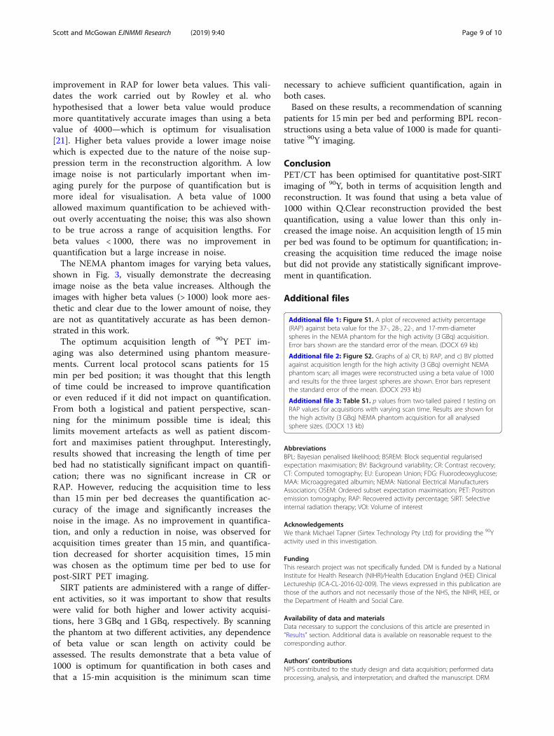
improvement in RAP for lower beta values. This vali-dates the work carried out by Rowley et al. whohypothesised that a lower beta value would producemore quantitatively accurate images than using a betavalue of 4000—which is optimum for visualisation[21]. Higher beta values provide a lower image noisewhich is expected due to the nature of the noise sup-pression term in the reconstruction algorithm. A lowimage noise is not particularly important when im-aging purely for the purpose of quantification but ismore ideal for visualisation. A beta value of 1000allowed maximum quantification to be achieved with-out overly accentuating the noise; this was also shownto be true across a range of acquisition lengths. Forbeta values < 1000, there was no improvement inquantification but a large increase in noise.The NEMA phantom images for varying beta values,
shown in Fig. 3, visually demonstrate the decreasingimage noise as the beta value increases. Although theimages with higher beta values (> 1000) look more aes-thetic and clear due to the lower amount of noise, theyare not as quantitatively accurate as has been demon-strated in this work.The optimum acquisition length of 90Y PET im-
aging was also determined using phantom measure-ments. Current local protocol scans patients for 15min per bed position; it was thought that this lengthof time could be increased to improve quantificationor even reduced if it did not impact on quantification.From both a logistical and patient perspective, scan-ning for the minimum possible time is ideal; thislimits movement artefacts as well as patient discom-fort and maximises patient throughput. Interestingly,results showed that increasing the length of time perbed had no statistically significant impact on quantifi-cation; there was no significant increase in CR orRAP. However, reducing the acquisition time to lessthan 15 min per bed decreases the quantification ac-curacy of the image and significantly increases thenoise in the image. As no improvement in quantifica-tion, and only a reduction in noise, was observed foracquisition times greater than 15 min, and quantifica-tion decreased for shorter acquisition times, 15 minwas chosen as the optimum time per bed to use forpost-SIRT PET imaging.SIRT patients are administered with a range of differ-
ent activities, so it was important to show that resultswere valid for both higher and lower activity acquisi-tions, here 3 GBq and 1 GBq, respectively. By scanningthe phantom at two different activities, any dependenceof beta value or scan length on activity could beassessed. The results demonstrate that a beta value of1000 is optimum for quantification in both cases andthat a 15-min acquisition is the minimum scan time
necessary to achieve sufficient quantification, again inboth cases.Based on these results, a recommendation of scanning
patients for 15 min per bed and performing BPL recon-structions using a beta value of 1000 is made for quanti-tative 90Y imaging.
ConclusionPET/CT has been optimised for quantitative post-SIRTimaging of 90Y, both in terms of acquisition length andreconstruction. It was found that using a beta value of1000 within Q.Clear reconstruction provided the bestquantification, using a value lower than this only in-creased the image noise. An acquisition length of 15 minper bed was found to be optimum for quantification; in-creasing the acquisition time reduced the image noisebut did not provide any statistically significant improve-ment in quantification.
Additional files
Additional file 1: Figure S1. A plot of recovered activity percentage(RAP) against beta value for the 37-, 28-, 22-, and 17-mm-diameterspheres in the NEMA phantom for the high activity (3 GBq) acquisition.Error bars shown are the standard error of the mean. (DOCX 69 kb)
Additional file 2: Figure S2. Graphs of a) CR, b) RAP, and c) BV plottedagainst acquisition length for the high activity (3 GBq) overnight NEMAphantom scan; all images were reconstructed using a beta value of 1000and results for the three largest spheres are shown. Error bars representthe standard error of the mean. (DOCX 293 kb)
Additional file 3: Table S1. p values from two-tailed paired t testing onRAP values for acquisitions with varying scan time. Results are shown forthe high activity (3 GBq) NEMA phantom acquisition for all analysedsphere sizes. (DOCX 13 kb)
AbbreviationsBPL: Bayesian penalised likelihood; BSREM: Block sequential regularisedexpectation maximisation; BV: Background variability; CR: Contrast recovery;CT: Computed tomography; EU: European Union; FDG: Fluorodeoxyglucose;MAA: Microaggregated albumin; NEMA: National Electrical ManufacturersAssociation; OSEM: Ordered subset expectation maximisation; PET: Positronemission tomography; RAP: Recovered activity percentage; SIRT: Selectiveinternal radiation therapy; VOI: Volume of interest
AcknowledgementsWe thank Michael Tapner (Sirtex Technology Pty Ltd) for providing the 90Yactivity used in this investigation.
FundingThis research project was not specifically funded. DM is funded by a NationalInstitute for Health Research (NIHR)/Health Education England (HEE) ClinicalLectureship (ICA-CL-2016-02-009). The views expressed in this publication arethose of the authors and not necessarily those of the NHS, the NIHR, HEE, orthe Department of Health and Social Care.
Availability of data and materialsData necessary to support the conclusions of this article are presented in“Results” section. Additional data is available on reasonable request to thecorresponding author.
Authors’ contributionsNPS contributed to the study design and data acquisition; performed dataprocessing, analysis, and interpretation; and drafted the manuscript. DRM
Scott and McGowan EJNMMI Research (2019) 9:40 Page 9 of 10

contributed to the study design, data acquisition, data interpretation, andmanuscript revision. Both authors read and approved the final manuscript.
Ethics approval and consent to participatePatient data was used in this project in the form of retrospectivelyreconstructed image datasets. This practice does not require informedconsent at Oxford University Hospitals nor was specific ethics approvalrequired.
Consent for publicationNot applicable.
Competing interestsThe authors declare that they have no competing interests.
Publisher’s NoteSpringer Nature remains neutral with regard to jurisdictional claims inpublished maps and institutional affiliations.
Received: 22 February 2019 Accepted: 23 April 2019
References1. Salem R, Thurston KG. Radioembolization with 90Yttrium microspheres: a
state-of-the-art brachytherapy treatment for primary and secondary livermalignancies. J Vasc Interv Radiol. 2006;17(9):1425–39.
2. Ahmadzadehfar H, Sabet A, Biermann K, Muckle M, Brockmann H, Kuhl C, etal. The significance of 99mTc-MAA SPECT/CT liver perfusion imaging intreatment planning for 90Y-microsphere selective internal radiationtreatment. J Nucl Med. 2010;51(8):1206–12.
3. Leung TWT, Lau WY, Ho SKW, Ward SC, Chow JHS, Chan MSY, et al.Radiation pneumonitis after selective internal radiation treatment withintraarterial 90Yttrium-microspheres for inoperable hepatic tumors. Int JRadiat Oncol Biol Phys. 1995;33(4):919–24.
4. Salem R, Parikh P, Atassi B, Lewandowski RJ, Ryu RK, Sato KT, et al. Incidenceof radiation pneumonitis after hepatic intra-arterial radiotherapy withYttrium-90 microspheres assuming uniform lung distribution. Am J ClinOncol. 2008;31(5):431–8.
5. Murthy R, Brown DB, Salem R, Meranze SG, Coldwell DM, Krishnan S, et al.Gastrointestinal complications associated with hepatic arterial Yttrium-90microsphere therapy. J Vasc Interv Radiol. 2007;18(4):553–61.
6. Gates VL, Atassi B, Lewandowski RJ, Ryu RK, Sato KT, Nemcek AA, et al.Radioembolization with Yttrium-90 microspheres: review of an emergingtreatment for liver tumors. Future Oncol. 2007;3:73–81.
7. Dezarn WA, Cessna JT, Dewerd LA, Feng W, Gates VL, Halama J, et al.Recommendations of the American Association of Physicists in Medicine ondosimetry, imaging, and quality assurance procedures for90Y microspherebrachytherapy in the treatment of hepatic malignancies. Med Phys. 2011;38(8):4824–45.
8. Garin E, Rolland Y, Laffont S, Edeline J. Clinical impact of 99mTc-MAA SPECT/CT-based dosimetry in the radioembolization of liver malignancies with90Y-loaded microspheres. Eur J Nucl Med Mol Imaging. 2016;43(3):559–75.
9. Garin E, Lenoir L, Rolland Y, Edeline J, Mesbah H, Laffont S, et al. Dosimetrybased on 99mTc-macroaggregated albumin SPECT/CT accurately predictstumor response and survival in hepatocellular carcinoma patients treatedwith 90Y-loaded glass microspheres: preliminary results. J Nucl Med. 2012;53(2):255–63.
10. Wondergem M, Smits MLJ, Elschot M, de Jong HWAM, Verkooijen HM, vanden Bosch MAAJ, et al. 99mTc-macroaggregated albumin poorly predictsthe intrahepatic distribution of 90Y resin microspheres in hepaticradioembolization. J Nucl Med. 2013;54(8):1294–301.
11. Jiang M, Fischman A, Nowakowski FS. Segmental perfusion differences onpaired Tc-99m macroaggregated albumin (MAA) hepatic perfusion imagingand Yttrium-90 (Y-90) bremsstrahlung imaging studies in SIR-sphereradioembolization: associations with angiography. J Nucl Med Radiat Ther.2012;03(01):1–5.
12. D’Arienzo M. Emission of β+ particles via internal pair production in the 0+− 0+ transition of 90Zr: historical background and current applications innuclear medicine imaging. Atoms. 2013;1(1):2–12.
13. Wright CL, Zhang J, Tweedle MF, Knopp MV, Hall NC. Theranostic imagingof Yttrium-90. Biomed Res Int. 2015;2015:1–11.
14. Kao Y-H, Steinberg JD, Tay Y-S, Lim GK, Yan J, Townsend DW, et al. Post-radioembolization yttrium-90 PET/CT - part 1: diagnostic reporting. EJNMMIRes. 2013;3(1):56.
15. Willowson KP, Tapner M, Bailey DL, Bailey DL. A multicentre comparison ofquantitative 90Y PET/CT for dosimetric purposes after radioembolizationwith resin microspheres. Eur J Nucl Med Mol Imaging. 2015;42(8):1202–22.
16. Miller TR, Wallis JW. Clinically important characteristics of maximum-likelihood reconstruction. J Nucl Med. 1992;33(9):1678–84.
17. Teoh EJ, McGowan DR, Macpherson RE, Bradley KM, Gleeson FV. Phantom andclinical evaluation of the Bayesian penalized likelihood reconstructionalgorithm Q.Clear on an LYSO PET/CT system. J Nucl Med. 2015;56(9):1447–52.
18. Teoh EJ, McGowan DR, Bradley KM, Belcher E, Black E, Gleeson FV. Novelpenalised likelihood reconstruction of PET in the assessment ofhistologically verified small pulmonary nodules. Eur Radiol. 2016;26(2):576–84.
19. Parvizi N, Franklin JM, McGowan DR, Teoh EJ, Bradley KM, Gleeson FV. Doesa novel penalized likelihood reconstruction of 18F-FDG PET-CT improvesignal-to-background in colorectal liver metastases? Eur J Radiol. 2015;84(10):1873–8.
20. Chilcott AK, Bradley KM, McGowan DR. Effect of a Bayesian penalizedlikelihood PET reconstruction compared with ordered subset expectationmaximization on clinical image quality over a wide range of patientweights. Am J Roentgenol. 2018.
21. Rowley LM, Bradley KM, Boardman P, Hallam A, McGowan DR. Optimizationof image reconstruction for 90 Y selective internal radiotherapy on alutetium yttrium Orthosilicate PET/CT system using a Bayesian penalizedlikelihood reconstruction algorithm. J Nucl Med. 2017;58(4):658–64.
22. Goedicke A, Berker Y, Verburg FA, Behrendt FF, Winz O, Mottaghy FM.Study-parameter impact in quantitative 90-yttrium PET imaging forradioembolization treatment monitoring and dosimetry. IEEE Trans MedImaging. 2013;32(3):485–92.
23. European Parliament. Council directive 2013/59/Euratom of 5 December2013 laying down basic safety standards for protection against the dangersarising from exposure to ionising radiation, and repealing directives 89/618/Euratom, 90/641/Euratom, 96/29/Euratom, 97/43/Euratom a. Off J EurCommun L13. 2014;1–73.
Scott and McGowan EJNMMI Research (2019) 9:40 Page 10 of 10