
Optical Imaging as Auxiliary Tool in Skin Cancer Diagnosiscdn.intechopen.com/pdfs/23034.pdf ·...
15
7 Optical Imaging as Auxiliary Tool in Skin Cancer Diagnosis S. Pratavieira, C. T. Andrade, A. G. Salvio, V.S. Bagnato and C. Kurachi Instituto de Física de São Carlos - Universidade de São Paulo, São Carlos, SP, Brasil 1. Introduction Clinical detection of early malignant and premalignant skin lesions is not simple due to their similar features with the more common benign lesions. The traditional skin cancer screening is based on careful whole body visual inspection, and if a suspected lesion is noticed, further examination with biopsy is required. Visual examination relies heavily on the experience and skills of the physician to identify and delineate the initial changes related to carcinogenesis and cancer progression. White light dermatoscope is the most used conventional tool for skin cancer screening, used by dermatologists for a magnified visualization of the skin. Optical imaging at widefield and microscopic levels has been presented as clinical tools for aiding lesion detection and discrimination. Optical techniques are non-invasive, with fast response through an in situ interrogation, and are potentially sensitive to biochemical and structural changes presented in skin cancer development. For skin cancer diagnostics the main used techniques are: widefield imaging, optical spectroscopy and microscopy imaging. Widefield imaging allows the examination of large areas and has the potential of improving detection of occult lesions, margin delimitation, and of guiding biopsy site determination. Optical spectroscopy presents more detailed information on tissue composition, through the light intensity for each collected emission wavelength, which might be later correlated to specific biomolecules. Microscopy imaging has a main advantage of the evaluation of the tissue characteristics at cellular level, but only a small fraction of the lesion volume is interrogated. Distinct optical techniques offer different tissue/cellular information that is complimentary, even though for cancer screening, widefield optical imaging may represent the first best option, when compared to point spectroscopy or optical microscopy. The combination of information from different imaging modalities has the potential of revealing tissue features resulting in an improved diagnostic performance, due to their distinct provided tissue information. In this chapter an overview of the optical clinical methods and the instruments developed for skin cancer detection of published studies will be discussed. 2. Interaction visible light – skin The electromagnetic radiation extends, in order of increasing energy, from radio waves with low energy (long wavelengths), to high-energy radiation (X-ray and gamma radiation). Include regions of radiation with intermediate energies between the ultraviolet and www.intechopen.com
Transcript of Optical Imaging as Auxiliary Tool in Skin Cancer Diagnosiscdn.intechopen.com/pdfs/23034.pdf ·...
7
Optical Imaging as Auxiliary Tool in Skin Cancer Diagnosis
S. Pratavieira, C. T. Andrade, A. G. Salvio, V.S. Bagnato and C. Kurachi Instituto de Física de São Carlos - Universidade de São Paulo, São Carlos, SP,
Brasil
1. Introduction
Clinical detection of early malignant and premalignant skin lesions is not simple due to their similar features with the more common benign lesions. The traditional skin cancer screening is based on careful whole body visual inspection, and if a suspected lesion is noticed, further examination with biopsy is required. Visual examination relies heavily on the experience and skills of the physician to identify and delineate the initial changes related to carcinogenesis and cancer progression. White light dermatoscope is the most used conventional tool for skin cancer screening, used by dermatologists for a magnified visualization of the skin. Optical imaging at widefield and microscopic levels has been presented as clinical tools for aiding lesion detection and discrimination. Optical techniques are non-invasive, with fast response through an in situ interrogation, and are potentially sensitive to biochemical and structural changes presented in skin cancer development. For skin cancer diagnostics the main used techniques are: widefield imaging, optical spectroscopy and microscopy imaging. Widefield imaging allows the examination of large areas and has the potential of improving detection of occult lesions, margin delimitation, and of guiding biopsy site determination. Optical spectroscopy presents more detailed information on tissue composition, through the light intensity for each collected emission wavelength, which might be later correlated to specific biomolecules. Microscopy imaging has a main advantage of the evaluation of the tissue characteristics at cellular level, but only a small fraction of the lesion volume is interrogated. Distinct optical techniques offer different tissue/cellular information that is complimentary, even though for cancer screening, widefield optical imaging may represent the first best option, when compared to point spectroscopy or optical microscopy. The combination of information from different imaging modalities has the potential of revealing tissue features resulting in an improved diagnostic performance, due to their distinct provided tissue information. In this chapter an overview of the optical clinical methods and the instruments developed for skin cancer detection of published studies will be discussed.
2. Interaction visible light – skin
The electromagnetic radiation extends, in order of increasing energy, from radio waves with low energy (long wavelengths), to high-energy radiation (X-ray and gamma radiation). Include regions of radiation with intermediate energies between the ultraviolet and
www.intechopen.com
Skin Cancers – Risk Factors, Prevention and Therapy 160
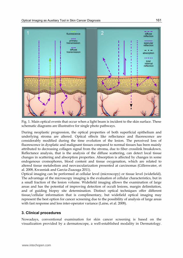
microwave, passing by the visible light. Each of these regions has distinct ways to be produced and detected, and each interacts with matter differently, which may reveal certain characteristics of the system investigated. Due to the distinct energy characteristics, different light/biological tissue interaction occurs. This is the reason, for instance, why the x-rays may induce DNA damage (Karagas, et al. 1996, McLaren, et al. 1975), and the violet light is used for jaundice treatment of new-borns (Bauer, et al. 2004). Optical diagnostic systems are the system that uses the visible light for diagnostics. The visible light or white light is composing by all colors, with the range of wavelength of 400 – 700 nanometers (when the light is understood as a wave). All optical diagnostic systems are based on the one or more types of light-tissue interactions to provide biochemical and/or structural tissue information. This idea relies on the fact that tissue changes alters light-tissue interaction, and if the re-emitted light exiting the tissue is collected and analyzed, a correlation may be obtained. Each specific light wavelength (or color) has a particular interaction with the tissue. When the light is incident in a biological tissue, the main following events occur: a. Specular scattering: the light is reflected at the tissue surface with the same incident
angle relative to the normal orientation. b. Diffuse scattering: the light is diffusely scattered inside the tissue. Due to the different
refractive indexes of biological components, the photon (when the light can be understood as constitute by photons) travelling within the tissue suffers multiple direction changes.
c. Transmission: the light is transmitted through the tissue and may exit without changing its incident direction.
d. Absorption: the light is absorbed by biological chromophores and this energy may result in molecular vibration (heat), and luminescent emission. The most important biological absorbers are hemoglobin and melanin.
e. Luminescence: after light absorption, some excited biological chromophores may lose this excess of energy through light emission (fluorescence and phosphorescence). The prevalent optical luminescent event in biological tissues is the fluorescence.
These are the most important events of the light-tissue interaction (figure 1), but there are many others phenomena that can be explored by an optical technique. Light polarization, Raman scattering, lifetime fluorescence, phase coherence, non-linear effects, are some examples. All these effects can be used to form an optical image. Biological tissues, especially the skin, are considered turbid media, where the diffuse scattering is the most important optical event in the visible range, i.e., the incident photons have multiple scattering before they are absorbed. Biological fluorophores will emit fluorescence in several directions, and these fluorescent photons will scattered and can also be absorbed by other chromophores. Tissue native fluorescence will result from the contribution of all fluorophores present in the interrogated tissue. This detected tissue fluorescence is also influenced by the presence and concentration of absorbers and scatterers. The influence of absorbance and structural tissue properties is even more relevant whereas the collected fluorescence is obtained at the tissue surface result from the exiting photons. The main endogenous fluorophores are tryptophan, nicotinamide adenine dinucleotide (NADH), flavin adenine dinucleotide (FAD), collagen, elastin, and keratin. Main absorbers non-fluorescent are hemoglobin and melanin; and the scatterers are the cell membrane, organelles, and collagen fibers, that indirectly modifies the intensity and shape of the fluorescence collected spectrum. For the imaging widefield devices, the most important light-tissue interactions are the fluorescence and diffuse reflectance (de Veld, et al. 2005).
www.intechopen.com
Optical Imaging as Auxiliary Tool in Skin Cancer Diagnosis 161
Fig. 1. Main optical events that occur when a light beam is incident to the skin surface. These schematic diagrams are illustrative for single photo pathways.
During neoplastic progression, the optical properties of both superficial epithelium and underlying stroma are altered. Optical effects like reflectance and fluorescence are considerably modified during the time evolution of the lesion. The perceived loss of fluorescence in dysplatic and malignant tissues compared to normal tissues has been mainly attributed to decreasing collagen signal from the stroma, due to fiber crosslink breakdown. Reflectance analysis, that is the analysis of the diffuse scattering, can detect local tissue changes in scattering and absorption properties. Absorption is affected by changes in some endogenous cromophores, blood content and tissue oxygenation, which are related to altered tissue metabolism and neovascularization presented at carcinomas (Gillenwater, et al. 2008, Kwasniak and Garcia-Zuazaga 2011). Optical imaging can be performed at cellular level (microscopy) or tissue level (widefield). The advantage of the microscopy imaging is the evaluation of cellular characteristics, but in a small fraction of the lesion volume. Widefield imaging allows the examination of large areas and has the potential of improving detection of occult lesions, margin delimitation, and of guiding biopsy site determination. Distinct optical techniques offer different tissue/cellular information that is complimentary, but widefield optical imaging may represent the best option for cancer screening due to the possibility of analysis of large areas with fast response and less inter-operator variance (Laine, et al. 2008).
3. Clinical procedures
Nowadays, conventional examination for skin cancer screening is based on the visualization provided by a dermatoscope, a well-established modality in Dermatology.
www.intechopen.com
Skin Cancers – Risk Factors, Prevention and Therapy 162
Dermatoscopy (or dermoscopy or epiluminescence microscopy) uses a magnifying lens through an oil/gel interface, the conventional immersion contact dermatoscopy, or cross-polarizing optical filters, the non-contact dermatoscopy. This last one method reduces the amount of reflected light, resulting in the visualization of deeper skin structures. The clinical features that are important for analysis using a dermatoscope are color, dimensions (symmetry), and the presence or absence of certain structures in the lesion. The main idea of this diagnostic tool is to improve the visualization of the skin lesions, based on magnification, so the physician can detect better the clinical features to discriminate and distinguish the lesion types. In this method, the white light is used for this visualization (Ferris and Divito 2010, Tanaka 2006). Total body investigation and time monitoring of suspicious lesions and benign nevi would be the ideal skin screening, but this is unpractical because of the required clinical time. Some studies have showed improved early detection of melanoma when the dermatoscopy is associated with time series photography. The disadvantage of this method, concerning health policies, is the high cost that is around thousands US$ per person. Incidence of new and changed nevi and melanomas detected using baseline images and dermoscopy in patients at high risk for melanoma (Banky, et al. 2003). When the clinician detects a lesion, its visual characteristics as color, architecture, texture, and its palpation are carefully evaluated under white light illumination, the ABCD (Asymmetry, Border, Color, Diameter) rule. If malignant features are suspected, a biopsy is indicated to provide tissue sample for histological analysis, the golden standard for skin cancer diagnostics. In the cases of punch biopsies, the biopsy site is chosen based on the individual clinician criteria to determine the more representative area of the lesion. This procedure is highly subjective and variable comparing dermatologists with distinct experience and training (Kittler, et al. 2011, Bono, et al. 1999). The biopsy is indicated when the clinician recognizes clinical features that are potential indicatives of a malignant condition. At non-homogenous and extent lesions, the biopsy site determination may not be simple. It is not unusual that a lesion shows distinct histological patterns, as different dysplasia degrees, carcinoma in situ, and even invasive carcinoma. As a result, the chosen lesion sample may not be representative, and initial carcinoma can be missed. On the other hand, multiple biopsies at once are not well accepted by the patient, since it is an invasive procedure. After tissue removal, the material is sent to a laboratory for processing and analysis by a pathologist. This method takes time and cannot be performed in situ, and besides the fresh frozen tissue protocol for resected specimens, the pathology result is usually obtained only after a few days (Patel, et al. 2008).
4. Optical systems for aid in skin cancer diagnostics
Visual examination and the methods of the last section relies heavily on the experience and skills of the physician to identify and delineate premalignant and early cancer changes, which is not simple due to the similar clinical characteristics compared to the more common benign lesions. Gold standard diagnostics is obtained only after a tissue sample removal and processing for microscopic evaluation and its classification based on morphological characteristics at cellular level. Enhancing the visual contrast between normal and altered skin has the potential to contribute for biopsy site determination, as well as for lesion margin delimitation before surgical resection. Optical widefield imaging has been presented as an attractive technique
www.intechopen.com
Optical Imaging as Auxiliary Tool in Skin Cancer Diagnosis 163
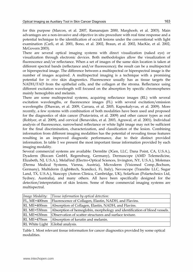
for this purpose (Marcon, et al. 2007, Ramanujam 2000, Marghoob, et al. 2003). Main advantages are a non-invasive and objective in situ procedure with real time response and a potential technique to the identification of occult lesions under the conventional with light examination (Carli, et al. 2001, Bono, et al. 2002, Braun, et al. 2002, MacKie, et al. 2002, McGovern 2003). There are several optical imaging systems with direct visualization (naked eye) or visualization through electronic devices. Both methodologies allow the visualization of fluorescence and/or reflectance. When a set of images of the same skin location is taken at different spectral bands (reflectance and/or fluorescence); the result can be a multispectral or hiperspectral image. The difference between a multispectral or hiperspectral image is the number of images acquired. A multispectral imaging is a technique with a promising potential for in vivo skin diagnostics. Fluorescence usually has as tissue targets the NADH/FAD from the epithelial cells, and the collagen at the stroma. Reflectance using different excitation wavelength will focused on the absorption by specific chromospheres mainly hemoglobin and melanin. There are some multispectral systems, acquiring reflectance images (RL) with several excitation wavelengths, or fluorescence images (FL) with several excitation/emission wavelengths (Dhawan, et al. 2009, Carrara, et al. 2005, Kapsokalyvas, et al. 2009). More recently, a few systems with combination of both modalities have been used and proposed for the diagnostics of skin cancer (Pratavieira, et al. 2009) and other cancer types as oral (Roblyer, et al. 2009), and cervical (Benavides, et al. 2003, Agrawal, et al. 2001). Individual analysis of fluorescence, narrowband reflectance or white light image may not be sufficient for the final discrimination, characterization, and classification of the lesion. Combining information from different imaging modalities has the potential of revealing tissue features resulting in an improved diagnostic performance, due to their distinct provided information. In table 1 we present the most important tissue information provided by each imaging modality. Several commercial systems are available: Dermlite (3Gen, LLC, Dana Point, CA, U.S.A.), Dyaderm (Biocam GmbH, Regensburg, Germany), Dermascope (AMD Telemedicine, Elizabeth, NJ, U.S.A.), MelaFind (Electro-Optical Sciences, Irvington, NY, U.S.A.), Molemax (Derma Medical Systems, Vienna, Austria), Microderm (Visiomed Comp.,Bochum, Germany), Multiderm (Light4tech, Scandicci, Fi, Italy), Nevoscope (Translite LLC, Sugar Land, TX, U.S.A.), Siascopy (Astron Clinica, Cambridge, UK), SolarScan (Polartechnics Ltd, Sydney, Australia), and many others. All have been specifically designed for the detection/interpretation of skin lesions. Some of those commercial imaging systems are multispectral. Image Modality: Tissue information by optical detection:
FL, ME=400nm Fluorescence of Collagen, Elastin, NADH, and Flavins.
RL ME=400nm Absorption of Collagen, Elastin, NADH, and Flavins.
RL ME=530nm Absorption of hemoglobin, morphology and identification of blood vessels.
RL ME=630nm Observation of scatter structures and surface texture.
RL ME=470nm Absorption of keratin and melanin.
RL White Light Global analysis.
Table 1. Most relevant tissue information for cancer diagnostics provided by some optical modalities.
www.intechopen.com
Skin Cancers – Risk Factors, Prevention and Therapy 164
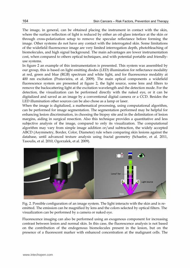
The image, in general, can be obtained placing the instrument in contact with the skin, where the surface reflection of light is reduced by either an oil-glass interface at the skin or through cross-polarization setup to remove the specular reflectance before forming the image. Other systems do not have any contact with the interrogated skin. Some limitations of the widefield fluorescence image are very limited interrogation depth, photobleaching of biomolecules, and high signal background. The main advantages are lower instrumentation cost, when compared to others optical techniques, and with potential portable and friendly-use systems. In figure 2 an example of this instrumentation is presented. This system was assembled by our group, this is based on light emitting diodes (LED) illumination for reflectance modality at red, green and blue (RGB) spectrum and white light, and for fluorescence modality at 400 nm excitation (Pratavieira, et al. 2009). The main optical components a widefield fluorescence system are presented at figure 2, the light source, some lens and filters to remove the backscattering light at the excitation wavelength and the detection mode. For the detection, the visualization can be performed directly with the naked eye, or it can be digitalized and saved as an image by a conventional digital camera or a CCD. Besides the LED illumination other sources can be also chose as a lamp or laser. When the image is digitalized, a mathematical processing, using computational algorithm, can be performed for image segmentation. The segmentation performed may be helpful for enhancing lesion discrimination, in choosing the biopsy site and in the delimitation of lesion margins, aiding in surgical resection. Also this technique provides a quantitative and less subjective analysis of the image, compared to only its visualization. The computational algorithm may vary from simple image addition or/and subtraction, the widely accepted ABCD (Asymmetry, Border, Color, Diameter) rule when comparing skin lesions against the database, until advanced texture analysis using fractal geometry (Schaefer, et al. 2011, Tasoulis, et al. 2010, Ogorzalek, et al. 2009).
Fig. 2. Possible configuration of an image system. The light interacts with the skin and is re-emitted. The emission can be magnified by lens and the colors selected by optical filters. The visualization can be performed by a camera or naked eye.
Fluorescence imaging can also be performed using an exogenous component for increasing contrast between lesion and normal skin. In this case, the fluorescence analysis is not based on the contribution of the endogenous biomolecules present in the lesion, but on the presence of a fluorescent marker with enhanced concentration at the malignant cells. The
www.intechopen.com
Optical Imaging as Auxiliary Tool in Skin Cancer Diagnosis 165
most common technique is the previous topical application of a 15-20% aminolevulinic acid (ALA) or metil-ALA cream, a pro-drug for photodynamic therapy. ALA or Me-ALA enhances the cellular production of protoporphyrin IX (PpIX), a fluorescent biomolecule. When the pretreated tissue with ALA or Me-ALA is visualized with widefield fluorescence imaging, a strong red fluorescence, originated from the produced PpIX, lesions discrimination is improved. This technique is known as photodynamic detection (Hewett, et al. 2000, Orenstein, et al. 1998, Hewett, et al. 2001).
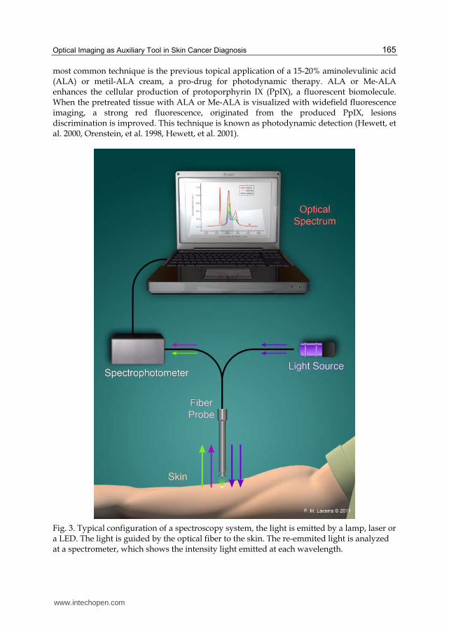
Fig. 3. Typical configuration of a spectroscopy system, the light is emitted by a lamp, laser or a LED. The light is guided by the optical fiber to the skin. The re-emmited light is analyzed at a spectrometer, which shows the intensity light emitted at each wavelength.
www.intechopen.com
Skin Cancers – Risk Factors, Prevention and Therapy 166
Another commonly investigated technique is the point spectroscopy by fluorescence (FL) and/or reflectance (RL), with single or multiple wavelengths. In this case, the result is not an image, but a graph showing the spectral information present at the re-emitted light. The spectroscopy methods show good specificity and sensitivity on cancer discrimination however, the obtained information is punctual, which makes the analysis difficult for large areas and the diagnostics is dependent on and specific for the interrogated site. Point spectroscopy uses optical fibers that deliver the excitation light to the tissue surface and collect the re-emitted light. Depending on the spectrometer and optical design, fluorescence or reflectance is investigated. The result showed after this optical measurement is a graph with the emitted light intensity as a function of the wavelength (Sterenborg, et al. 1994, Utz, et al. 1994, Kollias, et al. 2000, Ramanujam 2000, Panjehpour, et al. 2002, Panjehpour, et al. 2002). In figure 3 a typical configuration of this setup is presented. Microscopy imaging is another punctual technique. For skin investigation, common systems are confocal scanning microscopy, and multiphoton tomography, where a laser is used to excite the skin fluorophores, or scatterers components. For one or two photon excitation some commercial system are available such as Lucid's VivaScope (Rochester, NY, U.S.A.), and DermaInspect (JenLab GmbH, Jena, Germany). This technique has a great advantage in comparison with the others (widefield, spectroscopy) because in this method, the image resolution is at cellular level. For some instrumentation, the cellular and tissue features may be similar to what is visualized by histological slides. Characteristics used for conventional diagnostics as cellular shape, nuclei structure and tissue organization may be also obtained. The microscopy images has the advantage of very high resolution, very low out-of-focus signal, but the main limitation is still the high cost (Roberts, et al. 2011, Koehler, et al. 2011, Garbe, et al. 2011, Breunig, et al. 2010).
5. Widefield fluorescence image in skin cancer diagnostics
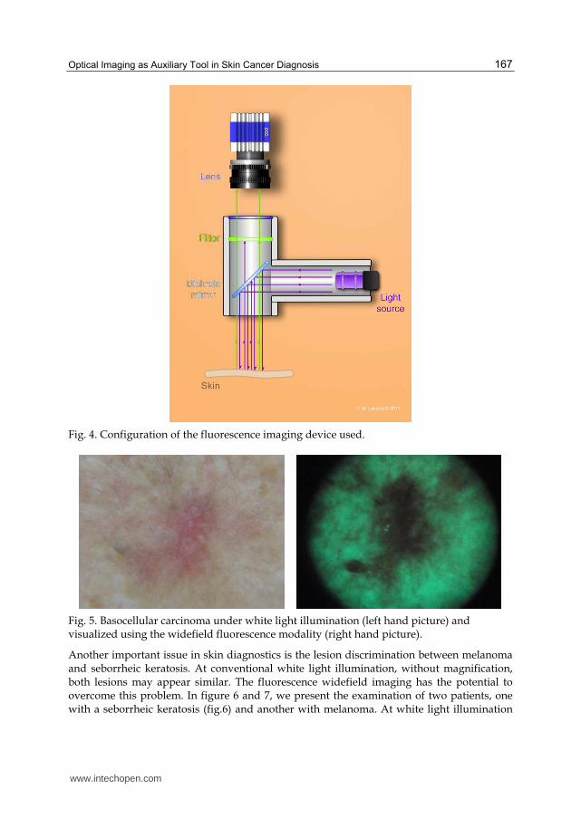
In this section, we are going to present a widefield fluorescence device assembled by our group and some of the images taken at patients with skin lesions. The illumination device is based on an array of two LEDs composing an UV-blue range light. This spectral region was chosen for its already well-known use to excite endogenous fluorophores at skin. Light intensity is around 50 mW/cm2. A handheld device enclose the illumination part and also the optical components to provide fluorescence imaging: a dichroic mirror, a longpass filter to remove the light at excitation wavelengths, and another filter to remove the yellow spectral range. This set of filters allows the fluorescence viewing in the green and red range (Costa, et al. 2010). A schematic diagram of the configuration of the widefield fluorescence image is presented at figure 4. An example of the obtained fluorescence image is showed at figure 5. A basocellular carcinoma lesion at a Caucasian patient was visualized using the system. The comparison between the discrimination provided at a white light illumination and the fluorescence imaging can be observed. The cancer lesion is usually visualized at the fluorescence image as a darker region, correlated with the less tissue fluorescence, mainly due to the changes on collagen structure induced by the carcinoma progression. The tumor margins can be better discriminate using the fluorescence, the contrast between lesion and normal tissue is enhanced.
www.intechopen.com
Optical Imaging as Auxiliary Tool in Skin Cancer Diagnosis 167
Fig. 4. Configuration of the fluorescence imaging device used.
Fig. 5. Basocellular carcinoma under white light illumination (left hand picture) and visualized using the widefield fluorescence modality (right hand picture).
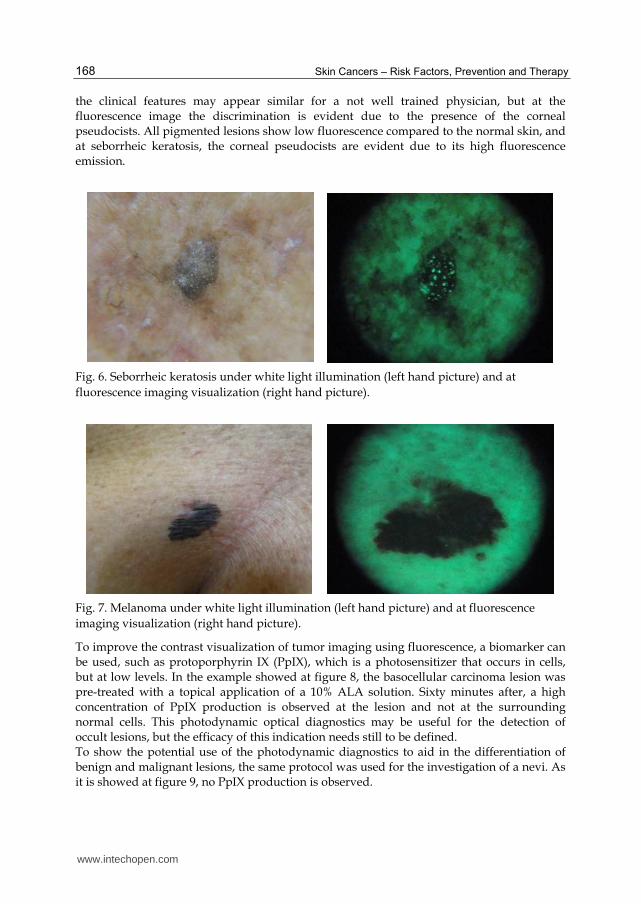
Another important issue in skin diagnostics is the lesion discrimination between melanoma and seborrheic keratosis. At conventional white light illumination, without magnification, both lesions may appear similar. The fluorescence widefield imaging has the potential to overcome this problem. In figure 6 and 7, we present the examination of two patients, one with a seborrheic keratosis (fig.6) and another with melanoma. At white light illumination
www.intechopen.com
Skin Cancers – Risk Factors, Prevention and Therapy 168
the clinical features may appear similar for a not well trained physician, but at the fluorescence image the discrimination is evident due to the presence of the corneal pseudocists. All pigmented lesions show low fluorescence compared to the normal skin, and at seborrheic keratosis, the corneal pseudocists are evident due to its high fluorescence emission.
Fig. 6. Seborrheic keratosis under white light illumination (left hand picture) and at
fluorescence imaging visualization (right hand picture).
Fig. 7. Melanoma under white light illumination (left hand picture) and at fluorescence
imaging visualization (right hand picture).
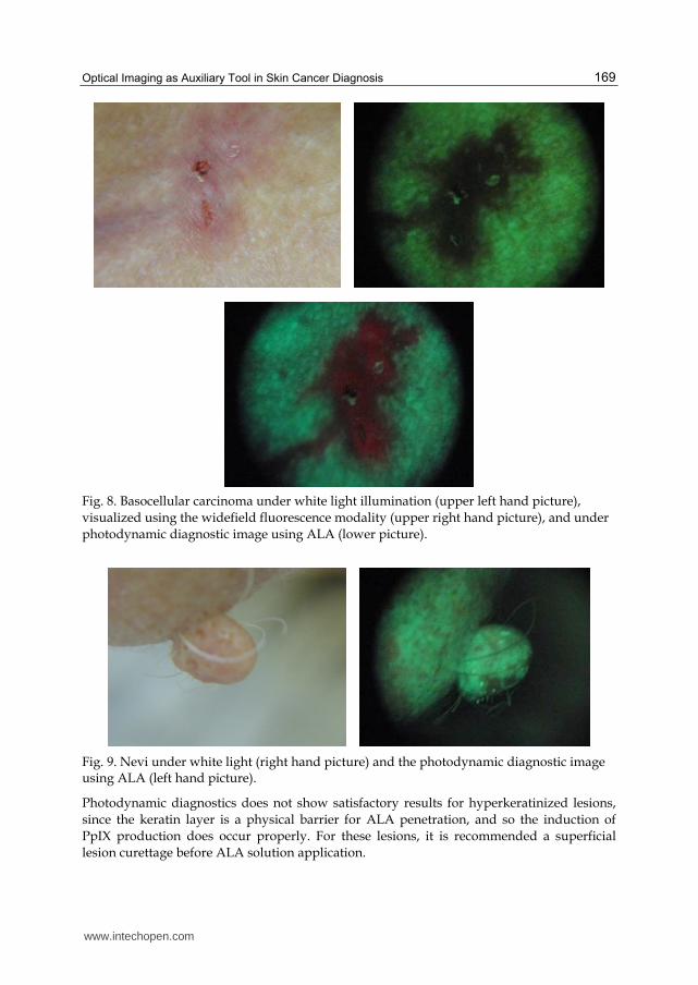
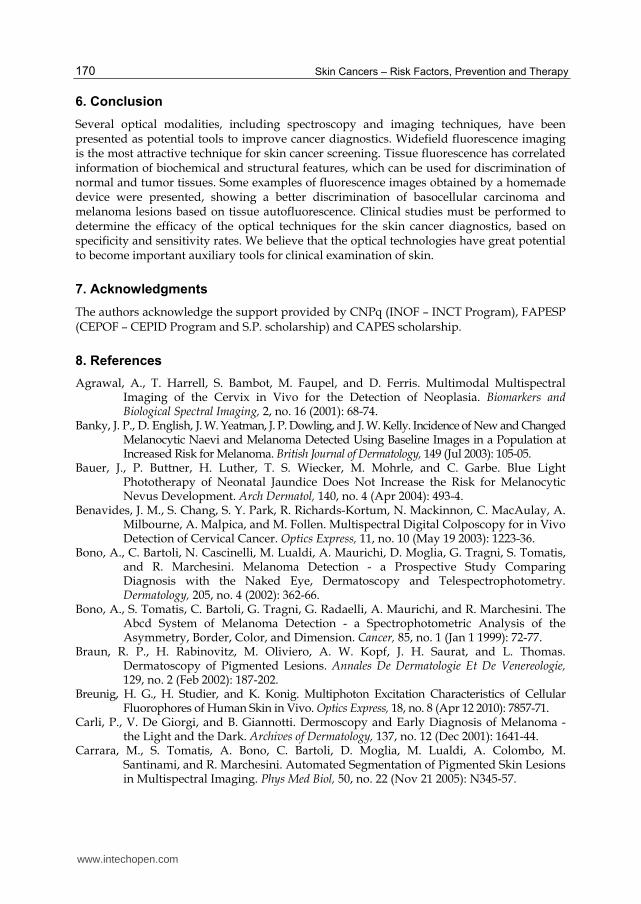
To improve the contrast visualization of tumor imaging using fluorescence, a biomarker can be used, such as protoporphyrin IX (PpIX), which is a photosensitizer that occurs in cells, but at low levels. In the example showed at figure 8, the basocellular carcinoma lesion was pre-treated with a topical application of a 10% ALA solution. Sixty minutes after, a high concentration of PpIX production is observed at the lesion and not at the surrounding normal cells. This photodynamic optical diagnostics may be useful for the detection of occult lesions, but the efficacy of this indication needs still to be defined. To show the potential use of the photodynamic diagnostics to aid in the differentiation of benign and malignant lesions, the same protocol was used for the investigation of a nevi. As it is showed at figure 9, no PpIX production is observed.
www.intechopen.com
Optical Imaging as Auxiliary Tool in Skin Cancer Diagnosis 169
Fig. 8. Basocellular carcinoma under white light illumination (upper left hand picture), visualized using the widefield fluorescence modality (upper right hand picture), and under photodynamic diagnostic image using ALA (lower picture).
Fig. 9. Nevi under white light (right hand picture) and the photodynamic diagnostic image using ALA (left hand picture).
Photodynamic diagnostics does not show satisfactory results for hyperkeratinized lesions, since the keratin layer is a physical barrier for ALA penetration, and so the induction of PpIX production does occur properly. For these lesions, it is recommended a superficial lesion curettage before ALA solution application.
www.intechopen.com
Skin Cancers – Risk Factors, Prevention and Therapy 170
6. Conclusion
Several optical modalities, including spectroscopy and imaging techniques, have been presented as potential tools to improve cancer diagnostics. Widefield fluorescence imaging is the most attractive technique for skin cancer screening. Tissue fluorescence has correlated information of biochemical and structural features, which can be used for discrimination of normal and tumor tissues. Some examples of fluorescence images obtained by a homemade device were presented, showing a better discrimination of basocellular carcinoma and melanoma lesions based on tissue autofluorescence. Clinical studies must be performed to determine the efficacy of the optical techniques for the skin cancer diagnostics, based on specificity and sensitivity rates. We believe that the optical technologies have great potential to become important auxiliary tools for clinical examination of skin.
7. Acknowledgments
The authors acknowledge the support provided by CNPq (INOF – INCT Program), FAPESP (CEPOF – CEPID Program and S.P. scholarship) and CAPES scholarship.
8. References
Agrawal, A., T. Harrell, S. Bambot, M. Faupel, and D. Ferris. Multimodal Multispectral Imaging of the Cervix in Vivo for the Detection of Neoplasia. Biomarkers and Biological Spectral Imaging, 2, no. 16 (2001): 68-74.
Banky, J. P., D. English, J. W. Yeatman, J. P. Dowling, and J. W. Kelly. Incidence of New and Changed Melanocytic Naevi and Melanoma Detected Using Baseline Images in a Population at Increased Risk for Melanoma. British Journal of Dermatology, 149 (Jul 2003): 105-05.
Bauer, J., P. Buttner, H. Luther, T. S. Wiecker, M. Mohrle, and C. Garbe. Blue Light Phototherapy of Neonatal Jaundice Does Not Increase the Risk for Melanocytic Nevus Development. Arch Dermatol, 140, no. 4 (Apr 2004): 493-4.
Benavides, J. M., S. Chang, S. Y. Park, R. Richards-Kortum, N. Mackinnon, C. MacAulay, A. Milbourne, A. Malpica, and M. Follen. Multispectral Digital Colposcopy for in Vivo Detection of Cervical Cancer. Optics Express, 11, no. 10 (May 19 2003): 1223-36.
Bono, A., C. Bartoli, N. Cascinelli, M. Lualdi, A. Maurichi, D. Moglia, G. Tragni, S. Tomatis, and R. Marchesini. Melanoma Detection - a Prospective Study Comparing Diagnosis with the Naked Eye, Dermatoscopy and Telespectrophotometry. Dermatology, 205, no. 4 (2002): 362-66.
Bono, A., S. Tomatis, C. Bartoli, G. Tragni, G. Radaelli, A. Maurichi, and R. Marchesini. The Abcd System of Melanoma Detection - a Spectrophotometric Analysis of the Asymmetry, Border, Color, and Dimension. Cancer, 85, no. 1 (Jan 1 1999): 72-77.
Braun, R. P., H. Rabinovitz, M. Oliviero, A. W. Kopf, J. H. Saurat, and L. Thomas. Dermatoscopy of Pigmented Lesions. Annales De Dermatologie Et De Venereologie, 129, no. 2 (Feb 2002): 187-202.
Breunig, H. G., H. Studier, and K. Konig. Multiphoton Excitation Characteristics of Cellular Fluorophores of Human Skin in Vivo. Optics Express, 18, no. 8 (Apr 12 2010): 7857-71.
Carli, P., V. De Giorgi, and B. Giannotti. Dermoscopy and Early Diagnosis of Melanoma - the Light and the Dark. Archives of Dermatology, 137, no. 12 (Dec 2001): 1641-44.
Carrara, M., S. Tomatis, A. Bono, C. Bartoli, D. Moglia, M. Lualdi, A. Colombo, M. Santinami, and R. Marchesini. Automated Segmentation of Pigmented Skin Lesions in Multispectral Imaging. Phys Med Biol, 50, no. 22 (Nov 21 2005): N345-57.
www.intechopen.com
Optical Imaging as Auxiliary Tool in Skin Cancer Diagnosis 171
Costa, M. M. ;, C. ; Kurachi, V. S. ; Bagnato, and L. Ventura. "Development of an Optical Fluorescence Imaging System for Medical Use." Paper presented at the 19th International Laser Physics Workshop, 2010, Foz do Iguaçu. BOOK OF ABSTRACTS, 2010. p. 123-123., 2010.
de Veld, D. C. G., M. Skurichina, M. J. H. Wities, R. P. W. Duin, H. J. C. M. Sterenborg, and J. L. N. Roodenburg. Autofluorescence and Diffuse Reflectance Spectroscopy for Oral Oncology. Lasers Surg Med, 36, no. 5 (Jun 2005): 356-64.
Dhawan, A. P., B. D'Alessandro, S. Patwardhan, and N. Mullani. Multispectral Optical Imaging of Skin-Lesions for Detection of Malignant Melanomas. Conf Proc IEEE Eng Med Biol Soc, 2009 (2009): 5352-5.
Ferris, L. K., and S. J. Divito. Advances and Short Comings in the Early Diagnosis of Melanoma. Melanoma Research, 20, no. 6 (Dec 2010): 450-58.
Garbe, C., D. Leupold, M. Scholz, G. Stankovic, J. Reda, S. Buder, R. Eichhorn, et al. The Stepwise Two-Photon Excited Melanin Fluorescence Is a Unique Diagnostic Tool for the Detection of Malignant Transformation in Melanocytes. Pigment Cell & Melanoma Research, 24, no. 3 (Jun 2011): 438-45.
Gillenwater, A., I. Pavlova, M. Williams, A. El-Naggar, and R. Richards-Kortum. Understanding the Biological Basis of Autofluorescence Imaging for Oral Cancer Detection: High-Resolution Fluorescence Microscopy in Viable Tissue. Clinical Cancer Research, 14, no. 8 (Apr 15 2008): 2396-404.
Hewett, J., T. McKechnie, and W. Sibbett. Fluorescence Detection of Superficial Skin Cancers. Journal of Modern Optics, 47, no. 11 (Sep 2000): 2021-27.
Hewett, J., V. Nadeau, J. Ferguson, H. Moseley, S. Ibbotson, L. Brancaleon, D. Birkin, W. Sibbett, and M. Padgett. Multispectral Monitoring of Fluorescence During Ala-Pdt of Superficial Skin Cancers. Optical Methods for Tumor Treatment and Detection: Mechanisms and Techniques in Photodynamic Therapy X, 2, no. 5 (2001): 68-75.
Kapsokalyvas, D., N. Bruscino, G. Cannarozzo, V. de Giorgi, T. Lotti, and F. S. Pavone. Multispectral Dermoscope. Clinical and Biomedical Spectroscopy, 7368 (2009).
Karagas, M. R., J. A. McDonald, E. R. Greenberg, T. A. Stukel, J. E. Weiss, J. A. Baron, and M. M. Stevens. Risk of Basal Cell and Squamous Cell Skin Cancers after Ionizing Radiation Therapy. For the Skin Cancer Prevention Study Group. J Natl Cancer Inst, 88, no. 24 (Dec 18 1996): 1848-53.
Kittler, H., C. Rosendahl, P. Tschandl, C. Med, and A. Cameron. Diagnostic Accuracy of Dermatoscopy for Melanocytic and Nonmelanocytic Pigmented Lesions. Journal of the American Academy of Dermatology, 64, no. 6 (Jun 2011): 1068-73.
Koehler, M. J., M. Speicher, S. Lange-Asschenfeldt, E. Stockfleth, S. Metz, P. Elsner, M. Kaatz, and K. Konig. Clinical Application of Multiphoton Tomography in Combination with Confocal Laser Scanning Microscopy for in Vivo Evaluation of Skin Diseases. Experimental Dermatology, 20, no. 7 (Jul 2011): 589-94.
Kollias, N., R. Gillies, G. Zonios, and R. R. Anderson. Fluorescence Excitation Spectroscopy Provides Information About Human Skin in Vivo. Journal of Investigative Dermatology, 115, no. 4 (Oct 2000): 704-07.
Kwasniak, L. A., and J. Garcia-Zuazaga. Basal Cell Carcinoma: Evidence-Based Medicine and Review of Treatment Modalities. International Journal of Dermatology, 50, no. 6 (Jun 2011): 645-58.
Laine, A. F., A. R. Kherlopian, T. Song, Q. Duan, M. A. Neimark, M. J. Po, and J. K. Gohagan. A Review of Imaging Techniques for Systems Biology. Bmc Systems Biology, 2 (Aug 12 2008).
www.intechopen.com
Skin Cancers – Risk Factors, Prevention and Therapy 172
MacKie, R. M., C. Fleming, A. D. McMahon, and P. Jarrett. The Use of the Dermatoscope to Identify Early Melanoma Using the Three-Colour Test. British Journal of Dermatology, 146, no. 3 (Mar 2002): 481-84.
Marcon, N. E., R. S. DaCosta, and B. C. Wilson. Fluorescence and Spectral Imaging. Thescientificworldjournal, 7 (2007): 2046-71.
Marghoob, A. A., L. D. Swindle, C. Z. Moricz, F. A. Sanchez Negron, B. Slue, A. C. Halpern, and A. W. Kopf. Instruments and New Technologies for the in Vivo Diagnosis of Melanoma. J Am Acad Dermatol, 49, no. 5 (Nov 2003): 777-97; quiz 98-9.
McGovern, V. Digital Diagnosis: New Tool for Detecting Skin Cancer. Environ Health Perspect, 111, no. 14 (Nov 2003): A770-3.
McLaren, J. R., A. J. Maruszczak, and Z. L. Olkowski. Short Term Effects of Ionizing Radiation on the Recurrent or Metastatic Skin Cancer--a Quantitative DNA Study. Strahlentherapie, 149, no. 2 (Feb 1975): 166-72.
Orenstein, A., G. Kostenich, C. Rothmann, Barshack I, and Z. Malik. Imaging of Human Skin Lesions Using Multipixel Fourier Transform Spectroscopy. Lasers in Medical Science, 13, no. 2 (1998): 112-18.
Panjehpour, M., C. E. Julius, M. N. Phan, T. Vo-Dinh, and S. Overholt. Laser-Induced Fluorescence Spectroscopy for in Vivo Diagnosis of Non-Melanoma Skin Cancers. Lasers in Surgery and Medicine, 31, no. 5 (2002): 367-73.
Panjehpour, M., C. Julius, M. N. Phan, T. Vo-Dinh, and S. Overholt. In Vivo Fluorescence Spectroscopy for Diagnosis of Skin Cancer. Biomedical Diagnostic, Guidance, and Surgical-Assist Systems Iv, 4615 (2002): 20-31.
Patel, J. K., S. Konda, O. A. Perez, S. Amini, G. Elgart, and B. Berman. Newer Technologies/Techniques and Tools in the Diagnosis of Melanoma. European Journal of Dermatology, 18, no. 6 (Nov-Dec 2008): 617-31.
Pratavieira, S., P. L. A. Santos, V. S. Bagnato, and C. Kurachi. "Development of a Widefield Reflectance and Fluorescence Imaging Device for the Detection of Skin and Oral Cancer." In Photodynamic Therapy: Back to the Future. Seattle: edited by David H. Kessel, Proceedings of SPIE Vol. 7380 (SPIE, Bellingham, WA 2009) 73805G, 2009.
Ramanujam, N. Fluorescence Spectroscopy of Neoplastic and Non-Neoplastic Tissues. Neoplasia, 2, no. 1-2 (Jan-Apr 2000): 89-117.
Roberts, MS Roberts, M. S., Y. Dancik, T. W. Prow, C. A. Thorling, L. L. Lin, J. E. Grice, T. A. Robertson, K. Konig, and W. Becker. Non-Invasive Imaging of Skin Physiology and Percutaneous Penetration Using Fluorescence Spectral and Lifetime Imaging with Multiphoton and Confocal Microscopy. European Journal of Pharmaceutics and Biopharmaceutics, 77, no. 3 (Apr 2011): 469-88.
Roblyer, D., C. Kurachi, A. M. Gillenwater, and R. Richards-Kortum. In Vivo Fluorescence Hyperspectral Imaging of Oral Neoplasia. Advanced Biomedical and Clinical Diagnostic Systems Vii, 7169 (2009).
Sterenborg, H. J. C. M., M. Motamedi, R. F. Wagner, M. Duvic, S. Thomsen, and S. L. Jacques. In-Vivo Fluorescence Spectroscopy and Imaging of Human Skin Tumors. Lasers in Medical Science, 9, no. 3 (Sep 1994): 191-201.
Tanaka, M. Dermoscopy. Journal of Dermatology, 33, no. 8 (Aug 2006): 513-17. Utz, S. R., Y. P. Sinichkin, I. V. Meglinski, and H. A. Pilipenko. Fluorescence Spectroscopy in
Combine with Reflectance Measurements in Human Skin Examination - What for and How. Optical Biopsy and Fluorescence Spectroscopy and Imaging, Proceedings Of, (1994): 125-36.
www.intechopen.com
Skin Cancers - Risk Factors, Prevention and TherapyEdited by Prof. Caterina La Porta
ISBN 978-953-307-722-2Hard cover, 272 pagesPublisher InTechPublished online 14, November, 2011Published in print edition November, 2011
InTech EuropeUniversity Campus STeP Ri Slavka Krautzeka 83/A 51000 Rijeka, Croatia Phone: +385 (51) 770 447 Fax: +385 (51) 686 166www.intechopen.com
InTech ChinaUnit 405, Office Block, Hotel Equatorial Shanghai No.65, Yan An Road (West), Shanghai, 200040, China
Phone: +86-21-62489820 Fax: +86-21-62489821
Skin cancers are the fastest growing type of cancer in the United States and represent the most commonlydiagnosed malignancy, surpassing lung, breast, colorectal and prostate cancer. In Europe, the British Isleshave been the highest rates of skin cancer in children and adolescents. The overall idea of this book is toprovide the reader with up to date information on the possible tools to use for prevention, diagnosis andtreatment of skin cancer. Three main issues are discussed: risk factors, new diagnostic tools for preventionand strategies for prevention and treatment of skin cancer using natural compounds or nano-particle drugdelivery and photodynamic therapy.
How to referenceIn order to correctly reference this scholarly work, feel free to copy and paste the following:
S. Pratavieira, C. T. Andrade, A. G. Salvio, V.S. Bagnato and C. Kurachi (2011). Optical Imaging as AuxiliaryTool in Skin Cancer Diagnosis, Skin Cancers - Risk Factors, Prevention and Therapy, Prof. Caterina La Porta(Ed.), ISBN: 978-953-307-722-2, InTech, Available from: http://www.intechopen.com/books/skin-cancers-risk-factors-prevention-and-therapy/optical-imaging-as-auxiliary-tool-in-skin-cancer-diagnosis