optical coherence tomography, and ultrasound biomicroscopy ...
4
Case Report Cogan-Reese syndrome: image analysis with specular microscopy, optical coherence tomography, and ultrasound biomicroscopy Denise Loya-Garcia, MD, Julio C. Hernandez-Camarena, MD, Jorge E. Valdez-Garcia, MD, PhD, and Alejandro Rodríguez-Garcia, MD Author affiliations: Tecnologico de Monterrey, School of Medicine and Health Sciences, Institute of Ophthalmology and Visual Sciences, Monterrey, Mexico Summary Iridocorneal endothelial (ICE) syndrome is a progressive clinical spectrum of corneal endothelial abnor‐ malities affecting the cornea, iris, and iridocorneal angle. Three clinical variations are recognized: essential (progressive) iris atrophy, Chandler syndrome, and Cogan-Reese syndrome. Direct slit-lamp visualization of the cornea and anterior segment in cases of ICE syndrome is inadequate for precise and objective assess‐ ment of the affected structures. We describe the evolution of corneal and anterior segment structural changes in a woman with Cogan-Reese syndrome using three different methods of image analysis: specular microscopy, anterior segment optical coherence tomography, and ultrasound biomicroscopy. Introduction Cogan-Reese syndrome is a clinical variation of the iri‐ docorneal endothelial (ICE) syndrome characterized by multiple small pedunculated round nodules on the ante‐ rior iris surface, iris atrophy, heterochromia, ectropion uveae, and ectopic pupil, as well as corneal edema and iridocorneal angle abnormalities. 1–5 Anterior segment slit-lamp visualization becomes limited as the disease progresses due to corneal stromal edema. The use of imaging technologies such as specular microscopy (SM), anterior segment spectral domain optical coher‐ ence tomography (SD-OCT), and ultrasound biomicro‐ scopy (UBM) allow an objective analysis of the progres‐ sion, grade, and staging of the disease. Hence, these imaging technologies can help with surgical planning in advanced cases. 6 Case Report A 46-year-old woman presented at the Institute of Oph‐ thalmology and Visual Sciences, Monterrey, Mexico, with a complaint of intense photophobia and blurred vision in the mornings in her right eye. Her medical his‐ tory was significant for glaucoma in her right eye, trea‐ ted with a trabeculectomy 5 years previously. At her ini‐ tial visit, she was on the following topical regimen: dor‐ zolamide and timolol twice daily, sodium chloride 5% three times daily, prednisolone acetate 1% six times daily, and nepafenac 0.1% twice daily. Corrected dis‐ tance visual acuity was 20/50 in the right eye and 20/20 in the left eye. Intraocular pressure (IOP) by Goldmann tonometry was 21 mm Hg in the right eye and 15 mm Hg in the left eye. On slit-lamp examination, the right eye showed a flat filtration bleb in the superior quadrant, mild corneal stromal edema, and a beaten-metal appear‐ ance in the posterior corneal surface, with central pre‐ dominance (Figure 1A); multiple pedunculated and pig‐ mented iris nodules surrounded by stromal atrophy areas were observed (Figure 1B–C). Gonioscopy revealed a narrow iridocorneal angle (Shaffer II, inferior and nasal quadrants; Shaffer III, temporal and superior quadrants), with peripheral anterior synechiae of the iris. Fundus examination of the right eye showed an optic nerve with a cup-to-disc ratio of 0.7 and no other alterations. The left eye examination was unremarkable. Corneal and anterior segment SD-OCT (RTVue-100, Optovue, Fremont, CA) and SM (Specular Microscopy EM-3000, Tomey Corp, Nagoya, Japan) were performed Published May 31, 2019. Copyright ©2019. All rights reserved. Reproduction in whole or in part in any form or medium without expressed written permission of the Digital Journal of Ophthalmology is prohibited. doi:10.5693/djo.02.2019.05.001 Correspondence: Alejandro Rodriguez-Garcia, MD, Instituto de Oftalmologia y Ciencias Visuales Centro Medico Zambrano Hellion, Av. Batal‐ lón de San Patricio No. 112. Col. Real de San Agustin, Garza Garcia, N.L. CP. 62670 Mexico (email: [email protected]). Digital Journal of Ophthalmology, Vol. 25 Digital Journal of Ophthalmology, Vol. 25
Transcript of optical coherence tomography, and ultrasound biomicroscopy ...
Case ReportCogan-Reese syndrome: image analysis with specular microscopy,optical coherence tomography, and ultrasound biomicroscopyDenise Loya-Garcia, MD, Julio C. Hernandez-Camarena, MD, Jorge E. Valdez-Garcia, MD, PhD, andAlejandro Rodríguez-Garcia, MDAuthor affiliations: Tecnologico de Monterrey, School of Medicine and Health Sciences, Institute of Ophthalmology and VisualSciences, Monterrey, Mexico
SummaryIridocorneal endothelial (ICE) syndrome is a progressive clinical spectrum of corneal endothelial abnor‐malities affecting the cornea, iris, and iridocorneal angle. Three clinical variations are recognized: essential(progressive) iris atrophy, Chandler syndrome, and Cogan-Reese syndrome. Direct slit-lamp visualizationof the cornea and anterior segment in cases of ICE syndrome is inadequate for precise and objective assess‐ment of the affected structures. We describe the evolution of corneal and anterior segment structuralchanges in a woman with Cogan-Reese syndrome using three different methods of image analysis: specularmicroscopy, anterior segment optical coherence tomography, and ultrasound biomicroscopy.
IntroductionCogan-Reese syndrome is a clinical variation of the iri‐docorneal endothelial (ICE) syndrome characterized bymultiple small pedunculated round nodules on the ante‐rior iris surface, iris atrophy, heterochromia, ectropionuveae, and ectopic pupil, as well as corneal edema andiridocorneal angle abnormalities.1–5 Anterior segmentslit-lamp visualization becomes limited as the diseaseprogresses due to corneal stromal edema. The use ofimaging technologies such as specular microscopy(SM), anterior segment spectral domain optical coher‐ence tomography (SD-OCT), and ultrasound biomicro‐scopy (UBM) allow an objective analysis of the progres‐sion, grade, and staging of the disease. Hence, theseimaging technologies can help with surgical planning inadvanced cases.6
Case ReportA 46-year-old woman presented at the Institute of Oph‐thalmology and Visual Sciences, Monterrey, Mexico,with a complaint of intense photophobia and blurredvision in the mornings in her right eye. Her medical his‐tory was significant for glaucoma in her right eye, trea‐ted with a trabeculectomy 5 years previously. At her ini‐
tial visit, she was on the following topical regimen: dor‐zolamide and timolol twice daily, sodium chloride 5%three times daily, prednisolone acetate 1% six timesdaily, and nepafenac 0.1% twice daily. Corrected dis‐tance visual acuity was 20/50 in the right eye and 20/20in the left eye. Intraocular pressure (IOP) by Goldmanntonometry was 21 mm Hg in the right eye and 15 mmHg in the left eye. On slit-lamp examination, the righteye showed a flat filtration bleb in the superior quadrant,mild corneal stromal edema, and a beaten-metal appear‐ance in the posterior corneal surface, with central pre‐dominance (Figure 1A); multiple pedunculated and pig‐mented iris nodules surrounded by stromal atrophy areaswere observed (Figure 1B–C). Gonioscopy revealed anarrow iridocorneal angle (Shaffer II, inferior and nasalquadrants; Shaffer III, temporal and superior quadrants),with peripheral anterior synechiae of the iris. Fundusexamination of the right eye showed an optic nerve witha cup-to-disc ratio of 0.7 and no other alterations. Theleft eye examination was unremarkable.
Corneal and anterior segment SD-OCT (RTVue-100,Optovue, Fremont, CA) and SM (Specular MicroscopyEM-3000, Tomey Corp, Nagoya, Japan) were performed
Published May 31, 2019.Copyright ©2019. All rights reserved. Reproduction in whole or in part in any form or medium without expressed written permission of theDigital Journal of Ophthalmology is prohibited.doi:10.5693/djo.02.2019.05.001Correspondence: Alejandro Rodriguez-Garcia, MD, Instituto de Oftalmologia y Ciencias Visuales Centro Medico Zambrano Hellion, Av. Batal‐lón de San Patricio No. 112. Col. Real de San Agustin, Garza Garcia, N.L. CP. 62670 Mexico (email: [email protected]).
Digital Journal of O
phthalmology, Vol. 25
Digital Journal of O
phthalmology, Vol. 25
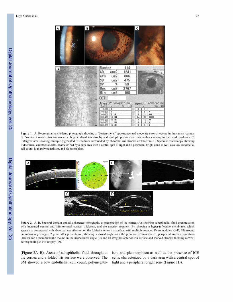
(Figure 2A–B). Areas of subepithelial fluid throughoutthe cornea and a folded iris surface were observed. TheSM showed a low endothelial cell count, polymegath‐
ism, and pleomorphism as well as the presence of ICEcells, characterized by a dark area with a central spot oflight and a peripheral bright zone (Figure 1D).
Figure 1. A, Representative slit-lamp photograph showing a “beaten-metal” appearance and moderate stromal edema in the central cornea.B, Prominent nasal ectropion uveae with generalized iris atrophy and multiple pedunculated iris nodules arising in the nasal quadrants. C,Enlarged view showing multiple pigmented iris nodules surrounded by abnormal iris stromal architecture. D, Specular microscopy showingiridocorneal endothelial cells, characterized by a dark area with a central spot of light and a peripheral bright zone as well as a low endothelialcell count, high polymegathism, and pleomorphism.
Figure 2. A–B, Spectral domain optical coherence tomography at presentation of the cornea (A), showing subepithelial fluid accumulationwith increased central and inferior-nasal corneal thickness, and the anterior segment (B), showing a hyper-reflective membrane, whichappears to correspond with abnormal endothelium on the folded anterior iris surface, with multiple rounded Reese nodules. C–D, Ultrasoundbiomicroscopy images, 2 years after presentation, showing a closed angle with the presence of broad-based, peripheral anterior synechiae(arrow) and a membranelike mound in the iridocorneal angle (C) and an irregular anterior iris surface and marked stromal thinning (arrow)corresponding to iris atrophy (D).
Loya-Garcia et al. 27
Digital Journal of O
phthalmology, Vol. 25
Digital Journal of O
phthalmology, Vol. 25
Topical treatment was modified with the addition oftravoprost daily, brinzolamide/timolol twice daily, lote‐prednol etabonate 0.5% every 6 hours, and sodiumchloride 5% twice daily. Follow-up consultationsshowed a remarkable reduction in the corneal stromaledema; IOP dropped to 12 mm Hg.
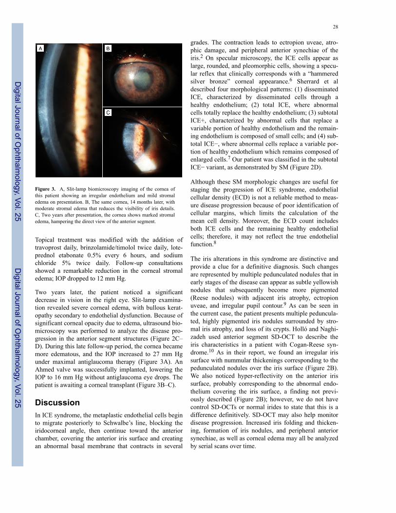
Two years later, the patient noticed a significantdecrease in vision in the right eye. Slit-lamp examina‐tion revealed severe corneal edema, with bullous kerat‐opathy secondary to endothelial dysfunction. Because ofsignificant corneal opacity due to edema, ultrasound bio‐microscopy was performed to analyze the disease pro‐gression in the anterior segment structures (Figure 2C–D). During this late follow-up period, the cornea becamemore edematous, and the IOP increased to 27 mm Hgunder maximal antiglaucoma therapy (Figure 3A). AnAhmed valve was successfully implanted, lowering theIOP to 16 mm Hg without antiglaucoma eye drops. Thepatient is awaiting a corneal transplant (Figure 3B–C).
DiscussionIn ICE syndrome, the metaplastic endothelial cells beginto migrate posteriorly to Schwalbe’s line, blocking theiridocorneal angle, then continue toward the anteriorchamber, covering the anterior iris surface and creatingan abnormal basal membrane that contracts in several
Figure 3. A, Slit-lamp biomicroscopy imaging of the cornea ofthis patient showing an irregular endothelium and mild stromaledema on presentation. B, The same cornea, 14 months later, withmoderate stromal edema that reduces the visibility of iris details.C, Two years after presentation, the cornea shows marked stromaledema, hampering the direct view of the anterior segment.
grades. The contraction leads to ectropion uveae, atro‐phic damage, and peripheral anterior synechiae of theiris.2 On specular microscopy, the ICE cells appear aslarge, rounded, and pleomorphic cells, showing a specu‐lar reflex that clinically corresponds with a “hammeredsilver bronze” corneal appearance.6 Sherrard et aldescribed four morphological patterns: (1) disseminatedICE, characterized by disseminated cells through ahealthy endothelium; (2) total ICE, where abnormalcells totally replace the healthy endothelium; (3) subtotalICE+, characterized by abnormal cells that replace avariable portion of healthy endothelium and the remain‐ing endothelium is composed of small cells; and (4) sub‐total ICE−, where abnormal cells replace a variable por‐tion of healthy endothelium which remains composed ofenlarged cells.7 Our patient was classified in the subtotalICE− variant, as demonstrated by SM (Figure 2D).
Although these SM morphologic changes are useful forstaging the progression of ICE syndrome, endothelialcellular density (ECD) is not a reliable method to meas‐ure disease progression because of poor identification ofcellular margins, which limits the calculation of themean cell density. Moreover, the ECD count includesboth ICE cells and the remaining healthy endothelialcells; therefore, it may not reflect the true endothelialfunction.8
The iris alterations in this syndrome are distinctive andprovide a clue for a definitive diagnosis. Such changesare represented by multiple pedunculated nodules that inearly stages of the disease can appear as subtle yellowishnodules that subsequently become more pigmented(Reese nodules) with adjacent iris atrophy, ectropionuveae, and irregular pupil contour.9 As can be seen inthe current case, the patient presents multiple peduncula‐ted, highly pigmented iris nodules surrounded by stro‐mal iris atrophy, and loss of its crypts. Holló and Naghi‐zadeh used anterior segment SD-OCT to describe theiris characteristics in a patient with Cogan-Reese syn‐drome.10 As in their report, we found an irregular irissurface with nummular thickenings corresponding to thepedunculated nodules over the iris surface (Figure 2B).We also noticed hyper-reflectivity on the anterior irissurface, probably corresponding to the abnormal endo‐thelium covering the iris surface, a finding not previ‐ously described (Figure 2B); however, we do not havecontrol SD-OCTs or normal irides to state that this is adifference definitively. SD-OCT may also help monitordisease progression. Increased iris folding and thicken‐ing, formation of iris nodules, and peripheral anteriorsynechiae, as well as corneal edema may all be analyzedby serial scans over time.
28
Digital Journal of O
phthalmology, Vol. 25
Digital Journal of O
phthalmology, Vol. 25
The progressive course of the disease leads to severecomplications, mainly irreversible corneal stromaledema and secondary glaucoma.2 See Figure 3. In ear‐lier stages of the disease, gonioscopy is very useful todetermine the grade and extent of the anterior chamberangle closure.3 However, once significant endothelialdysfunction occurs, visualization of the angle and theiris is only achieved with the aid of UBM.4 Anteriorsegment UBM is a useful tool for grading the extent ofiris atrophy, peripheral anterior synechiae, and prolifera‐tive changes in the angle structure.4,11 Zhang et al,12
reporting the results of UBM analysis of 21 patientswith ICE syndrome, showed that patients with Cogan-Reese syndrome presented more peripheral anterior syn‐echiae with higher IOP elevation, severe glaucoma, anda greater failure rate of filtering surgery than in the otherclinical variants of ICE syndrome. They concluded thatUBM is a fast and effective diagnostic method toexplore the extent of affected structures in this syndromeand its subtypes and found that UBM analysis has ahigher sensitivity than slit-lamp gonioscopy in detectingperipheral anterior synechiae and iris atrophy in patientswith advanced Cogan-Reese syndrome. Thus, we rec‐ommend conducting a baseline UBM early in this dis‐ease to use as a reference, unlike the present case, whereSD-OCT was used; although SD-OCT does providesome information, its utility in progressive disease islimited.The progression of Cogan-Reese clinical manifestationsshould be evaluated with advanced imaging technolo‐gies, which allow a sensitive and objective analysis ofthe corneal and anterior segment alterations seen inthese patients in order to facilitate surgical and therapeu‐tic decision making. To our knowledge, this is the firstreport of all three imaging modalities being used foranterior segment and corneal analysis for the diagnosisand assessment of a case of Cogan-Reese syndrome.
Literature SearchThe authors searched PubMed on January 8, 2018, forEnglish-Language results using the following terms sin‐
gly and in combination: Cogan-Reese syndrome, ICEsyndrome, specular microscopy, ultrasound biomicro‐scopy, and optical coherence tomography.
References1. Eagle R, Font R, Yanoff M, Fine B. Proliferative endotheliopathy
with iris abnormalities: the iridocorneal endothelial syndrome. ArchOphthalmol 1979;97:2104-11.
2. Shields MB. Progressive essential iris atrophy, Chandler’s syn‐drome, and the iris nevus (Cogan-Reese) syndrome: a spectrum ofdisease. Surv Ophthalmol 1979;24:3-20.
3. Campbell D, Shields M, Smith T. The corneal endothelium and thespectrum of essential iris atrophy. Am J Ophthalmol1978;86:317-24.
4. Scheie H, Yanoff M. Iris Nevus (Cogan-Reese) syndrome: a causeof unilateral glaucoma. Arch Ophthalmol 1975;93:963-70.
5. Cogan DG, Reese AB. A syndrome of iris nodules, ectopic Desce‐met’s membrane, and unilateral glaucoma. Doc Ophthalmol1969;26:424-33.
6. Ozdemir Y, Onder F, Coşar CB, Usubütün A, Kural G. Clinical andhistopathologic findings of iris nevus (Cogan-Reese) syndrome.Acta Ophthalmol Scand 1999;77:234-7.
7. Sherrard, Emil S.; Frangouils, Modea A.; Malcolm, Kerr Muir. Onthe morphology of cells of posterior cornea in the iridocorneal endo‐thelial syndrome. Cornea 1991;10:233-43.
8. Liu YK, Wang IJ, Hu FR, Hung T, Chang HW. Clinical and specularmicroscopic manifestations of iridocorneal endothelial syndrome.Jpn J Ophthalmol 2001;45:281-7.
9. Sacchetti M, Mantelli F, Marenco M, Macchi I, Ambrosio O, RamaP. Diagnosis and management of iridocorneal endothelial syndrome.Biomed Res Int 2015;2015:763093.
10. Holló G, Naghizadeh F. Optical coherence tomography characteris‐tics of the iris in Cogan-Reese syndrome. Eur J Ophthalmol2014;24:797-9.
11. Dada T, Gadia R, Sharma A, et al. Ultrasound Biomicroscopy inGlaucoma. Surv Ophthalmol 2011;56:433-50.
12. Zhang M, Chen J, Liang L, Laties aM, Liu Z. Ultrasound biomi‐croscopy of Chinese eyes with iridocorneal endothelial syndrome.Br J Ophthalmol 2006;90:64-9.
Loya-Garcia et al. 29
Digital Journal of O
phthalmology, Vol. 25
Digital Journal of O
phthalmology, Vol. 25