
Opthalmology - Rhode Island Medical Societyrimed.org/medhealthri/2008/2008-02.pdf51 The Quest To...
32
Volume 91 No. 2 February 2008 Opthalmology
Transcript of Opthalmology - Rhode Island Medical Societyrimed.org/medhealthri/2008/2008-02.pdf51 The Quest To...

Volume 91 No. 2 February 2008
�Opthalmology


41VOLUME 91 NO. 2 FEBRUARY 2008
Medicine and Health/Rhode Island (USPS 464-820), a monthly publication, is owned and published by the Rhode Island Medical Society, 235Promenade St., Suite 500, Providence, RI 02908, Phone: (401) 331-3207. Single copies $5.00, individual subscriptions $50.00 per year, and $100per year for institutional subscriptions. Published articles represent opinions of the authors and do not necessarily reflect the official policy of the Rhode IslandMedical Society, unless clearly specified. Advertisements do not imply sponsorship or endorsement by the Rhode Island Medical Society. Periodicals postagepaid at Providence, Rhode Island. ISSN 1086-5462. POSTMASTER: Send address changes to Medicine and Health/Rhode Island, 235 Promenade St.,Suite 500, Providence, RI 02908. Classified Information: RI Medical Journal Marketing Department, P.O. Box 91055, Johnston, RI 02919,phone: (401) 383-4711, fax: (401) 383-4477, e-mail: [email protected]. Production/Layout Design: John Teehan, e-mail: [email protected].
UNDER THE JOINTEDITORIAL SPONSORSHIP OF:
The Warren Alpert Medical School ofBrown UniversityEli Y. Adashi, MD, Dean of Medicine& Biological Science
Rhode Island Department of HealthDavid R. Gifford, MD, MPH, Director
Quality Partners of Rhode IslandRichard W. Besdine, MD, ChiefMedical Officer
Rhode Island Medical SocietyNick Tsiongas, MD, MPH, President
EDITORIAL STAFFJoseph H. Friedman, MD
Editor-in-ChiefJoan M. Retsinas, PhD
Managing EditorStanley M. Aronson, MD, MPH
Editor Emeritus
EDITORIAL BOARDStanley M. Aronson, MD, MPHJay S. Buechner, PhDJohn J. Cronan, MDJames P. Crowley, MDEdward R. Feller, MDJohn P. Fulton, PhDPeter A. Hollmann, MDSharon L. Marable, MD, MPHAnthony E. Mega, MDMarguerite A. Neill, MDFrank J. Schaberg, Jr., MDLawrence W. Vernaglia, JD, MPHNewell E. Warde, PhD
OFFICERSNick Tsiongas, MD, MPH
PresidentDiane R. Siedlecki, MD
President-ElectVera A. DePalo, MD
Vice PresidentMargaret A. Sun, MD
SecretaryMark S. Ridlen, MD
TreasurerBarry Wall, MD
Immediate Past President
DISTRICT & COUNTY PRESIDENTSGeoffrey R. Hamilton, MD
Bristol County Medical SocietyHerbert J. Brennan, DO
Kent County Medical SocietyRafael E. Padilla, MD
Pawtucket Medical AssociationPatrick J. Sweeney, MD, MPH, PhD
Providence Medical AssociationNitin S. Damle, MD
Washington County Medical SocietyJacques L. Bonnet-Eymard, MD
Woonsocket District Medical Society
RHODE ISLANDPUBLICATION OF THE RHODE ISLAND MEDICAL SOCIETY
Medicine � Health VOLUME 91 NO. 2 February 2008
COMMENTARIES42 Medical History
Joseph H. Friedman, MD
43 Can Privacy Survive In the Brave New WorldStanley M. Aronson, MD
CONTRIBUTIONSSPECIAL ISSUE: OphthalmologyGuest Editor: Elliot M. Perlman, MD
44 New Strategies for Common Eye DiseasesElliot M. Perlman, MD
45 Endothelial KeratoplastyElliot M. Perlman, MD
48 Presbyopic Intraocular LensesElliot M. Perlman, MD
51 The Quest To Conquer Age-Related Macular DegenerationRichard G. Bryan, MD
57 Clinical Update on Optic Neuritis and Multiple SclerosisMarjorie A. Murphy, MD
COLUMNS
60 HEALTH BY NUMBERS – Eye Injuries Treated In Rhode Island HospitalsEdward F. Donnelly, RN, MPH, and Jay S. Buechner, PhD
63 IMAGES IN MEDICINE – Incarcerated Inguinal Hernia As the PresentingFeaturing of Carcinoma of Unknown Primary SiteSamantha Nazareth, Yasmin Metz, MD, Samir A. Shah, MD, andEdward Feller, MD
64 PUBLIC HEALTH BRIEFING – The Rhode Island Survey of PhysicianEMR AdoptionJay S. Buechner, PhD, Rosa R. Baier, PhD, and David R. Gifford, MD, MPH
65 GERIATRICS FOR THE PRACTICING PHYSICIAN – Nutrition In the Older AdultTimothy Farrell, MD, and Ana Tuya, MD
67 PHYSICIAN’S LEXICON – Forty Days In LimboStanley M. Aronson, MD
67 Vital Statistics
68 February Heritage
Cover: “Joined at the Broken Places,”mixed media, by Lindy Tucker, an artistbased in Connecticut and Rhode Island.E-mail: [email protected]

42MEDICINE & HEALTH/RHODE ISLAND
ment disorders, one ofwhich was akathisia,the syndrome of mo-tor restlessness. It mayseem obvious to thereader, but wasn’t tome at the time, that studying a subjective phe-nomenon in people who were seriously men-tally ill, taking medications that slowed theirthought processes, was not going to be a veryproductive experience. And it wasn’t, so I gaveit up. People who were restless one minuteweren’t the next and even reported that theynever had been restless. And what did I meanby restless?
There are three points in this essay. Thefirst is the importance of Humpty Dumpty’stime-honored declamation, that “Words meanexactly what I want them to mean, nothingmore, nothing less.” The second is that themedical history needs to be taken for what it’sworth which is often not face value, some-times valuable, sometimes irrelevant or evenobfuscatory. While it is an error to discountthe reports of the patient and family, it can beequally counterproductive to base too great areliance on it. Thirdly, there is a need for stud-ies of the process of medical practice. We needto better understand when the history is use-ful, and when not; when to rely on objective(usually expensive) tests, and when to let ourjudgment be our guide (medical, not legal).There is a reason that certain symptom com-plexes are more challenging than others. It isbecause people are not machines, and we ex-perience our aches and pains in different ways.
There are reasons that computers willnever take over from doctors. Dizziness is oneof them.
– JOSEPH H. FRIEDMAN, MD
Disclosure of Financial InterestsJoseph Friedman, MD, Consultant: Acarta
Pharmacy, Ovation, Transoral; Grant ResearchSupport: Cephalon, Teva, Novartis, Boehringer-Ingelheim, Sepracor, Glaxo; Speakers’ Bureau:Astra Zeneca, Teva,Novartis, Boehringer-Ingelheim,GlaxoAcadia, Sepracor, Glaxo Smith Kline
Medical History�
Commentaries
In my medical school training takinga comprehensive medical history was sacrosanct.We had to memorize a huge list of symptomsfor each organ system and ask every patient aboutevery symptom in the specified order. As a resi-dent in a medical specialty I still had to fill outa form, but it was less complete.
Houston Merritt, MD, sole author ofMerritt’s Textbook of Neurology, and one of thegreat clinicians of the twentieth century, onthe downside of his prowess, but still teachingthird-year medical students in a weekly con-ference when I was a student, was renownedfor his ability to extract, like Sherlock Holmes,the important nuggets of a case, leaving thechaff behind as needless, confusing facts. Hisstyle of listening to a case presentation, thentapping a reflex, or asking one question, orchecking the eye movements, then pronounc-ing the solution, was legendary.
“Giant basilar artery,” he proclaimed.“A giant basilar artery aneurysm?” he was
asked.“What else could it be?” was his response,
in the era before CT and MRI imaging madesuch questions irrelevant.
When I moved to Rhode Island I met anexcellent dermatologist who told me that hedidn’t like to listen to the medical history. Ilaughed. This was inconceivable. He said itwas usually a waste of time. “When I tell theresidents that I don’t want to hear the history,they think I’m joking. It takes them a while torealize that I’m serious. If I want to know some-thing I’ll ask. Otherwise I just want to see thelesions.”
In my area of neurology, Parkinson’s dis-ease and movement disorders, unlike the casefor epilepsy or cerebrovascular disease, the his-tory generally isn’t very useful either. Some-times it is, of course, and I am still a diligenttaker of historical data.
In the November issue of Mayo ClinicProceedings, an article was published which, tome was a landmark for creativity and utility(Newman-Toker DE et al. Imprecision in Pa-tient Reports of Dizziness…Mayo Clin Proc2007;82:1329) It was like a step into anotherdimension, a sort of meta-medical analysis.
This article examined the value of the medicalhistory in the evaluation of the dizzy patientand concluded that it was relatively useless (“thequality of the patient’s dizziness symptomsshould be given less diagnostic weight than itcurrently receives.”).
This groundbreaking report evaluated ERpatients at a Johns Hopkins-affiliated Hospi-tal complaining of “dizziness.” Dizziness, is, ofcourse, a common problem in the ER, and achallenge to the clinician because of its manymeanings. It is generally subdivided into fourclassifications: vertigo, hypotension, disequi-librium, and “nonspecific.” It is so poorly de-fined that I do not allow housestaff or stu-dents to use the term unless quoting the pa-tient, since the term doesn’t even point to anorgan system at fault (inner ear, cardiovascular,gait instability, peripheral neuropathy, meta-bolic or drug effect, etc). The goal of the studywas to learn how to classify the symptom.Their findings were as follows. Patients usedpoorly localizing adjectives like “woozy” and“empty.” They generally used more than oneterm to describe their sensation, endorsingsymptoms from different subtypes in the clas-sification into etiologies. Of those “who didnot identify vertigo, spinning, or motion” whenasked to choose from a list of symptoms,70%…endorsed a sense of room spinning…”Most impressive to me was a test for reliabil-ity in which patients were asked, after a meantime delay of only six minutes, the same ques-tions about their symptoms and only 52%were consistent.
In the same month, an article in Neurol-ogy, the largest circulation American neurol-ogy journal, compared patient self-report forseizures, and actual seizures, measured withcomplete observation in a seizure ward, andfound that patients underestimated their spells,missing 80% of certain types of seizures (com-plex partial seizures).
Twenty years ago I worked part-time atRhode Island’s Institute for Mental Health(now the psychiatric branch of Eleanor SlaterHospital) as the neurology consultant for hos-pitalized psychiatric patients. I did this becauseof my interest in studying drug-induced move-

43VOLUME 91 NO. 2 FEBRUARY 2008
Can Privacy Survive In the Brave New World?�
Privacy is an old word describing the condition of beingwithdrawn from the company of others or, alternatively, por-traying a place of concealment and seclusion. These past defini-tions have now given way to a broader meaning: to define a per-sonal form of human liberty. And there are few liberties morefervently, more deeply held by Americans—or protected moreassertively—than their right of privacy. The privilege of insulat-ing one’s thoughts, one’s living space, one’s beliefs and apostasies,one’s integrity, one’s body, one’s very individuality represents, formany, the bedrock of fundamental freedom. And yet wordssuch as privacy and individuality are nowhere encountered ei-ther in the Declaration of Independence or in the Preamble andfirst ten Amendments of the US Constitution. Synonyms suchas “liberty”, “freedom of speech” and “the right of the people tobe secure in their persons” abound but privacy, as the innermostsanctuary of liberty, is not expressly mentioned.
Privacy must be distinguished from secrecy. Privacy is thenourishing of one’s inner thoughts to make them worthy ofguarding. Secrecy is the building of a protective wall to keepoutsiders from intruding—whether or not that which isguarded is worth guarding.
Of man’s many possessions, his personal thoughts musthave a protected sanctuary called privacy. Privacy by itself ismeaningless if it doesn’t hold something to be held private;empty privacy, like an empty vault, holds nothing worth pro-tecting. Privacy must safeguard something of substance and itthen represents the culmination of a lengthy process. Beforethere is freedom of speech, for example, there must be free-dom of thought. For unencumbered thought to be defensibleit must incubate over time, like a defenseless babe, until it canuphold itself. And thinking one’s independent thoughts, someperhaps novel or nonconforming, represents hard labor sinceit is unaided by the comforting ideas of the majority. Even theproblem with heresy requires that one has to think out, with-out help, one’s own unorthodoxies.
Some nations cherish the traits of privacy, individualityand nonconformity. Santayana observed that England was aparadise of individuality, eccentricity, heresy, anomalies, hob-bies and humors. And while the oft-quoted line, “For a man’shome is his castle,” was originally professed in Latin, it was firstuttered as policy in England in 1644. Other nations, how-ever, find privacy a deeply suspicious, alien quirk, with the cultof individuality to be suppressed rather than sheltered.
Three ancient professions—clergy, law and medicine—intrude upon the privacy of their constituents. But in each in-stance there is a solemn pledge to honor the secrecy of thatwhich is disclosed whether it be in the act of confession, thediscussions between an attorney and client or the many per-sonal secrets that a patient will share with his physician.
In medicine the bond of secrecy harkens back 2,400 yearswhen novitiates in the Aesclapians, the ancient guild of Greekphysicians, took a solemn pledge prior to membership, a cov-enant now called the Hippocratic Oath. Beyond mere moralsanction, it was a binding oath upon its members. The followingstatement appears in this brief pledge: “And whatsoever I shall
see or hear in the course of my profession, as well as outside mydealings with men, if it be what should not be published abroad,I will never divulge, holding such things to be holy secrets.” Itbound physicians to protect the right of privacy, but it alsofrowned upon gossip in a non-medical context.
To this day, newly graduating physicians take this solemnoath [or some revised oath] bearing the same promise to upholdprivacy. Yet some promises give way to what society calls “practi-cality.” In decades past, there was just the patient, the attendingphysician and an unstated, shared bond of secrecy. Today, thereare many intruders into this partnership-of-secrecy includingclinical laboratories, imaging facilities, electrophysiological cen-ters, countless accountants and clerical personnel—and hover-ing over all, are the insurance companies who pay for all of thesediverse clinical resources. Thus, instead of a private compact be-tween two, there is an Orwellian nightmare with a participatingcast of thousands despite the desperate efforts of all concernedto protect the tattered remnants of patient-privacy.
The forensic sciences have accelerated the drive to diminishprivacy and positively identify each person’s DNA and finger-prints, innocent or not. Some have suggested that the poetryengraved on the base of the Statue of Liberty now be appendedto: “Give me your tired, your poor, your fingerprints.” And thefuture may hold only a life of increased transparency with suc-ceeding generations deploring their kinship with the goldfishes.
The diversity of views on the merit of privacy is reflectedin the many, often conflicting, meanings imparted by the word“privacy.” In classical usage—as seen in cognate words such asprivy and privilege—privacy meant a highborn, empoweringstatus surviving in entities such as the Privy Council. Some-where in the middle are newer words such as privatization.And at the lower end of the spectrum are such derivative wordsas privation suggesting a divestment of something rather thanan act of addition or strengthening. And in modern armies,indeed, is there any rank lower than private ?
– STANLEY M. ARONSON, MD
Disclosure of Financial InterestsStanley M. Aronson, MD, has no financial interests to disclose.
CORRESPONDENCEe-mail: [email protected]

44MEDICINE & HEALTH/RHODE ISLAND
New Strategies for Common Eye Diseases�
This edition of Medicine & Health/Rhode Island deals withseveral recent advances in ophthalmology. I proposed thesetopics to Joe Friedman, the editor, and then solicited two of mycolleagues to add their input. My purpose was twofold:
1. I was inspired by the contributions of time and effort thatour colleagues have been making to this journal. Withthe ever-increasing pile of unread eye journals accumu-lating on my desk, I still manage to peruse Medicine &Health/Rhode Island. The articles are appropriately gearedto the Rhode Island physician readership and serve toenlighten me about the progress in other fields of medi-cine. In one of the monthly editorials the editor notedthat, “We receive some unsolicited material, but not a lot.We encourage these.” I was encouraged and decided toput together this issue.
2. The practice of ophthalmology has drifted farther andfarther from the practice of mainstream medicine, notjust in what we do, but also where we practice. Our spe-cialty is virtually all outpatient-based. With most of usoperating in ambulatory surgicenters and seeing patientsonly in our private offices, our physical presence in a hos-pital corridor is becoming a rarity. We don’t interact regu-larly with our non-ophthalmology colleagues. Conse-quently, I was hoping to bridge the gap a little and letother physicians know how far we’ve come in ophthal-mology. The topics I’ve chosen are those that I believeother physicians will find helpful in relating to their pa-tients with common eye problems.
Presbyopic Intraocular Lenses discusses the exciting devel-opments in what undoubtedly will become the standard of carein cataract surgery. Endothelial Keratoplasty highlights a tech-nique of corneal transplant that is already replacing the methodwe have been using since 1905! The Quest to Conquer Age-related Macular Degeneration by Richard G. Bryan, MD, dis-cusses how far we’ve come in averting blindness in our bur-geoning elderly population. Lastly, Clinical Update on OpticNeuritis and Multiple Sclerosis by Marjorie A. Murphy, MD,discusses new management strategies in dealing with a com-mon, often devastating disease in young people.
Elliot M. Perlman, MD, is Clinical Associate Professor ofSurgery (Ophthalmology), at the Warren Alpert Medical Schoolat Brown University.
CORRESPONDENCEElliot M. Perlman, MDRhode Island Eye Institute150 East Manning St.Providence, RI 02906Phone: 401-272-2020E-mail: [email protected]
Disclosure of Financial InterestsThe guest editor and author has no financial interests to
disclose.
Elliot M. Perlman, MD

45VOLUME 91 NO. 2 FEBRUARY 2008
Endothelial KeratoplastyElliot M. Perlman, MD�
There are about 40,000 full-thickness(also called penetrating) corneal trans-plants (PKP) performed in the UnitedStates each year; 68% are done becauseof failure of a single cell layer: the cor-neal endothelium.1 (Figure 1)
The cornea at birth has about 400000 hexagonal-shaped endothelial cellsforming a monolayer lining the posteriorcornea. Like cells in the central nervoussystem, the endothelium cannot undergocell division; thus, there is a slow loss ofendothelial cells throughout life.2 Certaindiseases, most notably Fuchs’ endothelialdystrophy, cause a premature loss of en-dothelial cells. Ocular trauma, especiallycataract surgery, can also be responsiblefor significant loss of endothelial cells.
The function of the endothelium isto maintain the clarity of the cornealstroma. The normal corneal stroma is clear,compact, and contains very little water(78% water vs. 98% water for the opaquesclera). Collagen fibrils run limbus-to-lim-bus in the stroma and are stacked in a veryregular fashion (Figure 2a). The fibrils aresurrounded by an extracellular matrix ofhydrophilic glycosaminoglycans. Thisstructure gives the cornea its unique opti-cal clarity. The normal endothelial cells areconnected to each other only by focal tightjunctions and thus form a “leaky” barrierto aqueous humor percolation into thestroma. The endothelial cells are highlymetabolic. They utilize Na+/K+ ATPaseion pumps to pump solutes out of thestroma; the water leaves the cornea follow-ing along the osmotic gradient.2
When endothelial cell function iscompromised, the corneal stroma be-comes edematous and thickened. Theevenly packed collagen fibrils spreadapart and incoming light is scattered. (Fig-ure 2b) Visual acuity drops. The only so-lution is to replace the endothelial cells.A reasonable question to ask is: why dowe need to transplant all the layers of thecornea just to replace a single cell layer?The answer is that PKP has several ad-vantages: it is relatively easy to do; it worksfor all types of corneal disease (not justendothelial disease); and it has the po-tential to produce very good visual results.
Traditional PKP is done by punch-ing out a full-thickness corneal buttonfrom donor tissue, typically about 7.5 to8.5 mm in diameter. A similar full-thick-ness trephination is done on the patient’scornea, and the donor button is suturedinto the opening using 10-0 monofila-ment nylon. The sutures can be running,interrupted or a combination of these.
There are, however, significantproblems with PKP. It typically takes 6months to a year to obtain optimal visionafter PKP. Sutures are usually removedselectively to improve vision over thecourse of multiple visits during this pe-riod. Not only is the optical clarity of thegrafted cornea necessary, but also its con-
tour is important. The cornea does two-thirds of the refraction of the eye. If thecornea is excessively steep, the eye be-comes myopic; if it is excessively flat, hy-peropia results. If the contour is anythingbut perfectly spherical, astigmatism willbe present. Thus, a grafted eye can bevery myopic or hyperopic, as well as haveexcessive astigmatism. Despite careful at-tention to these details, as many as 10%of patients cannot see well after PKP—even with a perfectly clear graft—be-cause the required glasses would be toothick and incompatible with the spec-tacle lens needed for the unoperated eye.
More serious problems can developfrom suture-related problems: suture
Figure 1. Corneal anatomy.
2a 2b
Figure 2a. Cross-section of collagen fibrils in normal corneal stroma (original magnificationx51800). Figure 2b. Cross-section of collagen fibrils in an edematous cornea (original
magnification x46700).

46MEDICINE & HEALTH/RHODE ISLAND
breakage causes discomfort and patientsneed to have them removed, leading tomany unscheduled office visits. Suturescan become infected, leading to cornealabscess and even endophthalmitis withprofound loss of vision. Another seriousissue with PKP is that full-thickness grafts
permanently weaken the cornea. Blunttrauma to the eye can lead to wound rup-ture, even years after the transplant. Theintraocular contents (iris, lens etc) canprolapse out of the eye if the rupture islarge enough, leading to permanent lossof vision.
In the past several years, recogniz-ing the inherent problems with PKP, cor-neal surgeons have been working on tech-niques to transplant only the deeper lay-ers of the cornea. Called collectively en-dothelial keratoplasty, these proceduresreplace only the innermost portion of thecornea (posterior stroma, Descemet’smembrane and the endothelium).
A variation of endothelial kerato-plasty is called DSEK (Descemet’s Strip-ping Endothelial Keratoplasty).3 InDSEK, the donor tissue is prepared byplacing the donor cornea in an artificialanterior chamber and securing it in place.A rapidly oscillating blade called akeratome is used to remove the top 60-70% of the donor cornea (epithelium and60-70% of the stroma), which is dis-carded. (Figure 3) The remaining 30-40% of the cornea is placed in a curvedTeflon block and a large disk is punched(typically 8.5 to 9.0 mm diameter).
DSEK is done with IV sedation, ei-ther with a peribulbar injection of anes-thetic or topical anesthesia. A small inci-sion is made at the limbus of the patient’scornea, and a small hook-like instrumentis used to strip off and remove the dis-eased Descemet’s membrane and endo-thelium. (Figure 4) Then, another 4 to 5mm incision is made at the limbus. Thedonor cornea is folded like a taco andinserted into the anterior chamber of thepatient. (Figure 5a,b) The incision isclosed with 2 10-0 nylon sutures. An airbubble is injected between the leaves ofthe taco to unfold it and force it into po-sition apposing the patient’s stromal tis-sue. The air bubble is left in this positionfor 10-60 minutes to allow adherence ofthe donor tissue to the recipient stroma.Finally most of the air is removed (Fig-ure 5c) and the patient is dischargedhome.
Over the course of the next severalweeks, vision improves as the grafted en-dothelium begins clearing the edema
Figure 3. DSEK: donor tissue preparation.
Figure 4. DSEK: recipient cornea preparation.
5a 5b 5c
Figure 5a. DSEK: Folded donor disk outside anterior chamber. Figure 5b. DSEK: Folded donor disk inside anterior chamber.Figure 5c. DSEK: Donor disk adhering to recipient stroma.

47VOLUME 91 NO. 2 FEBRUARY 2008
from the recipient cornea. The 2 suturesare removed at 6 weeks. Visual recoverycan come as soon as 1 month, but typi-cally takes several months. This is still con-siderably faster than after PKP.
Because the incision is small and islocated at the limbus—not directly in thecornea—the refractive status of the eyeis similar to what it was before the corneabecame edematous3. Not infrequently,the DSEK patient can see well withoutany glasses—a distinctly unusual eventafter PKP. DSEK essentially eliminatessuture problems. Because the corneal in-cision is small and peripheral, the cornealmechanical strength is nearly normal,and the threat from blunt trauma ismuch less.4 The donor disk in DSEK hasa larger diameter than a typical PKPgraft, so the number of endothelial cellstransplanted by DSEK is 15%-26%higher. Another intriguing possibility isthat the location of the DSEK disk (inthe anterior chamber) is farther from thelimbal host cells that are involved in graftrejection. A recent study showed a lowergraft rejection rate in DSEK patients com-pared to PKP patients (7.5% vs.13 % af-ter 2 years).5
As the procedure is relatively new,there are still post-operative problems.The donor disk may not stay adherent tothe host stroma, and an air bubble mighthave to be placed back into the anteriorchamber as an office procedure. Also,perhaps because of the extra manipula-tion of the donor tissue, there is a higherincidence of primary graft failure (thedonor disk fails to clear the edematouscornea, and a re-operation would be re-quired). Another concern with DSEK isthat it is difficult to end up with a per-fect “20/20” result. This mild limitationmay be due to the fact that there is aninterface present (between the hoststroma and the donor stroma) (Figure 6)or that the recipient’s stroma may not be
totally transparent, even after the excesshydration is gone.6 Lastly, the long-termresults of endothelial keratoplasty –andwhether the grafted endothelium sur-vives at least as well as with PKP— muststill be evaluated.
Endothelial keratoplasty has revolu-tionized corneal transplantation. It is al-ready replacing PKP in the treatment ofcorneal endothelial disease. Some cornealsurgeons are making the incisions smallenough that they do not require sutures.Others have been working on transfer-ring just Descemet’s membrane and theendothelium, a process hampered by thefragility of this thin tissue preparation.The first report of a successful transplantof this type was recently published.7
Lastly, laboratory research on endo-thelial cell regeneration may some dayallow removal of a recipient’s remainingendothelial cells, increase the cell densityin vitro and then re-transplant them backinto the recipient, thereby avoiding graftrejection.4
REFERENCES1. Kang PC, Klintworth, et al. Trends in the indica-
tions for penetrating keratoplasty, 1980-2001.Cornea 2005; 24:801-3.
2. Foster CS, Azar DT, , eds. Smolin and Thoft’s TheCornea, 4th ed. New York: Lippincott Williams &Wilkins, 2005.
3. Price FW, Price MO. Descemet’s stripping withendothelial keratoplasty in 50 eyes. J Refrac Surg2005; 21: 339-45.
4. Terry MA. Endothelial keratoplasty. Cornea2006;25: 873-8.
5. Allan BDS, Terry MA, et al. Corneal transplantrejection rate and severity after endothelial kerato-plasty. Cornea (in press).
6. Melles GRJ, Posterior lamellar keratoplasty: DLEKto DSEK to DMEK. Cornea 2006; 25: 879-81.
7. Melles GRJ, Ong TS et al. Descemet membraneendothelial keratoplasty (DMEK). Cornea2006;25: 987-90.
Elliot M. Perlman, MD, is ClinicalAssociate Professor of Surgery (Ophthalmol-ogy), at the Warren Alpert Medical Schoolat Brown University.
Disclosure of Financial InterestsElliot M. Perlman, MD, has no fi-
nancial interests to disclose.
Figure 6. In vivo imaging of eye after DSEK.

48MEDICINE & HEALTH/RHODE ISLAND
Presbyopic Intraocular LensesElliot M. Perlman, MD�
“Now, thanks to ReZoom…I’vegot my zoom back!”
– Gary Player,– Grand Slam– Golf Champion 1
Although this sounds more like an ad forViagra®, it refers to one of the new FDA-approved intraocular lenses (IOLs)which correct presbyopia.
In the not-too-distant past (the late70s), during my ophthalmology resi-dency, we waited until cataracts got “ripe”before we operated. The reason was notrelated to the technical difficulty of thesurgery, but rather because the visual re-sult was so poor that a patient would bebetter off with moderately dense cataractsthan coke-bottle-like aphakic spectacles.These thick lenses would distort andmagnify the image as well as cause suchperipheral vision problems that evenwalking with them was dangerous!
We have come a long way since, notjust in the technique of cataract surgery, butalso in the method we use for visual correc-tion afterward. IOL implantation at thetime of cataract surgery has become thestandard of care for the past 25 years. Ad-vances in these lenses were prompted byadvances in the techniques used for cata-ract surgery itself. Initially the lenses werenot flexible, requiring a large incision forinsertion. At that time, prior to the wide-spread use of phacoemulsification for cata-ract surgery, cataract incisions had to bemuch larger (5 to 7 mm) to allow removalof the intact nucleus (center) of the cata-ract.
Modern cataract surgery utilizesphacoemulsification. In phacoemulsification,a small titanium or steel tip on a hand piece
is inserted into the anterior chamberthrough a small incision (2.2 to 2.75 mm)in the peripheral cornea. The tip vibrates atultrasonic speeds to break up the cataractinto very fine pieces, which are aspirated outof the anterior chamber, usually through aport in the same hand piece. Becausephacoemulsification cataract surgery can bedone through a very small incision, intraocu-lar lens manufacturers began developingfoldable intraocular lenses. First introducedabout 15 years ago, they are made of a plas-tic polymer, such as silicone or acrylic.
A typical cataract operation today isdone under topical anesthetic with IV se-dation as needed. There is usually no needto stop any anticoagulants or anti-plateletmedication. After the peripheral cornealincision is made, the surgeon creates a cir-cular opening in the anterior capsule ofthe lens (capsulorhexis) to allow access tothe cataractous portion of the lens. Whilethe surgeon is viewing the procedurethrough an operating microscope, thecataract is broken up and removed usinga phacoemulsifying hand piece.
Cataract surgery removes most of thenatural lens of the eye. (Figure 1a) Whatremains is the posterior capsule of the lensand part of the anterior capsule, a con-figuration which resembles a bag, oftencalled the capsular bag. (Figure 1b) Tocorrect the vision after cataract surgery,the foldable IOL is placed into the cap-sular bag. (Figure 1c). To replace the con-vex natural lens, the IOL has a convexshape. The thicker the IOL is, the moreconverging power the lens has. Until re-cently, this lens has been a monofocallens, meaning that it has only one powerand focuses in only one plane. The por-tion of the IOL that does the focusing iscalled the optic, which is usually circularand about 6 mm in diameter. The IOL isplaced into an inserter resembling a smallpeashooter, which is used to position thelens into the capsular bag. The IOL doesnot have to be sutured into place. Becauseof the small incision size, suturing of theincision is not generally required either.The entire cataract operation with IOLimplant takes about 20 minutes.
Figure 1a. Eye with a cataract.Figure 1b. Eye after cataract surgery, showing capsular bag (C-shaped structure).
Figure 1c. Eye after cataract surgery with intraocular lens.
1a 1b 1c
2a 2b 2c
Figure 2a. Distance vision in the emmetropic eye. Figure 2b. Near vision without accommodation.Figure 2c. Near vision with accommodation.

49VOLUME 91 NO. 2 FEBRUARY 2008
Part of what makes modern cataract sur-gery so successful is the accurate determina-tion of the IOL power needed to permit thepseudophakic patient (a patient who has hada cataract operation and IOL implant) tosee well in the distance with little or no spec-tacle correction (emmetropia). When thenatural lens of the eye is removed, the onlyconverging lens in the eye is the cornea. Thesteeper the cornea is, the more powerful isits converging power. There are a numberof instruments that can very accurately mea-sure the curvature (and thus the power) ofthe cornea. The only other parameter weneed to determine the correct IOL powerto implant is the axial length of the eye (thelength of the eye from the front surface ofthe cornea to the retina). We also know howfar the IOL will be from the retina. Withthis information, it is a straightforward cal-culation to determine the exact power theIOL must be to achieve emmetropia.
It should be apparent from the abovediscussion that we can select the correct IOLpower to eliminate myopia at the time of cata-ract surgery. And, indeed, this is the case.
Modern cataract surgery is aform of refractive surgery,and many previously existingrefractive errors can be cor-rected at the time of cataractsurgery. If patients have sig-nificant astigmatism preop-eratively (the cornea isshaped more like the surfaceof spoon rather than asphere), they would still need
glasses in the distance, despite perfectly cor-rect monofocal lens placement. Many oph-thalmic surgeons use incisions in the periph-ery of the cornea (limbal relaxing incisions)at the time of cataract surgery (or after thesurgery) to minimize the astigmatism.
Implantation of a monofocal IOL atthe time of cataract surgery can allow theelderly eye with a cataract to have the vi-sual quality of a middle-aged (presbyopic)eye without a cataract. The remaining is-sue is that presbyopia still exists, and virtu-ally all patients who have had cataract sur-gery and see well in the distance withoutglasses will still need reading glasses.
To understand how presbyopic IOLswork, it is necessary to review some basicoptics in the normal eye.2 In the emmetropiceye, the incoming rays from a distant ob-ject are parallel and are focused by the cor-nea and natural lens to a point on the sur-face of the retina. Both the cornea and lensact as converging lenses. (Figure 2a) Whenthe same emmetropic eye views a near ob-ject, the incoming rays from the near ob-ject are actually diverging when they strike
the surface of the cornea and lens. With-out the effort of accommodation, the rayswould focus somewhere behind the retinaand the object would appear blurry. (Fig-ure 2b) Accommodation involves constric-tion of the ciliary muscle, which allows thelens to become thicker (more powerful) andthus cause more convergence of the rays.With the proper amount of convergence,the rays will be focused as a point on theretina. (Figure 2c)
As we age, the lens of the eye be-comes more rigid. Thus, accommodativeeffort becomes less effective. This progres-sive loss of accommodation—like deathand taxes—is almost universal. It is calledpresbyopia (“old eyes”). By the time mostof us are 55 years old, we’ve lost it all,and reading glasses are a necessity.
The monofocal IOL has made cata-ract surgery one of the most successfuland satisfying operations in all of surgery.In the past several years, however, withthe introduction of presbyopic IOLs,cataract surgery results have taken an-other quantum leap forward, making thepseudophakic eye even more like the eyeof a young phakic person.
The Food and Drug Administration(FDA) has approved 3 presbyopic lenses: TheReZoom Lens by AMO; the ReStor Lens byAlcon and the crystalens by eyeonics. TheReZoom and ReStor lenses are multifocalIOLs; the crystalens is an accommodatingIOL. In contrast to a monofocal lens (Figure3a), which focuses light at one plane, a mul-tifocal lens focuses light at more than oneplane simultaneously. (Figure 3b) TheReZoom Lens has a series of 5 concentricrings, some of which focus at near (14” fromthe eye) and some of which focus in the dis-tance. The central 2.5 mm of the lens fo-cuses for distance. (Figure 4) The ReStor Lenshas an “apodized diffractive center”. Thislens utilizes sophisticated diffractive optics inits central 3.6 mm to cause light to focus inthe distance and quite close simultaneously(about 10” away from the eye). (Figure 5)
I believe a more accurate description ofthese 2 multifocal lenses would be the term“bifocal lens”, since the lenses actually haveonly 2 focal points (distance and near). At anygiven time, some of the light passing througha multifocal lens will be coming from distantobjects, and some will be coming from nearobjects. Which object the eye sees clearly ispartially dependent on the size of the pupil atthe time. For instance, in a bright light, the
3a 3b
Figure 3a. Monofocal lens focusing.Figure 3b. Multifocal lens focusing.
4 5
Figure 4. ReZoom multifocal intraocular lens.Figure 5. ReStor multifocal intraocular lens.

50MEDICINE & HEALTH/RHODE ISLAND
pupil will be constricted (e.g. less than 2.5mm). The ReZoom lens will be seeing mostlya distant object in that lighting, since the cen-tral 2.5 mm of that lens focuses for distance.The ReStor lens could be seeing both distanceand near with that pupil size3.,4
An interesting aspect of these multifo-cal lenses is that the brain must be “trained”to sort out distance and near objects. Thislearning process, which can take severalmonths, is greatly aided when both eyeshave multifocal lenses implanted; hence,bilateral cataract surgery is preferred whenthese lenses are used3,4 According to Alcon’sstudies,5 80% of patients with bilateralReStor lenses never need glasses.
One of the difficulties with multifocallenses is that glare and haloes may be notice-able under subdued lighting (e.g. night driv-ing). Under those circumstances, the pupil
is large, and both distance and near compo-nents of the multifocal lenses are function-ing. In addition, since the lenses are set foreither distance or near, patients may havetrouble with intermediate range objects (e.g.20”-30” away, such as computer screens, gro-cery store shelves, or music stands).
The crystalens, the accommodatingpresbyopic IOL, has a 5 mm optic and 2haptics (“arms”). (Figure 6) The hapticseach have a horizontal hinged area, whichis readily flexed. The crystalens more closelyresembles the accommodation of the natu-ral lens. When the eye containing thecrystalens is not accommodating, the lensfocuses in the distance, similar to amonofocal lens. (Figure 7a) When the eyeaccommodates (when the ciliary musclecontracts), the crystalens is actually pushedforward a small amount. This movementmakes the lens transiently more powerful.In addition, the whole front surface of thecrystalens flexes and becomes more con-vex. Both these actions allow focusing ona near object. (Figure 7b)
Like the multifocal lenses, thecrystalens also takes some “training” so thatthe patient gets used to accommodating thecorrect amount. It also is helpful ifcrystalenses have been implanted bilater-ally. While accommodating IOLs may nothave as much multifocal IOL problemssuch as glare and haloes, these lenses maynot fully accommodate, and the patient maystill require some form of reading glasses.
The entire cost of traditional monofocalIOLs (about $50-$150) is covered by Medi-care (and other insurance carriers) as a part ofthe facility fee, and the patient has no finan-cial responsibility. The cost of presbyopic IOLsis about $900 per lens and is not covered byMedicare and other carriers. In addition tothis cost, the ophthalmic surgeon charges anadditional fee for these lenses. Although theyare not technically more difficult to insert thanmonofocal lenses, all measurements to deter-mine lens power must be exceedingly accu-rate, the ocular health must be perfect, andthe cataract surgery must be flawless. Also, ifpatients have even a small amount of astigma-tism, presbyopic lenses may not work well.Post-operative management can also be moretime-consuming and may require additionalrefractive procedures. Patient expectations,as in patients who have LASIK (elective re-fractive surgery), can be extremely high.
The total cost borne by the patient forthe use of these lenses is typically $2000 to
$2500 per eye. Medicare and other insur-ance carriers consider presbyopic lenses “cos-metic” and do not pay for them. Thus, thepatient – for the first time in many years –has a fair amount of out-of-pocket cost tohave bilateral cataract surgery with presby-opic lenses implanted. As I alluded to in thebeginning of this article, presbyopic IOLscompanies are pitching their ads to theViagra®-age consumers: the baby boomers.According to the presbyopic IOL compa-nies,6 78 million Americans were born be-tween 1946 and 1964. Every 7 seconds, aboomer reaches age 50, the average age.There is a high overlap between this agegroup and the appearance of cataracts: 33%of the population between the age of 45 and64 already have formation of early cataractsor about 13 million people in the US. Fa-voring the potential wide-spread use of pres-byopic IOLs are financial statistics regard-ing this age group: baby boomers accountfor more than half of US spending, andpeople over the age of 50 account for morethan half of all health care spending5.
Presbyopic IOLs can give the cataractpatient vision similar to someone under 40years old and, indeed, patients can “get theirzoom back.” The same cataract operationand presbyopic lenses are even being usedin patients without cataracts (so-called “clearlensectomy”) to correct refractive errors aswell as presbyopia. With many new pres-byopic lenses being developed, it is possiblethat the use of these lenses may supplantLASIK as the predominant refractive pro-cedure, even in patients without cataracts.
REFERENCES1. Advanced Medical Optics website:
www.rezoomiol.com2. Rubin ML. Optics for Clinicians. TRIAD pub-
lishers, Gainesville, FL. 1974.3. Chang CF, Bucci FA, et al. IOL options in 2007.
Cataract & Refractive Surgery Today 2007;7: 49-69.4. Kent C, Presbyopic IOLs. Rev Ophthalmol 2007;
14: 26-33.5. Data from Alcon Laboratories, 6201 South Free-
way, Fort Worth, TX6. From crystalens resource materials. eyeonics, inc,
Aliso Viejo, CA
Elliot M. Perlman, MD, is ClinicalAssociate Professor of Surgery (Ophthalmol-ogy), at the Warren Alpert Medical Schoolat Brown University.
Disclosure of Financial InterestsElliot M. Perlman, MD, has no fi-
nancial interests to disclose.
Figure 6. crystalens accommodatingintraocular lens.
7a
7b
Figure 7a. crystalens in a non-accommodating eye.
Figure 7b. crystalens in anaccommodating eye.
51VOLUME 91 NO. 2 FEBRUARY 2008
The Quest To Conquer Age-Related Macular DegenerationRichard G. Bryan, MD
A new era has dawned in the treatmentof the leading cause of irreversible visionloss in elderly patients: age-related macu-lar degeneration (AMD), a disease afflict-ing at least 1.75 million people in thiscountry. That number is likely to doubleby 2030. Until recently, our treatmentswere palliative at best, but we now havethe capacity to stabilize and even reversevision loss in many patients.
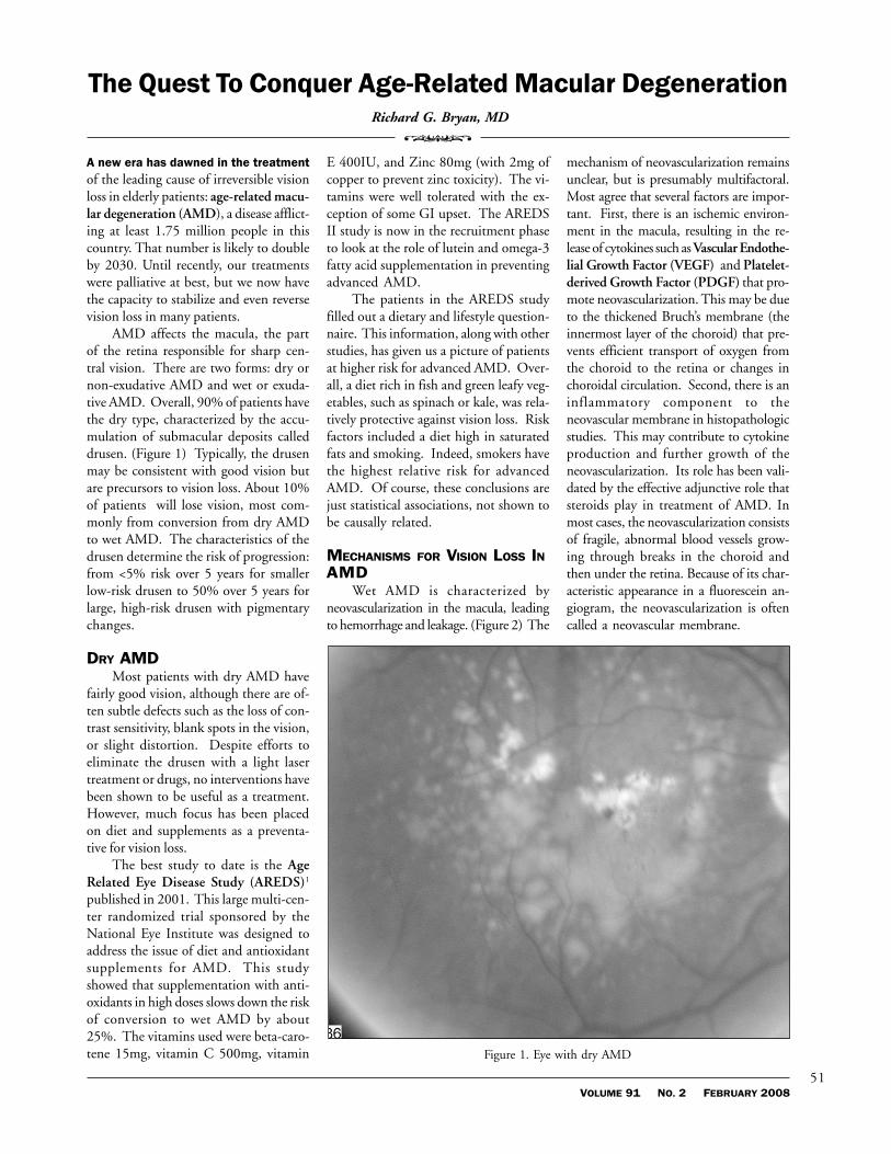
AMD affects the macula, the partof the retina responsible for sharp cen-tral vision. There are two forms: dry ornon-exudative AMD and wet or exuda-tive AMD. Overall, 90% of patients havethe dry type, characterized by the accu-mulation of submacular deposits calleddrusen. (Figure 1) Typically, the drusenmay be consistent with good vision butare precursors to vision loss. About 10%of patients will lose vision, most com-monly from conversion from dry AMDto wet AMD. The characteristics of thedrusen determine the risk of progression:from <5% risk over 5 years for smallerlow-risk drusen to 50% over 5 years forlarge, high-risk drusen with pigmentarychanges.
DRY AMDMost patients with dry AMD have
fairly good vision, although there are of-ten subtle defects such as the loss of con-trast sensitivity, blank spots in the vision,or slight distortion. Despite efforts toeliminate the drusen with a light lasertreatment or drugs, no interventions havebeen shown to be useful as a treatment.However, much focus has been placedon diet and supplements as a preventa-tive for vision loss.
The best study to date is the AgeRelated Eye Disease Study (AREDS)1
published in 2001. This large multi-cen-ter randomized trial sponsored by theNational Eye Institute was designed toaddress the issue of diet and antioxidantsupplements for AMD. This studyshowed that supplementation with anti-oxidants in high doses slows down the riskof conversion to wet AMD by about25%. The vitamins used were beta-caro-tene 15mg, vitamin C 500mg, vitamin
E 400IU, and Zinc 80mg (with 2mg ofcopper to prevent zinc toxicity). The vi-tamins were well tolerated with the ex-ception of some GI upset. The AREDSII study is now in the recruitment phaseto look at the role of lutein and omega-3fatty acid supplementation in preventingadvanced AMD.
The patients in the AREDS studyfilled out a dietary and lifestyle question-naire. This information, along with otherstudies, has given us a picture of patientsat higher risk for advanced AMD. Over-all, a diet rich in fish and green leafy veg-etables, such as spinach or kale, was rela-tively protective against vision loss. Riskfactors included a diet high in saturatedfats and smoking. Indeed, smokers havethe highest relative risk for advancedAMD. Of course, these conclusions arejust statistical associations, not shown tobe causally related.
MECHANISMS FOR VISION LOSS INAMD
Wet AMD is characterized byneovascularization in the macula, leadingto hemorrhage and leakage. (Figure 2) The
mechanism of neovascularization remainsunclear, but is presumably multifactoral.Most agree that several factors are impor-tant. First, there is an ischemic environ-ment in the macula, resulting in the re-lease of cytokines such as Vascular Endothe-lial Growth Factor (VEGF) and Platelet-derived Growth Factor (PDGF) that pro-mote neovascularization. This may be dueto the thickened Bruch’s membrane (theinnermost layer of the choroid) that pre-vents efficient transport of oxygen fromthe choroid to the retina or changes inchoroidal circulation. Second, there is aninflammatory component to theneovascular membrane in histopathologicstudies. This may contribute to cytokineproduction and further growth of theneovascularization. Its role has been vali-dated by the effective adjunctive role thatsteroids play in treatment of AMD. Inmost cases, the neovascularization consistsof fragile, abnormal blood vessels grow-ing through breaks in the choroid andthen under the retina. Because of its char-acteristic appearance in a fluorescein an-giogram, the neovascularization is oftencalled a neovascular membrane.
�
Figure 1. Eye with dry AMD

52MEDICINE & HEALTH/RHODE ISLAND
Another less common reason for vi-sion loss is a more advanced form of dryAMD: geographic atrophy. In this case,the subretinal drusen give way to loss ofthe overlying retinal pigment epithelium.This is a progressive process that typicallystarts off-center (often without symp-toms) then slowly expands to involve thecentral macula. This is responsible forabout 10% of dry AMD patients withsevere vision loss. Unfortunately, noth-ing can be done today to slow down orreverse the process, so I will not discussthe topic further.
EVOLUTION OF AMD TREATMENTSEffective treatment for most patients
with choroidal neovascularization was notavailable until recently. Destructive la-ser photocoagulation of the neovascularlesions was the only widely acceptedtreatment available until 2001, as dem-onstrated in the Macular Photocoagula-tion Study (MPS). Even though thestudy recommended treating all well-de-fined neovascular membranes, laser pho-tocoagulation was only widely used fortreatment of lesions where the centralmacula (the fovea) was spared. Thisgroup constitutes only about 17% ofnewly diagnosed patients. Even with asuccessful treatment, the recurrence ratewas at least 50%. Today, laser photoco-
agulation is still used, but only for lesionswell away from the macular center.
The first treatment with widespreadacceptance for subfoveal lesions began in2001 with the introduction of photody-namic therapy (PDT)2 with vertiporfin(Visudyne). This treatment involved in-travenous infusion of Visudyne, a drugthat selectively accumulates in leakyneovascular tissue, followed by treatmentwith a non-destructive laser that selec-tively activates the dye. The result wasendothelial cell damage and subsequentclosing of the neovascular complex. Thetreatment was somewhat disappointing,because it only slowed down vision lossrather than reversed it. A number of fac-tors were responsible for this. First, re-current leakage appeared in 80% of pa-tients, requiring multiple retreatments.Second, normal choroidal circulation isalso affected by the treatment, as evi-
denced by the general choroidalhypoperfusion on fluorescein angiogra-phy. After a few years of disappointingresults, many retina specialists combinedthis treatment with an intravitreal triam-cinolone injection to get a longer lastingeffect (see discussion below).
The second effective treatment forsubfoveal lesions was introduced in2004. Pegaptanib (Macugen, OSI[Eyetech] and Pfizer Pharmaceuticals) isan aptamer that selectively blocks the 165isoform of VEGF, the isoform most linkedto pathologic neovascularization in theeye.3 This treatment takes advantage ofVEGF’s important role in bothneovascularization and vascular perme-ability. It is given as an intravitreal injec-tion every 6 weeks to chronically suppressVEGF, as the neovascular lesions have atendency to come back once the medi-cation is gone. (Intravitreal injection is anin-office procedure, done using topicalanesthetic under sterile conditions.) Theresults from the Macugen trial were simi-lar to that seen with Visudyne, leavingretina specialists dissatisfied.
A turning point was reached with thewidespread off-label use of bevacizumab(Avastin, Genentech) in mid-late 2005and later ranibizumab (Lucentis,Genentech) that was approved by theFood and Drug Administration (FDA) inAugust, 2006.4 These compounds arehumanized monoclonal antibodies(Avastin) or an affinity-maturated Fab frag-ment (Lucentis) that block all isoforms ofVEGF. They are administered byintravitreal injection on a monthly basisfor up to 2 years, chronically suppressingVEGF in the eye. With either drug, theexudation from the neovascular complexusually dries up within a month or two,leaving a smaller dry scar. For the firsttime, patients maintained stable vision inmore than 90% and substantial improve-ment of vision in about 40%. It has beena remarkable advance. (Figure 3)
Because elderly patients with AMDoften have cardiovascular disease, thereis some concern about the safety ofchronic VEGF suppression. VEGF playsa crucial role in normal vascular and neu-ronal maintenance. Among patients over65 receiving standard chemotherapy andhigh dose Avastin intravenously every 2weeks for its FDA-approved indication—metastatic colon cancer—8.5% of them
Figure 2. Eye with wet AMD
…anti-VEGFmonotherapy will
probably remain themainstay of
treatment for mostpatients.

53VOLUME 91 NO. 2 FEBRUARY 2008
had vascular events, compared to 2.9%vascular events in patients on chemo-therapy alone. These vascular events in-cluded myocardial infarction, cere-brovascular accident, accelerated hyper-tension, venous thrombosis, and severehemorrhaging. Fortunately we use amuch smaller dose as an intravitreal in-jection, in theory limiting the systemicexposure.
There are several reasons to believethat there may be a systemic risk tointravitreal injection of Avastin andLucentis. First, systemic levels of Avastin(22 day half life) and Lucentis (half lifeof several hours) have been identified af-ter intravitreal injection at usual doses.Second, VEGF is well known as an im-portant mediator of repair of ischemicand damaged tissue.
Very little reliable data exist as to thecomplication rate of Avastin, as this is anoff-label use that was widely adopted af-ter a few very compelling case series werereported. A large randomized study isnow in the recruitment phase to deter-mine the level of risk, but it will be years
before data are available. Combined datafrom the ANCHOR and MARINA tri-als did not show any statistically signifi-cant increase in vascular events withLucentis use, although this was a smallstudy powered to determine efficacy andnot uncommon complications. Therewas a safety signal in these trials consist-ing of a few more cerebrovascular events,but these did not reach the level of statis-tical significance. The actual risk mayonly come out during the post-market-ing surveillance period.
Many questions remain unan-swered. First, how should a patient’s sys-temic health influence the use of Avastinand Lucentis? For example, how shoulda patient be treated who had a recentstroke or myocardial infarction? Howabout unstable angina? Since there is adocumented effect of bevacizumab onwound healing, should the treatment bewithheld in the peri-operative period ofmajor surgery? Second, what is the roleof the internist or cardiologist for patientswho are being treated? Is there any util-ity to preventative therapies such as anti-
coagulation or more intensive cardiovas-cular monitoring in at-risk patients?
ALTERNATE TREATMENT STRATEGIESAlthough the need for treatment of
exudative AMD is obvious, the questionsabout risk necessitate long discussionswith the patient and families. Most pa-tients wish to proceed with the treat-ment, but some are afraid. Even thosewith significant cardiovascular risk fac-tors have demanded treatment on thepremise that they would rather be deadthan blind. In my own practice, I amtreating a retired physician with cardio-vascular risk factors who had a minorstroke during a course of Avastin treat-ments. Even when told that I wouldwithhold treatment due to the seriouspotential extra risk, he absolutely de-manded that I continue. Fortunately,nothing further has happened.
An important pricing issue has re-cently surfaced regarding the use ofLucentis and Avastin (both Genentechproducts). The off-label use of Avastinby ophthalmologists has required com-pounding pharmacies to prepare smallsterile aliquots of the 4-ml vials. The costof a single intravitreal injection is about$40. However, Lucentis costs about$2000 for a single intravitreal injection.In October, 2007, Genentech sent a let-ter to all retinal surgeons, indicating thatit planned to stop distributing Avastin tocompounding pharmacies. The companycited FDA concerns about the particu-late level of this intravenous product whenused for intraocular injection. AlsoGenentech noted that Lucentis was de-veloped and FDA-approved specificallyfor ocular use. This ongoing controversymay limit the availability of the much lessexpensive product.
Besides systemic risks and pricing is-sues, there are other disincentives toLucentis/Avastin treatment as currentlyrecommended. The procedure itself onlytakes a few minutes and is minimally pain-ful, but it must be done monthly. Eachtime, the patient must deal with waittimes for dilation, imaging, and the pro-cedure that may reach an hour or two.They often have to bring a family mem-ber or friend to drive them. These fre-quent injections are not only a tremen-dous burden on the patient but also onour practices. Patients we used to see a
(Top) Figure 3a. Optical Coherence Tomography (OCT) showing cross-section of maculawith wet AMD before treatment with intra-vitreal Avastin.
(Bottom) Figure 3b. OCT showing dramatic recovery of normal macular appearance 1month after treatment with Avastin.


317VOLUME 90 NO. 10 OCTOBER 2007

56MEDICINE & HEALTH/RHODE ISLAND
couple of times a year now requiremonthly visits and procedures, cloggingup our schedules. Therefore, much re-cent investigation has focused on alter-native treatment strategies or new medi-cations to reduce the burden.
One strategy is to reduce the dosingschedule. The PIER study sponsored byGenentech was meant to address thisquestion by using Lucentis every threemonths instead of monthly. Unfortu-nately, the patients did not do as well aswith the monthly treatments, even thoughit was still better than photodynamictherapy or Macugen. Another strategyis the PRONTO protocol. This involvesan induction period of monthly injec-tions to dry up the lesion, followed byclose follow-up. If any recurrent exuda-tion appears and/or vision loss occurs,another injection is given. This strategywas shown in a relatively small trial tohave similar visual results to the AN-CHOR and MARINA phase III trialswith half the injections on average. Ofcourse, the patients still must come to theoffice monthly, even if they do not re-ceive an injection.
Yet another strategy is to use combi-nation treatments, usually involving somecombination of photodynamic therapyand another agent. The theory is to usethe vasculo-occlusive properties of thePDT with a vasculostatic agent such assteroids or anti-VEGF agents. The firstattempt at this was the use of intravitrealtriamcinolone acetate (Kenalog) to treatthe inflammatory component in combi-nation with PDT. This was proposedearly after the introduction of PDT in
response to the disappointing results ofPDT alone. Although this combinationwas initially superior to PDT, the resultswere more disappointing in long-termfollow-up. Additionally, the ocular sideeffects of Kenalog are significant, includ-ing glaucoma and cataract.
Today, most studies use an anti-VEGF agent and PDT with or withoutintravitreal steroid. Only a few trials havebeen done, but these combinations havebeen effective at reducing the need forrepeated treatments while still improvingvision. At the same time, they reducetheoretical ocular side effects and reducethe exposure to anti-VEGF agents.
To further address the safety and ef-ficacy problems, other more potent anti-VEGF products are in development. Onepromising agent is the VEGF trap, whichis just entering phase III trials. This is ahumanized soluble decoy VEGF receptor,which binds strongly to VEGF, therebypreventing VEGF from binding to the cellreceptor site. It appears that this drug mayrequire less frequent injections, up to ev-ery 3 months. However, the VEGF trapwill not be available for a few years.
Many retina specialists have not settledon a single treatment protocol for their pa-tients with exudative AMD. Indeed, thereare many permutations of available treat-ments. I expect that a more rational andindividualized approach to our AMD pa-tients will arise over the next few years,probably involving various combinationtreatments. Until then, anti-VEGFmonotherapy will probably remain themainstay of treatment for most patients.
SUMMARYAMD causes irreversible blindness
in the elderly. For years, treatment hasbeen non-existent or ineffective. With theadvent of anti-VEGF agents, ophthal-mologists can stabilize and even improvevisual function in these patients.
To accomplish this, however, requiresfrequent intraocular injections and fol-low-up, costly in time and money to thepatient and the health-care system. In thenear future, it is likely that newer strate-gies and newer drugs will make the treat-ment of AMD easier, more effective, andmore affordable.
REFERENCES1. Age-Related Eye Disease Research Group. A ran-
domized placebo-controlled trial of high-dosesupplementation with vitamins C and E:AREDS Report No. 4. Arch Ophthal2001;119:1417-36.
2. Bressler NM. Treatment of age-related maculardegeneration with photodynamic therapy. ArchOphthal 2001;119:198-2007.
3. Gragoudas ES, Adamis A, et al. Pegaptanib forneovascular age-related macular degeneration.NEJM 2004;351:2805-16.
4. Rosenfeld BJ, Rich RM. Ranibizumab. OphthalClin N Am 2006;19:361-72.
Richard G. Bryan, MD, is ClinicalAssistant Professor of Surgery (Ophthalmol-ogy), at the Warren Alpert Medical Schoolat Brown University.
Disclosure of Financial InterestsThe author has no financial inter-
ests to disclose.
Discussion of Investigational orOff-Label Use of Productbevacizumab triamcinolone acetate(Kenalog)
ACKNOWLEDGEMENTThe photographs and OCT images weremade by Michael Wright, the oph-thalmic photographer at Rhode IslandEye Institue.
CORRESPONDENCERichard G. Bryan, MDRhode Island Eye Institute150 East Manning St.Providence, RI 02906Phone: 401-272-2020E-mail: [email protected]

57VOLUME 91 NO. 2 FEBRUARY 2008
Clinical Update on Optic Neuritis and Multiple SclerosisMarjorie A. Murphy, MD�
Acute idiopathic optic neuritis is the mostcommon cause of optic neuropathy inyoung patients. It is an isolated inflamma-tory optic neuropathy secondary to demy-elination and is one of the clinically isolatedsyndromes suggestive of multiple sclerosis(MS).1 Optic neuritis is often the herald-ing manifestation of MS, and many patientswith MS develop optic neuritis at somepoint during the course of their disease.
The Optic Neuritis Treatment Trial(ONTT)2 has provided the best prospec-tive data regarding the clinical presentation,outcome with respect to treatment, and de-velopment of MS in patients with optic neu-ritis. This multi-centered study enrolled 448patients who were treated either with oralplacebo, IV steroids followed by an oral pla-cebo taper, or oral steroids alone. Patientswere followed for visual outcome as well asfor the development of clinically definite MS.
DEMOGRAPHICSPatients with optic neuritis are typically
young, with a peak incidence in the thirdand fourth decade, and more women thanmen are affected.3 In the ONTT, 77% ofthe patients were women, 85% were Cau-casian, and the mean age was 32. The an-nual incidence of optic neuritis in the US isapproximately 5 cases per 100,000 and theprevalence is 115 cases per 100,000.4
SYMPTOMSVisual loss generally occurs over a pe-
riod of hours to a few days, and may progressover 7 to 10 days. Progressive deterioration ofvision beyond 2 weeks is highly uncharacter-istic of optic neuritis.5 Reduced color vision iscommon, and patients frequently report adarkening of vision or desaturation of color.Pain, usually exacerbated by eye movement,is present in more than 90% of cases3 andmay precede or coincide with visual loss.Uhthoff’s phenomenon (transient worsen-ing of vision with elevation of body tempera-ture such as after exercise or a hot shower)may be seen, though it is nonspecific and isalso noted with other optic neuropathies.
SIGNSPatients typically have reduced visual
acuity ranging from nearly normal to no lightperception (NLP). In the ONTT, 10% ofthe patients were 20/20, 25% were between20/25 and 20/40, 29% were 20/50 to 20/
190, and 36% were 20/200 to NLP.2,3 Dys-chromatopsia can usually be identified bytesting with Ishihara pseudoisochromaticcolor plates and noting asymmetry betweeneyes. The typical visual field defect is a cen-tral scotoma but can be of any type. The opticnerve appears normal in the acute phase inabout two thirds of cases (retrobulbar opticneuritis) and is swollen in about one third ofcases (optic papillitis).2 In both cases, tem-poral pallor of the disc often develops after4 to 6 weeks from onset of visual loss. Peri-papillary hemorrhages and retinal exudatesare uncommon findings.3
DIAGNOSISThe diagnosis of optic neuritis is prima-
rily a clinical one. The ONTT showed thatroutine blood tests, including ANA, ACE,FTA-ABS, and chest x-ray are of no value intypical cases (young patients with subacutevision loss and pain on eye movement).6 Amore thorough assessment should be consid-ered when atypical features of optic neuritisare present, including a very swollen opticnerve, retinal exudates, absence of pain, andabsence of any recovery within 30 days.7
An MRI of the brain with gadoliniumshould be obtained in all patients with opticneuritis. This study is essential to evaluate therisk of MS, and it may be repeated over timebecause the most recent criteria for the diag-nosis of MS incorporate the presence of MRIfindings.8 In the ONTT, 59% of patients witha previously normal neurologic history hadclinically silent white matter lesions.9 The mosttypical findings are small T2-hyperintense le-sions in the periventricular white matter, sub-cortical white matter, and pons.10 Enhance-ment of the lesions on T1-imaging indicatesactive plaques. Both short-term inversion re-covery (STIR) and fluid-attenuated inver-sion recovery (FLAIR) sequences increase thesensitivity of detecting these white matter le-sions.11
Spinal cord imaging is usually nothelpful in patients with clinically isolatedoptic neuritis.12 Dedicated orbital views(thin sections with fat suppression and ga-dolinium administration) are only necessaryin atypical optic neuritis, as the documen-tation of optic nerve enhancement is notnecessary in most typical cases.1,14
CSF analysis was found by the ONTTto be unnecessary in the initial evaluationof patients with typical isolated optic neu-
ritis because it neither changed the diag-nosis nor added information to that ob-tained from MRI in predicting future de-velopment of MS.13 A lumbar punctureshould only be performed in selected atypi-cal cases of optic neuritis, especially for bi-lateral cases, in childhood, or when an in-fectious or inflammatory disorder is sus-pected.13
Visual evoked potentials (VEPs) areonly an extension of the ophthalmologicexamination and should not be used tomake a diagnosis of acute optic neuritisin the setting of unexplained visual loss.15
As the diagnosis of acute optic neuritis isclinical, ophthalmologists do not recom-mend VEPs in the routine diagnosis ormanagement of acute optic neuropathies.
An MRI of the brain is therefore theonly requisite test in typical cases of opticneuritis. Criteria for atypical optic neuritisinclude: 1) marked optic disc swelling, 2)vitritis, 3) evidence of orbital inflammationor infiltration 4) progressive visual loss after2 weeks, 5) lack of partial recovery within 4weeks of onset of visual loss, and 6) persis-tent pain.15,18 An MRI of the orbits with fatsuppression and administration of gado-linium will exclude compressive lesions, anda laboratory workup (RPR, FTA-ABS, Lymetiters, ACE, ESR, ANA, B12, c-ANCA, p-ANCA, and mitochondrial analysis) andlumbar puncture may be obtained.
VISUAL PROGNOSISThe ONTT confirmed that sponta-
neous visual recovery begins rapidly (within3 weeks) in approximately 80% of patientswith idiopathic acute optic neuritis andcontinues to improve for up to 1 year.16,17
At one-year follow-up, at least 95% of pa-tients had visual acuity better than 20/40in the affected eye. Although 50% of pa-tients had a visual acuity of 20/20 in theaffected eye, a majority of patients com-plained of permanent visual dysfunctionsuch as impaired contrast sensitivity, de-creased color vision, difficulty with motionperception, and diminished intensity oflight. At 10 years, visual acuity was > 20/20 in 74% of patients and > 20/40 in 92%,with only 3% worse than 20/200.17
Optic neuritis not uncommonly recursin either the same or fellow eye. In the ONTT,28% of patients developed recurrence within5 years16 and 35% at 10 years.17 Recurrent

58MEDICINE & HEALTH/RHODE ISLAND
…the standard ofpractice…is not to
treat idiopathicoptic neuritis withoral prednisone.
episodes were more frequent in patients whoeventually developed MS and in those whoreceived treatment with oral prednisone alone.
The ONTT provided documentationof the effect of corticosteroid therapy on vi-sual outcome in optic neuritis. Intravenousmethylprednisolone (IVMP) at a dose of250 mg four times a day for 3 days, followedby 11 days of oral prednisone at 1 mg/kg/dresulted in increased rates of visual recoveryduring the 15 days after vision loss. At suc-cessive follow-up examinations, however, thiseffect diminished. By 6 months, there wasminimal difference between treated and pla-cebo groups, and by 1 year and thereafter,there was no significant long-term benefitfor visual function.2,17 Hence, treatment withIVMP may speed recovery of vision in thefirst few weeks after onset but provides nolong-term benefit.
Oral prednisone alone at doses admin-istered in the ONTT (1 mg/kg/d) pro-duced no visual benefit, either for speed-ing recovery or for long-term visual func-tion. Furthermore, it was associated with asignificantly higher rate of recurrence in theaffected or fellow eye (27% vs. 13% in theIVMP and placebo groups) at 6 months,an effect that was borne out through 10years of follow-up.17 The continuing rec-ommendation from the ONTT and thestandard of practice in the US neuro-oph-thalmology community is not to treat idio-pathic optic neuritis with oral prednisone.18
RISK OF DEVELOPING MSIn the ONTT, the overall risk for the
development of clinically definite MS(CDMS) after an initial isolated episode ofidiopathic optic neuritis was 30% at 5-yearand 38% at 10-year follow-up.17 Numerousstudies have shown that brain MRI is themost powerful predictor of future develop-ment MS in patients with acute idiopathicoptic neuritis. This is in accordance with therecent modification of MS diagnostic crite-ria, which now include MRI changes.8
Although the presence of white matterabnormalities (demyelinating lesions) is not suf-ficient to make the diagnosis of CDMS, it doesprovide evidence of multifocal brain involve-ment, and in the clinical setting of optic neu-ritis, raises the risk significantly. In the ONTT,the 5-year risk for CDMS was 16% with anormal brain MRI (no lesions), comparedwith 37% with one or two lesions and 51%with three or more lesions.19 At 10 years, theincreased risk of CDMS with the presence ofwhite matter lesions was sustained, but the onlystatistically significant difference was betweenno lesions (22% risk) and one or more lesions
(56% risk); the gradually progressive risk withincreasing volume load of lesions seen at 5 yearswas not continued at 10 years.20
The ONTT did not show any demo-graphic or clinical features of optic neuritispredictive of developing MS in patients withabnormal baseline MRI. However, in pa-tients with normal baseline MRI, the risk ofdeveloping MS was three times lower formen than for women.20 The risk was alsolower for those who had optic nerve headedema. MS did not develop in any patientwith a normal MRI at 10-year follow-up whohad 1) painless visual loss, 2) absence of lightperception in the affected eye, 3) severe op-tic disc edema, 4) peripapillary hemorrhage,or 5) macular exudates.20 These findingsemphasize the importance of a dilated fun-duscopic examination by an ophthalmolo-gist in all patients with acute optic neuritis,as these findings should help identify a groupof patients with a very low risk of MS.7
BENEFITS OF THERAPYCorticosteroids
At 2-year follow-up in the ONTT(in patients with two or more white mat-ter lesions), the IVMP treatment groupwas found to show a significantly de-creased risk for the development of MS.However, the beneficial effect was notmaintained for 3 years.21 The lack of asignificant difference between the treat-ment groups in the subsequent develop-ment of MS was apparent regardless ofthe number of MRI abnormalities. Thislack of benefit was borne out in subse-quent 5- and 10-year follow-up studies.20
Immunomodulation TherapyThree types of immunomodulation
agents (IMAs) are available for the treatmentof relapsing-remitting multiple sclerosis(RRMS) : interferon beta -1b (Betaseron), in-terferon beta-1a (Avonex, Rebif), and the syn-thetic copolymer glatiramer acetate(Copaxone). Several large-scale phase IIImulti-center clinical trials have established thatthese agents are beneficial in reducing disabil-ity progression, acute demyelinating inflam-mation (active white matter lesions on T1-weighted MRI), total disease burden (cumu-
lative white matter lesions on T2-weightedMRI), and brain atrophy (overall parenchy-mal volume and “black holes” of focal atro-phy) in patients with established relapsing dis-ease.22-25 In 1998, the National MS Society ina consensus statement recommended thatIMA therapy be initiated immediately uponestablishing a diagnosis of RRMS.26
More recent studies have addressedIMAs’ effectiveness in reducing the risk ofdeveloping MS after a single demyelinatingepisode. The Controlled High-Risk SubjectsAvonex Multiple Sclerosis Prevention Study(CHAMPS) 27 was designed to evaluate theeffect of Avonex in lowering the rate of devel-oping MS after a single demyelinating event(optic neuritis, incomplete transverse myeli-tis, or brain-stem/cerebellar syndrome). Halfof the patients enrolled had isolated optic neu-ritis as this initial event. All subjects had two ormore white matter lesions on brain MRI, andall received IVMP followed by corticosteroidtherapy within 14 days of onset. They werethen randomized to weekly injections witheither intramuscular Avonex or placebo.
At 3 years after the onset of treatment,the cumulative probability of CDMS was35% in the Avonex group and 50% in theplacebo group.27 In addition, Avonex was as-sociated with a significant reduction of newMRI T2 lesions, gadolinium-enhanced le-sions, and T2 lesion volume. New clinicallysilent MRI signal abnormalities appearedwithin 18 months in 82% of the placebo-treated patients.27 Hence, this finding indi-cated that a large number of such high-riskpatients had ongoing silent demyelination.28,29
The follow-up study, the Controlled HighRisk Avonex Multiple Sclerosis PreventionStudy in Ongoing Neurologic Surveillance(CHAMPIONS), showed that these resultswere sustained at 5 years. It also suggested thatthere may be modest beneficial effects of im-mediate treatment with Avonex comparedwith delayed initiation of treatment.30
Subsequent trials were conducted withthe early use of other immunomodulatorydrugs. The Early Treatment of MS(ETOMS)31 study evaluated therapy withRebif in patients with a first neurologic epi-sode consistent with MS, assessing the effecton lowering the risk of subsequent CDMS.The study differed from the CHAMPS withregard to a number of features including theinconsistent initial use of IVMP and a lowerincidence of optic neuritis as the initial event(35% vs. 50%). However, the results con-firmed the findings of the CHAMPS: therisk of subsequent CDMS was reduced at2-year follow-up from 45% with placebo to34% with treatment.31,32 In addition, the

59VOLUME 91 NO. 2 FEBRUARY 2008
number of new T2-MRI lesions and the in-crease in lesion burden were significantlylower with active treatment. Similar findingswere noted in the Betaseron in NewlyEmerging Multiple Sclerosis for InitialTreatment (BENEFIT) study,33 in which28% of patients in the Betaseron-treatedgroup developed clinically definite MS, com-pared with 45% in the placebo group.
MANAGEMENT OF OPTIC NEURITISInitial therapeutic options
Acute treatment options for idiopathicoptic neuritis include intravenous methyl-prednisolone or observation alone. IVMPhastens visual recovery but has no effect onthe final visual outcome. The decision to useIVMP should be individualized, consideringsuch factors as the patient’s visual function,results of brain MRI, and side effects.7 Withregard to visual function, this regimen is onlyconsidered for those patients requiring fasterrecovery, such as monocular patients, thosewith severe bilateral visual loss, or those withvocational requirements for a high level of vi-sual acuity or depth perception.18 Althoughthe ONTT protocol involved daily IVMP individed doses, pulse therapy is now commonlyadministered as a single daily outpatient doseof methylprednisolone, 1 gram daily for 3days. The subsequent oral prednisone taper(1 mg/kg daily for 11 days with a 4-day taperthereafter) is still used by many but not allophthalmologists. Oral prednisone aloneshould not be used in the treatment of idio-pathic acute optic neuritis.
Long-term therapeutic optionsMS has traditionally been considered a
disease in which early inflammatory eventsinjured myelin but spared axons, with thecumulative effects of multiple episodes pro-ducing axonal damage and permanent neu-rologic disability only late in the course ofthe disease.18 However, recent pathologicaland MRI studies suggest that axonal dam-age occurs early in MS.34-37 Once this axonaldamage occurs, permanent neurologicaldeficits may result. The issue of axonal dam-age is at the center of an ongoing debateover whether to intervene early with diseaseIMAs in patients with clinically isolated syn-dromes,38,39 especially those predicted at highrisk for the subsequent development of MS.Results of the CHAMPS 27-29 and ETOMS31,32 studies suggest that patients with opticneuritis and abnormal baseline MRI (“highrisk patients”) should be considered for in-terferon beta-1a therapy. Moreover, theCHAMPIONS study30 suggested that suchtreatment should be initiated early after the
first demyelinating event. In addition, theresults of the BENEFIT study33 suggest thatinterferon beta-1b should be considered todelay progression to definite MS in patientswith a first clinically isolated syndrome, in-cluding acute idiopathic optic neuritis.
All patients presenting with idiopathicdemyelinating optic neuritis and a high-riskbrain MRI should be informed of the thera-peutic option of IMAs for reducing the riskof MS.18 The data on the benefits of IMAsapply only to patients with at least two typicalMRI white matter lesions; in those with lowerrisk based on MRI, the benefits of IMAs areunproved. Additional considerations includethe high cost of therapy, commitment to long-term weekly injections with associated side ef-fects, and the possibility that therapy in anyindividual may be unnecessary.5,18 At 10-yearfollow-up in the ONTT, 44% of patients weredisease-free without therapy, and there is evi-dence that the clinical course of MS that de-velops after optic neuritis may be less severethan other cases of MS.40 There is no consen-sus on this issue: expert recommendationsrange from treatment in all cases, to treatmentonly in cases with at least two MRI lesions, totreatment only for those who, on repeat MRI(3 to 6 months), show newly active lesions,suggesting ongoing demyelinating activ-ity.7,18,38,39 The decision to initiate IMA therapyafter initially isolated optic neuritis should beindividualized and requires a careful discus-sion of all aspects of therapy.
REFERENCES1. Frohman EM, Frohman TC, et al. Lancet Neurol
2005: 4: 111-21.2. Beck RW, Cleary PA, et al. NEJM 1992: 326: 581-8.3. Optic Neuritis Study Group. Arch Ophthalmol 1991;
109: 1673-8.4. Rodriguez M, Siva A, et al. Neurol 1995; 45: 244-50.5. Foroozen R, Buono LM, et al. Curr Opin Ophthalmol
2002 13: 375-80.6. Beck RW, Cleary PA, et al. NEJM 1993; 329: 1764-
9.7. Atkins EJ, Biousse V, Newman NJ. Semin Neurol 2007;
27: 211-20.8. Polman CH, Reingold SC, et al. Ann Neurol 2005:
58: 840-6.9. Beck RW, Arrington J, Murtagh FR. Arch Neurol 1993;
8: 841-6.10. Morrisey SP, Miller DH, et al. Brain 1993; 116; 135-
146.11. Tourbah A, Deschamps R,, et al.. J Neuroradiol 1996;
23: 217-222.12. Dalton CM, Brex PA, et al. J Neurol Neurosurg Psy-
chiatry 2003; 74: 1577-80.13. Rolak LA, Beck RW, et al. Neurol 1996; 46: 368-72.14. Purvin VA. Semin Neurol 2000; 20: 97-110.15. Liu GT, Volpe NJ, Galetta SL. Visual Loss: Optic
Neuropathies in: Neuro-ophthalmology: Diagnosis andManagement. WB Saunders. Philadelphia. 2001.
16. Optic Neuritis Study Group. Arch Ophthalmol 1997:115: 1545-52.
17. Optic Neuritis Study Group. Am J Ophthalmol 2004;137: 77-83.
18. Arnold AC. Am J Ophthalmol 2005; 139: 1101-8.
19. Optic Neuritis Study Group. Neurol 1997; 49:1404-14.
20. Optic Neuritis Study Group. Arch Ophthalmol 2003;121:944-9.
21. Optic Neuritis Study Group. Neurol 1997; 49:1404-13.
22. IFNB Multiple Sclerosis Study Group and the Uni-versity of British Columbia MS/MRI Analysis Group.Neurol 1995; 45:1277-85.
23. PRISMS (Prevention of Relapses and Disability byInterferon beta-1a Subcutaneously in Multiple Scle-rosis) Study Group. Neurol 2001; 56: 1628-36.
24. Li DKB, Paty DW, and the UBC MS/MRI AnalysisResearch Group and the PRISMS Study Group. AnnNeurol 1999; 46: 197-206.
25. Johnson KP, Brooks BR, et al. and the Copolymer 1Multiple Sclerosis Study Group. Neurol 1995: 45:1268-76.
26. Galetta SL, Markowitz C, Lee AG. Arch Int Med2002; 162: 2161-9.
27. Jacobs LD, Beck RW, et al. NEJM 2000; 343: 898-904.
28. CHAMPS Study Group. Am J Ophthalmol 2001;132: 463-71.
29. Beck RW, Chandler DL, et al. Ann Neurol 2002; 51:481-90.
30. CHAMPIONS Study Group. Neurol 2006; 66:678-684.
31. Comi G, Filippi M, et al. Lancet 2001; 357: 1576-82.
32. Comi G, Filippi M, Wolinsky JS and the European/Canadian Glatiramer Acetate Study Group. AnnNeurol 2001: 49: 290-7.
33. Kappos L, Polman C, et al. Neurol 2006: 76: 1242-1249.
34. De Stefano N, Narayanan S, et al. Arch Neurol 2001;58: 65-70.
35. Revesz T. Brain 2000; 123: 203-4.36. Ferguson B, Matyszak MK, et al. Brain 1997; 120:
393-9.37. Truyen L, van Waesberghe THTM, et al. Neurol 199;
47: 1469-76.38. Frohman EM, Racke M, van Den Noort S. Arch Neurol
2000; 57: 930-2.39. Balcer LJ. NEJM 2006; 354: 1273-80.40. Optic neuritis Study Group. Arch Neurol 2004; 61:
1386-9.
Marjorie A Murphy, MD, is a Clini-cal Associate Professor in the Departmentof Ophthalmology, Rhode Island Hospital/The Warren Alpert Medical School atBrown University, and Director of Neuro-Ophthalmalogy at Rhode Island Hospital.
Disclosure of Financial InterestsThe author has no financial inter-
ests to disclose.
CORRESPONDENCEMarjorie A. Murphy, MDDepartment of OphthalmalogyRhode Island HospitalAPC 7593 Eddy St.Providence, RI 02903Phone: (401) 444-4669E-mail: [email protected]

60MEDICINE & HEALTH/RHODE ISLAND
Eye injuries are an important cause of visual impairment.1 Thereported population-based rate of the incidence of eye injuryvaries depending on the data source and on the definition ofeye injury. One recent study reports an estimated nationalrate of eye injuries treated in hospital emergency departmentsof 3.15 per 1000 population in 2000.2
Information on the incidence of eye injuries among RhodeIsland residents comes primarily from thedata on inpatient (IP) discharges and emer-gency department (ED) visits reported regu-larly to the Rhode Island Department ofHealth by the state’s acute-care general hos-pitals since 1989 (IP) and 2005 (ED). Re-cent analyses of all injury and trauma treatedin hospital inpatient settings3 and hospitalemergency departments4 did not specificallyaddress eye injuries. This analysis presentsRhode Island data on ED patients treated foreye injuries during 2005 and on hospital in-patients treated for eye injuries during the five-year period 2002-2006, aggregated becauseof small annual numbers.
METHODSUnder licensure regulations, the eleven
acute-care general hospitals in Rhode Islandhave reported to the Department of Health’sCenter for Health Data and Analysis a de-fined set of data items on each IP dischargebeginning January 1, 1989, and on each EDvisit beginning January 1, 2005. The datareported include patient-level demographicand clinical information. This analysis coversIP discharges occurring January 1, 2002 –December 31, 2006, (including admissionsfrom all sources, not just the ED) and ED vis-its occurring January 1 – December 31, 2005(including only those ED visits where the pa-tient received treatment in the ED and wasnot admitted as an inpatient). Due to ongo-ing investigations into the manner in whichhospitals report their utilization data, thesedata are provisional and subject to change.
Cases of eye injury were identified bysearching all twenty-five fields for diagnos-tic codes provided in hospital IP records andall eleven fields for diagnostic codes providedin records of ED visits. Cases with an eyeinjury reported anywhere in the record weredivided into those with a principal diagnosisof eye injury and those with an additional
diagnosis of eye injury. In the latter group, the principal di-agnosis could be either another injury or a condition otherthan injury.
The included codes and code ranges, from the Interna-tional Classification of Diseases, 9th revision, Clinical Modifi-cation (ICD-9-CM)5, were grouped as follows:
Eye Injuries Treated in Rhode Island HospitalsEdward F. Donnelly, RN, MPH, and Jay S. Buechner, PhD
RHODE ISLAND DEPARTMENT OF HEALTH • DAVID GIFFORD, MD, MPH, DIRECTOR OF HEALTH EDITED BY JAY S. BUECHNER, PHD
Figure 1. Hospital inpatient discharges with a diagnosis of eye injury, by type ofinjury, Rhode Island, 2002-2006.
Figure 2. Hospital inpatient discharges with a diagnosis of eye injury, by age groupand sex, Rhode Island, 2002-2006

61VOLUME 91 NO. 2 FEBRUARY 2008
• Blowout fracture of the orbital floor [ICD-9-CM802.6, 802.7]
• Open wound to the adnexa [870]
• Open wound to the eyeball [871]
• Superficial injury (abrasion) to the eyeball and ad-nexa [918]
• Contusions of the eyeball and adnexa [921]
• Burn of the eye [940; 941 with 5th digit of ‘2’]
• Injury to nerves involved in vision and movement ofthe eyes [950, 951.0, 951.1, 951.3]
• Foreign body on external eye [930]
Groupings were based on those used in the Barell InjuryDiagnosis Matrix6 augmented to be comparable with a recentnational study.1 The mechanism and intentionality of eye inju-ries were examined using the ICD-9-CM external cause of in-
jury codes (E-codes) reported in both the IP andED databases.7
RESULTSIn the five-year period 2002-2006 there
were 1,088 hospital discharges with either aprincipal diagnosis or an additional diagnosis foreye injuries. Eye injuries were the principal dis-charge diagnosis in 200 of these cases. Openwounds to the eyeball were the most often cited(39.5%) as a principal diagnosis, with orbitalfloor fractures second (29.0%). Among all cases,contusions (36.2%) were the most commonlyreported eye-injury diagnoses in inpatientrecords. (Figure 1)
Eye injuries resulting in an inpatient hospi-talization occurred throughout the age span butfollowed very different patterns by sex. (Figure2) Males predominated in the age groupsthrough age 54, as is common with injury hos-pitalizations. Female and male numbers wereabout equal in the age groups 55-74. In per-sons 75 years of age and older who were hospi-
talized with eye injuries, females make up most of the cases(75.9%). The median age among IP hospitalizations was 39for males and 68 for females.
By mechanism of injury, eye injuries among inpatients werecaused primarily by falls (34.6%), motor vehicle crashes(21.6%), and being struck by an object or person (10.6%).Only 5% of hospital IP records reporting eye injuries lackedan external cause of injury code.
There were 3,425 visits to Rhode Island hospital EDs in2005 with diagnoses of eye injuries. For eye injuries for whichmedical care in the ED was sought, very few (3.5%) were ad-mitted for an overnight stay in the hospital. (There are nodata on how many of these injuries were referred for subse-quent inpatient treatment at the same or a different hospital.)Most of the eye injuries seen in the ED were classified as unin-tentional (85.8%) with a much smaller number due to assault(6.3%). Slightly more ED visits lacked a code for external cause(7.1%) than was true for inpatient discharges.
Among reported ED visits for eye injury in 2005, abra-sions (40.9%), foreign bodies on the externaleye (25.4%), and contusions (13.6%) predomi-nated. (Figure 3) Open wounds to the adnexa(12.6%) were common but not often admitted,while eye-injury patients with open wounds tothe eyeball and blowout fracture of the orbitalfloor were those most likely to be admitted.
In 2005, males accounted for 64.2% ofhospital ED visits involving an eye injury andmade up the majority of visits in age categoriesthrough age 74. (Figure 4) Seven percent ofmales and 5% of females reported assault as thecause of the injury. ED visits for eye injurieswere concentrated among children andyounger adults, with the number generally de-clining with increasing age.
Figure 4. Hospital emergency department visits with a diagnosis of eye injury, byage group and sex, Rhode Island, 2005
Figure 3. Hospital emergency department visits with a diagnosis of eye injury, bytype of injury, Rhode Island, 2005.

62MEDICINE & HEALTH/RHODE ISLAND
DISCUSSIONAlthough the number of eye injuries treated in hospitals
in Rhode Island annually reaches into the thousands, relativelyfew require immediate hospitalization as an inpatient. Never-theless, the consequences of eye injuries to the patient can besubstantial, as well as the costs to society, if lasting visual im-pairment results. The recent availability of data on ED visitsfor eye injuries in Rhode Island will support investigations intothe types of injuries, their causes, and the affected populations,data that can guide public health efforts to prevent and con-trol such injuries in our population.
Edward F. Donnelly, RN, MPH, is Senior Public HealthEpidemiologist, Center for Health Data and Analysis, and Clini-cal Teaching Associate, Department of Community Health, War-ren Alpert Medical School of Brown University.
Jay S. Buechner, PhD, is Chief, Center for Health Data andAnalysis, and Clinical Assistant Professor of Community Health,Warren Alpert Medical School of Brown University.
Disclosure of Financial InterestsThe authors have no financial interests to disclose.
REFERENCES1. McGwin G Jr, et al. Trends in eye injury in the United States, 1992-2001.
Investigative Ophthalmol & Visual Science 2006;47:521-7.2. MgGwin G Jr, Owsley C. Incidence of emergency department-treated eye
injury in the United States. Arch Ophthamol 2005;123:662-6.3. Buechner JS. Inpatient care for severe trauma in Rhode Island. Med Health R I
2004;87:115-6.4. Donnelly EF, Buechner JS. Injury visits to hospital emergency departments in
Rhode Island, 2005 Med Health RI. 2007;90:396-7.5. Public Health Service and Health Care Financing Administration. Interna-
tional Classification of Diseases, 9th Revision, Clinical Modification, 6th ed.Washington: Public Health Service, 1996.
6. Israeli Center for Trauma and Emergency Medicine Research, Gertner Insti-tute, Sheba Medical Center, Tel Hashomer Israel. The Barell Injury Matrix,Classification by Body Region and Nature of the Injury. www.cdc.gov/nchs/about/otheract/ice/barellmatrix.htm.
7. National Center for Injury Prevention and Control, Centers for Disease Con-trol and Prevention. Recommended framework of E-code groupings for pre-senting injury mortality and morbidity data (February 1, 2007). http://www.cdc.gov/ncipc/whatsnew/matrix2.htm.

63VOLUME 91 NO. 2 FEBRUARY 2008
A 73-year-old man presented to his physician three weeksafter discovery of a painless right groin mass. On examination,he was afebrile and a hard, smooth mobile, 4 cm, non-tender,irreducible mass was palpated above the right external ring.Abdominal CT confirmed an incarcerated right inguinal her-nia( figure ). Laparoscopic herniorrhaphy revealed that the masswas indurated omentum. Pathological exam of the specimenwith special stains revealed a metastatic adenocarcinoma ofpresumed GI origin Review of his CT showed no evidence ofpossible primary site. CT of the chest without contrast revealedno mass or adenopathy. Laboratory investigation revealed aslight , non-specific elevation of cancer associated antigen (CA19-9) = 43.6 U/mL (normal to 37 U/mL with metastatic dis-ease generally having values > 1000 U/mL) ; carcinoembryonicantigen (CEA) =normal. CA19-9 and CEA are tumor mark-ers commonly elevated in intra-abdominal malignancies. UGIendoscopy and colonoscopy were normal, MRI of the abdo-men and pelvis revealed omentum with some nodular stud-ding. The patient remained asymptomatic, declined empirictreatment, and died of disseminated, undiagnosed malignancy6 months post-diagnosis.
DISCUSSIONMalignancy is a rare cause of incarcerated hernia, reported
in 15 of 22,816 (0.07%) of cases in one large review. Similarly,incarcerated inguinal hernia is a very rare initial manifestationof carcinoma of unknown primary. Adenocarcinoma found ininguinal hernia sacs is most commonly gastrointestinal, ovarian,prostatic or due to tumors associated with ascites. Unusual be-nign lesions, including pancreratic pseudocysts or abdominalabscesses may also present as a groin hernia. A new, irreducible,rapidly expanding inguinal hernia or a long-standing hernia thatbecomes acutely incarcerated may, exceptionally, be due to ma-lignancy. Our patient’s non-specific presentation did not sug-gest malignancy. Physicians should be aware of incarcerated in-guinal hernia as the initial manifestation of cancer, includingcarcinoma of unknown primary site, a malignancy that com-monly eludes specific diagnosis, as in our patient.
REFERENCES1. Nicholson CP, Donohue JH, Lewis JE. A study of metastatic cancer found
during inguinal hernia repair. Cancer 1992; 69: 3008-11.2. Bunting D, Harshen R, et al. Unusual diagnosis presenting as incarcerated
inguinal hernia:. Int J Clin Pract 2006; 60: 1681-2.3. Boormans JL, Hesp et al. Carcinoma of sigmoid presenting as a right inguinal
hernia. Hernia 2006; 10: 93-6.4. Gurer A, Ozdogan M, et al. Uncommon content in groin hernia sac. Hernia
2006; 10: 152-5.
Samantha Nazareth is a 4th year student at The WarrenAlpert Medical School of Brown University.
Yasmin Metz, a Brown MD, is a resident in Internal Medi-cine at New York Hospital-Presbyterian Medical Center.
Samir A. Shah, MD, is Clinical Associate Professor of Medi-cine at the Warren Alpert Medical School at Brown University.
Edward Feller, MD, is Clinical Professor of Medicine atThe Warren Alpert Medical School at Bron University, and Di-rector, Division of Gastroenterology at Miriam Hospital.
Disclosure of Financial InterestsSamaantha Nazareth, Hasmin Metz, MD, and Edward
Feller, MD, have no financial interests to disclose.Samir A. Shah, MD. Speaker’s Bureau: Centocor, Abbott,
UGB, Proctor & Gamble, Taleda.
CORRESPONDENCEEdward Feller, MDe-mail: [email protected]
Incarcerated Inguinal Hernia As the PresentingFeature of Carcinoma of Unknown Primary Site
Samantha Nazareth, Yasmin Metz, MD, Samir A. Shah MD, and Edward Feller, MD
Images In Medicine

64MEDICINE & HEALTH/RHODE ISLAND
The widespread adoption of electronic medical recordselectronic medical recordselectronic medical recordselectronic medical recordselectronic medical records(EMR) systems by physicians has the potential to reduce theincidence of medical errors and increase the likelihood thatpatients with acute and chronic conditions will receive carethat meets recommended standards. Because of this potential,President Bush has established the goal that a majority of Ameri-cans will have access to electronic health records by 2014.1
The American Health Information Community, an advisorybody to the US Department of Health and Human Services,has identified the adoption of EMRs by physicians as the highestpriority in meeting that goal.2 As of 2006, an estimated 29.2%of physicians in office-based practice reported using an EMRthat was partly or fully electronic and 12.4% reported using a“comprehensive EMR” including four minimum features:computerized orders for prescriptions, computerized ordersfor tests, test results (laboratory and/or imaging), and clinicalnotes.3 Governor Carcieri has also made the adoption of EMRspart of his health care platform.
In Rhode Island, legislation passed in 2006 mandatedthat the Department of Health collect and make public in-dicators of the quality of care provided by individual licensedhealth care practitioners, building on similar legislation passedin 1998 covering licensed health care facilities.4 After con-sultation with stakeholder groups, the Department’s HealthCare Quality Program recommended that measures of theadoption and use of EMRs be collected and reported forphysicians, and the Program’s Steering Committee endorsedthat recommendation in July 2007.
The survey, currently collecting physician responsesthrough February 29, 2008, has several innovative features.To identify the appropriate survey population, theDepartment’s licensure database was accessed to identify phy-sicians (MDs and DOs) who reported that they provided anydirect patient care when they last renewed their license. Of4,573 licensed physicians on December 19, 2007, 2,125(46%) reported they provide direct patient care; they formedthe base survey population. The initial contact for most physi-cians was by e-mail (96.7% of the population provided an e-mail address on their re-licensure application), and the surveyis administered through a web-based survey system. Physicianswho previously opted out of receiving communication fromthe web-based survey system were excluded from the e-maildistribution. The survey content draws from similar efforts inMassachusetts5 and at the national level3 and covers physicianpractice arrangements, EMR system characteristics, and phy-sician use of selected EMR system features. An option avail-able to physicians who participate in Blue Cross of Rhode Is-land is to generate a printout of their responses that can besubmitted to Blue Cross and may qualify them for increasedreimbursement rates. Because of the usefulness of this featureto their reimbursement incentive program, Blue Cross is a co-
sponsor of the EMR survey.As the Department of Health’s licensure database may
not have the most current information on physicians’ e-mailaddresses, mailing addresses, or practice status, some prac-ticing physicians may not have been included in the initialsurvey mailings. Physicians who believe they should havebeen included may participate by accessing the surveythrough the Department of Health’s website(www.health.ri.gov) by clicking on the link “Physician EMRSurvey” appearing on the website’s home page.
After the data collection period concludes on February29, 2008, the Health Care Quality Program will treat thesurvey data as a pilot round, as it has for other settings. Thedata will be examined and reported in aggregate and evalu-ated for possible improvements in later iterations of the sur-vey. Information at the level of the individual physician willnot be publicly reported on the HEALTH website, but willbe shared with each reporting physician. Later iterations ofdata collection will be publicly reported at the physician level,thereby satisfying the 2006 legislative requirement placed onthe Health Care Quality Program by the Rhode Island Gen-eral Assembly.
Jay S. Buechner, PhD, is Chief, Center for Health Data andAnalysis, and Clinical Assistant Professor of Community Health,The Warren Alpert Medical School of Brown University.
Rosa R. Baier, MPH, is Associate Scientist at Quality Part-ners of Rhode Island.
David R. Gifford, MD, MPH, is Director of Health, RhodeIsland Department of Health, and Clinical Associate Professor ofMedicine, The Warren Alpert Medical School of Brown University.
REFERENCES1. American Health Information Community (AHIC) website: www.hhs.gov/
healthit/community/background/. Accessed 12/28/07.2. AHIC Electronic Records Workgroup website: www.hhs.gov/healthit/ahic/
healthrecords/. Accessed 12/28/07.3. Hing ES, Burt CW, Woodwell DA. Electronic medical record use by office-
based physicians and their pratices: United States, 2006. Advance Data fromVital and Health Statistics no. 393. Hyattsville, MD: National Center forHealth Statistics. 2007.
4. Rhode Island General Laws Chapter 23-17.17 Health Care Quality Program,Section 23-17.17-3 Establishment of Health Care Quality Performance Mea-surement and Reporting Program. See http://www.health.ri.gov/chic/perfor-mance/index.php.
5. Simon SR, Kaushal R, et al. Physicians and electronic health records. ArchIntern Med 2007;167:507-12.
Disclosure of Financial InterestsThe authors have no financial interests to disclose.
The Rhode Island Survey of Physician EMR AdoptionJay S. Buechner, PhD, Rosa R. Baier, MPH, and David R. Gifford, MD, MPH
DAVID GIFFORD, MD, MPH, DIRECTOR OF HEALTH
RHODE ISLAND DEPARTMENT OF HEALTH EDITED BY JOHN P. FULTON, PHD

65VOLUME 91 NO. 2 FEBRUARY 2008
Nutrition in the Older AdultTimothy Farrell, MD, and Ana Tuya, MD
GERIATRICS FOR THEPRACTICING PHYSICIANDivision of Geriatrics Quality Partners of RI
Department of Medicine EDITED BY ANA C. TUYA, MD
THE WARREN ALPERT MEDICAL SCHOOLOF BROWN UNIVERSITY
Mrs. N is a 93 year-old woman with no significant past medicalhistory who presents to the office with her daughter for herannual visit. She has never been hospitalized, takes no routinemedications except for a multi-vitamin, and has no active medi-cal problems. Mrs. N lives in an apartment in her daughterand son-in-law’s home and is entirely independent. She nolonger drives due to visual impairment, and ambulates with acane, but is otherwise independent in all activities of daily liv-ing. Her daughter takes her to the grocery store and all ap-pointments. Mrs. N prepares her own meals, does her ownhousework and only needs help with laundry because it is inthe main house. Her daughter is concerned because of Mrs.N’s recent weight loss, about 5 pounds over six months. Thepatient says she’s not concerned and that she’s eating to hersatisfaction and feels well. Her average intake consists of toastwith jam in the morning; tea, half of a sandwich or salad forlunch, usually with one slice of luncheon meat; and dinner,which usually consists of some meat or fish, vegetable, and riceor potato, though she frequently only picks at her plate. Shewill have an occasional sweet. She does not drink any alcohol.
Although many clinicians would readily identify the term“failure to thrive” with the pediatric population, this term alsoapplies to the elderly population. Failure to thrive, often due toundernutrition, is defined by weight loss and associated loss offunctional status, muscle loss, and cachexia. There is a lack ofconsensus regarding the extent and rapidity ofweight loss that constitute failure to thrive; how-ever, an unintentional weight loss of greater than10 pounds over 1 year, or a body mass index (BMI)<18.5 kg/m2, is often used in clinical practice. Therate of weight loss and the absolute change fromthe patient’s self-reported ideal body weight shouldbe considered as well. The characteristic weightloss in failure to thrive usually has a multi-factorialetiology, and is often associated with depression ordementia. It is thought that a chronic inflamma-tory state and lack of physiologic reserve may con-tribute to failure to thrive.
Protein and calorie malnutrition are commonin elders, especially over age 75 and in the pres-ence of chronic disease. Unintentional weight lossis found in 13% of community-dwelling elders,and 30-80% of nursing home residents. Age-re-lated physiologic changes lead to decreased appe-tite due to changes in the appearance, smell, taste,and texture of food. Delays in resumption of nor-mal eating after illness and diminished thirst per-ception (and ability to access fluids) reflect altered
regulation of food and fluid intake, respectively. Older patientsrequire fewer calories to meet energy requirements due to a de-crease in lean body mass. A daily multivitamin will meet re-quirements for almost all vitamins, except vitamin B12 and vita-min D (see table below for nutritional requirements).
The diagnostic approach to failure to thrive in elders shouldbe tailored to the individual patient, but a thorough history (in-cluding a dietary history) and physical examination, calorie count,and consultation with a nutritionist or dietician are invaluable.The initial workup usually includes a complete blood count withdifferential, glucose, electrolytes, liver function tests, thyroid func-tion test, and urinalysis. Albumin and prealbumin are both nega-tive acute phase reactants which limits their clinical utility, althoughthe shorter half-life of prealbumin (2-3 days versus 3 weeks foralbumin) favors the use of prealbumin to monitor the response tonutritional interventions. Serum cholesterol less than160 mg/dL,as well as hypoalbuminemia have been linked to poor clinical out-comes, but neither test is highly sensitive or specific for undernu-trition. Screening tests, such as the Mini-Nutritional Assessmentand Subjective Global Assessment, have not been studied exten-sively or adopted in clinical practice.
The treatment of failure to thrive should begin, as in mostgeriatric syndromes, with a consideration of non-pharmaco-logical approaches. The physician should encourage the pa-tient to participate in activities that promote socialization, es-
Daily Nutritional Requirements: Younger vs. Older AdultsYounger adult Elderly population(age 18-65)
Total calories 25-40 kcal/kg/day 25-30 kcal/kg/day% calories <30% 25-30%from fat% calories from >55% 50%carbohydratesProtein 0.6 g/kg/day 1.0-1.25 g/kg/day
(increased to 1.5 g/kg/dayin stress states)
Water 1.8-2.8 L/day 1.5-2.0 L/daySodium 1.5-2.3 g/day 3.5 g/day (<2 gm/day
(<2 g/day in CHF) in CHF)Fiber 28-34 g/day 25-35 g/dayCalcium 1000-1200 mg/day 1200 mg/dayVitamin D 200 IU/day 400-800 IU/dayVitamin B12 1.3 mg/day 2.4 mg/dayFolate 400 mg/day 400 mg/day

66MEDICINE & HEALTH/RHODE ISLAND
pecially in relation to meals, and to increase physical activity ifpossible. Impediments to socialization and activity, includinglimited mobility, financial problems, and depression, shouldbe addressed. Dental care should be updated, and dysphagiaor odynophagia should be addressed if present. High-densityoral caloric supplements, such as Boost and Ensure, are mostlikely to be consumed when offered between meals. Meal pre-sentation should be as attractive as possible; for example, mealsshould be served at the proper temperature to maximize palat-ability in the context of decreased overall gustatory sensationin older adults. Medications which might decrease taste (e.g.,HCTZ) or olfaction (e.g., enalapril) should be discontinued ifpossible.
The pharmacological armamentarium for the treatmentof undernutrition is limited, but mirtazapine (an antagonist ofthe 5-HT3 receptor) is generally the first agent chosen in suchinstances. A typical starting dose of mirtazapine, whose appe-tite-stimulating and sedative effects are inversely proportionalto increasing dosage, is 7.5 mg po at bedtime for older per-sons. Megestrol, a progesterone derivative which increasesadipose tissue but not muscle mass, should be avoided in pa-tients with thromboembolic disease; a typical starting dose is625 mg po daily. Dronabinol and androgen therapy are gen-erally not well tolerated by older adults. Agents that target theproposed inflammatory mechanism in failure to thrive, such asanti-TNF drugs, remain experimental. A discussion of feed-ing tubes and parenteral nutrition is beyond the scope of thisarticle, but unless a true, reversible cause of undernutrition isfound, these are generally not indicated.
Mrs. N is followed for 6 months, during which time shecontinues to have gradual weight loss. Medical causes are in-vestigated, but all studies are negative for a reversible cause ofanorexia and weight loss. She continues to report only de-creased appetite, without focal symptoms of any other prob-lems. Gradually she becomes weaker and begins to rely on herdaughter for help with meal preparation and house cleaning,starts to use her husband’s old walker for support, and can nolonger shower without becoming fatigued. She moves into themain house with her daughter, who helps her with activities ofdaily living. She begins to appear more cachectic and debili-tated on examination. Yet, no obviously treatable cause is found.A family meeting is held with the daughter and Mrs. N todiscuss the options for pursuing further workup of her contin-ued anorexia and weight loss. The patient is fully oriented andable to make her own decisions, but she looks to her daughterfor support and is open to her opinions, and wanted her presentfor the meeting. The patient stated that she has lived a goodlife, and is satisfied with her current quality of life. She is notexcited to pursue additional testing and is unsure if she wouldpursue treatment if a disease were found. She is satisfied withtaking the multivitamins, and would like to continue with thesupplements and dietary recommendations. Her daughterunderstands her perspective and agrees that they would notwant to pursue surgery, chemotherapy, or other aggressive treat-ments, should a malignancy be found upon workup of heranorexia. This has been a chronic and slowly progressive prob-lem, and they prefer to focus on the quality of her life for thetime she has left. These wishes coincide with the patient’s pre-
viously outlined living will. If the patient continues to declinethey would pursue palliative, comfort-focused care.
On the next follow-up visit in 3 months, Mrs. N, aftercontinuing to get weaker, is referred to hospice and is cared forat home until her death 15 weeks later.
REFERENCESLandefeld C, Palmer R, Johnson M, et al. Current Geriatric Diagnosis andTreatment. McGraw Hill, 2004.Hazzard W, Blass J, Halter J, et al. Principles of Geriatric Medicine & Gerontol-ogy. 5th Edition. McGraw Hill. 2003.Morrison G, Hark L. Medical Nutrition and Disease, 2nd Edition. BlackwellScience, 1999.Pompei P, Murphy JB, eds. Geriatrics review syllabus: A Core Curriculum inGeriatric Medicine, 6th Edition. American Geriatrics Society, 2006.USDA Dietary Guidelines for Americans 2005. https://www.health.gov/dietaryguidelines /dga2005/document/html/chapter8.htm
Timothy Farrell, MD, is a Geriatric Fellow, The WarrenAlpert School of Medicine at Brown University.
Ana Tuya, MD, is Assistant Professor, The Warren AlpertSchool of Medicine at Brown University.
Disclosure of Financial InterestsThe authors have no financial interests to disclose.
THE ANALYSES UPON WHICH THIS PUBLICATION IS BASED wereperformed under Contract Number 500-02-RI02, funded bythe Centers for Medicare & Medicaid Services, an agency ofthe U.S. Department of Health and Human Services. The con-tent of this publication does not necessarily reflect the viewsor policies of the Department of Health and Human Services,nor does mention of trade names, commercial products, ororganizations imply endorsement by the U.S. Government.The author assumes full responsibility for the accuracy andcompleteness of the ideas presented.
8SOW-RI-GERIATRICS – 022008

67VOLUME 91 NO. 2 FEBRUARY 2008
Forty Days in Limbo�
Physician’s Lexicon
Number (a)262182
354152
Number (a) Rates (b) YPLL (c)2,795 261.3 3,614.02,299 214.9 6,100.0
385 36.0 492.0542 50.7 8,251.0479 44.8 452.5
Reporting Period
12 Months Ending with February 2007February
2007
UnderlyingCause of Death
Live BirthsDeaths
Infant DeathsNeonatal Deaths
MarriagesDivorces
Induced TerminationsSpontaneous Fetal Deaths
Under 20 weeks gestation20+ weeks gestation
Number Number Rates1,179 13,664 12.8*
779 10,018 9.4*(7) (105) 7.7#(5) (74) 5.4#
865 6,972 6.5*252 3,128 2.9*326 4,699 343.9#
55 1,017 74.4#(48) (937) 68.6#
(7) (80) 5.9#
Reporting Period12 Months Ending with
August 2007August2007
Vital Events
Rhode Island MonthlyVital Statistics Report
Provisional OccurrenceData from the
Division of Vital Records
(a) Cause of death statistics were derived fromthe underlying cause of death reported byphysicians on death certificates.
(b) Rates per 100,000 estimated population of1,067,610
(c) Years of Potential Life Lost (YPLL)
Note: Totals represent vital events which occurred in RhodeIsland for the reporting periods listed above. Monthly pro-visional totals should be analyzed with caution because thenumbers may be small and subject to seasonal variation.
* Rates per 1,000 estimated population# Rates per 1,000 live births
RHODE ISLAND DEPARTMENT OF HEALTH
DAVID GIFFORD, MD, MPHDIRECTOR OF HEALTH EDITED BY COLLEEN FONTANA, STATE REGISTRAR
VITAL STATISTICS
Diseases of the HeartMalignant Neoplasms
Cerebrovascular DiseasesInjuries (Accidents/Suicide/Homicde)
COPD
The English medical vocabulary owesa great debt to the Italian language formany of its words including lazaretto,anemia, malaria, marijuana, influenza,belladonna, dengue and quarantine.
The word, quarantina, derived fromthe Italian term for forty [days], echoesman’s lengthy preoccupation with thenumeral forty. The Bible is replete withforties. For example, the reign of Solomon[I Kings 11:42] was forty years, as werethe reigns of his predecessors, Saul andDavid [II Samuel 5:4]. Noah’s floodlasted forty days and Moses, at age 40,climbed Mount Sinai and remained iso-lated for forty days before resuming hisleadership of the wandering Israelites.The periodic faithlessness of these no-madic Israelites forced them to wanderfor forty years in the vast deserts of the
Middle East before reaching their prom-ised land.
Jesus endured forty days in the wil-derness, emerging victorious over temp-tation [Matthew 4:2] and then preachedfor forty months. Both Muhammad andBuddha began their separate evangelicmissions at age forty. The number fortyappears in many of the funerary ritualsof the Fulani of Africa. In many Africantribes, the final mourning taboos arelifted after forty days of grieving. In cer-tain Asiatic tribes, a widow may seek anew husband but only after forty days ofcelibate mourning.
Forty, some anthropologists believe,represents an interval for the preparationof an inspired task or, alternatively, a cycleof days marking the end of one livingevent and the beginning of another. And,
of course, there is the medical student’saphorism of those most likely to be vic-timized by gall bladder disease: “female,fair, fat and forty.”
A lazaretto was the name given tohospices for the care of those with lep-rosy, the first bearing this name was es-tablished in Venice in 1403. The Bible[Luke 16:20] describes a certain beggar,with many sores, named Lazarus, who wasapparently afflicted with leprosy. Thoughhungry, he was not fed by a rich man atwhose gates Lazarus dwelt. The beggardied and “was carried by the angels intoAbraham’s bosom.” Thus medieval sanc-tuaries for the lepers—and later, for pa-tients with any contagious pestilence—were often called lazarettos.
– STANLEY M. ARONSON, MD

68MEDICINE & HEALTH/RHODE ISLAND
�
�
�
�VOLUME 1 PER YEAR $2.00NUMBER 1 SINGLE COPY, 25 CENTSPROVIDENCE, R.I., JANUARY, 1917
The Official Organ of the Rhode Island Medical SocietyIssued Monthly under the direction of the Publications Committee
NINETY YEARS AGO, FEBRUARY 1918William Benham Snow, MD, in “Poliomyelitis, Anterior,
Pathology, Symptoms, Indications and Treatment,” noted: “Re-cent investigations have demonstrated it to be infectious in char-acter, of varying intensity, not in all cases causing paralysis, andsometimes manifesting congestions of the mucous membranesof the nose and fauces, with involvement of the gastrointestinaltract. The identify of the particular germ of the disease and itsorigin and method of transmission are still unsettled questions.”Dr. Snow cited statistics from the recent epidemic in New Yorkto show that foreign-born were most affected: 3825 people bornin the United States; 5,180 born elsewhere. Furthermore, manyof the native-born had foreign-born parents. These data standin contrast to a 1912 Treasury Department Public Health Bulle-tin: “’poverty and unsanitary conditions…seem to have little ifany influence in determining infection.’”
Marion R. Durfee, from the Providence School of LipReading, contributed “The Value of Speech-Reading for theAdult Deaf.” The author asserted that the United States was “atrifle backward” in its endorsement of lip reading.
An Editorial, “The Laboratory in War,” exhorted: “Thebacteria of disease are to be defeated by methods no less highlyspecialized than are the other procedures of modern warfare.”
FIFTY YEARS AGO, FEBRUARY 1958Johannes Virkes, MD, in “Barbituate Poisoning,” estimated,
from national data, that Rhode Island had 70 cases per year. Fortreatment, there were “two opposing schools of thought:” oneadvocated “an active expectant attitude relying on supportivetreatment only;” the other relied on “potent analeptics.”
A.A.Savastano, MD, in ”Use of Hydrocortisone in OfficePractice,” discussed the use among his orthopedic patients. Hefollowed 1072 patients (with arthritis, Osgood Schlatter’s dis-ease, tenditis, etc.) for at least 3 months. Of the patients witharthritis, 25% “improved so much after 1 to 3 injections that nofurther treatment became necessary for 3 months or longer.”
The Division of Vital Statistics of the Rhode Island De-partment of Health submitted “A Special Report: InfluenzaEpidemic and 1957 Mortality in Rhode Island.” In 1950, thestate recorded 16 deaths; in 1951, 16 deaths; in 1956, 5 deaths;in 1957, 8 deaths. The authors urged Rhode Islanders to bevaccinated.
TWENTY-FIVE YEARS, FEBURARY 1983W. Christopher Ehmann, MD, and Tom J. Wachtel, MD,
contributed “Acute Gastric Dilation Associated with ViralHepatitis.” They discussed a 52 year-old man, living in a grouphome, where no other residents were ill. They concluded:“Treatment of gastric dilation associated with medical disor-ders is simple and effective.”
John P. Fulton, PhD, Steven A.Wartman, MD, PhD, TomJ. Wachtel, MD, Albert F. Wessen, PhD, Herbert Constantine,MD, and David B. Reuben, contributed “Problem of VagueAcademic Evaluations in Selection of Residents: A Case Study.”Using multiple regression analysis, they concluded: “Medicalschool grades when available and national board Part II resultsprovide the best criteria for evaluation.”
David A. Rochefort, MD, Donald C. Williams, MA, BruceC. Kelley, PhD, David M.Gute, PhD, MPH, and James K.Jackson discussed “Rhode Island’s Aging Population and TheUse of Medical Care, 1980-2020.”


(800) 652-1051 ● www.norcalmutual.com
Whatever it is that sustains you through the daily challengesof your profession, know that you have an ally in NORCAL.
integrity
whatdrivesyou?
Call RIMS Insurance Brokerage Corporation at (401) 272-1050 to purchase NORCAL coverage.


![Bridge effect of carbon nanotubes on the electrical …carbonlett.org/Upload/files/CARBONLETT/[51-55]-Note-2.pdf51 Bridge effect of carbon nanotubes on the electrical properties of](https://static.fdocuments.us/doc/165x107/5b37b9b67f8b9a40428caf12/bridge-effect-of-carbon-nanotubes-on-the-electrical-51-55-note-2pdf51-bridge-effect.jpg)


