Opportunities for novel therapeutic agents acting at chemokine receptors
13
REVIEWS therapeutic focus 80 1359-6446/99/$ – see front matter © Elsevier Science. All rights reserved. PII: S1359-6446(98)01280-X DDT Vol. 4, No. 2 February 1999 O ver the past five years there has been an explo- sion in the number of isolated low molecular weight proteins called chemokines (chemotactic cytokines) 1 . These proteins are involved in a variety of inflammatory responses via the chemoattraction and activation of leukocytes. In response to certain stimuli or insult to the immune system, chemokines are secreted by proinflammatory cells, leukocytes or endothelial cells to recruit new leukocytes from the circulation across the lumen and into the tissue (Fig. 1) and, as such, have earned the nickname ‘crossing guards of the immune sys- tem’. The universal in vitro biological property of these molecules is the ability to effect the chemotaxis of specific cell types, but other chemokine functions have more re- cently been uncovered. These include inhibition or pro- motion of angiogenesis, inhibition or induction of cellular proliferation and induction of integrin receptors (Table 1). Classification of chemokines The chemokine proteins are segmented into four families (two major and two minor) based on the location of the first conserved Cys residues within the protein. Across and within the families, these proteins are 20–90% homologous with each other (see table of chemokine sequence hom- ology and predicted secondary structure at the Chemo- kine Information Source: http://www.expasy.ch/cgi-bin/ ChemokineTop.pl). The CXC family, in which IL-8 (see Box 1 for glossary of abbreviations) is the representative member, is characterized by the two conserved Cys residues (C), near the N-terminus, separated by an amino acid (X). Some of the CXC chemokines belong to the ELR subfamily (i.e. containing Glu-Leu-Arg motif), which are important in the recruitment and activation of neutrophils. All members of this subfamily contain the ELR triad of amino acids immediately before the first N-terminal Cys in the chemokine protein. This group of amino acids is ex- tremely sensitive to modification. A point mutation in the ELR motif of IL-8 dramatically effects chemokine activity and is somewhat detrimental to receptor binding, with Arg Opportunities for novel therapeutic agents acting at chemokine receptors John Saunders and Christine M. Tarby John Saunders, Neurocrine Biosciences, 10555 Science Center Drive, San Diego, CA 92121, USA and Christine M. Tarby*, CombiChem, Inc., 9050 Camino Santa Fe, San Diego, CA 92121, USA. *tel: 11 619 530 0484, fax: 11 619 530 9998, e-mail: [email protected] Chemokines are proinflammatory mediators that primarily control leukocyte migration into selected tissues and upregulation of adhesion receptors. They also have a role in pathological conditions that require neovascularization and are implicated in the suppres- sion of viral replication. By interaction with their re- spective G-protein-coupled receptor, chemokines have a profound influence over the selective recruitment of specific cell types in acute inflammatory disease and, hence, inhibition of their action should be of therapeu- tic benefit. Only now are small molecule inhibitors becoming available to validate this speculation. In this review, without seeking to be comprehensive, the au- thors provide an introduction to chemokines, their re- ceptors and their role in certain disease processes. Also, recent disclosures claiming novel nonpeptide ligands for chemokine receptors are summarized.
-
Upload
john-saunders -
Category
Documents
-
view
213 -
download
0
Transcript of Opportunities for novel therapeutic agents acting at chemokine receptors

REVIEWS therapeutic focus
80 1359-6446/99/$ – see front matter © Elsevier Science. All rights reserved. PII: S1359-6446(98)01280-X DDT Vol. 4, No. 2 February 1999
Over the past five years there has been an explo-sion in the number of isolated low molecularweight proteins called chemokines (chemotacticcytokines)1. These proteins are involved in a
variety of inflammatory responses via the chemoattractionand activation of leukocytes. In response to certain stimuli
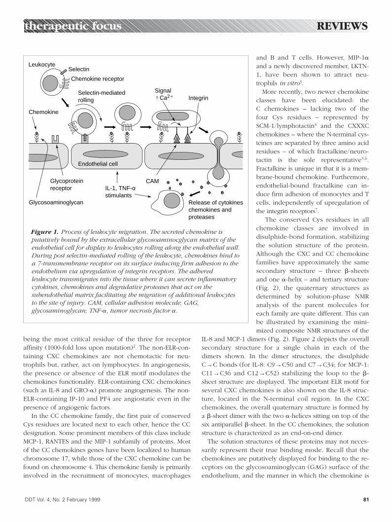
or insult to the immune system, chemokines are secretedby proinflammatory cells, leukocytes or endothelial cells torecruit new leukocytes from the circulation across thelumen and into the tissue (Fig. 1) and, as such, haveearned the nickname ‘crossing guards of the immune sys-tem’. The universal in vitro biological property of thesemolecules is the ability to effect the chemotaxis of specificcell types, but other chemokine functions have more re-cently been uncovered. These include inhibition or pro-motion of angiogenesis, inhibition or induction of cellularproliferation and induction of integrin receptors (Table 1).
Classification of chemokines
The chemokine proteins are segmented into four families(two major and two minor) based on the location of thefirst conserved Cys residues within the protein. Across andwithin the families, these proteins are 20–90% homologouswith each other (see table of chemokine sequence hom-ology and predicted secondary structure at the Chemo-kine Information Source: http://www.expasy.ch/cgi-bin/ChemokineTop.pl). The CXC family, in which IL-8 (seeBox 1 for glossary of abbreviations) is the representativemember, is characterized by the two conserved Cysresidues (C), near the N-terminus, separated by an aminoacid (X). Some of the CXC chemokines belong to the ELRsubfamily (i.e. containing Glu-Leu-Arg motif), which areimportant in the recruitment and activation of neutrophils.All members of this subfamily contain the ELR triad ofamino acids immediately before the first N-terminal Cys inthe chemokine protein. This group of amino acids is ex-tremely sensitive to modification. A point mutation in theELR motif of IL-8 dramatically effects chemokine activityand is somewhat detrimental to receptor binding, with Arg
Opportunities for noveltherapeutic agents acting atchemokine receptorsJohn Saunders and Christine M. Tarby
John Saunders, Neurocrine Biosciences, 10555 Science Center Drive, San Diego, CA 92121, USA and Christine M. Tarby*,CombiChem, Inc., 9050 Camino Santa Fe, San Diego, CA 92121, USA. *tel: 11 619 530 0484, fax: 11 619 530 9998, e-mail: [email protected]
Chemokines are proinflammatory mediators that
primarily control leukocyte migration into selected
tissues and upregulation of adhesion receptors. They
also have a role in pathological conditions that require
neovascularization and are implicated in the suppres-
sion of viral replication. By interaction with their re-
spective G-protein-coupled receptor, chemokines have
a profound influence over the selective recruitment of
specific cell types in acute inflammatory disease and,
hence, inhibition of their action should be of therapeu-
tic benefit. Only now are small molecule inhibitors
becoming available to validate this speculation. In this
review, without seeking to be comprehensive, the au-
thors provide an introduction to chemokines, their re-
ceptors and their role in certain disease processes.
Also, recent disclosures claiming novel nonpeptide
ligands for chemokine receptors are summarized.
being the most critical residue of the three for receptoraffinity (1000-fold loss upon mutation)2. The non-ELR-con-taining CXC chemokines are not chemotactic for neu-trophils but, rather, act on lymphocytes. In angiogenesis,the presence or absence of the ELR motif modulates thechemokines functionality. ELR-containing CXC chemokines(such as IL-8 and GRO-a) promote angiogenesis. The non-ELR-containing IP-10 and PF4 are angiostatic even in thepresence of angiogenic factors.
In the CC chemokine family, the first pair of conservedCys residues are located next to each other, hence the CCdesignation. Some prominent members of this class includeMCP-1, RANTES and the MIP-1 subfamily of proteins. Mostof the CC chemokines genes have been localized to humanchromosome 17, while those of the CXC chemokine can befound on chromosome 4. This chemokine family is primarilyinvolved in the recruitment of monocytes, macrophages
and B and T cells. However, MIP-1a
and a newly discovered member, LKTN-1, have been shown to attract neu-trophils in vitro3.
More recently, two newer chemokineclasses have been elucidated: the C chemokines – lacking two of the four Cys residues – represented by SCM-1/lymphotactin4 and the CXXXCchemokines – where the N-terminal cys-teines are separated by three amino acidresidues – of which fractalkine/neuro-tactin is the sole representative5,6.Fractalkine is unique in that it is a mem-brane-bound chemokine. Furthermore,endothelial-bound fractalkine can in-duce firm adhesion of monocytes and Tcells, independently of upregulation ofthe integrin receptors7.
The conserved Cys residues in allchemokine classes are involved indisulphide-bond formation, stabilizingthe solution structure of the protein.Although the CXC and CC chemokinefamilies have approximately the samesecondary structure – three b-sheetsand one a-helix – and tertiary structure(Fig. 2), the quaternary structures asdetermined by solution-phase NMRanalysis of the parent molecules foreach family are quite different. This canbe illustrated by examining the mini-mized composite NMR structures of the
IL-8 and MCP-1 dimers (Fig. 2). Figure 2 depicts the overallsecondary structure for a single chain in each of thedimers shown. In the dimer structures, the disulphideC→C bonds (for IL-8: C9→C50 and C7→C34; for MCP-1:C11→C36 and C12→C52) stabilizing the loop to the b-sheet structure are displayed. The important ELR motif forseveral CXC chemokines is also shown on the IL-8 struc-ture, located in the N-terminal coil region. In the CXCchemokines, the overall quaternary structure is formed bya b-sheet dimer with the two a-helices sitting on top of thesix antiparallel b-sheet. In the CC chemokines, the solutionstructure is characterized as an end-on-end dimer.
The solution structures of these proteins may not neces-sarily represent their true binding mode. Recall that thechemokines are putatively displayed for binding to the re-ceptors on the glycosoaminoglycan (GAG) surface of theendothelium, and the manner in which the chemokine is
therapeutic focus REVIEWS
DDT Vol. 4, No. 2 February 1999 81
Figure 1. Process of leukocyte migration. The secreted chemokine isputatively bound by the extracellular glycosoaminocglycan matrix of theendothelial cell for display to leukocytes rolling along the endothelial wall.During post selectin-mediated rolling of the leukocyte, chemokines bind toa 7-transmembrane receptor on its surface inducing firm adhesion to theendothelium via upregulation of integrin receptors. The adheredleukocyte transmigrates into the tissue where it can secrete inflammatorycytokines, chemokines and degradative proteases that act on thesubendothelial matrix facilitating the migration of additional leukocytesto the site of injury. CAM, cellular adhesion molecule; GAG,glycosaminoglycan; TNF-a, tumor necrosis factor a.
Leukocyte
Chemokine
Glycoproteinreceptor
Chemokine receptor
Selectin-mediatedrolling
Endothelial cell
Selectin
Glycosoaminoglycan
Signal Ca21 Integrin
Release of cytokineschemokines andproteases
CAMIL-1, TNF-αstimulants
REVIEWS therapeutic focus
82 DDT Vol. 4, No. 2 February 1999
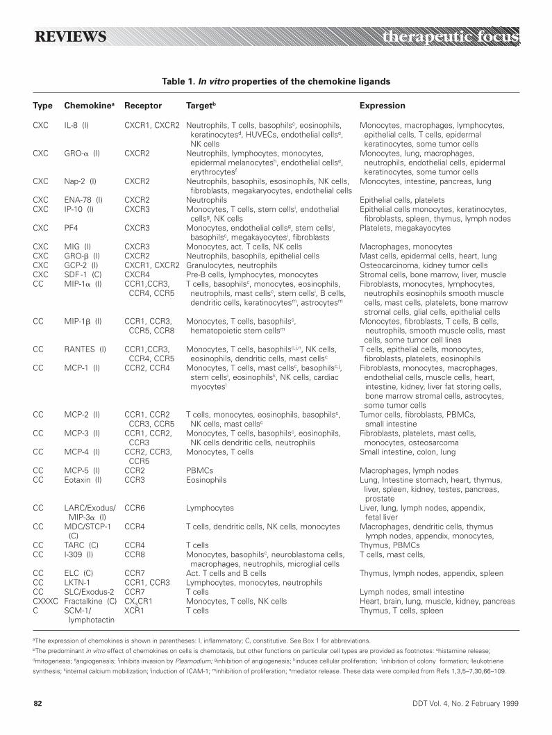
Table 1. In vitro properties of the chemokine ligands
Type Chemokinea Receptor Targetb Expression
CXC IL-8 (I) CXCR1, CXCR2 Neutrophils, T cells, basophilsc, eosinophils, Monocytes, macrophages, lymphocytes,keratinocytesd, HUVECs, endothelial cellse, epithelial cells, T cells, epidermal NK cells keratinocytes, some tumor cells
CXC GRO-a (I) CXCR2 Neutrophils, lymphocytes, monocytes, Monocytes, lung, macrophages, epidermal melanocytesh, endothelial cellse, neutrophils, endothelial cells, epidermalerythrocytesf keratinocytes, some tumor cells
CXC Nap-2 (I) CXCR2 Neutrophils, basophils, esosinophils, NK cells, Monocytes, intestine, pancreas, lungfibroblasts, megakaryocytes, endothelial cells
CXC ENA-78 (I) CXCR2 Neutrophils Epithelial cells, plateletsCXC IP-10 (I) CXCR3 Monocytes, T cells, stem cellsi, endothelial Epithelial cells monocytes, keratinocytes,
cellsg, NK cells fibroblasts, spleen, thymus, lymph nodesCXC PF4 CXCR3 Monocytes, endothelial cellsg, stem cellsi, Platelets, megakayocytes
basophilsc, megakayocytesi, fibroblastsCXC MIG (I) CXCR3 Monocytes, act. T cells, NK cells Macrophages, monocytesCXC GRO-b (I) CXCR2 Neutrophils, basophils, epithelial cells Mast cells, epidermal cells, heart, lungCXC GCP-2 (I) CXCR1, CXCR2 Granulocytes, neutrophils Osteocarcinoma, kidney tumor cellsCXC SDF -1 (C) CXCR4 Pre-B cells, lymphocytes, monocytes Stromal cells, bone marrow, liver, muscleCC MIP-1a (I) CCR1,CCR3, T cells, basophilsc, monocytes, eosinophils, Fibroblasts, monocytes, lymphocytes,
CCR4, CCR5 neutrophils, mast cellsc, stem cellsi, B cells, neutrophils eosinophils smooth muscledendritic cells, keratinocytesm, astrocytesm cells, mast cells, platelets, bone marrow
stromal cells, glial cells, epithelial cellsCC MIP-1b (I) CCR1, CCR3, Monocytes, T cells, basophilsc, Monocytes, fibroblasts, T cells, B cells,
CCR5, CCR8 hematopoietic stem cellsm neutrophils, smooth muscle cells, mastcells, some tumor cell lines
CC RANTES (I) CCR1,CCR3, Monocytes, T cells, basophilsc,j,n, NK cells, T cells, epithelial cells, monocytes, CCR4, CCR5 eosinophils, dendritic cells, mast cellsc fibroblasts, platelets, eosinophils
CC MCP-1 (I) CCR2, CCR4 Monocytes, T cells, mast cellsc, basophilsc,j, Fibroblasts, monocytes, macrophages,stem cellsi, eosinophilsk, NK cells, cardiac endothelial cells, muscle cells, heart,myocytesl intestine, kidney, liver fat storing cells,
bone marrow stromal cells, astrocytes,some tumor cells
CC MCP-2 (I) CCR1, CCR2 T cells, monocytes, eosinophils, basophilsc, Tumor cells, fibroblasts, PBMCs,CCR3, CCR5 NK cells, mast cellsc small intestine
CC MCP-3 (I) CCR1, CCR2, Monocytes, T cells, basophilsc, eosinophils, Fibroblasts, platelets, mast cells, CCR3 NK cells dendritic cells, neutrophils monocytes, osteosarcoma
CC MCP-4 (I) CCR2, CCR3, Monocytes, T cells Small intestine, colon, lungCCR5
CC MCP-5 (I) CCR2 PBMCs Macrophages, lymph nodesCC Eotaxin (I) CCR3 Eosinophils Lung, Intestine stomach, heart, thymus,
liver, spleen, kidney, testes, pancreas, prostate
CC LARC/Exodus/ CCR6 Lymphocytes Liver, lung, lymph nodes, appendix, MIP-3a (I) fetal liver
CC MDC/STCP-1 CCR4 T cells, dendritic cells, NK cells, monocytes Macrophages, dendritic cells, thymus(C) lymph nodes, appendix, monocytes,
CC TARC (C) CCR4 T cells Thymus, PBMCsCC I-309 (I) CCR8 Monocytes, basophilsc, neuroblastoma cells, T cells, mast cells,
macrophages, neutrophils, microglial cellsCC ELC (C) CCR7 Act. T cells and B cells Thymus, lymph nodes, appendix, spleenCC LKTN-1 CCR1, CCR3 Lymphocytes, monocytes, neutrophilsCC SLC/Exodus-2 CCR7 T cells Lymph nodes, small intestineCXXXC Fractalkine (C) CX3CR1 Monocytes, T cells, NK cells Heart, brain, lung, muscle, kidney, pancreasC SCM-1/ XCR1 T cells Thymus, T cells, spleen
lymphotactin
aThe expression of chemokines is shown in parentheses: I, inflammatory; C, constitutive. See Box 1 for abbreviations.bThe predominant in vitro effect of chemokines on cells is chemotaxis, but other functions on particular cell types are provided as footnotes: chistamine release;dmitogenesis; eangiogenesis; finhibits invasion by Plasmodium; ginhibition of angiogenesis; hinduces cellular proliferation; iinhibition of colony formation; jleukotriene
synthesis; kinternal calcium mobilization; linduction of ICAM-1; minhibition of proliferation; nmediator release. These data were compiled from Refs 1,3,5–7,30,66–109.

presented to the receptor might be quite different fromthat inferred by the solution structures. Because relativelyhigh concentrations of protein are needed for NMR struc-ture determination, the dimeric structure is favored, but atphysiologically relevant concentrations of chemokine, themonomeric protein is probably present, opening the de-bate as to which form of the chemokine is biologically relevant8.
In general, the body of the chemokine seems to be pri-marily responsible for binding to the receptor and theGAG matrix, while the N-terminal regions are importantfor signaling. To understand how chemokines interact withtheir receptors and to facilitate the discovery of small-mol-ecule inhibitors, several mutagenesis studies have eluci-dated key residues important for chemokine signaling andbinding to the receptor. In the ELR-containing CXCchemokines, removal of the Glu of the ELR motif affectsreceptor binding to some degree but, more importantly,causes a complete loss of the protein’s chemotactic andneutrophil-activating properties. As mentioned previously,Arg is the triad member most sensitive to mutation. Related
CXC chemokines such as PF4 and IP10 are devoid of theELR triad and have only weak activities for neutrophil IL-8receptors. Substitution of the natural sequence DRL forELR in PF4 confers IL-8-like behaviour on neutrophils.Even though mutation in the ELR triad has detrimental ef-fects on receptor binding, the ELR sequence alone is insuf-ficient for binding – short peptides carrying this motif donot compete with IL-8 at the CXCR1 receptor.
Site-directed mutagenesis studies on IL-8 have revealedanother set of amino acids in which mutations affect re-ceptor binding9. The second set (Phe17, Phe21, Ile22,Leu43) forms a solvent-accessible hydrophobic pocketthat, in theory, could entropically drive the binding of IL-8with CXCR1. In MCP-1 (a member of the CC family), theN-terminal deletion of the first eight amino acids results ina 9–76 mutant that is almost fully capable of binding toCCR2 (Ref. 10). However, no Ca21 flux is observed uponmutant binding, clearly indicating that, as for the CXCchemokines, the important residues for signaling at the receptor precede the first disulphide linkage along the N-terminus. A similar result can be shown for another b-chemokine, RANTES. N-terminal deletion of the first eight
therapeutic focus REVIEWS
DDT Vol. 4, No. 2 February 1999 83
Box 1. Glossary
ELC, Ebl1-ligand chemokineENA-78, epithelial-derived neutrophil attractant 78GCP-2, granulocyte chemotactic protein 2GRO, growth related oncogeneICAM-1, intracellular adhesion molecule 1IL -8, interleukin 8IFN-g, interferon gIP-10, (IFN-g)-inducible protein 10LARC, liver and activation-regulated chemokineLKTN-1, leukotactin 1LTB-4, leukotriene B4MAPK, mitogen-activated protein kinaseMCP, monocyte chemoattractant proteinMDC, macrophage-derived chemokineMGSA, melanoma growth-stimulating activityMIG, monokine induced by IFN-gMIP, macrophage inflammatory proteinNAP-2, neutrophil-activating protein 2NF -kB, nuclear factor k-bindingPBMC, peripheral blood mononucleocytesPF4, platelet factor 4PMN, polymorphonuclear leukocytesRANTES, regulated on activation, normal T expressedand secretedSCM-1 single cysteine motif 1SDF-1, stromal cell-derived factor 1SLC, secondary lymphoid tissue chemokineSTCP-1, stimulated T-cell chemotactic protein 1TARC, thymus and activation-regulated chemokine
Figure 2. Comparison of the solution structures of(a) interleukin 8 (IL-8) – a CXC ligand64 – and (b)monocyte chemotactic protein 1 (MCP-1) – a CCligand65. The figures of the monomer structures (top)were created from the dimer structures (bottom).These figure elements were retrieved from theBrookhaven data bank (http://www.pdb.bnl.gov/)and visualized by WebLab Viewer (MolecularSimulations, San Diego, CA, USA).

amino acids results in a mutant that prevents the infectionof activated PBMCs by the HIV-1 virus by inhibiting accessof the virus to CCR5 (Ref. 11).
RANTES is a typical CC chemokine that binds severalchemokine receptors including CCR1, CCR3 and CCR5.Site-directed mutagenesis studies of RANTES have indi-cated that different sets of amino acids on the protein areresponsible for interaction with the various receptors (i.e.for CCR1, R17; for CCR3, F12 and Y14; for CCR5, F12, P2and I15). With the exception of P2, all of these residuesare located on the N-loop, suggesting that the N-loop se-quences confer the binding affinity and selectivity to thechemokine11. The P2 residue, which is necessary forRANTES binding to CCR5, sits near the N-terminus of thechemokine. However, in the dimer structure, the N-termi-nal arm of each chain is located near the N-loop of itspartner. Perhaps this provides circumstantial evidence thatthe dimeric protein might be important to CCR5 binding.
Comparison of chemokine receptors
with other GPCRs
Receptor structure and putative ligand-binding site(s)By analogy with bacteriorhodopsin, members of the G-protein-coupled receptor (GPCR) superfamily (of whichthere are ~1000, based on genomic information) arethought to contain a seven-helical motif that traverses thecell membrane (Fig. 3) to form a central channel12.Together with the extracellular domains, this helical bun-dle is thought to provide the ligand-binding and activationsites, the precise location being determined by the natureof the ligand. All chemokine receptors share some com-mon features, some of which are also shared by manyother members of the GPCR superfamily. By inference,such features could be involved in ligand binding and receptor activation. All chemokine receptors have twoconserved Cys residues, one in the N-terminal domain andthe second within extracellular loop three; these seem to
REVIEWS therapeutic focus
84 DDT Vol. 4, No. 2 February 1999
Figure 3. The CCR1 receptor showing the extracellular loops (E), the putatively helical regions (H) that traverse thecell membrane and the intracellular loops (I). The amino acid residues are color coded according to the nature oftheir side-chains as shown in the key.
PFHLSCT
LAFVAHVI
TPCQKV
GFVWDD
AGFARQ L L P
S L V F
YLP
V L V L
VLI
L V G N
GIV
F Y L G
A L I A
SMP
G F T V
VIT
I I I S
WAL
F Q A L
L F G L
NLK
Y T G I
CII
L L V M
PLV
L S G
T G L Y
YYF
ITLL
F F I I
IES
F P L T
L L D S
FLF
I W
M NTSI
N L L Y
LAI
HESLREWKL
IKILL
F V S I
L N Y P
TIL
V F I L
IMI
L F F I
FWT
A V Q V
I A Y T
VET
F V G
YI
V N P V
CCH
RVAKS
R K
HHTFEWQ
SQECEHRHLDL
TFLFDQ
TKS
KLKYD
I
DAMC
DRYLA
KENPR
TRAR
K
N
GYDFET
E
DA
TTEDYDT
TEMH2N–
T
RKYQVKL
PN
RVAVH
PLWK
L
FLQRL
V
HR
ERFRK
Y
GEHEL
F –COOHG
S
PSTSS
A
ST
VDRLER
F
V
A
LS
H1
I1
E1 E2 E3
I2 I3
Cytosolicdomain5 Acidic
Amino acids:
5 Basic
5 Lipophilic
5 Aromatic
5 Hydrophilic
5 N or Q
5 G or P
5 C
Extracellulardomain
H2 H3 H4 H5 H6
H7
be structurally forming a C→C bond that is an integral partof the ligand-recognition site. There are also two con-served acidic residues (CXCR3 excepted) within the helicalbundle. The first, an Asp in helix 2, is intimately associatedwith receptor activation as with all GPCRs. However, un-like the GPCRs for monoamines that have a second Asp inhelix 3 close to the extracellular surface, chemokine recep-tors have a Glu residue in a similar position but on helix 7.In common with other superfamily members, there is aconserved DRY triad at the C-terminus of helix 3 – specu-latively involved in G-protein binding. Unique to thechemokine receptor subfamily, all have an acidic N-termi-nus with gross negative charges ranging from 21 to 26,and this might distinguish the initial recognition eventfrom that in other GPCRs.
The binding site(s) for endogenous ligands have begun to be mapped using single-point-mutation analysis andchimeric proteins, and the residues that are critical for recep-tor activation are being studied13–15. Of the two receptors forIL-8 (CXCR1 and CXCR2), CXCR1 has high affinity only forIL-8 (and more recently, weaker affinity for GCP-2), whereasCXCR2 binds other CXC ligands (GRO, NAP-2) equally well.Chimerae between CXCR1 and CXCR2 indicated that the ex-tracellular N-terminal domain determined ligand selectivitybecause receptors bearing the CXCR1 N-terminal regionwere selective for IL-8, while the CXCR2 N-terminal se-quence, either in a CXCR1 or CXCR2 background, displayedpromiscuity for other CXC chemokines. All acidic and basicresidues present on the ligand-accessible surface of CXCR1were systematically mutated to Ala, with the result that D11,R199, R203, D265, E275 and R280 were found to be criticalfor ligand binding13,14. Additional binding sites are found onthe second and third extracellular loops. The integrity of theC→C bond between the N-terminal region and extracellularloop 3 is also important and, together, these might constitutea major IL-8 binding site. Surprisingly, given the role of D89across the GPCR superfamily, little comment is available onthe role of D89 in ligand-induced receptor activation.
Chimeric CCR1 and CCR2 have helped to elucidate atwo-step mechanism for MCP-1 activation of CCR2(Ref. 15). Radiolabeled MCP-1 binds with high affinity towildtype CCR2 but not to CCR1; however, this profile isreversed in chimeras where the N-terminal sequences areinterchanged, and this effect is mirrored in signaling. Bycontrast, substitution of the wildtype CCR1 N-terminal do-main with that from CCR2 has little effect upon MIP-1a
binding. Thus, for CCR2 but not for CCR1 interacting withMIP-1a or RANTES, the N-terminal domain is importantboth for binding and signaling, as witnessed by a .10-fold and 30-fold increase in the Kd and the IC50 for adeny-
lyl cyclase inhibition, respectively. Progressive substitutionof the extracellular domains in CCR2 showed that eachdomain contributed to signal transduction: extracellularloops 1 and 2 contribute only modestly (,10-fold com-bined) but all three in concert contribute markedly (30-fold). Again, in CCR1, only the third extracellular domainwas especially critical for signaling.
Taken together, most studies point to a two-step modelfor chemokine receptor activation. The initial high-affinityrecognition event apparently tethers the ligand in closeproximity to the activation site from which receptor acti-vation is triggered. This is not unlike the model currentlyaccepted for thrombin receptor activation, albeit in that instance the ligand acts in cis.
There is considerable structural and sequence diversityacross the GPCR superfamily to accommodate functionaldifferences and also the disparate nature of the homolo-gous ligand, from simple amines (such as acetylcholineand dopamine) through adenosine, prostaglandins andsmall peptides (such as angiotensin II), to the larger li-gands represented by the chemokines. However, we spec-ulate that, while the recognition event is clearly divergent,activation is conserved and always involves the helical do-main most probably acting as a channel for cation (i.e. aproton or sodium ion) transfer from the extracellular fluid.
Intracelluar signalingChemokines signal by interaction with specific, cell-surfaceGPCRs and thence activation of heterotrimeric G-proteins,located intracellularly and protected from the surroundingchaos by the cell membrane. Given that there are .50chemokines reported to date and only a dozen or so recep-tors, there is significant redundancy in the ligand and, notsurprisingly, most ligands display promiscuity in theirbinding partners. The situation is further complicated bythe way in which each ligand is ‘presented’ to the receptorbecause chemokines are bound tightly to various extracel-lular matrix proteins, such as the GAGs, heparin, chon-droitin sulphate and dermatan sulphate. Thus, chemokineselectivity can be accurately mapped only in the exactphysiological environment. Signaling studies for each of thereceptors are far from complete and, again, experimentsusing convenient but nevertheless artificial systems involv-ing transfectants should be interpreted with some caution.
Chemotactic responses to chemokines are associated withrapid and transient elevations of intracellular Ca21. After acti-vation, chemokine receptors have altered sensitivity to re-peated stimulation with the appropriate ligand. As with allGPCRs, activated chemokine receptors couple to het-erotrimeric G-proteins consisting of abg subunits (Fig. 4). It
therapeutic focus REVIEWS
DDT Vol. 4, No. 2 February 1999 85
is the pertussis toxin-sensitive G protein16,17, Gi, that links thereceptor to PIP2 hydrolysis through the intermediacy of thedissociated bg subunit, that is implicated for all receptorsstudied to date. For example, cell lines stably transfectedwith CCR3 respond to eotaxin but not to any otherchemokine at reasonable concentrations (,10 nM), as ob-served by Ca21 flux measurements; at higher concentrations,RANTES and MCP-3 gave a similar response. When thesetransfectants were pretreated with pertussis toxin, the Ca21
flux response to eotaxin but not to ATP was abolished, sug-gesting the involvement of Gi in transduction. Sequentialstimulation of CCR3 by eotaxin but not any other chemokinecaused homologous desensitization. Thus, receptor regu-
lation represents another control pointin chemokine function. Expression ratesof receptor and receptor recycling alsodetermine the response to chemokineligands and might be modified toachieve a therapeutic benefit, as wit-nessed with the IL-8 receptor (seebelow). However, the pertussis-insensi-tive G protein, Gq, can also activate in-tracellular effector systems in othertransfectant studies18. Finally, it is nowclear that G proteins also play a pivotalrole in serine/threonine kinase path-ways, such as Ras/MAPK, and thatchemokines have been shown to acti-vate these processes (Fig. 4).
Role of chemokines in inflamma-
tory disease
IL -8-mediated processesWhile the chronic phase of the in-flammatory response is characterizedby extravasation and infiltration ofmacrophages and lymphocytes, theacute phase is mediated also by leuko-cytes such as neutrophils, eosinophilsand basophils19,20. In 1987 the endoge-nous peptide21, now known as IL-8,was first isolated from lipopolysaccha-ride-stimulated human monocytes andshown to be chemoattractive primarilyfor neutrophils, although it also affectsother leukocytes such as T and Bcells. Displacement studies with[125I]IL-8 have revealed two high-affin-ity sites on neutrophils for IL-8: the IL-8A site, which is specific for IL-8; and
the IL-8B site, at which the radiolabel can be displaced byother CXC ligands containing the ELR motif, such as NAP-2, GRO-a and MGSA. Subsequently, this was confirmed bythe cloning and characterization of two distinct IL-8 recep-tors (IL-8R) having 77% homology.
An IL-8 homologue in rats or mice does not exist; hence,experiments in these species have targeted the N51/KC receptor, which is highly homologous to IL-8RB (CXCR2).Using either specific antibodies for human IL-8 or IL-8 recep-tor homologue knock-out mice, it has been clearly demon-strated that IL-8 is involved in the recruitment of neutrophilsinto tissues in the acute phase of inflammation22. Thus, theneutrophils from IL-82/2 mice kill bacteria as effectively as
REVIEWS therapeutic focus
86 DDT Vol. 4, No. 2 February 1999
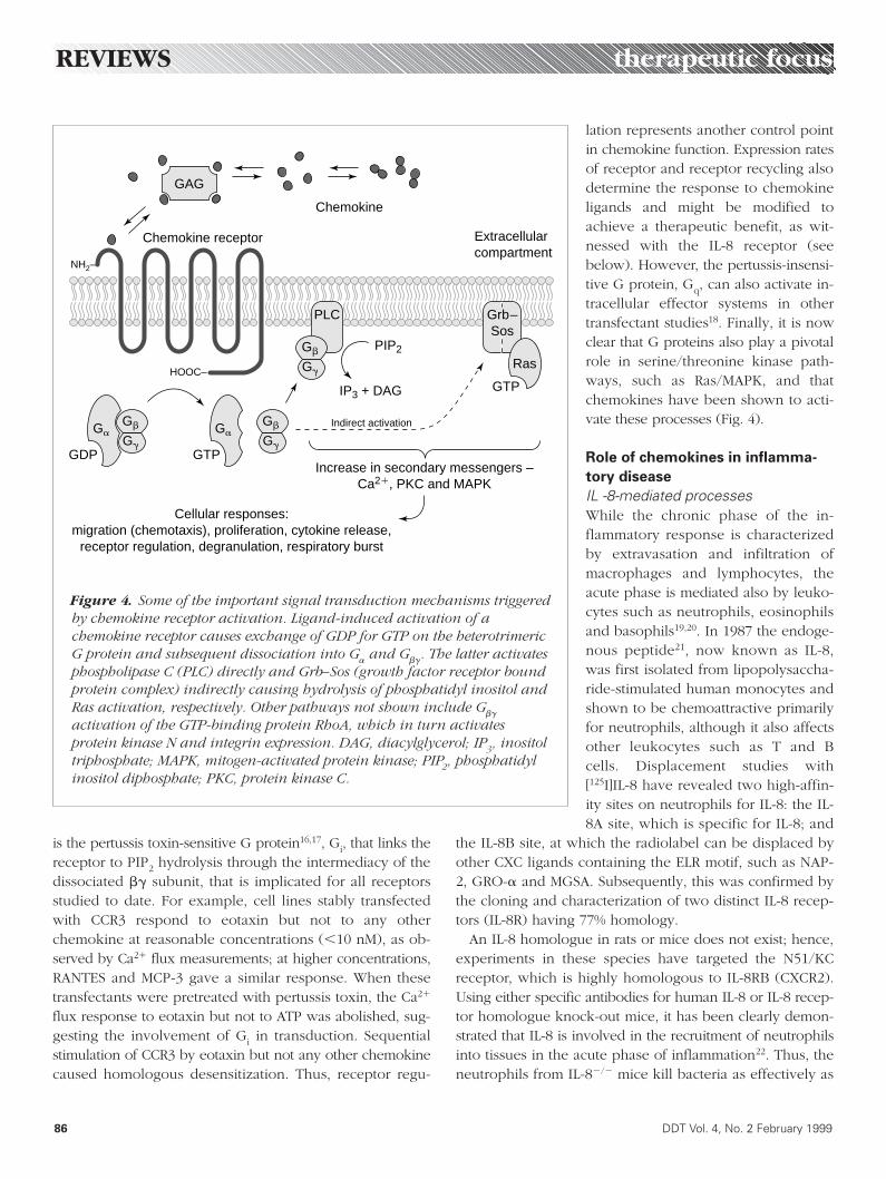
Figure 4. Some of the important signal transduction mechanisms triggeredby chemokine receptor activation. Ligand-induced activation of achemokine receptor causes exchange of GDP for GTP on the heterotrimericG protein and subsequent dissociation into Ga and Gbg. The latter activatesphospholipase C (PLC) directly and Grb–Sos (growth factor receptor boundprotein complex) indirectly causing hydrolysis of phosphatidyl inositol andRas activation, respectively. Other pathways not shown include Gbg
activation of the GTP-binding protein RhoA, which in turn activatesprotein kinase N and integrin expression. DAG, diacylglycerol; IP3, inositoltriphosphate; MAPK, mitogen-activated protein kinase; PIP2, phosphatidylinositol diphosphate; PKC, protein kinase C.
Indirect activation
NH2–
HOOC–
GAG
Chemokine
Chemokine receptor Extracellularcompartment
PIP2
PLC
IP3 + DAG
Increase in secondary messengers –Ca21, PKC and MAPK
Cellular responses:migration (chemotaxis), proliferation, cytokine release,
receptor regulation, degranulation, respiratory burst
GDP
GaGb
Gg
Gb
Gg
Gb
Gg
GTP
GTP
Ga
Ras
Grb–Sos

wild type but fail to infiltrate tissues; appar-ently this process cannot be substituted byother neutrophil chemoattractants such asthe complement protein C5a. Because IL-8has been detected at diverse inflammatorysites, it is still unclear what the primary indi-cation should be for compounds that inter-fere with aberrant IL-8 function. Diseasessuch as psoriasis, rheumatoid arthritis,glomerular nephritis, angiogenesis, adult res-piratory distress syndrome, ischemic reperfu-sion injury and pulmonary fibrosis have allbeen mentioned by various investigators.
In principle, as with all other chemokines,blockade of IL-8 function at many levels23 can be consid-ered (Fig. 4) from inhibition of IL-8 production through re-ceptor blockade to interference in one or more of the IL-8-mediated intracellular signaling pathways. For example,IL-8 production is controlled primarily by the transcriptionalfactor NF-kB – this step is cancelled by anti-inflammatorysteroids and other immunomodulators. However, agentsthat act at the receptor level are more specific. To date,most reports have focused on the ability of peptide frag-ments of IL-8 to act as functional antagonists, there beingfew small nonpeptide molecules described. Removal of theentire C-terminal helical domain (see above) after thefourth Cys residue attenuated, but did not eliminate, activ-ity. By contrast, deletion of the ELR motif close to the N-ter-minus completely destroyed both binding to and activationof the IL-8 receptor24. Closer investigation25 of truncated N-terminal peptides yielded receptor antagonists, the mostpotent being AA-IL-86–72, having a Kd of 8 nM (the Kd of IL-8 is 0.25 nM) and behaving as a functional antagonist of IL-8-induced elastase release. Subsequently, capped hexa- andheptapeptides, exemplified by Ac-RRWWCR-NH2 and all-D-Ac-RRWWCRC-NH2, respectively, were shown to be modestinhibitors of IL-8 binding, but in the mM range26.
A new nonpeptide inhibitor (1) was discovered by high-throughput screening27 and was optimized to produce 2(Fig. 5), having an IC50 of 22 nM against CXCR2 but failing toinhibit CXCR1 at 100-fold concentration. In human neu-trophils, which express almost equal numbers of each recep-tor, it is unclear whether chemotaxis is mediated by one orboth of these receptor subtypes. In these cells, 2 inhibitedGRO-a (CXCR2-selective) but not IL-8-stimulated Ca21 mobi-lization, presumably because IL-8 can bypass CXCR2 inhibi-tion by activating CXCR1. However, using human PMNs,chemotaxis to either IL-8 or GRO-a was inhibited (IC50 5
20 nM and 60 nM, respectively) indicating that neutrophilchemotaxis is mediated predominantly through CXCR2.
However, whether this series of compoundsacts by direct blockade at the receptor level orby a downstream event is still uncertain.
Role of chemokines and their receptors in infectious diseaseSeveral unrelated observations implicatechemokine receptors in viral pathogenesis,although these observations have yet to be translated into a therapeutic benefit.Cytomegalovirus (CMV) possesses threeopen reading frames that encode putativeGPCRs, designated US27, US28 and UL33,the functions of which were unknown until
it was realized that US28 is a functional chemokine recep-tor with limited sequence homology to CCR1. At present,the role of this receptor in the CMV replicative cycle is unknown, but it is well known that some chemokines, forinstance IL-8, can activate viral replication. Members of the poxvirus and herpesvirus families express GPCR-likereceptors that are sensitive to chemokines.
Co-infection of HIV-positive individuals by CMV is acommon clinical complication; indeed there is frequenthistochemical evidence of dually infected cells in brain,retina and lung. The product encoded by US28 is a func-tional chemokine receptor activated by RANTES, MIP-1a
and MIP-1b and was recently shown to permit HIV fusion(see below) with human cell lines bearing the CD4 receptorand cotransfected with US28 gene product. Whether US28plays a role in HIV pathogenesis remains controversial.
Researchers had known for over a decade that CD4 in itself was insufficient to support HIV fusion with the hostimmune cell, as mouse cells transfected with human CD4and expressed on the cell surface failed to become infec-ted with HIV. It was the discovery28 in 1995 that thechemokines RANTES, MIP-1a and MIP-1b were the majorHIV-suppressive factors produced by CD81 cells that initi-ated an explosion of new research to determine the mecha-nism and molecular target of these agents. In confirmationof their role, specific neutralizing antibodies to RANTES,MIP-1a or MIP-1b completely blocked their suppressive ac-tivity and, indeed, more recently it has been shown thatsome individuals who remain HIV-negative despite multi-ple high-risk exposures also have high levels of one ormore of these chemokines. With the cloning of ‘fusin’29,now known as CXCR4, the missing co-receptor predicted tobe necessary to complement the fusion process followingthe formation of the CD4–gp120 complex was identified.This receptor was surprisingly not sensitive to the CCchemokines listed above but, instead, was later shown30 to
therapeutic focus REVIEWS
DDT Vol. 4, No. 2 February 1999 87
Figure 5. NonpeptideCXCR2 antagonists:1, SK&F83589 and 2,SB225002.
NH
OOH
NH
O2N
R
1, R 5 H2, R 5 OH
be activated by the lymphocyte chemoattractant SDF-1,suggesting that a second co-receptor remained to be dis-covered. In the absence of CXCR4, CD4 expressed on thesurface of T helper (Th) cells or macrophages interacts withthe viral envelope glycoprotein gp120/gp41, but this is, initself, not permissive to fusion of the viral and host cellplasma membranes with subsequent viral replication. Morespecifically, CXCR4 supports the entry of HIV strains thatemerge at late clinical stages of the infection – strains thatare also distinguished by their ability to form syncytia inMT-2 cells and were previously called synctia-inducing.Because this receptor does not share RANTES, MIP-1a orMIP-1b as its ligand(s), another chemokine receptor wasalso thought to be involved in early-stage infection. Strainsof HIV that initially establish a persistent infection aremacrophage tropic, are not syncytium-inducing strains andwere shown31,32 to use CCR5 as a co-receptor for fusion.Even this explanation is a simplification because some pri-mary clinical isolates of HIV can use CCR3, which is nowknown not to be found exclusively on eosinophils but isalso expressed on Th2 cells and microglia. Finally, strainshave been isolated, largely dual tropic, that can induce syn-cytial formation on cells expressing CCR2b, CCR3, CCR5,CCR8, CXCR1, CXCR4 and LTB-4.
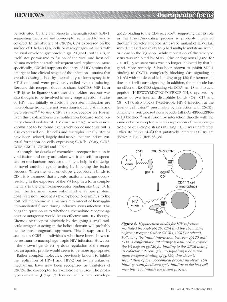
Although the details of chemokine receptor function inviral fusion and entry are unknown, it is useful to specu-late on mechanisms because this might help in the designof novel antiviral agents acting by blocking the fusionprocess. When the viral envelope glycoprotein binds toCD4, it is assumed that a conformational change occurs,resulting in the exposure of the V3 loop in a form comple-mentary to the chemokine-receptor binding site (Fig. 6). Inturn, the transmembrane subunit of envelope protein,gp41, can now present its hydrophobic N-terminus to thehost cell membrane in a manner reminiscent of hemagglu-tinin-mediated fusion during influenza virus infection. Thisbegs the question as to whether a chemokine receptor ag-onist or antagonist would be an effective anti-HIV therapy.Chemokine receptor blockade by designing a small-mol-ecule antagonist acting in the helical domain will probablybe the most pragmatic approach. This is supported bystudies on CCR52/2 individuals who have been shown tobe resistant to macrophage-tropic HIV infection. However,if the known ligands act by downregulation of the recep-tor, an agonist profile would seem to be more appropriate.
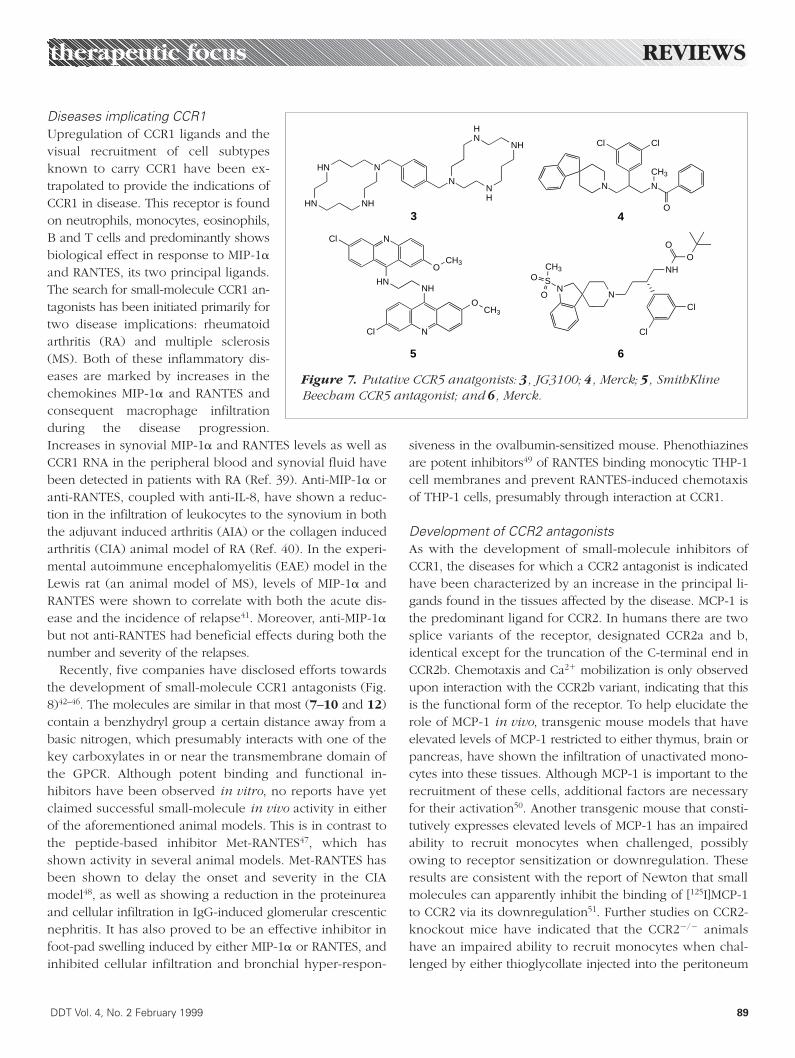
Rather complex molecules, previously known to inhibitthe replication of HIV-1 and HIV-2 but by an unknownmechanism, have now been recognized as inhibitors ofCXCR4, the co-receptor for T-cell-tropic viruses. The proto-type derivative 3 (Fig. 7) does not inhibit viral envelope
gp120 binding to the CD4 receptor33, suggesting that its rolein the fusion/uncoating process is probably mediatedthrough a cofactor receptor. An escape mutant of HIV-1 LAIwith decreased sensitivity to 3 had multiple mutations withinor close to the V3 loop. While replication of the wildtypevirus was inhibited by SDF-1 (the endogenous ligand forCXCR4), 3-resistant virus was no longer inhibited by that li-gand. More recently, 3 has been shown to inhibit SDF-1binding to CXCR4, completely blocking Ca21 signaling at0.1 nM with no detectable binding to gp120; furthermore, itdoes not itself cause signaling. In addition, the molecule hasno effect on RANTES signaling via CCR5. An 18-amino acidpeptide (H-RRWCYRKCYKGYCYRKCR-NH2), cyclized bymeans of two internal disulphide bonds (C4→C17 andC8→C13), also blocks T-cell-tropic HIV-1 infection at thelevel of cell fusion34, presumably by interaction with CXCR4.Similarly, a D-Arg-based nonapeptide (all D-Ac-RRRRRRRRR-NH2) blocked35 viral fusion by interaction directly with thesame cofactor receptor, whereas replication of macrophage-tropic or dual-tropic strains utilizing CCR5 was unaffected.Other structures (4–6) that putatively interact at CCR5 areshown in Fig. 7 (Refs 36–38).
REVIEWS therapeutic focus
88 DDT Vol. 4, No. 2 February 1999
Figure 6. Hypothetical model for HIV infectionmediated through gp120, CD4 and the chemokinecofactor receptor (either CXCR4, CCR5 or others).Following the initial interaction between gp120 andCD4, a conformational change is assumed to exposethe V3 loop on gp120 for binding to the GPCR actingas cofactor. Interestingly, no signaling is observedupon receptor binding of gp120, thus there isspeculation of the biochemical process involved. Thisbinding event facilitates gp41 binding to the host cellmembrane to initiate the fusion process.
HIVvirion
CXCR4 or CCR5
CD4
gp41
gp120
CD41 T cellor macrophage
HIVvirion
Diseases implicating CCR1Upregulation of CCR1 ligands and thevisual recruitment of cell subtypesknown to carry CCR1 have been ex-trapolated to provide the indications ofCCR1 in disease. This receptor is foundon neutrophils, monocytes, eosinophils,B and T cells and predominantly showsbiological effect in response to MIP-1a
and RANTES, its two principal ligands.The search for small-molecule CCR1 an-tagonists has been initiated primarily fortwo disease implications: rheumatoidarthritis (RA) and multiple sclerosis(MS). Both of these inflammatory dis-eases are marked by increases in thechemokines MIP-1a and RANTES andconsequent macrophage infiltrationduring the disease progression.Increases in synovial MIP-1a and RANTES levels as well asCCR1 RNA in the peripheral blood and synovial fluid havebeen detected in patients with RA (Ref. 39). Anti-MIP-1a oranti-RANTES, coupled with anti-IL-8, have shown a reduc-tion in the infiltration of leukocytes to the synovium in boththe adjuvant induced arthritis (AIA) or the collagen inducedarthritis (CIA) animal model of RA (Ref. 40). In the experi-mental autoimmune encephalomyelitis (EAE) model in theLewis rat (an animal model of MS), levels of MIP-1a andRANTES were shown to correlate with both the acute dis-ease and the incidence of relapse41. Moreover, anti-MIP-1a
but not anti-RANTES had beneficial effects during both thenumber and severity of the relapses.
Recently, five companies have disclosed efforts towardsthe development of small-molecule CCR1 antagonists (Fig.8)42–46. The molecules are similar in that most (7–10 and 12)contain a benzhydryl group a certain distance away from abasic nitrogen, which presumably interacts with one of thekey carboxylates in or near the transmembrane domain ofthe GPCR. Although potent binding and functional in-hibitors have been observed in vitro, no reports have yetclaimed successful small-molecule in vivo activity in eitherof the aforementioned animal models. This is in contrast tothe peptide-based inhibitor Met-RANTES47, which hasshown activity in several animal models. Met-RANTES hasbeen shown to delay the onset and severity in the CIAmodel48, as well as showing a reduction in the proteinureaand cellular infiltration in IgG-induced glomerular crescenticnephritis. It has also proved to be an effective inhibitor infoot-pad swelling induced by either MIP-1a or RANTES, andinhibited cellular infiltration and bronchial hyper-respon-
siveness in the ovalbumin-sensitized mouse. Phenothiazinesare potent inhibitors49 of RANTES binding monocytic THP-1cell membranes and prevent RANTES-induced chemotaxisof THP-1 cells, presumably through interaction at CCR1.
Development of CCR2 antagonistsAs with the development of small-molecule inhibitors ofCCR1, the diseases for which a CCR2 antagonist is indicatedhave been characterized by an increase in the principal li-gands found in the tissues affected by the disease. MCP-1 isthe predominant ligand for CCR2. In humans there are twosplice variants of the receptor, designated CCR2a and b,identical except for the truncation of the C-terminal end inCCR2b. Chemotaxis and Ca21 mobilization is only observedupon interaction with the CCR2b variant, indicating that thisis the functional form of the receptor. To help elucidate therole of MCP-1 in vivo, transgenic mouse models that haveelevated levels of MCP-1 restricted to either thymus, brain orpancreas, have shown the infiltration of unactivated mono-cytes into these tissues. Although MCP-1 is important to therecruitment of these cells, additional factors are necessaryfor their activation50. Another transgenic mouse that consti-tutively expresses elevated levels of MCP-1 has an impairedability to recruit monocytes when challenged, possiblyowing to receptor sensitization or downregulation. Theseresults are consistent with the report of Newton that smallmolecules can apparently inhibit the binding of [125I]MCP-1to CCR2 via its downregulation51. Further studies on CCR2-knockout mice have indicated that the CCR22/2 animalshave an impaired ability to recruit monocytes when chal-lenged by either thioglycollate injected into the peritoneum
therapeutic focus REVIEWS
DDT Vol. 4, No. 2 February 1999 89
Figure 7. Putative CCR5 anatgonists: 3, JG3100; 4, Merck; 5, SmithKlineBeecham CCR5 antagonist; and 6, Merck.
HN NH
NHN
NH
HN
NNH
N
HNNH
N
O
Cl
Cl
OCH3
CH3
ClCl
NN
CH3
O
Cl
Cl
NN
S
CH3O
O
3 4
5 6
NHO
O

or PPD-bead-induced lung granuloma. These studies havefurther suggested that activation of CCR2 might be involvedin Th1-type cytokine responses52.
Several inflammatory diseases are marked by specificsite elevations of MCP-1. These include atherosclerosis,RA, MS, glomuleronephritis, asthma and stroke. For exam-ple, in RA, the infiltation of monocytes and macrophagescan be observed in the synovial lining of the arthritic joint.The synovial fluid contains increases in a variety ofchemokines including MCP-1, a key ligand for CCR2.Elevated levels of this chemokine are probably responsiblefor the monocyte infiltration into the synovial tissue.Antibodies to MCP-1 have exhibited a reduction inswelling in both the AIA and CIA models of the disease inrat; however, the effect seen is relatively small (30% reduc-tion in swelling observed in CIA)53. A more dramatic effecthas been seen with the MCP-1 antagonist, MCP-19–76, in theMRL-lpr mouse arthritis model, providing a 50% reductionof the overall histopathological score 28 days after earlyonset of the disease54. In an animal model of crescenticnephritis, an influx of T cells, macrophages and neu-trophils to the kidney is observed along with the expres-sion of both MCP-1 and RANTES. The development of thedisease begins with inflammation of the kidney, the endo-thelial cells transform into crescents that, over time, be-come larger and fibrotic, ultimately squashing the glom-eruli. In humans, the inflammatory phase of the disease isrelative short, and the development of therapeutic agentsthat block the formation of the endothelial crescents andresulting fibrosis is critical. Although Met-RANTES is capa-ble of reducing renal inflammation, only anti-MCP-1 is able
to reduce both the inflammation and the development offibrosis in the kidney in an animal model of the disease55.Furthermore, anti-MCP-1 blocks renal cell proliferation. Itis hypothesized that a CCR2 antagonist might help reverseor halt the progression of the disease.
Three companies have reported studies in the initial de-velopment of CCR2 antagonists56–58. Compounds 14 and
REVIEWS therapeutic focus
90 DDT Vol. 4, No. 2 February 1999
Figure 8. CCR1 antagonists: 7, Banyu Pharmaceuticals [IC50 5 1.8 nM (binding)]; 8, Leukosite [IC50 5 200 nM (125I-RANTES), IC50 5 360 nM (125I-MIP-1a)]; 9, Berlex Biosciences [IC50 5 40 nM (125I-MIP-1a)]; 10, TakedaIndustries [IC50 5 50 nM (125I-RANTES), IC50 5 90 nM (125I-MIP-1a)]; 11, Takeda Industries [IC50 5 200 nM (125I-RANTES)]; and 12, Rhône-Poulenc Rorer.
O
NH
O
NS
CN
F
OHN
Cl
OH
CN
NH
N
O
ClOH
NO
OO
CH3
N1
HOS
NSN
OOH3C
H3C
NN
NHO
7 8 9
1011 12
Figure 9. CCR2 antagonists: 13, Roche Bioscience [Ki5 89 nM (MCP-1), IC50 5 210 nM (chemotaxis-THP-1cells)]; 14, Warner Lambert [IC50 5 1.1 mM (binding)];15, Teijin-CombiChem [IC50 5 1.8 mM (chemotaxis)].
HNO
O
NO
N
13
NCH3
N
N
NH
14
NN
SCH3
OO
15

15 (Fig. 9) are seemingly weak inhibitors of MCP-1 bind-ing, while 13 appears to be the most advanced of thethree. Again, similar to CCR1 antagonists, the compoundsshare hydrophobic groups some distance away from abasic nitrogen functionality. It has been postulated that thisbasic nitrogen interacts with a key anionic residue in ornear the 7-transmembrane region of the receptor, as foundwith antagonists of the monoamine receptors. This has re-cently been disclosed: site-directed mutagenesis studies in-dicated that 13 makes a key interaction with E291 on helix7 of CCR2 (Ref. 58). No successful in vivo studies have yetbeen reported with these small molecules.
Eotaxin receptor and asthmaThe accumulation of eosinophils in tissues is a characteris-tic feature of IgE-mediated allergic reactions such as aller-gic asthma, rhinitis and eczema. In the lung, the immediatebronchoconstriction in response to an allergic stimulus in-volves mast-cell degranulation and the release of constric-tor agents. After several hours there follows a massive in-flux of eosinophils and a marked hyperresponsiveness toconstrictor mediators, resulting in chronic inflammation ofthe airways. This late response might be inhibited by sup-pression of the lung eosinophilia.
It was a traditional pharmacological experiment that re-vealed the nature of an agent responsible for eosinophil re-cruitment into lung59. Fluid recovered by bronchoalveolarlavage of an inflammed guinea pig lung selectively inducedlocal accumulation of eosinophils when injected into theskin. The endogenous chemoattractant was shown to be eo-
taxin and it, together with its receptor, was later60–62 clonedfrom human cDNA. Unlike other CC chemokines, eotaxindisplays high fidelity for CCR3 and the receptor shows someselectivity for that ligand, albeit binding RANTES and MCP-3but with lower affinity. Taken together, this suggests ahighly discriminating mechanism by which eosinophils areselectively recruited in inflammation and an opportunity forintervention in those diseases where eosinophils contributeto pathogenesis. To date, only Merck63 and Banyu43 have re-ported potent antagonists of CCR3 (Fig. 10).
Concluding remarks
Will this become a fruitful area for the discovery of newdrugs against ‘inflammatory’ disease or will the earlypromise dissipate rapidly, as it has done in other fieldssuch as the prostaglandin research of the 1970s? As it isonly within the past year or so that potent, low molecularweight, nonpeptide antagonists have become available,further in vivo studies are necessary to determine theirtherapeutic potential. However, the design of ligands forGPCRs, both agonists and antagonists, has a long traditionof furnishing highly successful drugs (~40% of the top 200synthetic drugs act at GPCRs), unencumbered by cell per-meability issues. It is only a matter of time before newdrug entities will emerge from the clinical programs for thevariety of indications discussed above.
REFERENCES
001 Vaddi, K., Keller, M. and Newton, R.C. (1997) The Chemokine Facts
Book, Academic Press
002 Hébert, C.A. and Lowman, H.B. (1996) in Chemoattractant Ligands
and Their Receptors (Horuk, R., ed.), pp. 29–53, CRC Press
003 Youn, B.S. et al. (1997) J. Immunol. 159, 5201–5205
004 Kennedy, J. et al. (1995) J. Immunol. 155, 203–209
005 Bazan, J.F. et al. (1997) Nature 385, 640–644
006 Pan, Y. et al. (1997) Nature 387, 611–617
007 Imai, T. et al. (1997) Cell 91, 521–530
008 Paolini, J.F. et al. (1994) J. Immunol. 153, 2706–2717
009 Williams, G. et al. (1996) J. Biol. Chem. 271, 9579–9586
010 Gong, J.H. and Clark-Lewis, I. (1995) J. Exp. Med. 181, 631–640
011 Pakianathan, D.R. et al. (1997) Biochemistry 36, 9642.
012 Saunders, J. (1994) Drug Des. Discov. 9, 213–220
013 Hébert, C.A. et al. (1993) J. Biol. Chem. 268, 18549–18553
014 Leong, S.R. et al. (1994) J. Biol. Chem. 269, 19343–19348
015 Monteclaro, F.S. and Charo, I.F. (1996) J. Biol. Chem. 271, 19084–19092
016 Schall, T.J. and Bacon, K.B. (1994) Curr. Opin. Immunol. 6, 865–873
017 Kupper, R.W. et al. (1992) Biochem. J. 282, 429–434
018 Wu, D. et al. (1993) Science 261, 101–106
019 Cooper, K. and Neote, K. (1995) Annu. Rep. Med. Chem. 30, 209–219
020 Bacon, K.B. (1997) Drug News Perspect. 10, 133–143
therapeutic focus REVIEWS
DDT Vol. 4, No. 2 February 1999 91
Figure 10. CCR3 antagonists: 16, BanyuPharmaceuticals (IC50 5 0.74 nM); 17, Merck (bindsCCR3 and CCR5).
O
NH
O N1
16
Cl
Cl
Cl
Cl
N
O
CH3
Cl
Cl
N
N
O NH
H3C
17

021 Yoshimura, T. et al. (1987) Proc. Natl. Acad. Sci. U. S. A. 84, 9233–9237
022 Cacalano, G. et al. (1994) Science 265, 682–684
023 Harada, A. et al. (1996) Mol. Med. Today 2, 482–489
024 Suzuki, H. et al. (1994) J. Biol. Chem. 269, 18263–18266
025 Moser, B. et al. (1993) J. Biol. Chem. 268, 7125–7128
026 Hayashi, S. et al. (1995) J. Immunol. 147, 814–823
027 White, J.R. (1998) J. Biol. Chem. 273, 10095–10098
028 Cocchi, F. et al. (1995) Science 270, 1811–1815
029 Feng, Y. et al. (1996) Science 272, 872–877
030 Bleur, C.C. et al. (1996) Nature 382, 829–833
031 Deng, H. et al. (1996) Nature 381, 661–666
032 Dragic, T. et al. (1996) Nature 381, 667–673
033 Donzella, G.A. et al. (1998) Nat. Med. 4, 72–77
034 Murakami, T. et al. (1997) J. Exp. Med. 186, 1389–1393
035 Doranz, B.J. et al. (1997) J. Exp. Med. 186, 1395–1400
036 Bondinell, W.E., Reader, V.A. and Ku, T.W.F. (1998) WO Patent
98/30218
037 Mills, S.G., Springer, M.S. and MacCoss, M. (1998) WO Patent
98/25604
038 Mills, S.G., Springer, M.S. and MacCoss, M. (1998) WO Patent
98/25605
039 Kunkel, S.L. et al. (1996) J. Leukocyte Biol. 59, 6–12
040 Al-Mugales, J. et al. (1996) Clin. Exp. Immunol. 106, 230–236
041 Karpus, W.J. et al. (1995) J. Immunol. 155, 5003–5010
042 Hesselgesser, J. (1998) J. Biol. Chem. 273, 15687–15692
043 Kato, K. et al. (1997) WO Patent 97/24325
044 Naya, A. et al. (1998) WO Patent 98/04554
045 Schwender, C.F. et al. (1998) WO Patent WO 98/02151
046 Takeda Industries (1998) JP Patent 09255572
047 Proudfoot, A.E. et al. (1996) J. Biol. Chem. 271, 2599–2603
048 Plater-Zyberk, C. (1997) Immunol. Lett. 57, 117–120
049 Bright, C. et al. (1998) Bioorg. Med. Chem. Lett. 8, 771–774
050 Rollins, B.J. (1996) Mol. Med. Today 2, 198–204
051 Newton, R.C. (1996) IBC’s 4th Annual Industry Symposium on
Chemokines, 5–6 June, Baltimore, MD, USA
052 Boring, L. et al. (1997) J. Clin. Invest. 100, 2552–2561
053 Ogata, H. et al. (1997) J. Pathol. 182, 106–114
054 Gong, J.H., Ratkay, L.G., Waterfield, J.D. and Clark-Lewis, I. (1997)
J. Exp. Med. 186, 131–137
055 Lloyd, C.M. et al (1997) J. Exp. Med. 185, 1371–1380
056 Connor, D.T. et al. (1998) WO Patent 98/06703
057 Shiota, T. et al. (1997) WO Patent 97/44329
058 Lapierre, J.M. (1998) 26th National Medicinal Chemistry Symposium,
14–18 June, Richmond, VA, USA
059 Jose, P.J. et al. (1994) J. Exp. Med. 179, 881–887
060 Ponath, P.D. et al. (1996) J. Clin. Invest. 97, 604–612
061 Kitaura, M. et al. (1996) J. Biol. Chem. 271, 7725–7730
062 Daugherty, B.L. (1996) J. Exp. Med. 183, 2349–2354
063 Mills, S.G., Springer, M.S. and MacCoss, M. (1998) WO Patent
98/25617
064 Clore, G.M. et al. (1998) J. Biol. Chem. 264, 18907–18911
065 Handle, T.M. and Domaille, P.J. (1996) Biochemistry 35, 6569–6584
066 Godiska, R. et al. (1997) J. Exp. Med. 185, 1595–1604
067 Chang, Ms. (1997) J. Biol. Chem. 272, 25229–25237
068 Imai, T. et al. (1998) J. Biol. Chem. 273, 1764–1768
069 Baba, M. et al. (1997) J. Biol. Chem. 272, 14893–14898
070 Liao, F. et al. (1997) Biochem. Biophys. Res. Commun. 236, 212–217
071 Power, C.A. (1997) J. Exp. Med. 186, 825–835
072 Hieshima, K. et al. (1997) J. Biol. Chem. 272, 5846–5853
073 Hromas, R. et al. (1997) Blood 89, 3315–3322
074 Rovai, L.E., Herschmann, H.R. and Smith, J.B. (1997) J. Immunol.
158, 5257–5266
075 Wolf, M. et al. (1998) Eur. J. Immunol. 1, 164–170
076 Van Damme, J. (1997) J. Leukocyte Biol. 62, 563–569
077 Wuyts, A. et al. (1997) Biochemistry 36, 2716–2723
078 Stellato, C. et al. (1997) J. Clin. Invest. 5, 926–936
079 Berkhout, T.A. et al. (1997) J. Biol. Chem. 272, 16404–16413
080 Godiska, R. (1997) J. Leukocyte Biol. 61, 353–360
081 Uguccioni, M. (1996) J. Exp. Med. 83, 2379–2384
082 Ruffing, N. et al. (1998) Cell Immunol. 189, 160–168
083 Van Coillie, E. et al. (1997) Genomics 40, 323–331
084 Gong, X. et al. (1997) J. Biol. Chem. 272, 11682–11685
085 Harrison, J.K. (1998) Proc Natl. Acad. Sci. U. S. A. 95, 10896–10901
086 Garcia-Zepeda, E.A. (1996) Nat. Med. 2, 449–456
087 Daughtery, B.L. (1996) J. Exp. Med. 183, 2349–2354
088 Cook, D.N. (1996) J. Leukocyte Biol. 59, 61–66
089 Neville, L.F., Mathiak, G. and Bagasra, O. (1997) Cytokine Growth
Factor Rev. 8, 207–219
090 Farber, J.M. (1997) J. Leukocyte Biol. 61, 246–257
091 Weng, Y. et al. (1998) J. Biol. Chem. 273, 18288–18291
092 Bernardini, G. et al. (1998) Eur. J. Immunol. 28, 582–588
093 Oberlin, E. et al. (1996) Nature 382, 833–835
094 Heesen, M. et al. (1997) J. Immunol. 158, 3562–3564
095 Ueda, H. et al. (1997) J. Biol. Chem. 272, 24966–24970
096 D’Apuzzo, M. (1997) Eur. J. Immunol. 27, 1788–1793
097 Walz, A. et al. (1997) J. Leukocyte Biol. 62, 604–611
098 Hromas, R. et al. (1997) J. Immunol. 159, 2556–2558
099 Campbell, J.J. (1998) J. Cell Biol. 141, 1053–1059
100 Willimann, K. et al. (1998) Eur. J. Immunol. 28, 2025–2034
101 Hedrick, J.A. (1997) J. Immunol. 159, 1589–1593
102 Nagira, M. (1997) J. Biol. Chem. 272, 19518–19524
103 Yoshie, O., Imai, T. and Nomiyama, H. (1997) J. Leukocyte Biol. 62,
634–644
104 Hedrick, J.A. and Zlotnick, A. (1997) Methods Enzymol. 287,
206–215
105 Horuk, R. et al. (1998) J. Biol. Chem. 273, 386–391
106 Sarafi, M.N. et al. (1997) J. Exp. Med. 185, 99–109
107 Nomiyama, H. et al. (1997) Genomics 40, 211–213
108 Imai, T. et al. (1997) J. Biol. Chem. 272, 15036–15042
109 Yoshida, R. et al. (1997) J. Biol. Chem. 272, 13803–13809
REVIEWS therapeutic focus
92 DDT Vol. 4, No. 2 February 1999