Opportunities and Resistance to Signal-targeted Therapies ...
17
1 Opportunities and Resistance to Signal-targeted Therapies in Pancreatic Cancer: What Can We Learn from Proteomic Studies? Cintas C 1,2 , Douche T 1,2 , Guillermet-Guibert J 1,2 *. Affiliation 1 INSERM U1037, CRCT, Université Paul Sabatier, Toulouse; 2 Laboratoire d'Excellence TouCAN. * Corresponding author: Julie Guillermet-Guibert CRCT UMR1037 INSERM-matUniversité Toulouse 3 - ERL5294 CNRS; 2 avenue Hubert Curien; Oncopole de Toulouse; CS 53717; 31037 TOULOUSE CEDEX 1 - FRANCE Phone: +33-(0) 5 82 74 16 52 e-mail: [email protected] http://www.crct-inserm.fr/17-j-guillermet-guibert-sigdyn-group-pi3k-isoforms-signalling- cancerogenesis-559.html http://eupancreas.com/julie-guillermet-guibert Abstract In metastatic pancreatic cancer patients non eligible to surgery, signal-targeted therapies so far failed to show a significant amelioration of survival. These therapeutic options were tested in Phase II/III clinical trials mostly in combination with the reference treatment Gemcitabine. These innovative therapies aim at annihilating the oncogene dependency; they also aim at renormalizing the tumoral stroma to allow immune cell function or re-vascularisation. Transcriptomics and genomics large scale analysis show the great heterogeneity of pancreatic cancers and failed to clearly delineate specific oncogene dependency besides oncogenic Kras. In this review, we will describe the most recent proteomic data in pancreatic tumors and its metastasis, which could help at identifying their major signalling dependencies, as well as explain why they are intrinsically resistant to signal-targeted therapies. We will also discuss why PI3K signalling, as a paradigm of pro-tumorigenic cell signalling and of tumoral adaptative resistance to drugs, is a relevant target in this context. Key words Pancreatic cancer, proteomics, PI3K pathway, precision medicine, predictor of therapeutic response Preprints (www.preprints.org) | NOT PEER-REVIEWED | Posted: 1 February 2018 doi:10.20944/preprints201802.0011.v1 © 2018 by the author(s). Distributed under a Creative Commons CC BY license.
Transcript of Opportunities and Resistance to Signal-targeted Therapies ...

1
Opportunities and Resistance to Signal-targeted Therapies in Pancreatic Cancer:
What Can We Learn from Proteomic Studies?
Cintas C1,2, Douche T1,2, Guillermet-Guibert J1,2*.
Affiliation 1INSERM U1037, CRCT, Université Paul Sabatier, Toulouse; 2Laboratoire d'Excellence
TouCAN.
* Corresponding author: Julie Guillermet-Guibert
CRCT UMR1037 INSERM-matUniversité Toulouse 3 - ERL5294 CNRS; 2 avenue Hubert Curien;
Oncopole de Toulouse; CS 53717; 31037 TOULOUSE CEDEX 1 - FRANCE
Phone: +33-(0) 5 82 74 16 52
e-mail: [email protected]
http://www.crct-inserm.fr/17-j-guillermet-guibert-sigdyn-group-pi3k-isoforms-signalling-
cancerogenesis-559.html
http://eupancreas.com/julie-guillermet-guibert
Abstract
In metastatic pancreatic cancer patients non eligible to surgery, signal-targeted therapies so far
failed to show a significant amelioration of survival. These therapeutic options were tested in Phase
II/III clinical trials mostly in combination with the reference treatment Gemcitabine. These innovative
therapies aim at annihilating the oncogene dependency; they also aim at renormalizing the tumoral
stroma to allow immune cell function or re-vascularisation. Transcriptomics and genomics large scale
analysis show the great heterogeneity of pancreatic cancers and failed to clearly delineate specific
oncogene dependency besides oncogenic Kras. In this review, we will describe the most recent
proteomic data in pancreatic tumors and its metastasis, which could help at identifying their major
signalling dependencies, as well as explain why they are intrinsically resistant to signal-targeted
therapies. We will also discuss why PI3K signalling, as a paradigm of pro-tumorigenic cell signalling
and of tumoral adaptative resistance to drugs, is a relevant target in this context.
Key words
Pancreatic cancer, proteomics, PI3K pathway, precision medicine, predictor of therapeutic response
Preprints (www.preprints.org) | NOT PEER-REVIEWED | Posted: 1 February 2018 doi:10.20944/preprints201802.0011.v1
© 2018 by the author(s). Distributed under a Creative Commons CC BY license.

2
Introduction
So far, management of surgically resected pancreatic cancer provides the best chance of cure for
the patients. This curative approach is only proposed in 15 to 20% of cases. Despite tumor resection,
there is nevertheless a high rate of relapse. Local recurrence rates are greater than 50% after
surgery. The 5-year overall survival in resected patients is 28% and the median survival is 18 months.
This high rate of recurrence is associated with the presence of micrometastasis at the time of
surgery. This is why adjuvant chemotherapy is applied and significantly increases the survival of
patients. A very recent meta-analysis of 14 articles generally shows that adjuvant treatment
(chemotherapy or chemoradiotherapy) improves the survival of patients with pancreatic ductal
adenocarcinoma (PDAC) [1]. One can therefore ask the question of the use of targeted therapies in
this context, including those targeting the PI3K/AKT pathway [2]. Indeed, the lipid kinase PI3Kα was
shown to drive pancreatic cancer initiation downstream the main driving oncogene of this cancer
[3-6], oncogenic Kras, found mutated in more than 80% of all pancreatic cancer patients.
For the 80-85% of non-operable patients with locoregional or distant metastases (mainly liver and
lung), chemotherapeutic treatment is applied to improve patients quality of life and survival by
relieving symptoms of disease. Since 1997, gemcitabine monotherapy remains the standard palliative
chemotherapy for patients with metastatic PDAC [7-10], given the many clinical failures to combine it
with other agents [11]. Recently, the combination of gemcitabine and nab-paclitaxel (albumin-bound
paclitaxel, the former allowing an improved pharmacokinetics, greater specificity of distribution in
the tumor, higher intratumoral concentration, better efficacy) was superior to treatment with
gemcitabine alone (8.5 months versus 6.7 months of survival), but with a higher toxicity [12 , 13]. The
first major advance in the palliative treatment of pancreatic cancer dates from 2011 with
FOLFIRINOX, a combination chemotherapy combining 5-fluorouracil acid, irinotecan and oxaliplatin.
Its use has become a standard in the treatment of metastatic PDAC for patients in good general
condition. Its efficacy is superior to that of gemcitabine (mean overall survival of 11.1 months versus
6.7 months), but with more toxic side effects [14 , 15]. Similarly, for these patients, how to select the
most efficient targeted therapy is also an open question of the field.
The American Society of Clinical Oncology (ASCO) has published, in 2016, recommendations for
the management of potentially curable PDACs, locally advanced PDACs, and metastatic PDACs
(www.asco.org/guidelineswiki). Their recommendations for therapeutic interventions, including
FOLFORINOX, irradiation and/or gemcitabine in combination with nab-paclitaxel, are based on
relevant articles published between 2004 at 2015. However, it remains necessary and urgent to
discover new targets and associated therapeutics, improve irradiation protocols and current
therapeutic strategies by apprehending the mechanisms of intrinsic pancreatic resistance to anti-
tumor drugs including targeted therapies. Here, we will discuss how the use of proteomic strategies
will help to achieve these goals.
For this, key aspects will have to be answered by wider proteomics studies in the future. To name
but a few:
I. Can we detect earlier PDAC with proteomics? Are the earlier detected tumors more
sensitive to targeted therapies towards PDAC oncogenic dependency (e.g. PI3K)?
Preprints (www.preprints.org) | NOT PEER-REVIEWED | Posted: 1 February 2018 doi:10.20944/preprints201802.0011.v1

3
II. Can we refine the current (epi)-genetic and genomic characterization of PDAC to better
stratify patients with proteomics? This should also include a better understanding of its
metastatic disease.
III. Can we identify new targets which take into account tumor-stroma heterotypic signalling
with proteomics?
IV. Can we understand the specific resistance to targeted therapies of PDAC patients at the
targetable protein level/modification (phosphorylation, ubiquitination) with proteomics?
Targeted therapies in pancreatic cancer - what can we learn from the current clinical trials?
Anticancer drugs fall into four broad categories based on their pharmacological action:
conventional chemotherapy, radiopharmaceuticals, immunotherapies and inhibitors of (mostly)
oncogenic mechanisms (but also angiogenesis) that include targeted therapies and hormone
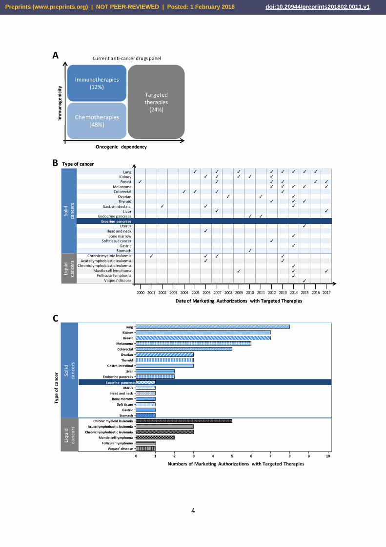
therapies (Figure 1A).
Figure1: Evolution of the use of targeted therapies in cancers for example in France. (A)
Contribution in percentage of targeted therapies as compared to the therapeutic arsenal authorized
in cancers excluding radiotherapies. Distribution (B) and number (C) of Marketing Authorization for
Targeted Therapies. Adapted from INCa 2015 and completed with clinicaltrials.gouv.
Preprints (www.preprints.org) | NOT PEER-REVIEWED | Posted: 1 February 2018 doi:10.20944/preprints201802.0011.v1
4
Immunotherapies(12%)
Chemotherapies(48%)
Oncogenic dependency
Targeted therapies
(24%)
Imm
un
oge
nic
ity
A
B
C
Type of cancer
Date of Marketing Authorizations with Targeted Therapies
2009 2010 2011 2012 2013 2014 2015
LungKidneyBreast
MelanomaColorectal
OvarianThyroid
Gastro-intestinalLiver
Endocrine pancreasExocrine pancreas
UterusHead and neck
Bone marrowSoft tissue cancer
GastricStomach
Chronic myeloid leukemiaAcute lymphoblastic leukemia
Chronic lymphoblastic leukemia Mantle cell lymphoma
Follicular lymphomaVaquez' disease
Solid
ca
nce
rsLi
qu
id
can
cers
✓
✓
✓
✓ ✓
✓✓
✓
✓
✓✓
✓
✓
✓✓ ✓
✓ ✓
✓
✓
✓✓
✓
✓
✓
✓
✓
✓
✓
✓✓
✓✓✓
✓
✓
✓
✓✓
✓
✓ ✓
✓
✓
✓
✓
✓✓
✓
✓
✓
✓
✓
✓
2000 2001 2002 2003 2004 2005 2006 2007 2008 2016 2017
✓
✓
✓
✓ ✓✓
Current anti-cancer drugs panel
Numbers of Marketing Authorizations with Targeted Therapies
0 1 2 3 4 5 6 7 8 9 10
Vaquez' desease
Follicular lymphoma
Mantle cell lymphoma
Chronic lymphobastic leukemia
Acute lymphobastic leukemia
Chronic myeloïd leukemia
Stomach
Gastric
Soft tissue
Bone morrow
Head and neck
Uterus
Exocrine pancreas
Endocrine pancreas
Liver
Gastro-intestinal
Thyroid
Ovarian
Colorectal
Melanoma
Breast
Kidney
Lung
So
lid
can
cers
Liq
uid
can
cers
Typ
e o
f ca
nce
r
Exocrine pancreas
Exocrine pancreas
Preprints (www.preprints.org) | NOT PEER-REVIEWED | Posted: 1 February 2018 doi:10.20944/preprints201802.0011.v1

5
Targeted anti-cancer therapies are strategies that aim to block the growth and/or spread of tumor
cells by specifically addressing some of their abnormalities. Their main mode of action goes through
an inhibition of the mechanisms of oncogenesis with a higher specificity towards cancer cells or their
microenvironment. These may be intracellular inhibitors (small chemical molecules such as protein or
lipid kinase inhibitors) or extracellular inhibitors (biological drugs such as monoclonal antibodies to
receptor tyrosine kinase RTK or their ligands) (Table 1).
Table 1: List of the 47 anti-cancer targeted therapies authorized for example in France. Adapted from
INCa data and completed with Vidal.fr, updated in January 2018.
Intracellular inhibitors Extracellular inhibitors
Inhibitors of protein(s) kinase(s) Ab diriged against RTK(s)
Name Target(s) Name Target(s)
Afatinib EGFR Cetuximab Ab anti-EGFR
Axitinib VEGFR Panitumumab Ab anti-EGFR
Osimertinib EGFR Pertuzumab Ab anti-HER2
Bosutinib Bcr-Abl, Src Ramucirumab Ab anti-VEGF
Cabozantinib MET, AXL, VEGFR, GAS6, RET, ROS1, FLT3,
Tie2 Trastuzumab Ab anti-HER2
Ceritinib ALK Trastuzumab emtansine Ab anti-HER2
Cobimetinib MEK Ab directed against ligand(s)
Crizotinib ALK et MET Aflibercept Ab anti-VEGF
Dabrafenib RAF BevAbizumab Ab anti-VEGF
Dasatinib Bcr-Abl, Src Denosumab Ab anti-RANKL
Erlotinib EGFR
Everolimus mTOR
Gefitinib EGFR
Ibrutinib BTK
Idelalisib p110δ (PI3K)
Imatinib Bcr-Abl, c-Kit, DDR1/2, CSF-1R, PDGFR
Lapatinib EGFR, ErbB2
Lenvatinib VEGFR, FGFR, PDGFR
Nilotinib Bcr-Abl
Nintedanib PDGFR, FGFR, VEGFR, FLT3, Lck, Lyn, Src
Olaparib PARP
Osimertinib EGFR
Palbociclib CDK4/6
Pazopanib VEGFR, c-Kit, PDGFR
Ponatinib Bcr-Abl
Regorafenib VEGFR, c-Kit, PDGFR
Ribociclib Cyclin D1/CDK4, CDK6
Ruxolitinib JAK1/2
Sonidégib SMO
Preprints (www.preprints.org) | NOT PEER-REVIEWED | Posted: 1 February 2018 doi:10.20944/preprints201802.0011.v1

6
Sorafenib RAF, VEGFR, FGFR, c-Kit, PDGFR
Sunitinib VEGFR, c-Kit, c-Kit, CSF-1R, RET, PDGFR
Temsirolimus mTOR
Tivozanib VEGF
Trametinib MEK1/2
Vandetanib VEGFR, EGFR, RET
Venetoclax Bcl2
Vemurafenib ERK, BRAF
Vismodégib SMO
Targeted therapies are part of what is called "precision medicine". This term refers to a medicine
that is based on a better knowledge of the biological mechanisms leading to the appearance and
development of tumors. The use of these treatments is therefore guided, as far as possible, by the
molecular characteristics of the tumor of each patient (for example: the state of differentiation of
the tumor, genetic alterations such as mutations/overexpression of oncogenes, loss of function of
tumor suppressor genes). The majority of targeted therapies are currently used as monotherapy
(62% in France). The first targeted therapy was approved there in 2000. This was trastuzumab, an
antibody targeting the extracellular domain of the HER2 receptor in the treatment of HER2-positive
metastatic breast cancer in monotherapy in patients already treated with at least two
chemotherapies for their metastatic disease. End 2015, the French Cancer Health Institute INCa has
identified 47 targeted therapies which have a Marketing Authorization (MA) in France for the
treatment of cancer. One molecule only has an indication in PDAC (Figure 1B, 1C): inhibitors of the
RTK EGFR. However, the efficiency of these molecules in PDAC remains modest, if compared to the
spectacular action of targeted therapies in other aggressive solid tumors such as BRAF inhibitors in
melanoma or EGFR inhibitors in lung cancers. Although overall survival (OS) and progression-free
survival (PFS) improved slightly (case of EGFR inhibitor), combination with Gemcitabine individually
provides no significantly different objective response (measurable response) as compared to placebo
plus Gemcitabine [16 , 17]. At present, in France, 38 clinical trials testing one or more anti-cancer
molecules are in progress in pancreatic cancer (excluding neuroendocrine tumors), including novel 20
targeted therapies mostly used in combination with chemotherapy (Table 2). Targeted therapies
against pancreatic microenvironment which are thought to contribute to pancreatic aggressiveness
are not yet in phase I clinical trials.
Table 2: Ongoing clinical trials in pancreatic cancer in France. In gray: clinical trials associating a
targeted therapy with a chemotherapy. In blue: clinical trials using a targeted therapy only. Adapted
from clinical.gouv.fr, updated in August 2017.
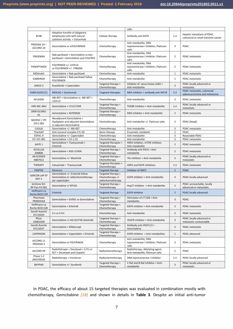
Name of the study
Molecule tested Type of therapy Type of drug Phase Pathologies
D081FC00001-POLO
Olaparib vs placebo Targeted therapy Inhibitor of PARP 3 PDAC metastatic with BRCA mutation
SIRINOX Oxaliplatin + Irinotecan Chemotherapy Platinum salts, DNA topoisomerase I inhibitor
1 Digestive adenocarcinoma (pancreas, esophagus, stomach, small intestine and biliary tract)
PRODIGE 29 FOLFIRINOX vs Gemcitabine Chemotherapy Anti-metabolite, DNA topoisomerase I inhibitor, Platinum salts
3 PDAC locally advanced
PAMELA-70 FOLFIRINOX Chemotherapy Anti-metabolite, DNA topoisomerase I inhibitor, Platinum
2 PDAC metastatic
Preprints (www.preprints.org) | NOT PEER-REVIEWED | Posted: 1 February 2018 doi:10.20944/preprints201802.0011.v1
7
salts
RC48 Adoptive transfer of allogeneic lymphocyte cells with natural cytotoxic activity + Cetuximab
Cellular therapy Antibody anti-EGFR 1-2 Hepatic metastasis of PDAC, colorectal or small intestine cancer
PRODIGE 24 – ACCORD 24
Gemcitabine vs mFOLFIRINOX Chemotherapy Anti-metabolite, DNA topoisomerase I inhibitor, Platinum salts
3 PDAC
FIRGEMAX Nab-paclitaxel + Gemcitabine vs ban-paclitaxel + Gemcitabine puis FOLFIRI3
Chemotherapy Anti-metabolite, DNA topoisomerase I inhibitor, Platinum salts
2 PDAC metastatic
PANOPTIMOX FOLFIRINOX +/- LV5FU2 vs FOLFIRINOX +/- FIRGEM
Chemotherapy Anti-metabolite, DNA topoisomerase I inhibitor, Platinum salts
2 PDAC metastatic
MOAnab1 Gemcitabine + Nab-paclitaxel Chemotherapy Anti-metabolite 1 PDAC metastatic
GABRINOX Gemcitabine + Nab-paclitaxel follow FOLFIRINOX
Chemotherapy Anti-metabolite 1 PDAC metastatic
JANUS-2 Ruxolitinib + Capecitabin Targeted therapy + Chemotherapy
Inhibitor of Janus kinase (JAK) + Anti-metabolite
3 PDAC locally advanced or metastatic
CMEK162X2111 MEK162 + Ganitumab Targeted therapies MEK inhibitor + antibody anti-IGF1R 1-2 PDAC metastatic, colorectal adenocarcinoma and melanoma
AFUGEM ABI-007 + Gemcitabine vs ABI-007 + LV5FU2
Chemotherapy Anti-metabolite 2 PDAC metastatic
H9H-MC-JBAJ Gemcitabine + LY2157299 Targeted therapy + Chemotherapy
TGFβR inhibitor + Anti-metabolite 1-2 PDAC locally advanced or metastatic
2009-011992-61
Gemcitabine + AS703026 Targeted therapy + Chemotherapy
MEK inhibitor + Anti-metabolite 2 PDAC metastatic
NEOPAC / IPC 2011-002
Neoadjuvant Gemcitabine + Oxaliplatin and adjuvant Gemcitabine vs adjuvant Gemcitabine
Chemotherapy Anti-metabolite +/- Platinum salts 3 PDAC (Head)
CAOU6 Gemcitabine +/- ABI-007 Chemotherapy Anti-metabolite 3 PDAC metastatic
TherGAP Anti-tumoral complex CYL-02 Genic therapy Enzymatic metabolic 1 PDAC
ESPAC-4 Gemcitabine +/- Capecitabin Chemotherapy Anti-metabolite 3 Resectable PDAC
CO-101-001 Gemcitabine + CO-1.01 Chemotherapy Anti-metabolite 2 PDAC metastatic
GATE 1 Gemcitabine + Trastuzumab + Erlotinib
Targeted therapies + Chemotherapy
HER2 inhibitor, mTOR inhibitor, Anti-metabolite
2 PDAC metastatic
ASTELLAS 200800
Gemcitabine + AGS-1C4D4 Targeted therapy + Chemotherapy
Antibody anti-PSCA + Anti-metabolite
2 PDAC metastatic
AB SCIENCE AB07012
Gemcitabine +/- Masitinib Targeted therapy + Chemotherapy
TKs inhibitor + Anti-metabolite 3 PDAC locally advanced or metastatic
THERAPY Cetuximab + Trastuzumab Targeted therapies + Chemotherapy
HER2 and EGFR inhibitors 1-2 PDAC metastatic
PANTER Efavirenz Targeted therapy Inhibitor of INNTI 2 PDAC
GERCOR LAP 07 D07-1
Gemcitabine +/- Erlotinib follow Gemcitabine or radiochemiotherapy par capecitabin
Targeted therapy + Chemotherapy +/- radiochemotherapy
EGFR inhibitor + Anti-metabolite 3 PDAC locally advanced
SciClone SCI-RP-Pan-P2-001
Gemcitabine +/-RP101 Targeted therapy + Chemotherapy
Hsp27 inhibitor + Anti-metabolite 2 PDAC unresectable, locally advanced or metastatic.
Hoffmann-La Roche BO21129
Erlotinib Targeted therapy EGFR inhibitor 2 PDAC locally advanced
Pharmexa PRIMOVAX
Gemcitabine + GV001 vs Gemcitabine Targeted therapy + Chemotherapy
Stimulator of LT CD8 + Anti-metabolite
3 PDAC
Hoffmann-La Roche BO21128
Gemcitabine + Erlotinib Targeted therapy + Chemotherapy
EGFR inhibitor + Anti-metabolite 2 PDAC metastatic
Sanofi-Aventis EFC10203
S-1 vs 5-FU Chemotherapy Anti-metabolite 3 PDAC metastatic
Pfizer A4061028
Gemcitabine +/-AG-013736 (Axitinib) Targeted therapy + Chemotherapy
VEGFR inhibitor + Anti-metabolite 3 PDAC locally advanced or metastatic unresectable
Sanofi-Aventis EFC10547
Gemcitabine + Aflibercept Targeted therapy + Chemotherapy
Antibody anti-VEGF1/2 + Gemcitabine
3 PDAC metastatic
CAPERGEM Gemcitabine + Capecitabin + Erlotinib Targeted therapy + Chemotherapy
EGFR inhibitor + Anti-metabolites 1 PDAC advanced
ACCORD 11 PRODIGE 4
Gemcitabine vs FOLFIRINOX Chemotherapy Anti-metabolite, DNA topoisomerase I inhibitor, Platinum salts
3 PDAC metastatic
ACCORD 09 Radiothérapie + Docetaxel + 5-FU or RCT + Docetaxel and Cisplatin
Radiochemotherapy Radiotherapy, Alkylating agent, Anti-metabolite, Platinum salts
2 PDAC
Phase 1-2 (RECF0016)
Radiotherapy + Irinotecan Radiochemotherapy DNA topoisomerase I inhibitor 1-2 PDAC locally advanced
BAYPAN Gemcitabine +/- Sorafenib Targeted therapy + Chemotherapy
C-Raf and B-Raf inhibitor + Anti-metabolite
3 PDAC locally advanced or metastatic
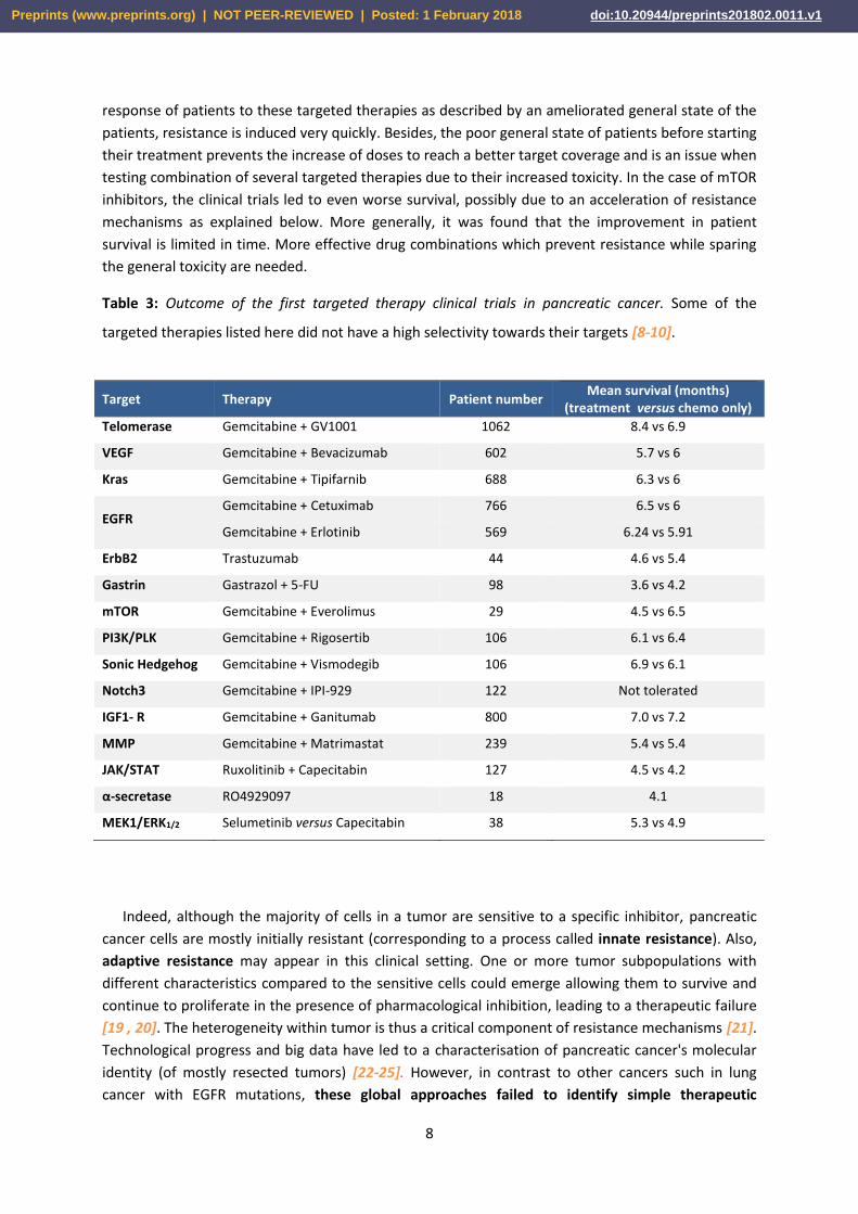
In PDAC, the efficacy of about 15 targeted therapies was evaluated in combination mostly with
chemotherapy, Gemcitabine [18] and shown in details in Table 3. Despite an initial anti-tumor
Preprints (www.preprints.org) | NOT PEER-REVIEWED | Posted: 1 February 2018 doi:10.20944/preprints201802.0011.v1
8
response of patients to these targeted therapies as described by an ameliorated general state of the
patients, resistance is induced very quickly. Besides, the poor general state of patients before starting
their treatment prevents the increase of doses to reach a better target coverage and is an issue when
testing combination of several targeted therapies due to their increased toxicity. In the case of mTOR
inhibitors, the clinical trials led to even worse survival, possibly due to an acceleration of resistance
mechanisms as explained below. More generally, it was found that the improvement in patient
survival is limited in time. More effective drug combinations which prevent resistance while sparing
the general toxicity are needed.
Table 3: Outcome of the first targeted therapy clinical trials in pancreatic cancer. Some of the
targeted therapies listed here did not have a high selectivity towards their targets [8-10].
Target Therapy Patient number Mean survival (months)
(treatment versus chemo only)
Telomerase Gemcitabine + GV1001 1062 8.4 vs 6.9
VEGF Gemcitabine + Bevacizumab 602 5.7 vs 6
Kras Gemcitabine + Tipifarnib 688 6.3 vs 6
EGFR Gemcitabine + Cetuximab 766 6.5 vs 6
Gemcitabine + Erlotinib 569 6.24 vs 5.91
ErbB2 Trastuzumab 44 4.6 vs 5.4
Gastrin Gastrazol + 5-FU 98 3.6 vs 4.2
mTOR Gemcitabine + Everolimus 29 4.5 vs 6.5
PI3K/PLK Gemcitabine + Rigosertib 106 6.1 vs 6.4
Sonic Hedgehog Gemcitabine + Vismodegib 106 6.9 vs 6.1
Notch3 Gemcitabine + IPI-929 122 Not tolerated
IGF1- R Gemcitabine + Ganitumab 800 7.0 vs 7.2
MMP Gemcitabine + Matrimastat 239 5.4 vs 5.4
JAK/STAT Ruxolitinib + Capecitabin 127 4.5 vs 4.2
α-secretase RO4929097 18 4.1
MEK1/ERK1/2 Selumetinib versus Capecitabin 38 5.3 vs 4.9
Indeed, although the majority of cells in a tumor are sensitive to a specific inhibitor, pancreatic
cancer cells are mostly initially resistant (corresponding to a process called innate resistance). Also,
adaptive resistance may appear in this clinical setting. One or more tumor subpopulations with
different characteristics compared to the sensitive cells could emerge allowing them to survive and
continue to proliferate in the presence of pharmacological inhibition, leading to a therapeutic failure
[19 , 20]. The heterogeneity within tumor is thus a critical component of resistance mechanisms [21].
Technological progress and big data have led to a characterisation of pancreatic cancer's molecular
identity (of mostly resected tumors) [22-25]. However, in contrast to other cancers such in lung
cancer with EGFR mutations, these global approaches failed to identify simple therapeutic
Preprints (www.preprints.org) | NOT PEER-REVIEWED | Posted: 1 February 2018 doi:10.20944/preprints201802.0011.v1

9
strategies based on a stratification of PDAC patients. Recent integrated omics approaches on all
cancers points out that the search of genetic or genomic alterations is not sufficient to predict which
patients will benefit from targeted therapies [26][69]. In contrast, proteomics appears better in
predicting sensitivity to a targeted therapy (PI3K inhibitors) [26], suggesting that proteomic
approaches could be a worthwhile strategy to investigate and to better understand the resistance to
treatment of pancreatic cancer patients, so as to predict which therapy will be more efficient (Figure
2). Recent pioneering data which need to be complemented by wider studies argue for the effort to
develop such challenging strategies in pancreatic cancer.
Figure 2: Proteomics for a better clinical care of pancreatic cancer patients.
Proteomics as a new way to improve clinical management of pancreatic cancer
Proteomics consists of studying all the proteins of an organism, a biological fluid, an organ, a cell
or even a cellular compartment. This set of proteins is called "proteome". The latter is a dynamic and
complex entity. The proteome contains a much larger number of proteins than the genome contains
genes. In human cells, an estimated 22.000 genes can yield up to one million proteins. Despite these
approximations, it is considered that proteins represent about 60% of a cell. The study of proteins
has grown dramatically during the 1990s, with the advent of mass spectrometers (Nobel Prize in
Chemistry in 2002 to John Fenn and Koichi Tanaka) [27]. Mass spectrometry (MS) is an analytical
method that aims to identify and separate molecules to be analyzed (small molecules, proteins,
drugs...) with a very good resolution and sensitivity. It allows the qualitative and quantitative analysis
of complex biological samples, which may contain thousands of proteins, some of which are present
in small quantities. Post-translational modifications such as phosphorylation but also ubiquitination
can be studied by these techniques. These post-translational alterations are key indicators of the
activity of the proteins.
In pancreatic cancer, proteomics provides insight into proteome-related changes in the disease,
such as observed protein changes in abundance, subcellular localization, post-translational
modifications and cell signaling. The detection of these changes thus constitutes research interests
ranging from the study of the mechanisms of initiation of the disease to the discovery of biomarkers.
Heterogeneity New targets Targeted therapy
sensibility
Therapeutic resistance
Predictive and precision medicine
Proteomics
Preprints (www.preprints.org) | NOT PEER-REVIEWED | Posted: 1 February 2018 doi:10.20944/preprints201802.0011.v1

10
Published proteomic results for pancreatic cancer are derived from fluid samples (blood, plasma,
serum, pancreatic juice, cystic fluid, in vitro cells conditions media) and/or solid samples (tumor
tissue) from healthy versus sick patients (resected patients, representing a subpopulation of patients,
see introduction), or patients at risk of developing pancreatic cancer (e.g. chronic pancreatitis CP) or
murine models of PDAC. Tumor tissue is the main source of investigation of protein alterations
associated with pancreatic cancer. These samples are mainly studied by mass spectrometry [28-32].
Nevertheless, some articles report the use of a targeted proteomic/phosphoproteomic technique,
called Reverse Phase Protein Array (RPPA) or "Reversed Phase Protein Chips" [33-35].
Proteomic approaches to search new biomarkers of early diagnosis
Early detection of pancreatic cancer offers hope for healing. Currently, tumor markers used
clinically lack sensitivity and specificity. For example, the carbohydrate antigen marker CA-19-9
(secreted by exocrine cells and tumors) present in the serum makes it possible to estimate the
pancreatic tumor progression during a treatment [36], but does not provide sufficient precision for
the diagnosis of pancreatic cancer, because it is not very specific (high concentration in the sera of
patients with acute and chronic pancreatitis, hepatitis and biliary obstruction).
Numerous studies report new specific biomarkers in the diagnosis of pancreatic cancer identified
from pancreatic tissue lysate or liquid biopsies (such as secretome) [28, 29, 35, 37, 38, 39 , 40-44].
Some biomarkers have been characterized and evaluated alone or in combination with CA19-9 on
their ability to diagnose [45]. A major step forward has been reached very recently with the access to
preneoplastic samples. So far, these samples were difficult to obtain due to the late diagnosis of this
disease. Thrombospondin 2 detected in plasma as a biomarker alone or in combination with CA19-9
was validated (98% combination specificity, 87% sensitivity), to distinguish all the stages of the
disease [45]. Studies on one type of the precursor lesions, intra-ductal papillary mucinous neoplasm
(IPMN) and cystic precursor lesion of the pancreas, should lead to further insights towards the early
diagnosis of pancreatic cancer [46, 47]. Proteomic studies of biomarkers in patients at risk to develop
pancreatic cancer, such as those with neo-onset diabetes, show that this condition impacts the
diagnostic performance of CA19-9 [48]. These advances in early diagnosis are expected to increase
the efficiency of the targeted therapies.
Proteomics to grade the disease including the metastatic sites and identify new targets
The majority of proteomic studies performed on pancreatic cancer to identify biomarkers does
not discriminate the different grades of the tumors studied. However, 2D-DIGE technology
performed on microdissections of human pancreatic tumors identified the calcium-binding protein
S110A6 in moderately or poorly differentiated tumors [49]. In 2009, Sitek and its collaborators
identified 86 differentially regulated proteins involved in pancreatic tumor progression only using
microdissections [50]. These results are complemented by proteomic analysis in 2D-DIGE of
microdissected murine PanIN cells (PanIN or Pancreatic Intraepithelial Neoplasia is a precursor lesion
of PDAC) and plasma samples corresponding to different precancerous stages of genetically modified
mice by Dufresne M et al [51]. The identification of peptide signatures specific to each type of
Preprints (www.preprints.org) | NOT PEER-REVIEWED | Posted: 1 February 2018 doi:10.20944/preprints201802.0011.v1

11
precancerous lesions would thus make it possible to discriminate them from normal pancreatic
tissue. Similarly, other studies proposes classifications of pancreatic cancer cell lines based on the
levels of tyrosine phosphorylation of RTKs, potentially identifying three groups of cell lines as low
pTyr, enriched in RTK and mixed [52]. These data indicate that a combination of RTK is usually
activated in pancreatic cancer, suggesting that single agent strategies towards RTKs are likely to be
inefficient. If this classification could be performed in patient biopsies, this could allow predicting
which targeted therapy could be efficient. To develop novel strategies targeting pancreatic cancer
stem cells, large scale proteome analysis in cells or their secretome showed the importance of fatty
acid synthesis and mevalonate pathways, as well as glycolysis, in these subtype of cells [53, 54].
These cells being possibly at the origin of tumor relapse under chemotherapy, discovering specific
targets of this pool of cells could improve the current treatments. Recent data show that surface
proteome of circulating exosomes provides unique opportunities to analyze the heterogeneity of the
metastatic disease though the easy-to-access liquid biopsies [55]. Proteomics studies bring insight in
metastatic PDAC biology, leading to the discovery of novel targets.
Proteomic approaches to search new biomarkers of predictive response
Proteomics is an adapted tool for predicting the response to targeted therapies by studying tumor
heterogeneity and changes in signaling pathways during treatment [21]. Kim and his collaborators
studied and apprehended the heterogeneity of three metastatic sites of pancreatic cancer (liver, lung
and peritoneum) by creating three lines from each organ in the same patient [56]. This heterogeneity
is characterized by changes in expression of the entire proteome and tyrosine kinase activity, in the
three sites of metastasis. It is involved in differences in the sensitivity of neoplastic cells to targeted
therapies. In contrast, in a large scale genomic and genetic analysis, genetic alterations in metastasis
sites were found to be maintained as compared to the primary site of tumorigenesis [21]. Thus, this
study highlights the interest of a personalized therapeutic combination targeting all the subclonal
features of metastases, using proteomics to guide the therapeutic choice.
Also, proteomics is a powerful tool in the study of tumor cell/microenvironment cell signaling
interactions and modifications. For example, Jorgensen's team has recently demonstrated from Kras-
mutated pancreatic cancer cells that this oncogenic tumor signaling activates a cell-autonomous
signaling network but also non-autonomous activation of oncogenic signaling of stromal cells.
Conversely, stromal cells can in turn modify and amplify oncogenic signaling in the same or other
tumor cells. Thus, oncogenic signaling is no longer limited only to tumor cells but to the entire tumor
compartment [57].
Hence, prediction of sensitivity to targeted therapies is influenced by, besides genetic and
genomic alterations, the heterotypic tumor-stroma signalling; proteomic assessment in patient-
derived samples including all the cellular partners at stake appear to be the only way to assess it.
Proteomic approaches to identify resistance mechanisms
Global studies to understand the adaptative responses to targeted therapies in pancreatic cancer
are starting to be published. Adaptative and reversible resistance to Kras inhibition in pancreatic
Preprints (www.preprints.org) | NOT PEER-REVIEWED | Posted: 1 February 2018 doi:10.20944/preprints201802.0011.v1

12
cancer cells includes phosphorylation of focal adhesion pathway components, while strikingly no
significant mutational or transcriptional changes were observed [58]. Temporal effects of paclitaxel
on pancreatic cells via large scale proteomics highlight protein involved in mitochondrial function,
survival (PI3K pathway) and cell cycle arrest as key resistance mechanism [59]. In PDAC, several
negative feedback mechanisms of mTORC inhibitors have been identified using proteomics,
explaining the disappointing results of the clinical trials on this target [38, 39, 57,59]. In other cancer
settings, Hsu and colleagues have shown, for example, through a global phosphoproteome analysis
approach (SILAC) that the mTORC1 complex is able to inhibit and degrade insulin and IGF-1 receptors
through phosphorylation of the adapter protein Grb10 (Growth Factor Bound Protein 10) [60].
mTORC1 leads to the phosphorylation of Grb10 then to the ubiquitination and degradation of insulin
and IGF receptors [61]. It is now generally agreed that to target PI3K/AKT/mTOR pathway, which is
hyperactivated in 50% of all PDAC patients and associated with poor prognosis [4], hitting the
upstream PI3K is a better strategy to prevent positive feedbacks due to mTOR inhibition. While some
mechanisms of resistance to PI3K inhibitors were found [62, 63], it remains to be investigated in an
integrated approach which mechanisms of resistance could still occur to allow a better efficiency of
these innovative targeted therapies [2].
Conclusion / Discussion / Perspectives
To conclude, only more comprehensive knowledge on resistance mechanisms observed at the
level of the protein (target) induced by targeted therapies will allow the researchers and clinicians to
develop effective therapeutic strategies adapted to each target / oncogenic pathway in each
environment specific to each patient, abolishing, preventing or delaying the appearance of
resistance.
Proteomics, and in particular phosphoproteomics (see Figure 2), are powerful and promising
tools in:
i. the identification of early diagnostic and prognostic biomarkers;
ii. the identification of deregulated proteins and signaling pathways in the
pathology;
iii. the identification of biomarkers predictive of the response to treatment;
iv. the identification of early resistance mechanism leading to the development of
adapted combinatorial treatments.
It is difficult to perform in-depth and large-scale clinical studies on PDAC. However, recent
advances in methods can advance PDAC proteomics both at the fundamental and clinical research
stage. Besides the better understanding of oncogenic dependency of PDAC [64], current
development of MS-based methods which need less material and are more quantitative [65], which
are coupled to imaging [66, 67], or of non-MS based methods which are robust targeted proteomic
approaches [68] together with the improvement of patient-derived ex vivo cultures better mimicking
each patient situation will be instrumental to improve the management of pancreatic cancer
patients.
Early diagnosis using proteomics is a growing field with already promising leads. The next
challenge for PDAC proteomics will be to identify the appropriate biomarkers indicating which is the
Preprints (www.preprints.org) | NOT PEER-REVIEWED | Posted: 1 February 2018 doi:10.20944/preprints201802.0011.v1
13
best targeted therapy strategy to use for each pancreatic cancer patient, and thus, to develop
stratifications of patients according to each therapeutic approach.
Abbreviations
- ALK: Anaplastic Lymphoma Kinase;
- ASCO: American Society of Clinical Oncology;
- BTK: Bruton's tyrosine kinase;
- CA 19-9: Cancer Antigen 19-9;
- CDK4/6: Cyclin-Dependent Kinase;
- CSF-1R: Colony-stimulating factor 1 receptor;
- DDR1/2: Discoidin domain receptor tyrosine kinase 1/2;
- EGFR: Epidermal Growth Factor Receptor;
- FGFR: Fibroblast Growth Factor Receptor;
- FLT3: Fms-related tyrosine kinase 3;
- GAS6: Growth arrest-specific 6 ;
- Grb10: Growth Factor Bound Protein 10;
- Hsp27: Heat Shock Protein 27;
- IGF-1(R): Insulin Growth Factor (Receptor);
- IPMN: Intraductal papillary mucinous neoplasm;
- JAK: Janus Kinase;
- Lck: Lymphocyte-specific protein tyrosine kinase;
- MA: Marketing Authorization;
- MEK: Mitogen-Activated Protein Kinase;
- MET: Hepatocyte growth factor receptor;
- MS : Mass spectrometry;
- mTOR: mammalian target of Rapamycin;
- PanIN: Pancreatic Intraepithelial Neoplasia;
- PARP: Enzyme poly ADP ribose polymerase;
- PDAC: Pancreatic ductal adenocarcinoma;
- PDGFR: Platelet-derived growth factor receptor
- PI3K: Phosphoinositide 3-kinase;
- PSCA : Prostate stem cell antigen;
- OS: Overall survival;
- PFS: Progression-free survival;
- RANKL: Receptor activator of nuclear factor kappa-Β ligand
- ROS1: c-ros oncogene 1;
- RPPA: Reverse Phase Protein Array;
- RTK: Receptor tyrosine kinase;
- Tie2: Tyrosine kinase with immunoglobulin and EGF homology domains;
- TGFβR: Transforming growth factor β receptor
- TK: Tyrosine Kinase;
- SILAC: Stable Isotope Labeling with Amino acids in Cell culture;
- SMO: Smoothened kinase;
- VEGF(R): Vascular endothelial growth factor (receptor);
- Vs: Versus
Preprints (www.preprints.org) | NOT PEER-REVIEWED | Posted: 1 February 2018 doi:10.20944/preprints201802.0011.v1

14
Glossary
Heterotypic signalling
Statement of conflict of interest
The authors declare no conflict of interests.
Author contributions
CC and JGG wrote the article; CC, TD and JGG performed bibliographic search.
Funding
JGG's laboratory belongs to Toucan, Laboratoire d’Excellence, ANR, an integrated research program
on Signal-targeted Drug Resistance. CC’s salary was funded by French Charity Ligue Nationale Contre
le Cancer (GB/MA/VSP-10443). JGG's laboratory for this topic was/is funded by Europe EU-ERG FP7
(270696 PaCa/PI3K), ARC (PJA20171206596), Toucan and MSCA-ITN/ETN PhD-PI3K (Project ID:
675392).
Acknowledgments
JGG is a member of COST action EU-Pancreas BM1204.
Bibliography
1. Xu, J., Bin Jiang, Ya Chen, Fu-Zhen Qi, Jian-Huai Zhang and Hang Yuan, Optimal adjuvant chemotherapy for resected pancreatic adenocarcinoma: a systematic review and network meta-analysis. Oncotarget, 2017.
2. Pons-Tostivint, E., B. Thibault, and J. Guillermet-Guibert, Targeting PI3K Signaling in Combination Cancer Therapy. Trends Cancer, 2017. 3(6): p. 454-469.
3. Baer, R., et al., Pancreatic cell plasticity and cancer initiation induced by oncogenic Kras is completely dependent on wild-type PI 3-kinase p110alpha. Genes Dev, 2014. 28(23): p. 2621-35.
4. Baer, R., et al., Implication of PI3K/Akt pathway in pancreatic cancer: When PI3K isoforms matter? Adv Biol Regul, 2015. 59: p. 19-35.
5. Eser, S., et al., Selective requirement of PI3K/PDK1 signaling for Kras oncogene-driven pancreatic cell plasticity and cancer. Cancer Cell, 2013. 23(3): p. 406-20.
6. Wu, C.Y., et al., PI3K regulation of RAC1 is required for KRAS-induced pancreatic tumorigenesis in mice. Gastroenterology, 2014. 147(6): p. 1405-16 e7.
7. Burris, H.A., 3rd, et al., Improvements in survival and clinical benefit with gemcitabine as first-line therapy for patients with advanced pancreas cancer: a randomized trial. J Clin Oncol, 1997. 15(6): p. 2403-13.
8. Adamska, A., A. Domenichini, and M. Falasca, Pancreatic Ductal Adenocarcinoma: Current and Evolving Therapies. Int J Mol Sci, 2017. 18(7).
Preprints (www.preprints.org) | NOT PEER-REVIEWED | Posted: 1 February 2018 doi:10.20944/preprints201802.0011.v1

15
9. Danovi, S.A., H.H. Wong, and N.R. Lemoine, Targeted therapies for pancreatic cancer. Br Med Bull, 2008. 87: p. 97-130.
10. Barati Bagherabad, M., et al., Targeted Therapies in Pancreatic Cancer: Promises and Failures. J Cell Biochem, 2017.
11. Paulson, A.S., et al., Therapeutic advances in pancreatic cancer. Gastroenterology, 2013. 144(6): p. 1316-26.
12. Von Hoff, D.D., et al., Gemcitabine plus nab-paclitaxel is an active regimen in patients with advanced pancreatic cancer: a phase I/II trial. J Clin Oncol, 2011. 29(34): p. 4548-54.
13. De Vita, F., et al., NAB-paclitaxel and gemcitabine in metastatic pancreatic ductal adenocarcinoma (PDAC): from clinical trials to clinical practice. BMC Cancer, 2016. 16(1): p. 709.
14. Conroy, T., et al., FOLFIRINOX versus gemcitabine for metastatic pancreatic cancer. N Engl J Med, 2011. 364(19): p. 1817-25.
15. Gunturu, K.S., et al., FOLFIRINOX for locally advanced and metastatic pancreatic cancer: single institution retrospective review of efficacy and toxicity. Med Oncol, 2013. 30(1): p. 361.
16. Moore, M.J., et al., Erlotinib plus gemcitabine compared with gemcitabine alone in patients with advanced pancreatic cancer: a phase III trial of the National Cancer Institute of Canada Clinical Trials Group. J Clin Oncol, 2007. 25(15): p. 1960-6.
17. Rougier, P., et al., Randomised, placebo-controlled, double-blind, parallel-group phase III study evaluating aflibercept in patients receiving first-line treatment with gemcitabine for metastatic pancreatic cancer. Eur J Cancer, 2013. 49(12): p. 2633-42.
18. Di Marco, M., et al., State of the art biological therapies in pancreatic cancer. World J Gastrointest Oncol, 2016. 8(1): p. 55-66.
19. Burrell, R.A. and C. Swanton, Tumour heterogeneity and the evolution of polyclonal drug resistance. Mol Oncol, 2014. 8(6): p. 1095-111.
20. Almendro, V., A. Marusyk, and K. Polyak, Cellular heterogeneity and molecular evolution in cancer. Annu Rev Pathol, 2013. 8: p. 277-302.
21. Makohon-Moore, A.P., et al., Limited heterogeneity of known driver gene mutations among the metastases of individual patients with pancreatic cancer. Nat Genet, 2017. 49(3): p. 358-366.
22. Waddell, N., et al., Whole genomes redefine the mutational landscape of pancreatic cancer. Nature, 2015. 518(7540): p. 495-501.
23. Bailey, P., et al., Genomic analyses identify molecular subtypes of pancreatic cancer. Nature, 2016. 531(7592): p. 47-52.
24. Sivakumar, S., et al., Master Regulators of Oncogenic KRAS Response in Pancreatic Cancer: An Integrative Network Biology Analysis. PLoS Med, 2017. 14(1): p. e1002223.
25. Collisson, E.A., et al., Subtypes of pancreatic ductal adenocarcinoma and their differing responses to therapy. Nat Med, 2011. 17(4): p. 500-3.
26. Zhang, Y., et al., A Pan-Cancer Proteogenomic Atlas of PI3K/AKT/mTOR Pathway Alterations. Cancer Cell, 2017. 31(6): p. 820-832 e3.
27. Fenn, J.B., et al., Electrospray ionization for mass spectrometry of large biomolecules. Science, 1989. 246(4926): p. 64-71.
28. Wehr, A.Y., et al., Relative quantification of serum proteins from pancreatic ductal adenocarcinoma patients by stable isotope dilution liquid chromatography-mass spectrometry. J Proteome Res, 2012. 11(3): p. 1749-58.
29. Gronborg, M., et al., Biomarker discovery from pancreatic cancer secretome using a differential proteomic approach. Mol Cell Proteomics, 2006. 5(1): p. 157-71.
30. Lee, Y.Y., et al., Phosphoproteomic analysis identifies activated MET-axis PI3K/AKT and MAPK/ERK in lapatinib-resistant cancer cell line. Exp Mol Med, 2013. 45: p. e64.
31. Wei, W., et al., Single-Cell Phosphoproteomics Resolves Adaptive Signaling Dynamics and Informs Targeted Combination Therapy in Glioblastoma. Cancer Cell, 2016. 29(4): p. 563-73.
Preprints (www.preprints.org) | NOT PEER-REVIEWED | Posted: 1 February 2018 doi:10.20944/preprints201802.0011.v1
16
32. Chen, R., et al., Comparison of pancreas juice proteins from cancer versus pancreatitis using quantitative proteomic analysis. Pancreas, 2007. 34(1): p. 70-9.
33. Huang, Y.J., et al., Reverse-phase protein array analysis to identify biomarker proteins in human pancreatic cancer. Dig Dis Sci, 2014. 59(5): p. 968-75.
34. Grote, T., et al., Validation of reverse phase protein array for practical screening of potential biomarkers in serum and plasma: accurate detection of CA19-9 levels in pancreatic cancer. Proteomics, 2008. 8(15): p. 3051-60.
35. Mustafa, S., et al., Comparison of the tumor cell secretome and patient sera for an accurate serum-based diagnosis of pancreatic ductal adenocarcinoma. Oncotarget, 2017. 8(7): p. 11963-11976.
36. Wakabayashi, T., et al., Diagnostic significance of cancer-associated carbohydrate antigen (CA19-9) concentrations in pancreatic juice: analysis in pure pancreatic juice collected by endoscopic aspiration and immunohistochemical study in chronic pancreatitis. Pancreas, 1993. 8(2): p. 151-9.
37. Fakelman, F., et al., New pre-analytical approach for the deep proteome analysis of sera from pancreatitis and pancreas cancer patients. Arch Physiol Biochem, 2010. 116(4-5): p. 208-17.
38. Sun, C., et al., Proteome-based biomarkers in pancreatic cancer. World J Gastroenterol, 2011. 17(44): p. 4845-52.
39. Takano, S., et al., Increased circulating cell signalling phosphoproteins in sera are useful for the detection of pancreatic cancer. Br J Cancer, 2010. 103(2): p. 223-31.
40. Lin, C., et al., ITRAQ-based quantitative proteomics reveals apolipoprotein A-I and transferrin as potential serum markers in CA19-9 negative pancreatic ductal adenocarcinoma. Medicine (Baltimore), 2016. 95(31): p. e4527.
41. Park, J., et al., Large-scale clinical validation of biomarkers for pancreatic cancer using a mass spectrometry-based proteomics approach. Oncotarget, 2017.
42. Saraswat, M., et al., Comparative proteomic profiling of the serum differentiates pancreatic cancer from chronic pancreatitis. Cancer Med, 2017.
43. Britton, D., et al., Quantification of pancreatic cancer proteome and phosphorylome: indicates molecular events likely contributing to cancer and activity of drug targets. PLoS One, 2014. 9(3): p. e90948.
44. Crnogorac-Jurcevic, T., et al., Proteomic analysis of chronic pancreatitis and pancreatic adenocarcinoma. Gastroenterology, 2005. 129(5): p. 1454-63.
45. Jenkinson, C., et al., Evaluation in pre-diagnosis samples discounts ICAM-1 and TIMP-1 as biomarkers for earlier diagnosis of pancreatic cancer. J Proteomics, 2015. 113: p. 400-2.
46. Ilies, M., et al., Plasma protein profiling of patients with intraductal papillary mucinous neoplasm of the pancreas as potential precursor lesions of pancreatic cancer. Clin Chim Acta, 2018. 477: p. 127-134.
47. Jabbar, K.S., et al., Highly Accurate Identification of Cystic Precursor Lesions of Pancreatic Cancer Through Targeted Mass Spectrometry: A Phase IIc Diagnostic Study. J Clin Oncol, 2018. 36(4): p. 367-375.
48. Jenkinson, C., et al., Decreased Serum Thrombospondin-1 Levels in Pancreatic Cancer Patients Up to 24 Months Prior to Clinical Diagnosis: Association with Diabetes Mellitus. Clin Cancer Res, 2016. 22(7): p. 1734-1743.
49. Shekouh, A.R., et al., Application of laser capture microdissection combined with two-dimensional electrophoresis for the discovery of differentially regulated proteins in pancreatic ductal adenocarcinoma. Proteomics, 2003. 3(10): p. 1988-2001.
50. Sitek, B., et al., Analysis of the pancreatic tumor progression by a quantitative proteomic approach and immunhistochemical validation. J Proteome Res, 2009. 8(4): p. 1647-56.
51. Ligat, L., et al., Pancreatic preneoplastic lesions plasma signatures and biomarkers based on proteome profiling of mouse models. Br J Cancer, 2015. 113(11): p. 1590-8.
52. Humphrey, E.S., et al., Resolution of Novel Pancreatic Ductal Adenocarcinoma Subtypes by Global Phosphotyrosine Profiling. Mol Cell Proteomics, 2016. 15(8): p. 2671-85.
Preprints (www.preprints.org) | NOT PEER-REVIEWED | Posted: 1 February 2018 doi:10.20944/preprints201802.0011.v1

17
53. Brandi, J., et al., Secretome protein signature of human pancreatic cancer stem-like cells. J Proteomics, 2016. 136: p. 1-12.
54. Brandi, J., et al., Proteomic analysis of pancreatic cancer stem cells: Functional role of fatty acid synthesis and mevalonate pathways. J Proteomics, 2017. 150: p. 310-322.
55. Castillo, J., et al., Surfaceome profiling enables isolation of cancer-specific exosomal cargo in liquid biopsies from pancreatic cancer patients. Ann Oncol, 2018. 29(1): p. 223-229.
56. Kim, M.S., et al., Heterogeneity of pancreatic cancer metastases in a single patient revealed by quantitative proteomics. Mol Cell Proteomics, 2014. 13(11): p. 2803-11.
57. Tape, C.J., et al., Oncogenic KRAS Regulates Tumor Cell Signaling via Stromal Reciprocation. Cell, 2016. 165(7): p. 1818.
58. Chen, P.Y., et al., Adaptive and reversible resistance to Kras inhibition in pancreatic cancer cells. Cancer Res, 2017.
59. Wang, X., et al., Temporal Effects of Combined Birinapant and Paclitaxel on Pancreatic Cancer Cells Investigated via Large-scale, Ion-Current-Based Quantitative Proteomics (IonStar). Mol Cell Proteomics, 2018.
60. Hsu, P.P., et al., The mTOR-regulated phosphoproteome reveals a mechanism of mTORC1-mediated inhibition of growth factor signaling. Science, 2011. 332(6035): p. 1317-22.
61. Yu, Y., et al., Phosphoproteomic analysis identifies Grb10 as an mTORC1 substrate that negatively regulates insulin signaling. Science, 2011. 332(6035): p. 1322-6.
62. Alagesan, B., et al., Combined MEK and PI3K inhibition in a mouse model of pancreatic cancer. Clin Cancer Res, 2015. 21(2): p. 396-404.
63. Junttila, M.R., et al., Modeling targeted inhibition of MEK and PI3 kinase in human pancreatic cancer. Mol Cancer Ther, 2015. 14(1): p. 40-7.
64. Ying, H., et al., Genetics and biology of pancreatic ductal adenocarcinoma. Genes Dev, 2016. 30(4): p. 355-85.
65. Dalla Pozza, E., et al., Trichostatin A alters cytoskeleton and energy metabolism of pancreatic adenocarcinoma cells: An in depth proteomic study. J Cell Biochem, 2017.
66. Gruner, B.M., et al., MALDI imaging mass spectrometry for in situ proteomic analysis of preneoplastic lesions in pancreatic cancer. PLoS One, 2012. 7(6): p. e39424.
67. Gruner, B.M., et al., Modeling Therapy Response and Spatial Tissue Distribution of Erlotinib in Pancreatic Cancer. Mol Cancer Ther, 2016. 15(5): p. 1145-52.
68. Hilhorst, R., et al., Peptide microarrays for detailed, high-throughput substrate identification, kinetic characterization, and inhibition studies on protein kinase A. Anal Biochem, 2009. 387(2): p. 150-61.
Preprints (www.preprints.org) | NOT PEER-REVIEWED | Posted: 1 February 2018 doi:10.20944/preprints201802.0011.v1